Embed Size (px)
Citation preview
Clinical Information
Transfus Med Hemother 2013;40:50–62DOI: 10.1159/000345752
Prof. Dr. med. Rainer SeitzPaul-Ehrlich-InstitutPaul-Ehrlich-Straße 51–59, 63225 Langen, [email protected]
© 2013 S. Karger GmbH, Freiburg1660-3796/13/0401-0050$38.00/0
Accessible online at: www.karger.com/tmh
Fax +49 761 4 52 07 [email protected]
Received: June 1, 2012Accepted: June 2, 2012Published online: January 7, 2013
Arbonematodes – Nematode Infections Transmissible by ArthropodsArbeitskreis Blut, Untergruppe «Bewertung Blutassoziierter Krankheitserreger»*
where they deposit their eggs in the tissue after penetrating the wall of the blood vessel. The eggs are excreted with urine and feces. The larvae which develop from the eggs are in-gested with food or water or acquired via skin passage, and do not fall into the category of arbonematodes. They are not transfusion-relevant. Therefore this article will not provide more detailed information on this species [4].
Tissue roundworms include Trichinella spiralis, Dracuncu-lus medinensis and Toxocara canis, which remain within tis-sues and neither release eggs nor microfilariae into the blood; they are not transmitted via arthropods. Nematodes which are transmitted to humans via arthropods include lymphatic filaria such as W. bancrofti, Brugia malayi and B. timori, as well as Mansonella, Onchocerca and Loa loa which live in tissues.
1.1. Characteristics
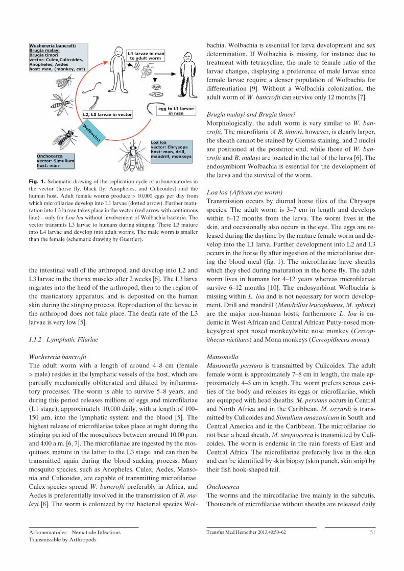
1.1.1 Life CycleArbonematodes are hermaphroditic worms with a reproduc-tion cycle in humans or primates and a maturation cycle of the larva in the appropriate arthropods (fig. 1). After the sting of a female mosquito or horse fly, who need blood for the matu-ration of their eggs, the L3 larvae leave the salivary gland of the arthropod and pass from the surface of the skin through the penetration canal into the subcutis [5]. From there, they pass into the lymphatic vessels, develop into L4 larvae, and, after another 6–9 months, develop into sexually mature male and female worms. The worms stay in the afferent lymphatic vessels of the extremities and of the male genital and in the skin (fig. 1). The fertilized female worm releases around 10–20,000 eggs daily into the lymphatic system or blood stream. The L1 larvae, called microfilariae, hatch from these eggs. If an infected human is stung by a mosquito or horse fly, the L1 larvae are consumed with the blood meal, penetrate through
Introduction
Some alphaviruses and flaviviruses, such as the transfusion-relevant Chikungunya and West-Nile virus, are transmitted by mosquitoes. Such viruses are called arboviruses. Bacteria typi-cally transmitted by ticks include Coxiella burnetii [1], Borre-lia, Ehrlichia, and Anaplasma. These are referred to as arbo-bacteria [2]. Protozoae include plasmodia which are transmit-ted via mosquitoes, leishmania transmitted via sand flies (Phlebotomus), and for example trypanosoma transmitted via kissing bugs (Triatoma). The latter are grouped under the name arboprotozoa [3]. The following nematodes are trans-mitted by arthropods as vectors to humans: microfilaria of Wuchereria bancrofti via mosquitoes (Culex), Onchocera (Onchocera volvulus in Africa, O. caecutiens in South Amer-ica) via black fly (Simulium) and Loa loa (African eye worm) via horse flies (Tabanidae) – therefore these agents are here grouped under the name arbonematodes.
1. Current Knowledge about the Pathogen
Helminths (worms) are subdivided into nematodes (round-worms), trematodes (flukes), and cestodes (tapeworms). Among nematodes, a distinction is made between intestinal and tissue roundworms. Intestinal nematodes are subdivided into the filarial worms, such as Ankylostoma duodenale, As-caris lumbricoides and Strongyloides stercoralis, which pass through the lung during maturation and re-enter the intestine thereafter; they only have a short hematogenic phase. In the case of trematodes, the larvae of schistosoma (Schistosoma mansoni, S. haematobium and S. japonicum) live only in the portal vein. Like intestinal nematodes, they achieve maturity by passing through the lung, and thus their larvae can also be present in the blood for a short period. The worms migrate through the veins to the bladder and rectum at night time
Transfus Med Hemother 2013;40:50–62Arbonematodes – Nematode Infections Transmissible by Arthropods
51
bachia. Wolbachia is essential for larva development and sex determination. If Wolbachia is missing, for instance due to treatment with tetracycline, the male to female ratio of the larvae changes, displaying a preference of male larvae since female larvae require a denser population of Wolbachia for differentiation [9]. Without a Wolbachia colonization, the adult worm of W. bancrofti can survive only 12 months [7].
Brugia malayi and Brugia timoriMorphologically, the adult worm is very similar to W. ban-crofti. The microfilaria of B. timori, however, is clearly larger, the sheath cannot be stained by Giemsa staining, and 2 nuclei are positioned at the posterior end, while those of W. ban-crofti and B. malayi are located in the tail of the larva [6]. The endosymbiont Wolbachia is essential for the development of the larva and the survival of the worm.
Loa loa (African eye worm)Transmission occurs by diurnal horse flies of the Chrysops species. The adult worm is 3–7 cm in length and develops within 6–12 months from the larva. The worm lives in the skin, and occasionally also occurs in the eye. The eggs are re-leased during the daytime by the mature female worm and de-velop into the L1 larva. Further development into L2 and L3 occurs in the horse fly after ingestion of the microfilariae dur-ing the blood meal (fig. 1). The microfilariae have sheaths which they shed during maturation in the horse fly. The adult worm lives in humans for 4–12 years whereas microfilariae survive 6–12 months [10]. The endosymbiont Wolbachia is missing within L. loa and is not necessary for worm develop-ment. Drill and mandrill (Mandrillus leucophaeus, M. sphinx) are the major non-human hosts; furthermore L. loa is en-demic in West African and Central African Putty-nosed mon-keys/great spot nosed monkey/white nose monkey (Cercop-ithecus nictitans) and Mona monkeys (Cercopithecus mona).
MansonellaMansonella perstans is transmitted by Culicoides. The adult female worm is approximately 7–8 cm in length, the male ap-proximately 4–5 cm in length. The worm prefers serous cavi-ties of the body and releases its eggs or microfilariae, which are equipped with head sheaths. M. perstans occurs in Central and North Africa and in the Caribbean. M. ozzardi is trans-mitted by Culicoides and Simulium amazonicum in South and Central America and in the Caribbean. The microfilariae do not bear a head sheath. M. streptocerca is transmitted by Culi-coides. The worm is endemic in the rain forests of East and Central Africa. The microfilariae preferably live in the skin and can be identified by skin biopsy (skin punch, skin snip) by their fish hook-shaped tail.
OnchocercaThe worms and the mircofilariae live mainly in the subcutis. Thousands of microfilariae without sheaths are released daily
the intestinal wall of the arthropod, and develop into L2 and L3 larvae in the thorax muscles after 2 weeks [6]. The L3 larva migrates into the head of the arthropod, then to the region of the masticatory apparatus, and is deposited on the human skin during the stinging process. Reproduction of the larvae in the arthropod does not take place. The death rate of the L3 larvae is very low [5].
1.1.2 Lymphatic Filariae
Wuchereria bancroftiThe adult worm with a length of around 4–8 cm (female > male) resides in the lymphatic vessels of the host, which are partially mechanically obliterated and dilated by inflamma-tory processes. The worm is able to survive 5–8 years, and during this period releases millions of eggs and microfilariae (L1 stage), approximately 10,000 daily, with a length of 100–150 m, into the lymphatic system and the blood [5]. The highest release of microfilariae takes place at night during the stinging period of the mosquitoes between around 10:00 p.m. and 4:00 a.m. [6, 7]. The microfilariae are ingested by the mos-quitoes, mature in the latter to the L3 stage, and can then be transmitted again during the blood sucking process. Many mosquito species, such as Anopheles, Culex, Aedes, Manso-nia and Culicoides, are capable of transmitting microfilariae. Culex species spread W. bancrofti preferably in Africa, and Aedes is preferentially involved in the transmission of B. ma-layi [8]. The worm is colonized by the bacterial species Wol-
Fig. 1. Schematic drawing of the replication cycle of arbonematodes in the vector (horse fly, black fly, Anopheles, and Culicoides) and the human host. Adult female worms produce > 10,000 eggs per day from which microfilariae develop into L1 larvae (dotted arrow). Further matu-ration into L3 larvae takes place in the vector (red arrow with continuous line) – only for Loa loa without involvement of Wolbachia bacteria. The vector transmits L3 larvae to humans during stinging. These L3 mature into L4 larvae and develop into adult worms. The male worm is smaller than the female (schematic drawing by Guertler).
52 Transfus Med Hemother 2013;40:50–62 Arbeitskreis Blut
1.2. Infection and Infectious Disease
1.2.1 Pathogenesis
Wuchereria bancrofti, Brugia malayi and B. timori Typical symptoms in humans do not develop before the age of 10 years and only in a minority of infected individuals, sug-gesting that in addition to immunological factors, genetic fac-tors are involved in the manifestation of the disease [15, 16, 7]. The infection always takes a chronic course. Co-infections such as malaria and tuberculosis exacerbate immunosuppres-sive effects caused by filariae [17]. The filaria infection re-duces Th1 and Th17 lymphocyte actions against Mycobacte-rium tuberculosis [17]. In addition, a part of the pathogenesis is due to the Wolbachia released by the dying worm. Compo-nents of Wolbachia cause activation of macrophages and com-plement, and attract neutrophil granulocytes. The worm is surrounded by an inflammatory granuloma. Th1 and Th17 helper lymphocytes are activated by Wolbachia components [7]. Nematode components induce a Th2 response with aller-gic reactions such as eosinophilia and IgE production. After years of infection, damage to the lymphatic vessels will lead to hydrocele and lymph edema with elephantiasis. The symp-toms, especially on the skin, are exacerbated by opportunistic infectious agents. Increased levels of vascular endothelial growth factor-A (VEGF-A) are associated with the manifes-tation of the hydrocele, and VEGF-C is associated with lymph edema. In Ghana, homozygosity of C/C in position 260 of VEGF-A is associated with the frequency of the hydrocele [15]. Treatment with tetracycline will reduce VEGF-A levels, which indicates that VEGF-A production is induced by Wol-bachia. Blood coagulation is impaired in patients with chronic microfilaria infection [18].
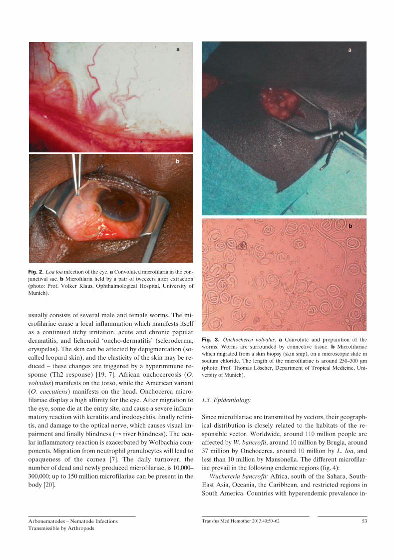
Loa loaAfter transmission of L. loa by horse fly sting, the first symp-toms occur after 5 months, but clinical latency can persist up to 13 years. In the case of colonization with low numbers of worms and in children, the infection is often asymptomatic. If the eyes are infected, a worm takes around 30–60 min, in some cases up to a day, to enter the eye (figs. 2 a and b). Inva-sion is accompanied by photophobia, itching, pain, and swell-ing. Overheating of the eye may induce migration of the worm. Finally lytic processes in the eye may lead to blindness. Calabar swelling, a short-term, painless, and partly itchy edema mostly of the forearm, is caused by a subcutaneous al-lergic reaction to L. loa, which occurs in particular in migrants in endemic regions. Calabar swelling is accompanied by eosi-nophilia [10].
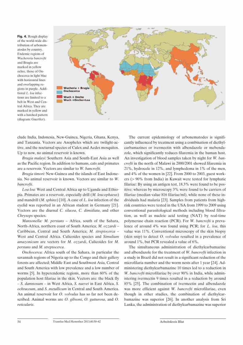
OnchocercaAfter transmission, pathogenic damage initially occurs pref-erentially in the skin. Thousands of microfilariae per day are released from the worm convolute (figs. 3 a and b) which
by the adult female worm (size around 40 0.03 cm). The male worm is smaller (around 3 0.03 cm) and survives in subcutaneous nodules which are easily palpable [5, 7]. The mi-crofilariae survive in humans for 12–18 months. When they die, they cause an itchy chronic inflammation due to the re-lease of Wolbachia. The microfilariae are 200 m in length; if the eye is affected, they can be detected directly or using a slit lamp. Wolbachia maintains and accelerates inflammatory processes by chemokine production, which leads to infiltra-tion with neutrophil granulocytes at the dying site of the mi-crofilariae. Furthermore, immune suppression and T-helper lymphocyte-dependent Th2 response is caused by endosym-biosis with Wolbachia. Onchocera from savannah regions, where river blindness (onchocercosis) is wide-spread and en-demic, are infested with Wolbachia to a considerably higher extent than in other regions so that the presence of the bacte-ria appears to play an essential role for the manifestation of blindness.
Dirofilaria repensDirofilaria repens is a parasite which infects dogs and cats. In the eyes of humans, the filariae undergo an incomplete devel-opment [5]. In most cases, they remain in the human skin. Dirofilaria also occurs in Southern Europe, clusters of high frequency have been described in some regions of Serbia [11]. Humans are accidental hosts. Therefore, dirofilaria will not be described in more detail.
Setaria labiatopapillosa, S digitataThese microfilariae occur in domestic animals in East Asia and Southern Europe. They are occasionally transmitted to humans where they cause subconjunctival inflammation. En-cephalomyelitis caused by Setaria has been described [12]. Se-taria has no significance for transfusion medicine.
VectorsIn rural regions, particularly Anopheles mosquitos transmit microfilariae. Species include Anopheles gambiae, Anopheles funestus, Anopheles punctulatus, and 40 other species. Species that occur in urban regions include Culex quinquefasciatus, Culex pipiens, and further species, as well as Aedes poly-nesiensis and Mansonia and Ochlerotatus [8, 13].
Among horse flies (Tabanidae), the species that transmits parasites belong to the genus Chrysops, in particular Chrys-ops silacea and Chrysops imidiata, which are diurnal and an-thropophilic, furthermore Chrysops distinctipennis, Chrysops zahrai, Chrysops centurionis and Chrysops longicornis, de-pending on the region [10].
The development of Simulium larvae is dependent on oxy-gen-rich water; therefore the black fly is found in the vicinity of small waterfalls and rapids. Such regions include the gallery forests in savannahs, in particular in West Africa and South America [14]. The black fly and Onchocerca are adapted to humans.
Transfus Med Hemother 2013;40:50–62Arbonematodes – Nematode Infections Transmissible by Arthropods
53
1.3. Epidemiology
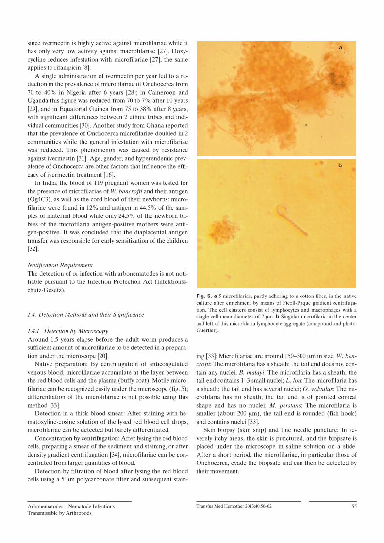
Since microfilariae are transmitted by vectors, their geograph-ical distribution is closely related to the habitats of the re-sponsible vector. Worldwide, around 110 million people are affected by W. bancrofti, around 10 million by Brugia, around 37 million by Onchocerca, around 10 million by L. loa, and less than 10 million by Mansonella. The different microfilar-iae prevail in the following endemic regions (fig. 4):
Wuchereria bancrofti: Africa, south of the Sahara, South-East Asia, Oceania, the Caribbean, and restricted regions in South America. Countries with hyperendemic prevalence in-
usually consists of several male and female worms. The mi-crofilariae cause a local inflammation which manifests itself as a continued itchy irritation, acute and chronic papular dermatitis, and lichenoid ‘oncho-dermatitis’ (scleroderma, erysipelas). The skin can be affected by depigmentation (so-called leopard skin), and the elasticity of the skin may be re-duced – these changes are triggered by a hyperimmune re-sponse (Th2 response) [19, 7]. African onchocercosis (O. volvulus) manifests on the torso, while the American variant (O. caecutiens) manifests on the head. Onchocerca micro-filariae display a high affinity for the eye. After migration to the eye, some die at the entry site, and cause a severe inflam-matory reaction with keratitis and irodocyclitis, finally retini-tis, and damage to the optical nerve, which causes visual im-pairment and finally blindness ( river blindness). The ocu-lar inflammatory reaction is exacerbated by Wolbachia com-ponents. Migration from neutrophil granulocytes will lead to opaqueness of the cornea [7]. The daily turnover, the number of dead and newly produced microfilariae, is 10,000–300,000; up to 150 million microfilariae can be present in the body [20].
Fig. 2. Loa loa infection of the eye. a Convoluted microfilaria in the con-junctival sac. b Microfilaria held by a pair of tweezers after extraction (photo: Prof. Volker Klaus, Ophthalmological Hospital, University of Munich).
Fig. 3. Onchocherca volvulus. a Convolute and preparation of the worms. Worms are surrounded by connective tissue. b Microfilariae which migrated from a skin biopsy (skin snip), on a microscopic slide in sodium chloride. The length of the microfilariae is around 250–300 m (photo: Prof. Thomas Löscher, Department of Tropical Medicine, Uni-versity of Munich).
54 Transfus Med Hemother 2013;40:50–62 Arbeitskreis Blut
The current epidemiology of arbonematodes is signifi-cantly influenced by treatment using a combination of diethyl-carbamazines or ivermectin with albendazole or mebenda-zole, which significantly reduces filaremia in the human host. An investigation of blood samples taken by night for W. ban-crofti in the north of Malawi in 2000/2001 showed filaremia in 21%, hydrocele in 12%, and lymphedema in 1% of the men, and 4% of the women in [22]. From 2000 to 2003, guest work-ers (> 90% from India) in Kuwait were tested for lymphatic filariae: By using an antigen test, 18.3% were found to be pos-itive; whereas by microscopy 3% were found to be carriers of filariae (median value 816 filariae/ml), while none of these in-dividuals had malaria [23]. Samples from patients from high-risk countries were tested in the USA from 1999 to 2009 using conventional parasitological methods including blood filtra-tion, as well as nucleic acid testing (NAT) by real-time polymerase chain reaction (PCR). For W. bancrofti a preva-lence of around 4% was found using PCR; for L. loa, this value was 11%. Conventional microscopy of the skin biopsy (skin snip) to detect O. volvulus resulted in a prevalence of around 1%, but PCR revealed a value of 6%.
The simultaneous administration of diethylcarbamazine and albendazole for the treatment of W. bancrofti infection in a study in Brazil did not result in a significant reduction of the microfilaria number and the worm nests after 1 year [24]. Ad-ministering diethylcarbamazine 10 times led to a reduction in W. bancrofti microfilariae by over 90% in India, while admin-istering ivermectin 9 times resulted in a reduction by around 85% [25]. The combination of ivermectin and albendazole was more efficient against W. bancrofti microfilariae, even though in other studies, the combination of diethylcar-bamazine was superior [26]. In another analysis from Sri Lanka, the administration of diethylcarbamazine was superior
clude India, Indonesia, New-Guinea, Nigeria, Ghana, Kenya, and Tanzania. Vectors are Anopheles which are twilight-ac-tive, and the nocturnal species of Culex and Aedes mosquitos. Up to now, no animal reservoir is known.
Brugia malayi: Southern Asia and South-East Asia as well as the Pacific region. In addition to humans, cats and primates are a reservoir. Vectors are similar to W. bancrofti.
Brugia timori: New-Guinea and the islands of East Indone-sia. No animal reservoir is known. Vectors are similar to W. bancrofti.
Loa loa: West and Central Africa up to Uganda and Ethio-pia. Primates are a reservoir, especially drill (M. leucophaeus) and mandrill (M. sphinx) [10]. A case of L. loa infection of the eyelid was reported in an African student in Germany [21]. Vectors are the diurnal C. silacea, C. dimidiata, and other Chrysops species.
Mansonella: M. perstans – Africa, south of the Sahara, North-Africa, northern coast of South America; M. ozzardi – Caribbean, Central and South America; M. streptocerca – West and Central Africa. Culicoides species and Simulium amazonicum are vectors for M. ozzardi, Culicoides for M. perstans and M. streptocerca.
Onchocerca: Africa south of the Sahara, in particular the savannah regions of Nigeria up to the Congo and their gallery forests are affected; Middle East and Southwest Asia, Central and South America with low prevalence and a low number of worms [5]. In hyperendemic regions, more than 60% of the population host filariae in the skin. Vectors are: the black fly – S. damnosum – in West Africa, S. naevei in East Africa, S. ochraceum, and S. metallicum in Central and South America. An animal reservoir for O. volvulus has so far not been de-scribed. Animal worms are O. gibsoni, O. gutturosa, and O. reticularis.
Fig. 4. Rough display of the world-wide dis-tribution of arbonem-atodes by country. Endemic regions of Wuchereria bancrofti and Brugia are marked in yellow ochre, those of On-chocerca in light blue with horizontal lines and overlapping re-gions in purple. Addi-tional L. loa infec-tions are limited to a belt in West and Cen-tral Africa. They are marked in yellow and with a hatched pattern (diagram: Guertler).
Transfus Med Hemother 2013;40:50–62Arbonematodes – Nematode Infections Transmissible by Arthropods
55
ing [33]: Microfilariae are around 150–300 m in size. W. ban-crofti: The microfilaria has a sheath; the tail end does not con-tain any nuclei; B. malayi: The microfilaria has a sheath; the tail end contains 1–3 small nuclei; L. loa: The microfilaria has a sheath; the tail end has several nuclei; O. volvulus: The mi-crofilaria has no sheath; the tail end is of pointed conical shape and has no nuclei; M. perstans: The microfilaria is smaller (about 200 m), the tail end is rounded (fish hook) and contains nuclei [33].
Skin biopsy (skin snip) and fine needle puncture: In se-verely itchy areas, the skin is punctured, and the biopsate is placed under the microscope in saline solution on a slide. After a short period, the microfilariae, in particular those of Onchocerca, evade the biopsate and can then be detected by their movement.
since ivermectin is highly active against microfilariae while it has only very low activity against macrofilariae [27]. Doxy-cycline reduces infestation with microfilariae [27]; the same applies to rifampicin [8].
A single administration of ivermectin per year led to a re-duction in the prevalence of microfilariae of Onchocerca from 70 to 40% in Nigeria after 6 years [28]; in Cameroon and Uganda this figure was reduced from 70 to 7% after 10 years [29], and in Equatorial Guinea from 75 to 38% after 8 years, with significant differences between 2 ethnic tribes and indi-vidual communities [30]. Another study from Ghana reported that the prevalence of Onchocerca microfilariae doubled in 2 communities while the general infestation with microfilariae was reduced. This phenomenon was caused by resistance against ivermectin [31]. Age, gender, and hyperendemic prev-alence of Onchocerca are other factors that influence the effi-cacy of ivermectin treatment [16].
In India, the blood of 119 pregnant women was tested for the presence of microfilariae of W. bancrofti and their antigen (Og4C3), as well as the cord blood of their newborns: micro-filariae were found in 12% and antigen in 44.5% of the sam-ples of maternal blood while only 24.5% of the newborn ba-bies of the microfilaria antigen-positive mothers were anti-gen-positive. It was concluded that the diaplacental antigen transfer was responsible for early sensitization of the children [32].
Notification RequirementThe detection of or infection with arbonematodes is not noti-fiable pursuant to the Infection Protection Act (Infektionss-chutz-Gesetz).
1.4. Detection Methods and their Significance
1.4.1 Detection by MicroscopyAround 1.5 years elapse before the adult worm produces a sufficient amount of microfilariae to be detected in a prepara-tion under the microscope [20].
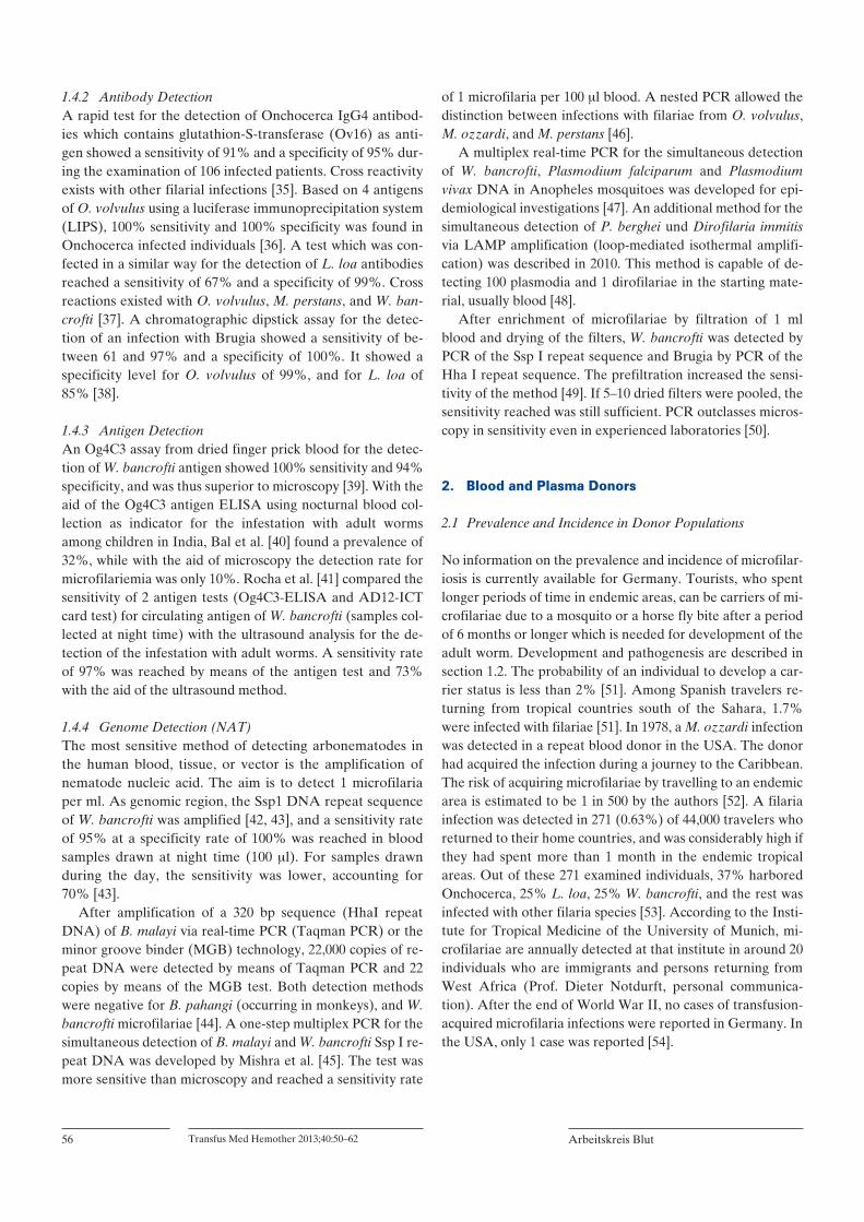
Native preparation: By centrifugation of anticoagulated venous blood, microfilariae accumulate at the layer between the red blood cells and the plasma (buffy coat). Motile micro-filariae can be recognized easily under the microscope (fig. 5); differentiation of the microfilariae is not possible using this method [33].
Detection in a thick blood smear: After staining with he-matoxyline-eosine solution of the lysed red blood cell drops, microfilariae can be detected but barely differentiated.
Concentration by centrifugation: After lysing the red blood cells, preparing a smear of the sediment and staining, or after density gradient centrifugation [34], microfilariae can be con-centrated from larger quantities of blood.
Detection by filtration of blood after lysing the red blood cells using a 5 m polycarbonate filter and subsequent stain-
Fig. 5. a 5 microfilariae, partly adhering to a cotton fiber, in the native culture after enrichment by means of Ficoll-Paque gradient centrifuga-tion. The cell clusters consist of lymphocytes and macrophages with a single cell mean diameter of 7 m. b Singular microfilaria in the center and left of this microfilaria lymphocyte aggregate (compound and photo: Guertler).
56 Transfus Med Hemother 2013;40:50–62 Arbeitskreis Blut
of 1 microfilaria per 100 l blood. A nested PCR allowed the distinction between infections with filariae from O. volvulus, M. ozzardi, and M. perstans [46].
A multiplex real-time PCR for the simultaneous detection of W. bancrofti, Plasmodium falciparum and Plasmodium vivax DNA in Anopheles mosquitoes was developed for epi-demiological investigations [47]. An additional method for the simultaneous detection of P. berghei und Dirofilaria immitis via LAMP amplification (loop-mediated isothermal amplifi-cation) was described in 2010. This method is capable of de-tecting 100 plasmodia and 1 dirofilariae in the starting mate-rial, usually blood [48].
After enrichment of microfilariae by filtration of 1 ml blood and drying of the filters, W. bancrofti was detected by PCR of the Ssp I repeat sequence and Brugia by PCR of the Hha I repeat sequence. The prefiltration increased the sensi-tivity of the method [49]. If 5–10 dried filters were pooled, the sensitivity reached was still sufficient. PCR outclasses micros-copy in sensitivity even in experienced laboratories [50].
2. Blood and Plasma Donors
2.1 Prevalence and Incidence in Donor Populations
No information on the prevalence and incidence of microfilar-iosis is currently available for Germany. Tourists, who spent longer periods of time in endemic areas, can be carriers of mi-crofilariae due to a mosquito or a horse fly bite after a period of 6 months or longer which is needed for development of the adult worm. Development and pathogenesis are described in section 1.2. The probability of an individual to develop a car-rier status is less than 2% [51]. Among Spanish travelers re-turning from tropical countries south of the Sahara, 1.7% were infected with filariae [51]. In 1978, a M. ozzardi infection was detected in a repeat blood donor in the USA. The donor had acquired the infection during a journey to the Caribbean. The risk of acquiring microfilariae by travelling to an endemic area is estimated to be 1 in 500 by the authors [52]. A filaria infection was detected in 271 (0.63%) of 44,000 travelers who returned to their home countries, and was considerably high if they had spent more than 1 month in the endemic tropical areas. Out of these 271 examined individuals, 37% harbored Onchocerca, 25% L. loa, 25% W. bancrofti, and the rest was infected with other filaria species [53]. According to the Insti-tute for Tropical Medicine of the University of Munich, mi-crofilariae are annually detected at that institute in around 20 individuals who are immigrants and persons returning from West Africa (Prof. Dieter Notdurft, personal communica-tion). After the end of World War II, no cases of transfusion-acquired microfilaria infections were reported in Germany. In the USA, only 1 case was reported [54].
1.4.2 Antibody DetectionA rapid test for the detection of Onchocerca IgG4 antibod-ies which contains glutathion-S-transferase (Ov16) as anti-gen showed a sensitivity of 91% and a specificity of 95% dur-ing the examination of 106 infected patients. Cross reactivity exists with other filarial infections [35]. Based on 4 antigens of O. volvulus using a luciferase immunoprecipitation system (LIPS), 100% sensitivity and 100% specificity was found in Onchocerca infected individuals [36]. A test which was con-fected in a similar way for the detection of L. loa antibodies reached a sensitivity of 67% and a specificity of 99%. Cross reactions existed with O. volvulus, M. perstans, and W. ban-crofti [37]. A chromatographic dipstick assay for the detec-tion of an infection with Brugia showed a sensitivity of be-tween 61 and 97% and a specificity of 100%. It showed a specificity level for O. volvulus of 99%, and for L. loa of 85% [38].
1.4.3 Antigen DetectionAn Og4C3 assay from dried finger prick blood for the detec-tion of W. bancrofti antigen showed 100% sensitivity and 94% specificity, and was thus superior to microscopy [39]. With the aid of the Og4C3 antigen ELISA using nocturnal blood col-lection as indicator for the infestation with adult worms among children in India, Bal et al. [40] found a prevalence of 32%, while with the aid of microscopy the detection rate for microfilariemia was only 10%. Rocha et al. [41] compared the sensitivity of 2 antigen tests (Og4C3-ELISA and AD12-ICT card test) for circulating antigen of W. bancrofti (samples col-lected at night time) with the ultrasound analysis for the de-tection of the infestation with adult worms. A sensitivity rate of 97% was reached by means of the antigen test and 73% with the aid of the ultrasound method.
1.4.4 Genome Detection (NAT)The most sensitive method of detecting arbonematodes in the human blood, tissue, or vector is the amplification of nematode nucleic acid. The aim is to detect 1 microfilaria per ml. As genomic region, the Ssp1 DNA repeat sequence of W. bancrofti was amplified [42, 43], and a sensitivity rate of 95% at a specificity rate of 100% was reached in blood samples drawn at night time (100 l). For samples drawn during the day, the sensitivity was lower, accounting for 70% [43].
After amplification of a 320 bp sequence (HhaI repeat DNA) of B. malayi via real-time PCR (Taqman PCR) or the minor groove binder (MGB) technology, 22,000 copies of re-peat DNA were detected by means of Taqman PCR and 22 copies by means of the MGB test. Both detection methods were negative for B. pahangi (occurring in monkeys), and W. bancrofti microfilariae [44]. A one-step multiplex PCR for the simultaneous detection of B. malayi and W. bancrofti Ssp I re-peat DNA was developed by Mishra et al. [45]. The test was more sensitive than microscopy and reached a sensitivity rate
Transfus Med Hemother 2013;40:50–62Arbonematodes – Nematode Infections Transmissible by Arthropods
57
transmitted, are removed from the blood stream by the re-sponse of natural immunity (innate immunity) [54].
3.3 Severity and Course of the Disease
Intravenous (i.v.) transmission of L1 microfilariae to hu-mans (self-test by Gönnert and artificial infection of pa-tients) lead 16–24 h after transmission to fatigue, faintness, dizziness, headache, and rise in temperature to up to > 39 °C for a few days; the febrile reaction was accompanied by eosinophilia [55, 56]. While the L1 microfilariae of W. ban-crofti were removed from the blood within only 4–5 days [56], the L1 microfilariae of M. perstans remained in the re-cipient for up to 37 months without causing any symptoms [55]. In another study, W. bancrofti microfilariae were no longer detectable in humans after 12–18 days after i.v. injec-tion. After i.v. injection into mice, they were eliminated from the blood stream after only 40 h; after intraperitoneal and subcutaneous injection into mice or rats, they survived for 14 days [57].
Years may pass after transmission of L3 larvae of filariae by the vector to humans before typical symptoms become evi-dent (cf. 1.2 Pathogenesis). As one of the first symptoms al-lergic reactions occur which manifest themselves particularly as itching and partly severe pain if the eyes are affected. Worm clusters develop in the skin, which are visible and palpable; after a number of years, an inflammation occurs with dilation of the lymphatic vessels together with subsequent elephantia-sis. When the adult worms die, this can cause strong inflam-matory reactions due to the release of Wolbachia (see above 1.2). The disease is curable after several treatment cycles with doxycycline and diethylcarbamazine or ivermectin and alben-dozol or mebendazol (cf. 3.4).
3.4 Therapy and Prophylaxis
3.4.1 ChemotherapyDoxycycline, diethylcarbamazine, ivermectin, and albenda-zole are available against the microfilariae and the worms, but ivermectin does not show any efficacy against macrofilariae, and albendazole is effective only against the adult worm. The vital symbiosis of Wuchereria, Brugia, and Onchocerca with the bacterium Wolbachia is restricted or inhibited by the use of tetracycline (doxycycline), rifampicine, or azithromycine [58, 10, 8]. Doxycycline significantly reduces the release of VEGF and the dilatation of the lymphatic blood vessels [15] (cf. 1.2 Pathogenesis). The aim of the treatment is to prevent the clinical symptoms of lymphedema, hydrocele and el-ephantiasis, and, from an epidemiological point of view, to re-duce the transmission of the filariae by reducing the produc-tion of microfilariae.
2.2 Definition of Exclusion Criteria
The deferral period of 6 months for individuals returning from tropical regions because of malaria infections is not a sufficient exclusion criterion for a possible transmission of microfilariae, since filariemia may develop with even longer delay (cf. 1.2). Typical symptoms of infestation in-clude itching and intermittent visual impairments. Donors with the respective travelling history and symptoms as described above should be deferred from donating blood until a laboratory analysis has ruled out an infestation with microfilariae.
2.3 Donor Testing and Significance
Donor testing for microfilariae by a sensitive PCR is possible, however, is not justified because of the low incidence.
2.4 Donor Interviews
A specific donor interview concerning the history of travelling takes place, but an interview on the risk of infection with filar-iae does not take place and is not required based on the low prevalence and incidence.
2.5 Donor Information and Counseling
Donor information and counseling is available from the Insti-tutes of Tropical Medicine and other centers for infectious diseases and specialized outpatient wards for tropical medicine.
3. Recipient
3.1 Prevalence and Incidence of Blood-Associated Infections and Infectious Diseases in Recipient Populations
No publications are available on the prevalence and incidence for Germany. As has been mentioned above about individuals returning from endemic regions (cf. 2.1), recipients can also be infected with filariae after staying in endemic regions be-fore a blood transfusion.
3.2 Immune Status (Resistance, Existing Immunity, Immune Response, Age, Exogenous Factors)
Microfilariae can be acquired through a sting of the infected vector at any age. Low quantities of L1 microfilariae, which may be present in a blood component and which may then be
58 Transfus Med Hemother 2013;40:50–62 Arbeitskreis Blut
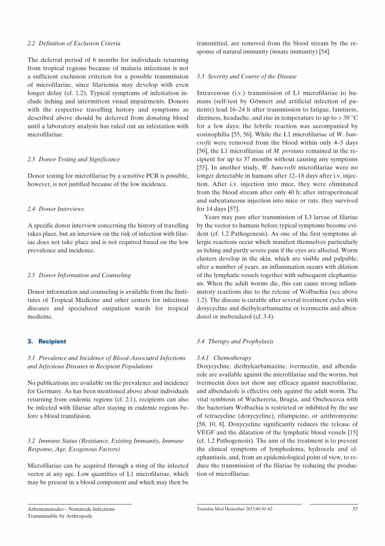
against microfilariae, could be introduced as new medicinal product for the treatment of macrofilariae [61]. Figure 6 shows a dead microfilaria in the skin – a so-called larva mi-grans – in a Cameroonian woman.
3.4.4 Prophylaxis of Arthropod Stings
Treatment of the Breeding Grounds of the VectorsDDT (dichlorodiphenyltrichloroethane) is no longer recom-mended because of the neurological adverse effects in hu-mans and the resistance of some mosquito species. In areas near human settlements it is recommended to cover free water surfaces, including open latrines, with polystyrene beads in order to suffocate the developing mosquito larvae. This measure helped to reduce the stinging rate in a commu-nity from around 25,000 per year to around 500 per year [62]. Another very effective measure is the removal of potential breeding grounds of mosquitoes by draining.
Mosquito Bite Prophylaxis Avoiding mosquito bites is the most effective prophylaxis in many regions in which arboviruses, plasmodiae, and micro-filariae are transmitted simultaneously by mosquitoes [13]. Anopheles stings can be prevented in twilight which, in Af-rica, lasts from 4:00 to 6:30 am and from 4:00 to 6:30 pm. The bites of Culicoides with diurnal and nocturnal activity are hard to prevent, while Culex and Aedes are active during the night.
Prevention of Horse Fly BitesHorse flies sting in the daytime. They are found in shady areas, waiting for an opportunity to sting. Animals, including monkeys, attract horse flies. Such areas should therefore be avoided. Clothes that are light in color and have stripes re-duce the probability to attract horse flies. A horse fly sting is very painful and is therefore noticed.
3.4.2 Wuchereria bancrofti, Brugia malayi, Brugia timori (lymphatic filariae), and Onchocerca volvulus, Mansonella perstansTreatment starts with doxcyline (200 mg/day) for 4–6 weeks, then a single dose of diehthycarbazamine (6 mg/kg) plus albendozole (400 mg) is administered, and if there is a simul-taneous presence of Onchocercerca and Wuchereria which is true of various regions, ivermectin plus albendozole (400 mg) is used [8, 59]. The treatment regimen is repeated annually for 5 years, and the success of the treatment is monitored by de-termining the blood and tissue burden of filariae. A reinfec-tion should be prevented through the control and reduction of the spreading of the vector within the first year. For children over 10 years and pregnant women, only treatment with doxy-cycline, if any, is recommended at present (caution: damage to tooth and bone development by doxycycline).
The efficacy of the doxycycline treatment against M. per-stans was very high in Mali with a decrease in the microfilaria count to about 23% in the blood after 6 months and to 0% after 12 months [59], whereas a decrease to 62% after 6 months and to 50% after 12 months was measured in the un-treated control group. The 12-month efficacy of the doxycy-cline treatment was 97% (67 of 69 treated individuals), com-pared with the untreated group with 16% (10 of 63) and with the ivermectin/albendazole group with 19% (5 of 27) [59].
Adverse effects include fever, headache, itching, myalgia, which is high in particular in regions such as Cameroon where L. loa is endemic simultaneously. Serious adverse effects with progressive loss of neurological functions and encephalopathy occurred in 1 of 800,000 treated patients [8]. Preliminary treatment with doxycycline prevents the occurrence of severe inflammatory reactions such as acute dermatolymphangioad-enitis. Vomiting is a frequent adverse reaction to doxycyline treatment [59]. Adverse effects of diethylcarbazamine treat-ment were more severe the higher the number of microfilar-iae in the tissue and the more interleukin-6 and lipopolysac-charide-binding protein was released [60].
3.4.3 Loa loaDiethylcarbamazine is effective only against the adult worm which is killed. Death of the worm is partially associated with a severe Jarisch-Herxheimer reaction which may be accompa-nied by serious adverse reactions such as encephalopathy which is partly fatal and is caused by microembolisms in the capillaries of the brain [10]. The recommended treatment today is a single administration of ivermectin, 50–200 g/kg plus albendazole (400 mg), which can be repeated after sev-eral months. If the combination of ivermectine and albanda-zole is used, serious adverse reactions are rare. In patients with a microfilaria burden of < 8,000/ml, the occurrence is very low. Allergic adverse reactions can be suppressed by the administration of cortisone. A simultaneous administration of tetracycline is not required since no Wolbachia are present in L. loa [7, 10]. Moxidectine, which shows strong efficacy
Fig. 6. Subcutaneously remaining dead L. loa microfilaria – picture of larva migrans. The only clinical symptoms which persist are the indura-tion and hyperpigmentation (photo: Dr. R. Krippner, German Embassy, Yaounde, Cameroon, 2006).
Transfus Med Hemother 2013;40:50–62Arbonematodes – Nematode Infections Transmissible by Arthropods
59
diethylcarbamazine or ivermectin and albendazole or meben-dazole in endemic regions is a more effective method of pro-tection than vaccination [31].
3.5 Transmissibility
3.5.1 Blood TransfusionIn acid-citrate-dextrose (ACD) blood, L1 microfilariae of W. bancrofti survived for 12 days in a state of good mobility at 4–6 °C. After 21 days they were immobile but could be reacti-vated by increasing the temperature. After 25 days, they were dead [70]. In citrate blood, microfilariae of W. bancrofti stored at 4 °C survived for at least 39 days, those of B. malayi sur-vived 47 days, and those of L. loa 15 days [71]. Around a third of the microfilariae of L. loa survived for 21 days in ACD blood [54]. Consequently, microfilariae present in a blood donation are theoretically able to survive up to the time of transfusion.
Transmission of filariosis by transfusion still does not occur when L1 microfilariae are transfused [70], since the further maturation into L3 microfilariae is processed only in the vec-tor but not in humans. M. perstans filariae, which were trans-mitted by transfusion, were rapidly eliminated from the re-cipient’s blood, and no symptoms or eosinophilia occurred after 4 months [72, 73]. On the other hand, Gönnert [55] and also Nutman [74] described that M. perstans microfilariae were detectable in the recipient up to 3 years after transfu-sion. In an analysis of the transmission of microfilariae by blood transfusion in India, the recipients of 11,572 transfu-sions were followed up for 15 months. 5 of the donors were carriers of filariae, 4 of the recipients were infected, and in 2 of the recipients and the donors, filariae were detectable dur-ing the entire observation period. The conclusion was that even if transmission of the microfilariae does not lead to de-velopment of the adult worm, blood donors with known filaria infections should be excluded from donating blood [75]. In the USA, the transfusion of blood from a regular donor trans-mitted M. ozzardi after the donor had spent a holiday in the Caribbean [52], as discussed in 1.3 and 2.1.
As for plasma, transmission of microfilariae by fresh frozen plasma (FFP) can be ruled out since microfilariae do not sur-vive the freeze-thaw cycle. In the case of stab injury and oc-cupational infection, so far no transmission of microfilaria by injury during surgical intervention or needle stick has been reported.
3.6 Frequency of Administration of the Blood Products and Amount Administered
FFP is not infectious in the case of filariae since the parasites are killed during the freeze-thaw cycle (cf. 3.5). As for red blood cell concentrates, no statements can be made on the
Prevention of the Bite of the Black Fly The habitat of the black fly is in gallery forests of the savan-nah and the surroundings of rapidly flowing streams/rivers in the rain forest. The black fly occurs in swarms, is found in the shade of the trees, and is aggressive in the daytime. The lar-vae develop at sites of flowing water with a high concentra-tion of oxygen, i.e. which normally show an open flow of water. Humans and animals are frequent hosts so that a human cycle can develop which, however, can be broken by treating the population affected by administration of iver-mectin and albendazole. A black fly sting is extremely pain-ful. Black fly larvae can partly be killed by means of the toxin of Bacillus sphaericus or Bacillus thuringiensis. However, Ba-cillus toxin-resistant mosquito larvae have developed in West Africa [63].
VaccinationThe vaccination of jirds (rodents similar to gerbils) with a re-combinant protein from maltose binding protein (BM5-MBP) and paramyosin of L3 larvae from B. malayi led to immunity after 2 administrations so that after infection with 100 L3 lar-vae, the number of developing adult worms was reduced, and the latter were smaller [64]. The immunization of BALB/c mice and jirds with maltose binding protein linked to tropo-myosine from O. volvulus resulted after infection with micro-filariae in a reduction of the growth of the adult worms of around 45% [65]. In its amino acid composition, tropomyo-sine from O. volvulus showed 91% homology with that of other nematodes; therefore, this antigen was used for the im-munization of mice. The immunity against microfilariae de-pended on the formation of IgE and could be transferred to other mice [66].
A cellular immune response against L. loa was induced in mandrills which were immunized with 50 irradiated L3 larvae. The vaccinated animals showed a Th1 response against L3 whereas non-vaccinated animals showed a Th2 response. After infection with 100 live L3 microfilariae, some animals did not show microfilaremia, which was rated as a protective effect [67]. The vaccination of mandrills with 150 irradiated L3 larvae and artificial infection 60 days later initially did not result in any difference between vaccinated and non-vacci-nated mandrills. It was only after 245 days that the micro-filaremia was considerably less severe in the vaccinated man-drills [68]. Rats and gerbils were immunized 3 times with re-combinant B. malayi myosine (molecular weight 73 kDa). After artificial infection with 100 larvae 1 week after the last vaccination, the vaccinated animals showed a reduction in the amount of microfilariae in the blood by 76% and a reduction of worm infestation by around 50% compared to the control animals [69].
These studies showed that a certain degree of immunity can be achieved. However, they indicate that a human vaccine will not be available in the near future. Prevention of micro-filaria transmission by prophylactic annual administration of
60 Transfus Med Hemother 2013;40:50–62 Arbeitskreis Blut
Treatment with 5 mg/ml diethylcarbamazine in vitro caused disintegration of microfilariae of W. bacrofti [85], and treat-ment with 2–10 mol/l mefloquine (Lariam®, Hoffmann–La Roche, Basel, Switzerland) used for malaria prophylaxis killed microfilariae of B. malayi after 10 h – 3 days [86].
4.3 Practicability and Validation of Procedures for Removal/Inactivation of Infectious Agents
4.3.1 Susceptibility in Animal ExperimentAnimals that can be experimentally infected with L. loa include the drill monkey (M. leukophaeus) and the mandrill (M. sphinx). In Gabon, 7 mandrills were infected with differ-ent amounts of L3 larvae of L. loa. In 1 of the animals in-fected with 1,000 L3 larvae, no filariae were found, while in another 4 animals filariemia was detected after approximately 150 days. In the remaining 2 animals whose mothers were in-fected with microfilariae, the filariae were only detected after 200 days. The filariae burden varied, with up to 10,000 micro-filariae/ml; however, after 300 days this amount decreased to 10–100 microfilariae/ml in all 6 animals. The monkeys showed no clinical symptoms, no leukocytosis, and no eosinophilia [87]. In conclusion, a sufficiently validated animal model is not available. Antigen tests and PCR (cf. 1.4 Detection meth-ods) are suitable for detecting a reduction in the pathogen burden. However, they are not suitable for ruling out residual infectivity.
5. Assessment
Arbonematodes are widespread in tropical regions with rain forests or gallery forests. They can be present in high num-bers in infected individuals. Filariae are very rarely found in Germany, and up to now, no reports exist stating that these parasites were transmitted by blood transfusions in Germany. The larvae of the filariae present in a blood donation, if any, are L1 larvae which, depending on the amount and type, die in the new human host within a few weeks up to several months, and are unable to mature into worms. From this point of view, the transmission of microfilariae by blood transfusion would be a self-limiting event and thus does not require any further action for the protection of the recipient. Whether the prevalence of microfilariae in the mosquito and horse fly vectors will increase in the course of global warm-ing, which would make transmission to humans in Germany more likely, should be monitored. Asian primates and cats are known as non-human hosts for Brugia, and African pri-mates for L. loa. From an epidemiological point of view, these animals are not important for a transmission cycle in Germany. Routine testing for microfilariae or an extension of the deferral for blood donors after travelling in endemic re-gions is not required.
frequency and amount since figures are not available for Ger-many. Since L1 larvae can survive in humans for 6 weeks and up to 37 months, the deferral of blood donors for 6 months due to the risk of acquiring malaria after a stay in a tropical region is not sufficient to exclude filaremia, e.g. by Man-sonella [76]. In the case of platelet concentrates, transmission of microfilariae has so far not been reported. L1 larvae are likely to survive the manufacturing process but do not lead to development of the worm since further maturation to the L3 larvae is hampered.
4. Blood Products
4.1 Burden in the Starting Material and Testing Methods
In 1987, in endemic areas such as Ibadan, Nigeria, 3.5% of the blood donors tested had L. loa microfilaremia while 7.8% were infected with P. falciparum [77]. None of 364 blood do-nors in Maiguguri, Nigeria, had a filaria infection [78]. Out of 1,259 blood donors in Ile-Ife, Nigeria, 2 (0.16%) were carriers of microfilariae [79]. In the blood bank at a maximum care hospital in Bangkok, Thailand, no filarial infections were de-tected in blood donors from 1999 to 2009 [80]. The prevalence in other endemic areas is described in Section 1.3 ‘Epidemiol-ogy’. The microfilaria burden in the starting material there-fore varies considerably in endemic areas but should be gen-erally considered as very low if at all present. If needed, mi-crofilariemia could be detected in blood donations by NAT (cf. 1.4 Genome detection).
4.2 Possibility of Removal and Inactivation of the Infectious Agent
Microfilariae normally circulate in the blood. On day 1 of a blood collection, 80% of L. loa microfilariae were retained by filtration via a 40 m blood filter, while on day 21 this rate was 92% [54].
Microfilariae in the L1 or L3 stadium could be kept alive in medium containing reduced glutathion, human cells and human serum (specific batches are required) [81], and could be used for spike assays. Herbal extracts impairing the viabil-ity of microfilariae have been investigated using this method [82]. Spike assays could therefore be performed with a high workload to test removal or inactivation.
The inactivation of microfilariae under the aspect of plasma or plasma derivatives has so far not been described. For non-cellular blood products, a freeze-thaw procedure is sufficient to kill the microfilariae. Only around 6% of the mi-crofilariae are killed during cryopreservation in the presence of 6% dimethyl sulfoxide (DMSO) and 15% serum, as used for the storage of stem cells [83]. Irradiation with gamma rays up to 10.5 Gy does not kill microfilariae of W. bancrofti [84].
Transfus Med Hemother 2013;40:50–62Arbonematodes – Nematode Infections Transmissible by Arthropods
61
Dr. Margarethe HeidenProf. Dr. Martin HildebrandtProf. Dr. Dr. Bernd JansenDr. Thomas Montag-LessingDr. Ruth OffergeldProf. Dr. Georg PauliProf. Dr. Rainer SeitzDr. Uwe SchlenkrichDr. Volkmar SchottstedtDr. Johanna StrobelDr. Hannelore Willkommen
This paper was completed on November 22, 2011, and approved by the German Advisory Committee Blood (Arbeitskreis Blut) on March 30, 2012. It was compiled by the members of the subgroup ‘Assessment of Pathogens Transmissible by Blood’ of the German Advisory Committee Blood (Arbeitskreis Blut):
Prof. Dr. Lutz GürtlerDr. Ursula BauerfeindDr. Johannes BlümelProf. Dr. Reinhard BurgerProf. Dr. Christian DrostenDr. Albrecht Gröner
References
1 Blümel J, Burger R, Gerlich W, Gürtler L, Heiden M, Hitzler W, Jansen B, Klamm H, Lefèvre H, Ludwig WD, Montag-Lessing T, Offergeld R, Paessens A, Pauli G, Seitz R, Schlenkrich U, Schottstedt V, Willkommen H, Baljer G: Coxiella burnetii – pathogen of the Q (Query) fever. Trans-fus Med Hemother 2005;32:218–226.
2 Blümel J, Burger R, Drosten C, Gröner A, Gürtler L, Heiden M, Hitzler W, Jansen B, Klamm H, Ludwig WD, Montag-Lessing T, Offergeld R, Pauli G, Seitz R, Schlenkrich U, Schottstedt V, Willkom-men H: Arbobakterien (über Arthropoden über-tragbare Bakterien). Bundesgesundheisbl Ges-forsch Gesschutz 2007;50:1192–1207.
3 Gürtler L, Blümel J, Burger R, Drosten C, Gröner A, Heiden M, Hildebrandt M, Jansen B, Montag-Lessing T, Offergeld R, Pauli G, Seitz R, Schlenk-rich U, Schottstedt V, Strobel J, Willkommen H, Wirsing von König CH: Arboprotozoen. Bundes-gesbl Gesundforsch Gesundschutz 2009;52:123–146.
4 Maguire JH: Introduction to helminth infections; in Mandell D (ed): Benett’s Principles and Practice of Infectious Diseases, 7th ed. Philadelphia, PA, Churchill Livingstone, 2010, pp. 3573–3575.
5 Kazura JW: Tissue nematodes, including Trichinel-losis, Dracunculiasis and the Filariases; in Mandell D (ed): Bennett’s Principles and Practice of Infec-tious Diseases. 7th ed. Philadelphia, PA, Churchill Livingstone, 2010, pp. 3587–3594.
6 Peeters W, Gilles HM: Colour Atlas of Tropical Medicine and Parasitology, 3rd ed. London, Wolfe Med Pub, 1989, pp. 65–82.
7 Taylor M, Hoerauf A, Backarie M: Lymphatic filaria-sis and onchocerciasis. Lancet 2010;376:1175–1185.
8 Bockarie MJ, Taylor MJ, Gyapong JO: Current practices in the management of lymphatic filariasis. Expert Rev Anti Infect Ther 2009;7:595–605.
9 Arumugam S, Pfarr KM, Hoerauf A: Infection of the intermediate mite host with Wolbachia-de-pleted Litomosoides sigmodontis microfilariae: im-paired L1 to L3 development and subsequent sex-ratio distortion in adult worms. Int J Parasitol 2008; 38:981–987.
10 Boussinesq M: Loiasis. Ann Trop Med Parasitol 2006;100:715–731.
11 Dzamic AM, Colovic IV, Arsic-Arsenijevic VS, Stepanovic S, Boricic I, Dzamic Z, Mitrovic SM, Rasic DM, Stefanovic I, Latkovic Z, Krajcic-Zec IF: Human Dirofilaria repens infection in Serbia. J Helminthol 2009;83:129–137.
12 Panaitescu D, Freda A, Bain O, Vasile-Bugarin AC: Four cases of human filariosis due to Setaria labiatopapillosa found in Bucharest, Romania. Roum Arch Microbiol Immunol 1999;58:203–207.
13 Manguin S, Bangs MJ, Pothikasikorn J, Chareon-viriyaphap T: Review on global co-transmission of human Plasmodium species and Wuchereria ban-crofti by Anopheles mosquitoes. Infect Genetic Evolution 2010;10:159–177.
14 Grove DI: Tissue nematodes including trichinosis, dracunculiasis, and the filariasis; in Mandell D (ed): Bennett’s Principles and Practice of Infec-tious Diseases, 6th ed. Philadelphia, PA, Elsevier Churchill Livingstone, 2005, pp. 3267–3276.
15 Debrah AY, Mand S, Toliat MR, Marfo-Debrekyei Y, Batsa L, Nürnberg P, Lawson B, Adjei O, Hoerauf A, Pfarr K: Plasma vascular endothelial growth factor A (VEGF-A) and VEGF-A polymorphism are asso-ciated with hydrocele development in lymphatic filariasis. Am J Trop Med Hyg 2007;77:601–608.
16 Pion SD, Grout L, Kamgno J, Nana-Djeunga H, Boussinesq M: Individual host factors associated with Onchocerca volvulus microfilarial densities 15, 80 and 180 days after a first dose of ivermectin. Acta Trop 2011;120(suppl 1):S91–99.
17 Babu S, Bhat SQ, Kumar NP, Jayantasri S, Ruk-mani S, Kumaran P, Gopi PG, Kolappan C, Ku-maraswani V, Nutman TB: Human type 1 and 17 response in latent tuberculosis are modulated by coincident filarial infection through cytotoxic T-lymphocyte antigen-4 and programmed death-1. J Infect Dis 2009;200:288–298.
18 Krushna NSA, Shiny C, Verma P, Nithya D, Basker P, Elango S, Babu S, Narayanan RB: Wuchereria bancrofti: diminished platelet activation in filarial patients. Exp Parasitol 2010;125:114–123.
19 Brattig NW: Pathogenesis and host response in human onchocercosis: impact of Onchocerca filar-iae and Wolbachia endobacteria. Microbes Infect 2004;6:113–128.
20 Udall DN: Recent updates on onchocerciasis: diag-nosis and treatment. Clin Infect Dis 2007;44:53–60.
21 Sbeity ZH, Jaksche A, Martin S, Loeffler KU: Loa loa microfilariasis in the eyelid: case report of the first periocular subcutaneous manifestation in Germany. Graefe’s Arch Clin Exp Ophthalmol 2006;244:883–884.
22 Ngwira BM, Jabu CH, Kanyongoloka H, Mponda M, Crampin AC, Branson K, Alexander ND, Fine PE: Lymphatic filariasis in the Karonga district of northern Malawi: a prevalence survey. Ann Trop Med Parasitol 2002;96:137–144.
23 Igbal J, Sher A: Determination of the prevalence of lymphatic filariasis among migrant workers in Ku-wait by detecting circulating filarial antigen. J Med Microbiol 2006;55:401–405.
24 Dreyer G, Addiss D, Williamson J, Noroes J: Effi-cacy of co-administered diethylcarbamazine and albendazole against Wuchereria bancrofti. Trans R Soc Trop Med Hyg 2006;100:1118–1125.
25 Ramaiah KD, Das PK, Vanamail P, Pani SP: Im-pact of 10 years of diethylcarbamazine and iver-mectin mass administration on infection and trans-mission of lymphatic filariasis. Trans R Soc Trop Med Hyg 2007;101:555–563.
26 Tisch DJ, Michael E, Kazura JW: Mass chemother-apy options to control lymphatic filariasis: a sys-tematic review. Lancet Infect Dis 2005;5:514–523.
27 Sumadhya DF, Chaturaka R, Senaka R: Current evidence on the use of antifilarial agents in the management of bancroftian filariasis. J Trop Med 2011;2011:175941.
28 Opara KN, Fagbemi BO: Population dynamics of onchocerca volvulus microfilariae in human host after six years of drug control. J Vector Borne Dis 2008;45:29–37.
29 Katabarwa M, Eyamba A, Habomugisha P, Lakwo T, Ekobo S, Kamgno J, Kuete T, Ndyomugyenyi R, Onapa A, Salifou M, Ntep M, Richards FO: After a decade of annual dose mass ivermectin treatment in Cameroon and Uganda, onchercosis transmission continues. Trop Med Int Health 2008;13:1196–2003.
30 Mas J, Ascaso C, Escaramis G, Abellana R, Duran E, Sima A, Sánchez MJ, Nkogo PR, Nguema R, Untoria MD, Echeverra MA, Ardevol MM, Jiménez Anta MT: Reduction in the prevalence and intensity of infection in Onchocerca volvulus microfilariae according to ethnicity and community after 8 years of ivermectin treatment on the island of Bioko, Equatorial Guinea. Trop Med Int Health 2006;11:1082–1091.
31 Osei-Atweneboana MY, Eng JKL, Bookye DA, Gyapong JO, Prichard RK: Prevalence and inten-sity of Onchocerca volvulus infection and efficacy of ivermectin in endemic communities in Ghana: a two-phase epidemiological study. Lancet 2007;369: 2021–2029.
32 Bal MS, Mandal NN, Das MK, Kar SK, Sarangi SS, Beuria MK: Transplacental transfer of filarial anti-gens from Wuchereria bancrofti-infected mothers to their offspring. Parasitology 2010;137:669–673.
33 Dietrich M, Kern P: Tropenlabor – Diagnostik für die ärztliche Praxis mit einfacher Laborausrüstung. Stuttgart, Gustav Fischer Verlag, 1983, pp. 31–34, p. 124.
34 El Bassiouny AE, el Gammal NE, Mahmoud AM: Isolation and concentration of microfilariae from peripheral blood of Wuchereria bancrofti infected patients by density gradient centrifugation. J Egypt Soc Parasitol 1993;23: 255–261.
35 Weil GJ, Steel C, Liftis F, Li BW, Mearns G, Lobos E, Nutman TB: A rapid-format antibody card test for diagnosis of onchocerciasis. J Infect Dis 2000; 182:1796–1799.
62 Transfus Med Hemother 2013;40:50–62 Arbeitskreis Blut
36 Burbelo PD, Leahy HP, Iadarola MJ, Nutman TB: A four-antigen mixture for rapid assessment of Onchocerca volvulus infection. PLoS Negl Trop Dis 2009;3:e438.
37 Burbelo PD, Ramanathan R, Klion AD, Iadarola MJ, Nutman TB: Rapid, novel, specific, high-throughput assay for diagnosis of Loa loa infection. J Clin Microbiol 2008;46:2298–2304.
38 Ramah N, Shenoy RK, Nutman TB, Weiss N, Gil-mour K, Maizels RM, Yazdanbaksh M, Sartono E: Multicentre laboratory evaluation of Brugia rapid dipstick test for detection of brugian filariasis. Trop Med Int Health 2003;8:895–900.
39 Wattal S, Dhariwal AC, Ralhan PK, Tripathi VC, Regu K, Kamal S, Lai S: Evaluation of Og4C3 anti-gen ELISA as a tool for detection of bancroftian filariasis under lymphatic filariasis elimination pro-gramme. J Commun Dis 2007;39:75–84.
40 Bal MS, Beuria MK, Mandal NN, Das MK: Anti-genemia in young children living in Wuchereria bancrofti-endemic areas of Orissa, India. Trans R Soc Trop Med Hyg 2009;103:262–265.
41 Rocha A, Braga C, Belém M, Carrera A, Aguiar-Santos A, Oliviera P, Texeira MJ, Furtado A: Comparison of tests for the detection of circulating filarial antigen (Og4C3 ELISA and AD12-ICT) and ultrasound in diagnosis of lymphatic filariasis in individuals with microfilariae. Mem Inst Os-waldo Cruz 2009;104:621–625.
42 Zhong M, McCarthy JS, Bierwert L, Lizotte-Warniewski M, Chanteau S, Nutman TB, Ottesen EA, Williams SA: A polymerase chain reaction assay for detection of the parasite Wuchereria ban-crofti in human blood samples. Am J Trop Med Hyg 1996;54:357–363.
43 McCarthy JS, Zhong M, Gopinath R, Ottesen EA, Williams SA, Nutman TB. Evaluation of a polymerase chain reaction-based assay for diagno-sis of Wuchereria bancrofti infection J Infect Dis 1996;173:1510–1514.
44 Rao RU, Weil GJ, Fischer K, Supali T, Fischer P: Detection of Brugia parasite DNA in human blood by real time PCR. J Clin Microbiol 2006;44:3887–3893.
45 Mishra K, Raj DK, Hazra RK, Dash AP, Supakar PC: The development and evaluation of a single step multiplex PCR method for simultaneous de-tection of Brugia malayi and Wuchereria bancrofti. Mol Cell Probes 2007;21:355–362.
46 Tang TH, López-Vélez R, Lanza M, Shelley AJ, Rubio JM, Luz SL: Nested PCR to detect and dis-tinguish the sympatric filarial species Onchocerca volvulus, Mansonella ozzardi and Mansonella per-stans in the Amazon region. Mem Inst Oswaldo Cruz 2010;105:823–828.
47 Rao RU, Bockarie MJ, Susapu M, Laney SJ, Weil GJ: A qPCR-based multiplex assay for the detec-tion of Wuchereria bancrofti, Plasmodium falci-parum and Plasmodium vivax DNA. Trans R Soc Trop Med Hyg 2009;103:365–370.
48 Aonuma H, Yoshimura A, Kobayashi T, Okado K, Badolo A, Nelson B, Kanuka H, Fukumoto S: A single fluorescence-based LAMP reaction for iden-tifying multiple parasites in mosquitoes. Exp Para-sitol 2010;125:179–183.
49 Supali T, Ismid IS, Wibowo H, Djuardi Y, Majawati E, Ginanjar P, Fischer P: Estimation of the preva-lence of lymphatic filariasis by a pool screen PCR assay using blood spots collected on filter paper. Trans R Soc Trop Med Hyg 2006;100:753–759.
50 Fink DL, Fahle GA, Fischer S, Fedorko DF, Nut-man TB: Toward molecular parasitologic diagno-sis: enhanced diagnostic sensitivity for filarial infec-tions in mobile populations. J Clin Microbiol 2011; 49:42–47.
51 Zamarron Fuertes P, Perez-Ayala A, Perez-Molina JA, Norman FF, Monge-Maillo B, Navarro M, Lopez-Vélez R: Clinical and epidemiological char-acteristics of imported infectious diseases in Span-ish travellers. J Travel Med 2010;17:303–309.
52 Weller PF, Simon HB, Parhurst BH, Medrek TF: Tourism-acquired Mansonella ozzardi micro-filaremia in a regular blood donor. J Am Med Ass 1978;240:858–859.
53 Lipner EM, Law MA, Barnett E, Keystone JS, von Sonnenburg F, Loutan L, Prevots DR, Kilon AD, Nutman TB: Filariasis in travelers presenting to the GeoSentinel Surveillance Network. PLoS Negl Trop Dis 2007;1:e88.
54 AuBuchon JP, Dzik WH: Survival of Loa loa in banked blood. Lancet 1983;321:647–648.
55 Gönnert R: Zur Lebensdauer menschlicher Mikro-filarien. Zentrbl Bakt Parasit Infek Orig 1943;149: 75–81.
56 Knott J: The periodicity of the microfilaria of Wuchereria bancrofti. Preliminary report of some injection experiments. Trans R Soc Trop Med Hyg 1935;29:59–64.
57 Hawking F: The transference of microfilaria ban-crofti into natural and unnatural hosts. Ann Trop Med Parasit 1940;34:121–129.
58 Rao R, Weil GJ: In vitro effects of antibiotics on Brugia malayi worm survival and reproduction. J Parasitol 2002;88:605–611.
59 Coulibaly YI, Dembele B, Diallo AA, Lipner EM, Doumbia SS, Coulibaly SY, Konate S, Diallo DA, Yalcouye D, Kubofcik J, Doumbo OK, Traore AK, Keita AD, Fay MD, Traore SF, Nutman TB, Kion AD: A randomized trial of doxycyline for Man-sonella perstans infection. N Engl J Med 2009;361: 1448–1458.
60 Haarbrink M, Abadi GK, Buurman WA, Dentener MA, Terhell AJ, Yazdanbakhsh M: Strong associa-tion of interleukin-6 and lipopolysaccharide-bind-ing protein with severity of adverse reactions after diethylcarbazamine treatment of microfilaremic patients. J Infect Dis 2000;182:564–569.
61 Cotreau MM, Warren S, Ryan JL, Fleckenstein L, Vanapalli SR, Brown KR, Rock D, Chen CY, Schwertschlag US: The antiparasitic moxidectin safety, tolerability, and pharmocokinetics in hu-mans. J Clin Pharm 2003;43:1108–1115.
62 Maxwell CA, Curtis CT, Haji H, Kisumku S, Thalib AI, Yahya SA: Control of bancroftian filariasis by integrating therapy with vector control using polystyrene beads in wet pit latrines. Trans R Soc Trop Med Hyg 1990;84:709–714.
63 Drobniewski FA: The safety of bacillus species as insect vector control agents. J Appl Bacteriol 1994; 76:101–109.
64 Li BW, Chandrashekar R, Well GJ: Vaccination with recombinant filarial paramyosin induces par-tial immunity to Brugia malayi infections in jirds. J Immunol 1993;50:1881–1885.
65 Taylor MJ, Jenkins RE, Bianco AE: Protective im-munity induced by vaccination with Onchocerca volvulus tropomyosin in rodents. Parasite Immunol 1996;18:219–225.
66 Jenkins RE, Taylor MJ, Gilvary NJ, Bianco AE: Tropomyosin implicated in host protective re-sponses to microfilariae in onchocerciasis. Proc Natl Acad Sci USA 1998;95:7550–7555.
67 Ungeheur M, Elissa N, Morelli A, Georges AJ, Deloron P, Debre P, Bain O, Millet P: Cellular re-sponses to Loa loa experimental infection in man-drills (Mandrillus sphinx) vaccinated with irradiated infective larvae. Parasite Immunol 2000;22:173–183.
68 Akue JP, Morelli A, Moukagni R, Moukana H, Blampain AG: Parasitological and immunological effects induced by immunization of Mandrillus sphinx against the human filarial Loa loa using in-fective stage larvae irradiated at 40 Krad. Parasite 2003;10:263–268.
69 Vedi S, Dangi A, Hajela K, Misra- Bhattacharya S: Vaccination with 73kDa recombinant heavy chain myosin generates high level of protection against Brugia malayi challenge in jird and mastomys model. Vaccine 2008;26:5997–6005.
70 Bird GWG, Menon KK: Survival of microfilaria bancrofti in stored blood. Lancet 1961;278:721.
71 Kremer M: Note sur la survie des microfilaires en sang conservé. Rev Fr Transfus 1969;12:281–282.
72 Bregani ER, Balzarini L, Ghiringhelli, Tarsia P: Transfusional Mansonella perstans microfilariasis. Parasitologia 2003;45:71–72.
73 Bregani ER: Filariasis due to blood transfusion. Blood Transfus 2010;8:129.
74 Nutman TB: Experimental infections of humans with filariae. Rev Infect Dis 1991;13:1018–1022.
75 Choudhury N, Murthy PK, Chatterjee RK, Khan MA, Ayyagari A: Transmission of filarial infection through blood transfusion. Indian J Pathol Micro-biol 2003;46:367–370.
76 Janssens PG, van Bogaert T, Tverdy G, Wanson M: Réfexion sur le sort des microfilaires de Loa loa dans l’organisme humain parasité. Manifestations viscé-rales provoquées par leur infiltration dans les tissus. Bull Société Pathol Exotique 1958;51:632–645.
77 Akinboye DO, Ogunrinade AF: Malaria and loia-sis among blood donors at Ibadan, Nigeria. Trans R Soc Trop Med Hyg 1987;81:398–399.
78 Chikwem JO, Mohammed I, Okara GC, Ukwandu NC, Ola TO: Prevalence of transmissible blood in-fections among blood donors at the University of Maiducuri Teaching Hospital, Maiguguri. East Afr Med J 1997;74:213–216.
79 Salawu L, Murainah HA: Pre-donation screening of intending blood donors for antibodies to infec-tious agents in a Nigerian tertiary health institu-tion: a pilot study. Afr J Med Sci 2006;35:453–456.
80 Wiwanitkit V: Filariasis due to blood transfusion: a topic in tropical medicine. Blood Transfus 2009;7:151.
81 Cupp MS: Perspectives on the in vitro culture of filariae. In Vitro Cell Dev Biol 1991;27A:505–508.
82 Khunkitti W, Fujimaki Y, Aoki Y: In vitro anti-filarial activity of extracts of the medicinal plant Cardiospermum halicacabum against Brugia pa-hangi. J Heminthol 2000;74:241–246.
83 Wang SH, Zheng HJ: Survival and infectivity of Brugia malayi microfilariae after cryopreservation. Southeast Asian J Trop Med Public Health 1991; 22:165–167.
84 Rao RU, Atkinson LJ, Vanderwall RP, Weil GJ: Brugia malayi: effects of gamma irradiation on adult worms and their intracellular Wolbachia species. Exp Parasitol 2005;109:87–93.
85 Pexioto CA, Rocha A, Aguiar-Santos A, Florencio MS: The effects of diethylcarbamazine on the ul-trastructure of microfilariae of Wuchereria ban-crofti in vivo and in vitro. Parasitol Res 2004;92: 513–517.
86 Walter RD, Wittich RM, Kuhlow F: Flaricidal ef-fect of mefloquine on adults and microfilariae of Brugia patei and Brugia malayi. Trop Med Parasi-tol 1987;38:55–56.
87 Leroy E, Baize S, Wahl G, Egwang TG, Georges AJ: Experimental infection of a non-human pri-mate with Loa loa induces transient strong immune activation followed by peripheral unresponsiveness of helper T cells. Infect Immun 1997;65:1876–1882.