Embed Size (px)
Citation preview

Arabidopsis MYB26/MALE STERILE35 Regulates SecondaryThickening in the Endothecium and Is Essential forAnther Dehiscence W OA
Caiyun Yang,a Zhengyao Xu,b Jie Song,a Katie Conner,a Gema Vizcay Barrena,a and Zoe A. Wilsona,1
a Plant Sciences Division, School of Biosciences, University of Nottingham, Sutton Bonington Campus, Loughborough,
Leicstershire LE12 5RD, United Kingdomb Institute of Genetics, Queen’s Medical Centre, University of Nottingham, Nottingham NG7 2UH, United Kingdom
The Arabidopsis thaliana MYB26/MALE STERILE35 (MS35) gene is critical for the development of secondary thickening in the
anther endothecium and subsequent dehiscence. MYB26 is localized to the nucleus and regulates endothecial development
and secondary thickening in a cell-specific manner in the anther. MYB26 expression is seen in anthers and also in the style and
nectaries, although there is no effect on female fertility in the ms35 mutant. MYB26 expression in anthers occurs early during
endothecial development, with maximal expression during pollen mitosis I and bicellular stages, indicating a regulatory role in
specifying early endothecial cell development. Overexpression of MYB26 results in ectopic secondary thickening in both
Arabidopsis and tobacco (Nicotiana tabacum) plants, predominantly within the epidermal tissues. MYB26 regulates a number
of genes linked to secondary thickening, including IRREGULAR XYLEM1 (IRX1), IRX3, IRX8, and IRX12. Changes in expression
were also detected in two NAC domain genes, NAC SECONDARY WALL–PROMOTING FACTOR1 (NST1) and NST2, which have
been linked to secondary thickening in the anther endothecium. These data indicate that MYB26 regulates NST1 and NST2
expression and in turn controls the process of secondary thickening. Therefore, MYB26 appears to function in a regulatory role
involved in determining endothecial cell development within the anther and acts upstream of the lignin biosynthesis pathway.
INTRODUCTION
In Arabidopsis thaliana, as in most flowering plant species, the
anther wall comprises four cell layers: the epidermis, endothe-
cium, middle layer, and tapetum (Dawson et al., 1993). Anther
dehiscence is a multiple-step process that includes the lytic
opening of a longitudinal line of weakness in the epidermis,
known as the stomium, and retraction of the anther wall to widen
the stomium and permit pollen release. During microspore mat-
uration, cellulose and lignified thickenings are deposited in the
endothecium (Dawson et al., 1999). The cells of the stomium and
circular cell cluster (present in tobacco [Nicotiana tabacum]) are
then enzymatically lysed (Goldberg et al., 1993). The endothe-
cium is thought to be critical in generating the forces required for
dehiscence. First, swelling of the endothecium cells provides an
inwardly directed force, causing the weakened stomium to
rupture. Then, desiccation of the endothecium causes differen-
tial shrinkage of thickened and unthickened parts of the cell wall,
resulting in an outwardly bending force, leading to retraction of
the anther wall and full opening of the stomium (Keijzer, 1987;
Bonner and Dickinson, 1989).
A number of Arabidopsis dehiscence mutants have been char-
acterized, such as dde1/opr3 (Sanders et al., 2000; Stintzi and
Browse, 2000), coi1 (Feys et al., 1994; Xie et al., 1998), dad1
(Ishiguro et al., 2001), and aos/dde2-2 (Park et al., 2002; von
Malek et al., 2002), which disrupt the jasmonic acid (JA) biosyn-
thesis/signaling pathway. Analysis of these mutants suggests
that water transport into the anthers is regulated by JA/ethylene
(Ishiguro et al., 2001). The SUC1 protein, a plasma membrane
Hþ-sucrose symporter, accumulates in the connective cells
surrounding the vascular tissue during the final stages of anther
development, and it has been suggested that this causes an
accumulation of sucrose in these tissues that results in increased
water uptake (Stadler et al., 1999). It is speculated that JA may be
needed for the expression of SUC1 and other genes associated
with water transport in anthers (Ishiguro et al., 2001).
The ms35/myb26 mutation appears to disrupt a pathway
distinct from that defined by the JA dehiscence mutants. Anther
and pollen development follows the same pattern as in wild-type
plants, with lysis of the cells forming the stomium, leaving no
bridge between the epidermal/endothecial cells on either side of
the stomium. However, wall thickenings are not formed in the
ms35 endothecium, resulting in the endothecial cells becoming
flattened and distorted (Dawson et al., 1999). The lack of thick-
ening means that the outward bending of the anther wall fails to
occur, the endothecial cells collapse, and the anther fails to
open. However, the pattern of lignification in the vascular tissue
of the anthers, stems, and leaves is the same in wild-type and
ms35/myb26 plants. Therefore, the ms35 mutation appears to
specifically affect secondary wall deposition in the endothecium.
1 To whom correspondence should be addressed. E-mail [email protected]; fax 44-115-9516334.The author responsible for distribution of materials integral to thefindings presented in this article in accordance with the policy describedin the Instructions for Authors (www.plantcell.org) is: Zoe A. Wilson([email protected]).W Online version contains Web-only data.OA Open Access articles can be viewed online without a subscription.www.plantcell.org/cgi/doi/10.1105/tpc.106.046391
The Plant Cell, Vol. 19: 534–548, February 2007, www.plantcell.org ª 2007 American Society of Plant Biologists

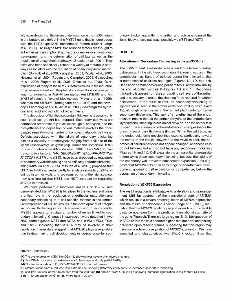
Figure 1. Phenotypic Characterization of Arabidopsis ms35 Mutant and Arabidopsis Pro35S:MYB26 Overexpression Lines.
(A) to (F) Anthers were stained with acridine orange/ethidium bromide and visualized by confocal microscopy (excitation, 590 nm).
(A) Ler wild-type partially dehisced anther from open flowers. Secondary thickening is visible in the endothecium (arrow).
(B) ms35 mutant nondehiscent mature anther from open flowers. Secondary thickening is absent in the endothecium (arrow).
(C) Mature anther from open flowers of a MYB26 overexpression line.
(D) Close-up of (A).
(E) Close-up of (B).
(F) Close-up of (C). Secondary thickening is visible in the endothecium (arrow) and as ectopic islands in the epidermal tissues (arrowheads).
(G) to (J) Sections through wild-type anthers ([G] and [I]) and ms35 mutant anthers ([H] and [J]).
(G) During pollen mitosis I, endothecial cells begin to expand (arrow) in the wild type.
(H) At the corresponding stage in the ms35 mutant, minimal endothecium expansion is seen (arrow).
(I) During pollen mitosis II, endothecium expansion continues in the wild type and secondary thickening is clearly evident (arrow).
(J) At the corresponding stage in the mutant, the endothecium cells fail to fully expand and no secondary thickening occurs (arrow).
(K) to (M) and (O) MYB26 overexpression lines.
MYB26 Regulator of Secondary Thickening 535
We have shown that the failure of dehiscence in the ms35 mutant
is attributable to a defect in the MYB26 gene that is homologous
with the R2R3-type MYB transcription factors (Steiner-Lange
et al., 2003). R2R3-type MYB transcription factors are thought to
act either as transcriptional activators or repressors, controlling
development and the determination of cell fate as well as the
regulation of biosynthetic pathways (Stracke et al., 2001). They
have also been specifically linked to a variety of metabolic path-
ways associated with the regulation of phenylpropanoid metab-
olism (Borevitz et al., 2000; Yang et al., 2001; Patzlaff et al., 2003;
Newman et al., 2004; Rogers and Campbell, 2004; Goicoechea
et al., 2005; Rogers et al., 2005; Deluc et al., 2006). Over-
expression of many of these MYB factors results in the induction
of genes associated with the phenylpropanoid biosynthesis path-
way; for example, in Antirrhinum majus, Am MYB305 and Am
MYB340 regulate flavanol biosynthesis (Moyano et al., 1996),
whereas Am MYB308 (Tamagnone et al., 1998) and the Arabi-
dopsis homolog At MYB4 (Jin et al., 2000) downregulate hydro-
cinnamic acid and monolignol biosynthesis.
The deposition of lignified secondary thickening is usually only
seen once cell growth has stopped. Secondary cell walls are
composed predominantly of cellulose, lignin, and xylan, and the
biosynthesis and deposition of wall material involves the coor-
dinated regulation of a number of complex metabolic pathways.
Defects associated with the failure of secondary thickening
exhibit a diversity of phenotypes, ranging from collapse of the
xylem vessels (irregular xylem [irx]) (Turner and Somerville, 1997)
to loss of dehiscence (Mitsuda et al., 2005). Two NAC domain
transcription factors, NAC SECONDARY WALL–PROMOTING
FACTOR1 (NST1) and NST2, have been proposed as regulators
of secondary wall thickening and specifically endothecium thick-
ening (Mitsuda et al., 2005). Mitsuda et al. (2005) proposed that
NST1 and NST2 act redundantly to regulate secondary cell thick-
enings in anther walls and are required for anther dehiscence;
they also implied that NST1 and NST2 may act by regulating
MYB26 expression.
We have performed a functional analysis of MYB26 and
demonstrated that MYB26 is localized to the nucleus and plays
a critical role in the regulation of endothecial maturation and
secondary thickening in a cell-specific manner in the anther.
Overexpression of MYB26 results in the development of ectopic
secondary thickening in both Arabidopsis and tobacco plants.
MYB26 appears to regulate a number of genes linked to sec-
ondary thickening. Changes in expression were detected in two
NAC domain genes, NST1 and NST2, and in IRX1, IRX3, IRX8,
and IRX12, indicating that MYB26 may be involved in their
regulation. These data suggest that MYB26 plays a regulatory
role in determining cell development, or competence for sec-
ondary thickening, within the anther and acts upstream of the
lignin biosynthesis pathway, possibly via NST1 and NST2.
RESULTS
Alterations in Secondary Thickening in the ms35 Mutant
The ms35 mutant is male-sterile as a result of a failure of anther
dehiscence. In the wild type, secondary thickening occurs in the
endothecium as bands of striated spring-like thickening that
is composed of cellulose and lignin (Figures 1A, 1D, and 1N).
Deposition commences during pollen mitosis I and is maximal by
the end of pollen mitosis II (Figures 1G and 1I). Secondary
thickening is absent from the surrounding cell layers of the anther
and is necessary to create the shearing force required for anther
dehiscence. In the ms35 mutant, no secondary thickening or
lignification is seen in the anther endothecium (Figures 1B and
1E), although other tissues in the mutant plant undergo normal
secondary thickening. This lack of strengthening of the endo-
thecium means that as the anther dehydrates the endothecium
layer distorts, shearing forces do not develop, and the anther fails
to open. The appearance of the endothecium changes before the
onset of secondary thickening (Figure 1G). In the wild type, as
the endothecial cells develop they expand, particularly toward
the center of the locule. However, in the ms35 mutant, the en-
dothecial cell number does not appear changed, and these cells
do not fully expand and do not have any secondary thickening
(Figures 1H and 1J). Cell expansion is an essential prerequisite
before laying down secondary thickening, because the rigidity of
the secondary wall prevents subsequent expansion. This sug-
gests that MYB26 acts at an early stage of endothecium devel-
opment, governing cell expansion or competence, before the
deposition of secondary thickening.
Regulation of MYB26 Expression
The ms35 mutation is attributable to a deletion and rearrange-
ment 1288 bp upstream of the translational start of MYB26,
which results in a severe downregulation of MYB26 expression
and the failure of dehiscence (Steiner-Lange et al., 2003), indi-
cating that the MYB26 regulatory region extends a considerable
distance upstream from the predicted translational start site of
the gene (Figure 2). There is a large region (8.124 kb) upstream of
MYB26 before the next annotated gene that does not contain any
predicted open reading frames, suggesting that this region may
have some role in the regulation of MYB26 expression. We have
identified and characterized two SALK knockout lines that
Figure 1. (continued).
(K) The overexpression (OEx) line OEx4-8, showing less severe phenotypic changes.
(L) Line OEx8-1, showing an extreme dwarf phenotype and only partial fertility.
(M) Nuclear localization of Pro35S:MYB26:GFP OEx in root tissues.
(O) Mature silique from a reduced less extreme line, showing deformity attributable to increased secondary thickening.
(N) and (P) Overview of mature anthers from the wild type (N) and a MYB26 OEx line (P) showing increased lignification in the MYB26 OEx line.
Bars ¼ 40 mm except in (G) to (J), where bars ¼ 20 mm.
536 The Plant Cell
contain insertions upstream of MYB26 (SALK_056264) and
within the coding sequence (SALK_112372) (Figure 2). Homozy-
gous knockout lines were generated and screened for the pres-
ence of insertions and phenotypic changes; both lines were
male-sterile as a result of a lack of secondary thickening in the
endothecium and the failure of anther dehiscence. These lines
were crossed using pollen from heterozygous ms35MS35 plants;
the resulting progeny from each cross segregated 1:1 for male
sterility:fertility (data not shown), indicating that they were allelic
to the ms35 mutation. These knockout lines gave the same
phenotype of full sterility attributable to a lack of dehiscence; this
was expected in the case of SALK_112372, which lies within the
third exon of the MYB26 transcript. However, the flanking se-
quence from the SALK_056264 insertion maps to �1696 bp
upstream of the translational start (Figure 2). Therefore, the
MYB26 regulatory region may extend as far as �1696 bp up-
stream from the translational start site, and this region is required
for functional expression of the MYB26 transcript. Bioinformatic
analysis of this region has not indicated any definitive motifs that
may be responsible for this regulation.
A promoter:b-glucuronidase (GUS) fusion was constructed to
localize the expression pattern of MYB26; the 3.1-kb region
upstream of the transcriptional start site was fused to the GUS
reporter gene and transformed into Landsberg erecta (Ler) wild-
type plants. Expression of ProMYB26:GUS is seen only in floral
tissues, with signal observed in the anthers and filaments of the
flowers; strong signal is also detected in the style and nectaries
of the flowers (Figure 3). Expression is observed in young
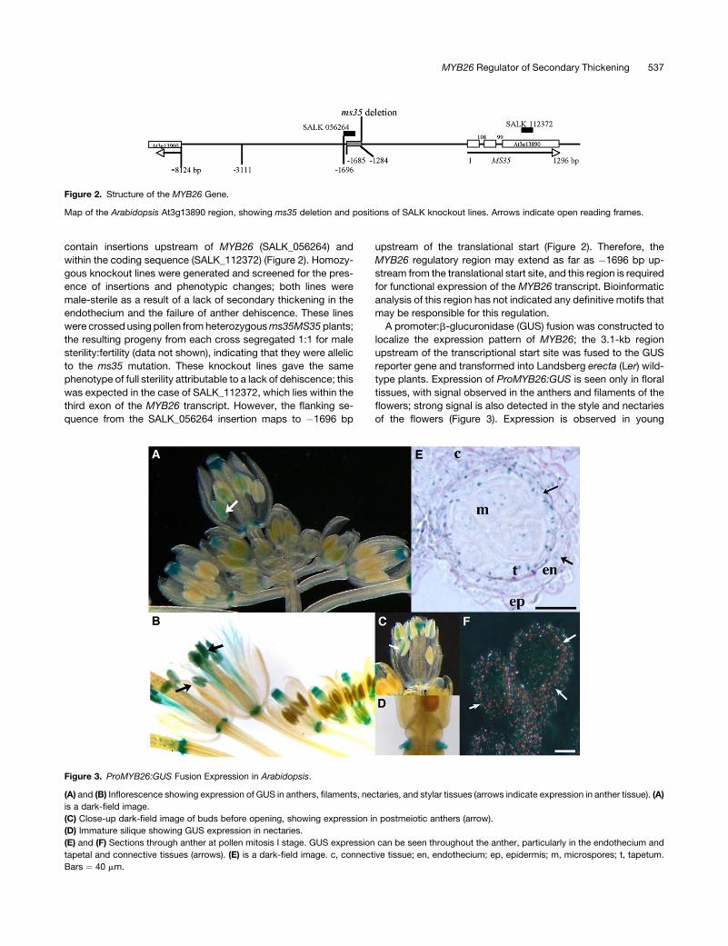
Figure 2. Structure of the MYB26 Gene.
Map of the Arabidopsis At3g13890 region, showing ms35 deletion and positions of SALK knockout lines. Arrows indicate open reading frames.
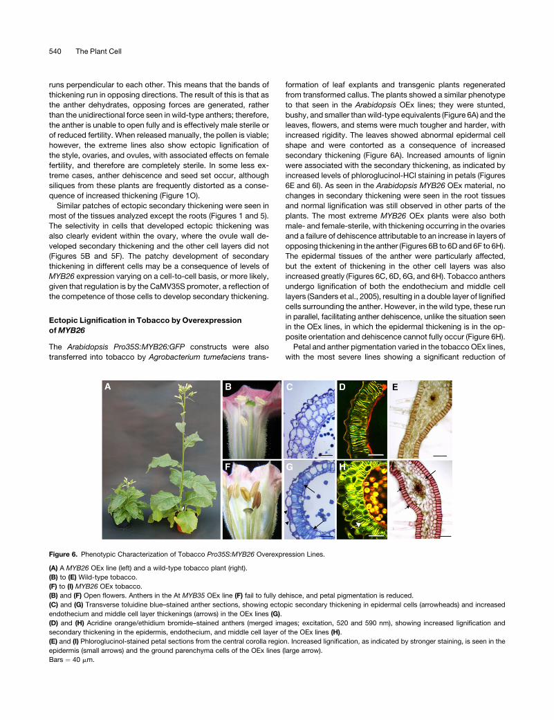
Figure 3. ProMYB26:GUS Fusion Expression in Arabidopsis.
(A) and (B) Inflorescence showing expression of GUS in anthers, filaments, nectaries, and stylar tissues (arrows indicate expression in anther tissue). (A)
is a dark-field image.
(C) Close-up dark-field image of buds before opening, showing expression in postmeiotic anthers (arrow).
(D) Immature silique showing GUS expression in nectaries.
(E) and (F) Sections through anther at pollen mitosis I stage. GUS expression can be seen throughout the anther, particularly in the endothecium and
tapetal and connective tissues (arrows). (E) is a dark-field image. c, connective tissue; en, endothecium; ep, epidermis; m, microspores; t, tapetum.
Bars ¼ 40 mm.
MYB26 Regulator of Secondary Thickening 537

postmeiotic buds through to open flowers (Figures 3A and 3C).
Expression is seen at a low level throughout the anther in the
tapetum, endothecium, and connective tissues (Figure 3B) and
also in the filament tissues (Figures 3A and 3D). Strong GUS
staining is also visible in the stylar tissue and nectaries after
fertilization, during early seed development (Figure 3E). No signal
is detected in vegetative tissues, and no vegetative expression
is detected by RT-PCR analysis (data not shown). Although
strong GUS staining is seen in the style and nectaries of the
ProMYB26:GUS transgenic plants, no significant phenotypic
differences associated with an alteration of lignification are ob-
served in these tissues in the ms35 mutant. A slight reduction in
lignification is apparent in ms35 mutant styles, although lignifi-
cation is still present and no change in female fertility is detected,
because full seed set can be obtained by crossing the male-
sterile mutant with pollen from wild-type plants.
The ProMYB26:GUS fusion data were supported by quantita-
tive PCR analysis of MYB26 expression in staged flowers and
anthers (Figure 4). High levels of MYB26 expression were
detected in excised anthers at pollen mitosis I and bicellular
stages (Figure 4). Very low levels of expression were detected
before pollen mitosis I and in pollen mitosis II, but at a signif-
icantly lower level than in pollen mitosis I and bicellular stages.
Quantitative RT-PCR analysis of expression was performed in
buds, which showed expression in older buds (tricellular pollen
stage) and open flowers (see Supplemental Figure 1 online),
indicating that this expression corresponds to that seen in
ProMYB26:GUS fusion lines in the nectaries and stylar tissue
and not to anther expression. Other genes associated with
secondary thickening in the anther were also analyzed by quan-
titative RT-PCR (Figure 4); these show expression at stages after
MYB26, indicating that they may be regulated by MYB26 or
induced by changes in the cell competence linked to MYB26
expression. The timing of MYB26 expression, before secondary
thickening formation, and the altered appearance of the endo-
thecium cells implies that MYB26 may play a regulatory role at an
earlier stage associated with the determination of cell expansion
or maturation rather than by direct regulation of secondary cell
wall thickening.
Ectopic Lignification Attributable to Overexpression
of MYB26
The MYB26 cDNA was cloned from closed buds into the pGWB5
vector (kindly provided by Tsuyoshi Nakagawa, Shimane Uni-
versity) under the control of the cauliflower mosaic virus (CaMV)
35S promoter with a C-terminal green fluorescent protein (GFP)
sequence to analyze the effect of ectopic overexpression. This
construct was transferred into the wild-type Ler background
and also into heterozygous ms35 gl1 plants. The Pro35S:MYB26
transcript partially complemented the ms35 mutation, causing
some restoration of lignification within the endothecium that was
sufficient to allow partial anther dehiscence and limited self-
fertilization, although complete endothecial thickening did not
occur. The fact that endothecial thickening was induced by the
Pro35S:MYB26:GFP construct implies that the GFP tag does not
affect the function of the MYB26 protein; rather, the CaMV35S
promoter was unable to regulate expression in the anther to the
same extent as seen with the native MYB26 promoter.
The pGWB5:MYB26 construct contains a C-terminal GFP tag,
and expression of the GFP could be visualized by confocal mi-
croscopy within the nuclei, confirming that MYB26 is localized to
the nucleus (Figure 1M), as predicted for its role as a putative
transcription factor. Transgenic lines were tested by semiquan-
titative RT-PCR and quantitative RT-PCR to identify those show-
ing overexpression of MYB26. Different levels of expression were
seen between the different lines and in the different tissues
(see Figures 7 and 8 below), and varying degrees of phenotypic
changes were observed between different lines (Figures 1K and
1L). In all cases, the plants overexpressing MYB26 were reduced
in size, which appeared to be attributable to a premature ces-
sation of cell expansion rather than to altered cell number. This
variation in size ranged from partially reduced to extremely
stunted (Figures 1K and 1L). Changes in the thickening of tissues
was evident in many organs of the plant, particularly leaves,
petals, and ovules (Figure 5); however, no changes were de-
tected in the root tissues, despite obvious expression in the roots
(Figure 1M). Levels of fertility varied, with reduced fertility corre-
lating with levels of ectopic thickening in the reproductive tissues
(e.g., OEx8-1 and OEx8-3); very extreme lines were very stunted
and completely sterile.
In the floral and reproductive tissues, the ectopic thickening
resembled the striated spring-like bands of thickening that are
seen in the xylem tissues (Fukuda, 1997) and the anther endo-
thecium (Dawson et al., 1999) (Figures 5A and 5B). However, in
the leaf tissues, ectopic lignification of the epidermal cells
appeared as a dense net-like pattern that covered the epider-
mal cell walls (Figures 5C and 5D). The leaves of the plants
tended to be distorted and twisted as a consequence of the
changes in the thickening of these tissues. In all cases, the
thickening was caused by modifications in both secondary
thickening and lignification, as indicated by phloroglucinol-HCl
and also acridine orange/ethidium bromide staining; however,
normal lignification of xylem tissues still occurred in these lines
(Figures 5A and 5B).
Figure 4. Quantitative RT-PCR Expression in Wild-Type Arabidopsis
Staged Excised Anthers.
Relative expression levels were determined compared with actin ex-
pression and expressed as fold changes relative to actin expression
(Stratagene Mx3005P). The data pool consisted of two replicates re-
peated on at least two separate occasions; error bars show SD of
expression changes. PM I, pollen mitosis I; PM II, pollen mitosis II.
538 The Plant Cell

Ectopic secondary thickening and lignification are frequently
associated with the epidermal cells (Figures 1 and 5) and involve
specific patches of cells within the different tissues rather then all
cells within a tissue. This is particularly evident in the epidermal
tissues of the anther. In the wild type, secondary thickening oc-
curs in the anther endothecium as bands of striated spring-like
thickening (Figures 1A, 1D, 1I, and 1N); this is not seen in the
surrounding epidermal cell layers of the anther and is absent in
the ms35 mutant (Figures 1B, 1E, and 1J). However, in the lines
overexpressing MYB26, patches of epidermal cells within the
anther also develop these spring-like secondary thickenings
(Figures 1C, 1F, and 1P). These bands of thickening run along the
longitudinal axis of the cells; in the resultant architecture, the
thickening in the two cell layers, endothecium and epidermis,
Figure 5. Ectopic Secondary Thickening in Different Tissues in the Arabidopsis MYB26 Overexpression Lines and Wild-Type Ler.
Tissues ([A] to [D], At MYB OEx; [E] to [G], wild type) were stained with acridine orange/ethidium bromide and visualized by confocal microscopy
(590 nm). Bars ¼ 40 mm.
(A) Petal from a MYB26 OEx line, showing normal xylem development (arrow) and ectopic spring-like secondary thickening (arrowheads).
(B) Immature ovule from a MYB26 OEx line, showing normal xylem development (arrow) and ectopic secondary thickening in the epidermal tissues of
the ovule (arrowheads).
(C) and (D) Leaf tissues from a MYB26 OEx line, showing net-like ectopic secondary thickening. A more detailed view is shown in (D).
(E) Wild-type petal, showing normal xylem development.
(F) Corresponding wild-type ovule.
(G) Wild-type leaf tissue.
MYB26 Regulator of Secondary Thickening 539
runs perpendicular to each other. This means that the bands of
thickening run in opposing directions. The result of this is that as
the anther dehydrates, opposing forces are generated, rather
than the unidirectional force seen in wild-type anthers; therefore,
the anther is unable to open fully and is effectively male sterile or
of reduced fertility. When released manually, the pollen is viable;
however, the extreme lines also show ectopic lignification of
the style, ovaries, and ovules, with associated effects on female
fertility, and therefore are completely sterile. In some less ex-
treme cases, anther dehiscence and seed set occur, although
siliques from these plants are frequently distorted as a conse-
quence of increased thickening (Figure 1O).
Similar patches of ectopic secondary thickening were seen in
most of the tissues analyzed except the roots (Figures 1 and 5).
The selectivity in cells that developed ectopic thickening was
also clearly evident within the ovary, where the ovule wall de-
veloped secondary thickening and the other cell layers did not
(Figures 5B and 5F). The patchy development of secondary
thickening in different cells may be a consequence of levels of
MYB26 expression varying on a cell-to-cell basis, or more likely,
given that regulation is by the CaMV35S promoter, a reflection of
the competence of those cells to develop secondary thickening.
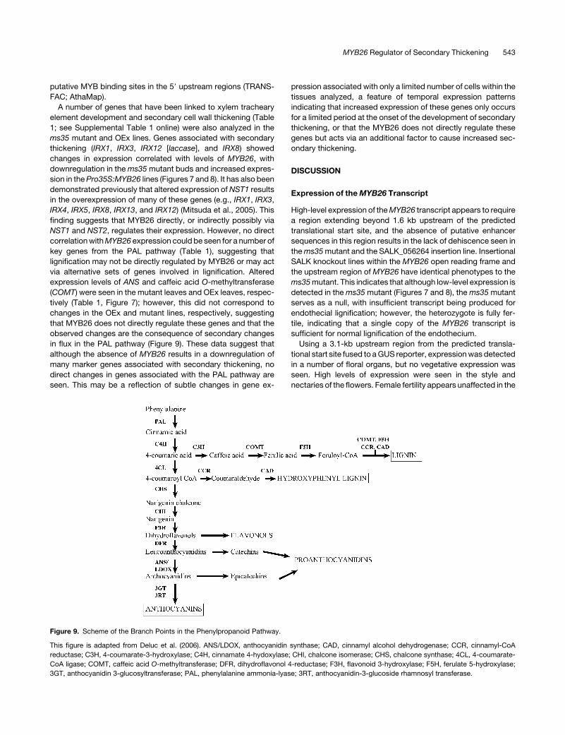
Ectopic Lignification in Tobacco by Overexpression
of MYB26
The Arabidopsis Pro35S:MYB26:GFP constructs were also
transferred into tobacco by Agrobacterium tumefaciens trans-
formation of leaf explants and transgenic plants regenerated
from transformed callus. The plants showed a similar phenotype
to that seen in the Arabidopsis OEx lines; they were stunted,
bushy, and smaller than wild-type equivalents (Figure 6A) and the
leaves, flowers, and stems were much tougher and harder, with
increased rigidity. The leaves showed abnormal epidermal cell
shape and were contorted as a consequence of increased
secondary thickening (Figure 6A). Increased amounts of lignin
were associated with the secondary thickening, as indicated by
increased levels of phloroglucinol-HCl staining in petals (Figures
6E and 6I). As seen in the Arabidopsis MYB26 OEx material, no
changes in secondary thickening were seen in the root tissues
and normal lignification was still observed in other parts of the
plants. The most extreme MYB26 OEx plants were also both
male- and female-sterile, with thickening occurring in the ovaries
and a failure of dehiscence attributable to an increase in layers of
opposing thickening in the anther (Figures 6B to 6D and 6F to 6H).
The epidermal tissues of the anther were particularly affected,
but the extent of thickening in the other cell layers was also
increased greatly (Figures 6C, 6D, 6G, and 6H). Tobacco anthers
undergo lignification of both the endothecium and middle cell
layers (Sanders et al., 2005), resulting in a double layer of lignified
cells surrounding the anther. However, in the wild type, these run
in parallel, facilitating anther dehiscence, unlike the situation seen
in the OEx lines, in which the epidermal thickening is in the op-
posite orientation and dehiscence cannot fully occur (Figure 6H).
Petal and anther pigmentation varied in the tobacco OEx lines,
with the most severe lines showing a significant reduction of
Figure 6. Phenotypic Characterization of Tobacco Pro35S:MYB26 Overexpression Lines.
(A) A MYB26 OEx line (left) and a wild-type tobacco plant (right).
(B) to (E) Wild-type tobacco.
(F) to (I) MYB26 OEx tobacco.
(B) and (F) Open flowers. Anthers in the At MYB35 OEx line (F) fail to fully dehisce, and petal pigmentation is reduced.
(C) and (G) Transverse toluidine blue–stained anther sections, showing ectopic secondary thickening in epidermal cells (arrowheads) and increased
endothecium and middle cell layer thickenings (arrows) in the OEx lines (G).
(D) and (H) Acridine orange/ethidium bromide–stained anthers (merged images; excitation, 520 and 590 nm), showing increased lignification and
secondary thickening in the epidermis, endothecium, and middle cell layer of the OEx lines (H).
(E) and (I) Phloroglucinol-stained petal sections from the central corolla region. Increased lignification, as indicated by stronger staining, is seen in the
epidermis (small arrows) and the ground parenchyma cells of the OEx lines (large arrow).
Bars ¼ 40 mm.
540 The Plant Cell
pigmentation in the petals (Figure 6F). This finding suggests that
in tobacco overexpression of the pathway regulating secondary
thickening also affects flavonoid development; no such changes
were evident in the Arabidopsis OEx lines.
Expression of Genes Associated with Secondary Thickening
in Arabidopsis Overexpression Lines
Overexpression of MYB26 resulted in ectopic secondary thick-
ening and lignification in various organs and cell types within
the transgenic lines. The Arabidopsis lines carrying the Pro35S:
MYB26:GFP construct and the ms35 mutant were analyzed by
semiquantitative RT-PCR for alterations in the expression of
selected genes linked to secondary thickening (Table 1). These
genes fall into a number of distinct classes, including those
associated with the regulation of secondary thickening, to key
steps in the phenylalanine ammonia-lyase (PAL) pathway, or to
cellulose synthesis and secondary thickening (Table 1; see Sup-
plemental Table 1 online). Quantitative RT-PCR analysis was also
conducted on a subset of these genes, and the data were
analyzed by normalization and relative expression to actin con-
trols using the 2�DDCT method (Livak and Schmittgen, 2001)
(Figure 8); the observed expression patterns mimic those seen by
semiquantitative RT-PCR analysis.
As expected, MYB26 expression was decreased in the ms35
mutant buds and increased in the MYB26 OEx lines. The OEx
lines showed varying levels of expression in the buds and in all
cases showed ectopic expression in the leaf tissues, which
normally do not express MYB26 (Figures 7 and 8). No changes in
At MYB32 and At MYB61 expression were observed in the OEx
lines, although the At MYB32 transcript increased slightly in the
ms35 mutant buds. These MYB transcription factors have been
linked to the regulation of phenylpropanoid metabolism and
ectopic lignification in Arabidopsis (Newman et al., 2004; Preston
et al., 2004) respectively. MYB32 has been shown to be ex-
pressed at high levels in the tapetum and to be important for
stamen development (Preston et al., 2004; Mandaokar et al.,
2006). Therefore, the expression change seen in the ms35 buds,
and the associated lack of change in the OEx lines, are likely to be
secondary consequences of changes in anther development and
not of direct MYB26 regulation, indicating that MYB26 does not
act via a pathway involving either of these transcription factors.
We also observed a slight increase in expression of the ANTHO-
CYANIDIN SYNTHASE (ANS) gene in ms35 leaves; no pheno-
typic changes were seen in the ms35 mutant leaves, so this is
likely to represent secondary changes in the regulation of path-
ways attributable to the lack of secondary thickening in the
anther.
Two NAC domain genes, NST1 and NST2, have been linked to
secondary thickening of the anther tissues (Mitsuda et al., 2005),
with the overexpression of NST1 and NST2 reported to induce
ectopic secondary thickening of the anther tissues. Mitsuda et al.
(2005) suggested that they may be involved in regulating MYB26
expression; however, no data were presented to support this.
By contrast, our analysis supports the reverse situation, with
downregulation of NST1 and NST2 in the ms35 mutant and a
corresponding increase of NST1 and NST2 expression in the
Pro35S:MYB26 lines (Figures 7 and 8). Analysis of NST1 and
Table 1. Summary of Semiquantitative RT-PCR of Genes Associated with Lignin and Secondary Thickening
Gene of
Interest AGI Code
Wild-Type Expression ms35 Mutant OEx LinesChange in
ms35 Mutant
Change in
OEx Lines
Putative Role
of MYB26Leaves Buds Leaves Buds Leaves Buds
NST1 At2g46770 * *** ** ** *** *** Decrease in buds Increase in leaves Regulation
NST2 At3g61910 – ** * * ** ** Decrease in buds Increase in leaves Regulation
MYB32 At4g34990 ** ** ** *** ** ** Increase in buds No change Secondary
effect
MYB61 At1g09540 * ** * ** * * No change No change No effect
IRX8 At5g54690 * ** * * *** *** Decrease in buds Increase Regulation
IRX1 At4g18780 * ** * ** ** ** Slight decrease Increase in leaves Regulation
IRX3 At5g17420 * ** * ** ** ** No change Increase Regulation
IRX5 At5g44030 ** *** ** *** ** *** No change No change No effect
IRX13 At5g03170 * ** * ** * ** No change No change No effect
IRX12
(laccase)
At2g38080 ** ** * * ** ** Decrease Increase Regulation
XCIP At4g35350 ** ** ** ** ** ** No change No change No effect
ANS At2g38240 ** ** *** ** ** ** Increase in leaves No change No effect
C4H At2g30490 ** ** ** ** ** ** No change No change No effect
DFR At1g08200 *** *** *** *** *** *** No change No change No effect
COMT At1g67980 ** ** ** ** *** ** No change Increase in leaves Secondary
effect
PAL2 At3g53260 ** ** ** ** ** ** No change No change No effect
IRX4 At1g15950 *** *** *** *** *** *** No change No change No effect
At3g62160 At3g62160 ** ** NT ** ** ** No change No change No effect
AtHB-8 At4g32880 ** ** NT ** ** ** No change No change No effect
These data were determined using overexpression lines OEx8-1, OEx8-3, and OEx4-8 (Figure 7). ***, high level of expression; **, moderate level of
expression; *, low level of expression; –, no expression detected. AGI, Arabidopsis Genome Initiative; NT, not tested.
MYB26 Regulator of Secondary Thickening 541

NST2 expression in wild-type anthers showed that these genes
are expressed after MYB26 expression at the bicellular and
pollen mitosis II stages, although some expression of NST2 is
also evident at the tricellular stage (Figure 4). No significant
expression of NST1 and NST2 is seen in wild-type leaf tissues,
although induction is observed in leaves when MYB26 is ectop-
ically expressed. In general, the level of NST1 and NST2 induc-
tion in the different overexpression lines correlates with the level
of ectopic MYB26 expression. In buds, line OEx4-8 shows fewer
changes in expression of MYB26 compared with the wild type
and the other overexpressing lines and a corresponding reduced
enhancement of NST1 and NST2 expression; however, it also
shows minimal changes in floral phenotype (Figure 1K). In other
lines, such as OEx8-3, the induction of NST1 and NST2 is more
pronounced, particularly for NST2 (Figure 8A). The same trends
are observed in the leaf tissue, with greater expression of MYB26
resulting in more NST1 and NST2 expression (Figure 8B); this is
particularly evident for NST2 expression. However, the level of
change between the two genes, NST1 and NST2, is not always
constant. NST1 and NST2 have already been shown to act
redundantly (Mitsuda et al., 2005); therefore, the level of each is
unlikely to be critical at inducing ectopic secondary thickening.
This variability of induction indicates that other factors may in-
fluence NST1 and NST2 expression, particularly because some
expression of NST1 and NST2 is still seen in the ms35 mutant,
but it may also reflect variation in a cell-specific and temporal
manner. NST1 and NST2 expression is reduced significantly in
the ms35 mutant; however, a low level of NST1 and NST2 ex-
pression was still detectable by quantitative PCR, suggesting
that there may be additional regulators of their expression. These
data suggest that MYB26 acts upstream of these two NAC
genes, NST1 and NST2, and in turn the formation of thickening in
the endothecium. It is unclear at present whether this regulation
occurs directly or indirectly; however, analysis of the promoter
regions of NST1 and NST2 suggests that there are a number of
Figure 7. RT-PCR Analysis of Genes Associated with Secondary Thick-
ening in Arabidopsis Wild Type, ms35 Mutant, and MYB26 Overexpres-
sion Lines.
Overexpression lines were OEx8-1, OEx8-3, and OEx4-8 (Table 1; see
Supplemental Table 1 online). M, 1-kb marker; Wt, wild type; L, leaf
tissue; B, buds; gDNA, genomic DNA control.
Figure 8. Quantitative RT-PCR of Selected Genes Showing Altered
Regulation in the Arabidopsis MYB26 Overexpression Lines OEx8-1,
OEx8-3, and OEx4-8.
Data are shown for bud tissues (A) and leaves (B). Relative expression
levels were determined compared with actin leaf expression using the
2�DDCT analysis method and are expressed as fold changes relative to
actin. The data shown are from two replicates repeated on at least two
separate occasions; error bars show SD of expression changes.
542 The Plant Cell
putative MYB binding sites in the 59 upstream regions (TRANS-
FAC; AthaMap).
A number of genes that have been linked to xylem tracheary
element development and secondary cell wall thickening (Table
1; see Supplemental Table 1 online) were also analyzed in the
ms35 mutant and OEx lines. Genes associated with secondary
thickening (IRX1, IRX3, IRX12 [laccase], and IRX8) showed
changes in expression correlated with levels of MYB26, with
downregulation in the ms35 mutant buds and increased expres-
sion in the Pro35S:MYB26 lines (Figures 7 and 8). It has also been
demonstrated previously that altered expression of NST1 results
in the overexpression of many of these genes (e.g., IRX1, IRX3,
IRX4, IRX5, IRX8, IRX13, and IRX12) (Mitsuda et al., 2005). This
finding suggests that MYB26 directly, or indirectly possibly via
NST1 and NST2, regulates their expression. However, no direct
correlation with MYB26 expression could be seen for a number of
key genes from the PAL pathway (Table 1), suggesting that
lignification may not be directly regulated by MYB26 or may act
via alternative sets of genes involved in lignification. Altered
expression levels of ANS and caffeic acid O-methyltransferase
(COMT) were seen in the mutant leaves and OEx leaves, respec-
tively (Table 1, Figure 7); however, this did not correspond to
changes in the OEx and mutant lines, respectively, suggesting
that MYB26 does not directly regulate these genes and that the
observed changes are the consequence of secondary changes
in flux in the PAL pathway (Figure 9). These data suggest that
although the absence of MYB26 results in a downregulation of
many marker genes associated with secondary thickening, no
direct changes in genes associated with the PAL pathway are
seen. This may be a reflection of subtle changes in gene ex-
pression associated with only a limited number of cells within the
tissues analyzed, a feature of temporal expression patterns
indicating that increased expression of these genes only occurs
for a limited period at the onset of the development of secondary
thickening, or that the MYB26 does not directly regulate these
genes but acts via an additional factor to cause increased sec-
ondary thickening.
DISCUSSION
Expression of the MYB26 Transcript
High-level expression of the MYB26 transcript appears to require
a region extending beyond 1.6 kb upstream of the predicted
translational start site, and the absence of putative enhancer
sequences in this region results in the lack of dehiscence seen in
the ms35 mutant and the SALK_056264 insertion line. Insertional
SALK knockout lines within the MYB26 open reading frame and
the upstream region of MYB26 have identical phenotypes to the
ms35 mutant. This indicates that although low-level expression is
detected in the ms35 mutant (Figures 7 and 8), the ms35 mutant
serves as a null, with insufficient transcript being produced for
endothecial lignification; however, the heterozygote is fully fer-
tile, indicating that a single copy of the MYB26 transcript is
sufficient for normal lignification of the endothecium.
Using a 3.1-kb upstream region from the predicted transla-
tional start site fused to a GUS reporter, expression was detected
in a number of floral organs, but no vegetative expression was
seen. High levels of expression were seen in the style and
nectaries of the flowers. Female fertility appears unaffected in the
Figure 9. Scheme of the Branch Points in the Phenylpropanoid Pathway.
This figure is adapted from Deluc et al. (2006). ANS/LDOX, anthocyanidin synthase; CAD, cinnamyl alcohol dehydrogenase; CCR, cinnamyl-CoA
reductase; C3H, 4-coumarate-3-hydroxylase; C4H, cinnamate 4-hydoxylase; CHI, chalcone isomerase; CHS, chalcone synthase; 4CL, 4-coumarate-
CoA ligase; COMT, caffeic acid O-methyltransferase; DFR, dihydroflavonol 4-reductase; F3H, flavonoid 3-hydroxylase; F5H, ferulate 5-hydroxylase;
3GT, anthocyanidin 3-glucosyltransferase; PAL, phenylalanine ammonia-lyase; 3RT, anthocyanidin-3-glucoside rhamnosyl transferase.
MYB26 Regulator of Secondary Thickening 543

ms35 mutant, indicating that the style is fully functional. Lignin
development, although reduced, still occurs within the style,
implying that there may be redundant genes regulating second-
ary thickening within the style, so that secondary thickening still
occurs in the absence of MYB26, albeit at a slightly reduced
level. Nectaries have a role in the production of nectar for insect
attraction to facilitate pollination; however, because Arabidop-
sis is predominantly self-fertile, the nectaries may not have a
functional role in pollination and fertilization. Expression of the
MYB26 transcript is detected in these tissues, although no
significant differences in secondary thickening were observed,
which may either reflect a lack of functional MYB26 protein in
these tissues or indicate a redundancy of gene regulation. Ex-
pression of a number of genes linked to PAL metabolism (Thoma
et al., 1994) and dehiscence (Rajani and Sundaresan, 2001) has
also been observed in Arabidopsis nectaries, although altera-
tions in phenotype have not been linked to these expression
patterns. Expression, as indicated by GUS promoter fusion data
and quantitative RT-PCR, is seen in the anthers, from the start of
pollen mitosis I through to bicellular pollen, although expression
in the nectaries and style is maintained beyond these stages.
This pattern of anther expression coincides with the differentia-
tion of cell types within the anther and the expansion of the
endothecial cells immediately before the initiation of secondary
thickening in the endothecium and vascular tissues in the fila-
ment. Immediately after MYB26 expression, other genes directly
linked to secondary thickening are expressed, implying that
MYB26 acts upstream of secondary thickening via the regulation
of genes such as the NAC domain genes NST1 and NST2 and
may direct cell expansion and the change of cell competence
toward secondary thickening, rather than directly inducing the
expression of genes in the secondary thickening pathways.
Induction of Secondary Thickening
Ectopic secondary thickening was induced in both Arabidopsis
and tobacco plants by overexpression of the MYB26 transcript.
In general, the severity of the phenotype correlated with the level
of MYB26 transcript, suggesting that the amount of MYB26
protein is linked to the extent of phenotypic changes. Increased
secondary thickening and lignification were seen in these lines.
A number of MYB factors have been linked to PAL metabolism
and secondary thickening. Eucalyptus grandis Eg MYB2, which
has homology with Arabidopsis At MYB83 and Populus tremuloides
Pt MYB4, has been shown to regulate expression by binding to
MYB binding sites in the promoters of cinnamoyl-CoA reductase
and cinnamyl alcohol dehydrogenase genes (Goicoechea et al.,
2005). Tobacco plants overexpressing Eucalyptus MYB2 showed
increased secondary cell wall thickness and altered lignification
profiles, indicating that Eucalyptus MYB2 has a role in the coordi-
nated control of genes involved in the monolignol-specific pathway;
however, other core PAL genes remain unaffected (Goicoechea
et al., 2005). Arabidopsis MYB61 has also been linked to the ec-
topic lignification and dark photomorphogenic phenotype of de-
etiolated3 (Newman et al., 2004). Arabidopsis MYB32 has also
been shown to regulate genes in the PAL pathway, with down-
regulation of the MYB32 gene increasing transcript levels of the
COMT gene and reducing levels of the DIHYDROFLAVONOL
4-REDUCTASE (DFR) and ANS genes (Preston et al., 2004).
Despite anther expression, the pattern of MYB32 expression is
very different from that of MYB26; MYB32 is expressed in all
major organs, but with a high level of expression in the tapetum
and the stigma (Preston et al., 2004), whereas expression of
MYB26 occurs specifically within the endothecium tissues, style,
and nectaries. MYB32 does not appear to affect the dehiscence
process but is thought to alter pollen wall formation by affecting
wall development, resulting in collapsed inviable pollen (Preston
et al., 2004). No change of expression was seen in either of these
two MYB factors in the MYB26 overexpression lines, indicating
that MYB26 is upstream of, or does not induce, secondary
thickening via MYB32 or MYB61.
IRX1, IRX3, IRX8, and IRX12 expression appears to be corre-
lated directly with levels of MYB26, suggesting direct or indirect
regulation by MYB26. Similar increased expression of these
genes was also observed in the Pro35S:NST1-overexpressing
lines (Mitsuda et al., 2005). IRX1, IRX3, and IRX5 have been
shown to be coexpressed in secondary cell walls and to encode
essential components of the cellulose-synthesizing complex
(Taylor et al., 1999, 2003). Transcriptomic analysis has shown
that IRX1 and IRX3 exhibited similar expression patterns during
hypocotyl and stem development, and their expression pattern
has been linked by expression profiling to a number of genes
associated with secondary thickening, including IRX8 (a glucosyl
transferase involved in pectin synthesis) and IRX12 (laccase)
(Brown et al., 2005). IRX3 and laccase (IRX12) have both been
shown to be regulated by VND7 during transdifferentiation of
xylem vessel elements (Kubo, 2005). The altered expression of
these genes in the Pro35S:MYB26:GUS lines suggests that
MYB26 directly or indirectly regulates a group of key genes
associated with secondary wall formation, possibly via regulation
of NST1 and NST2 expression.
Lignin is derived from the dehydrogenative polymerization of
hydroxycinnamyl alcohols (monolignols) and other phenylpropa-
noids (Raes et al., 2003). Biosynthesis of monolignols requires
enzymes of the phenylpropanoid (PAL) pathway (Figure 9) using
derivatives of Phe. Lignin is a major component of secondary
thickening of cell walls, resulting in the development of large
amounts of biomass and providing physical strength for plants
combined with the physical forces required for dehiscence, pod
shatter, etc. Many mutants have been identified in biosynthetic
pathways of lignin, but only recently have genes been identified
associated with the regulation of this pathway (see Supplemental
Table 1 online). Lignification is usually seen in sclerified cells and
requires the coordinated expression of a number of genes from
the PAL pathway and those associated with the polymerization
of monolignols. Therefore, there is a need for a central regulatory
system for the induction of lignin development, which then
switches on the other components required for lignification.
MYB proteins have been linked to such a regulatory role; two
Antirrhinum MYB transcription factors have been shown to regu-
late the expression of genes in the PAL pathway (Tamagnone
et al., 1998). MYB46 is a predicted ortholog of pine (Pinus) MYB4,
a positive regulator of the lignification of xylem cells and phloem
fibers in loblolly pine (Pinus taeda) (Patzlaff et al., 2003), and
MYB52, a predicted ortholog of Populus tremula 3 P. tremuloides
MYB21a, is a repressor of caffeoyl-CoA 3-O-methyltransferase
544 The Plant Cell

expression (Karpinska et al., 2004). Expression of NAC and MYB
factors has been associated with the transcriptome of xylem and
phloem development in Arabidopsis root hypocotyls (Zhao et al.,
2005). Conserved activator elements, which are critical for the
regulation of expression, have been identified in promoters of PAL
pathway genes (Neustaedter et al., 1999) and are similar to the
motifs recognized by MYB proteins. In the case of the PAL
promoter, these have been shown to bind and regulate GUS
reporter expression (Sablowski et al., 1994). No changes were
evident in pigment formation and flavonoid gene expression in the
Arabidopsis OEx material, although changes in pigmentation in
petal and anther were seen in the tobacco OEx lines, suggesting
that there may be subtle alterations in the regulation of compo-
nents of the PAL pathway in tobacco compared with Arabidopsis.
Similar effects were observed in grapevine (Vitis vinifera) by
overexpression of Vv MYB5a, which resulted in an increase in
pigment formation in stamens, enhanced anthocyanin biosynthe-
sis, and increased expression of genes in the flavonoid biosyn-
thetic pathway, such as CHALCONE SYNTHASE, CHALCONE
ISOMERASE, FLAVANONE 3-HYDROXYLASE, and DFR (del Rio
et al., 2004).
The fact that ectopic production of lignin is seen in the MYB26
OEx Arabidopsis and tobacco lines suggests that MYB26 has a
key role in the regulation of genes in the PAL and monolignol
polymerization pathways and that the regulation of this pathway
appears to be evolutionarily conserved between tobacco and
Arabidopsis. However, no direct increase in gene expression of a
number of specific genes from the PAL pathway was seen in OEx
MYB26 lines, although changes in IRX12 (laccase) expression
were seen. This finding suggests that the regulation of PAL path-
way genes is either extremely transient and low-level or that the
role of MYB26 in lignification may be indirect or via alternative
members of these pathways, because there is a high level of
redundancy in the PAL pathway. Our data indicate that MYB26
acts by inducing the expression of the NAC domain genes NST1
and NST2, which were previously linked to expression changes
associated with secondary thickening and lignification (Mitsuda
et al., 2005). The NAC transcription family has been linked to
maintaining tissue boundaries, regulating the control of growth
from cell division to expansion, and other developmental pro-
cesses such as senescence (Zhao et al., 2005). NST1 and NST2
appear to act redundantly in the regulation of secondary cell
walls in the anther (Mitsuda et al., 2005), and a similar phenotype
to that seen in the Pro35S:MYB26 lines of ectopic secondary
thickening was observed when NST1 or NST2 was expressed
using the CaMV35S promoter. It was suggested previously that
NST1 and/or NST2 may regulate the expression of MYB26
(Mitsuda et al., 2005); however, this contradicts the changes of
NST1 and NST2 expression seen in our data. NST1 and NST2
exhibit different but overlapping expression patterns: NST1 is
seen in many tissues of the plant, including inflorescence stems,
mid ribs of leaves and anthers, filaments, stamens, and carpels,
whereas NST2 shows a much more specific pattern, with ex-
pression in anther walls and pollen grains (Mitsuda et al., 2005).
This is in contrast with the observations of MYB26 expression,
which is seen only in the reproductive tissues and principally
in the filament, anther, nectaries, and style. Therefore, MYB26
may play a role in establishing cell competence and determining
which cells are capable of undergoing secondary thickening by
regulating NST1 and NST2 expression in these tissues.
The induction of ectopic secondary thickening does not occur
in all cells, but it is evident predominantly in epidermal cells of
both Arabidopsis and tobacco MYB26 OEx lines. The epidermis
has been shown to be a highly metabolically active cell type; for
example, in Catharanthus roseus, concomitant expression of
genes from at least four separate metabolic pathways, including
those from the PAL pathway, was seen in the epidermal tissues
(Mahroug et al., 2006). The development of thickening predom-
inantly in the epidermis of the OEx lines may reflect the compe-
tence of the epidermal tissue for such metabolic activity.
However, not all cells undergo thickening; some were visible as
ectopic islands among cells showing normal development. This
sporadic thickening has also been described in other reports of
ectopic secondary thickening (Mitsuda et al., 2005). This may be
a consequence of differences in the cell-specific expression of
the transgene, but more likely it reflects a competence for sec-
ondary thickening in those cells that is associated either with the
cessation of cell growth or with the ability to respond to signals
required for secondary thickening.
This work has shown that the processes of secondary thick-
ening are conserved between Arabidopsis and tobacco and that
overexpression of MYB26 results in ectopic induction of sec-
ondary thickening and lignification. This is seen in selected cells
within a variety of tissues, suggesting that some aspect of com-
petence is required for the induction of thickening. These data
suggest that MYB26 may either regulate genes associated with
secondary thickening in the endothecium or else function in
specifying cell competence and determining which cells undergo
secondary thickening. MYB26 appears to act by regulating the
expression of NST1 and NST2, which may directly or indirectly
affect the levels of genes associated with the development of
secondary thickening. The ability to regulate secondary thicken-
ing in a conserved manner has significant commercial applica-
tions for the wood and paper industries.
METHODS
Plant Growth Conditions
Seeds of Arabidopsis thaliana var Ler and the ms35gl mutant were sown
onto a compost mix of Levington M3:vermiculite (3:1) and grown in a
glasshouse at 21/178C (day/night) with a 22/2-h photoperiod as described
previously (Dawson et al., 1999). The T-DNA insertion lines SALK_056264
and SALK_112372 were generated by the Salk Institute Genomic Analysis
Laboratory and obtained from the Nottingham Arabidopsis Stock Centre
(NASC). Both T-DNA insertion lines and wild-type (ecotype Columbia)
control plants were grown as described previously. Plants were genop-
typed by PCR using SALK_LBb1 (59-GCGTGGACCGCTTGCTGCAACT-39),
SALK_112372_RP (59-CATTGAGCTTCACAGCATTCTTGG-39), and
SALK_112372_LP (59-GTCCACAAGAGATTGGCGACG-39) primers for
SALK_112372 and SALK_LBb1 (59-GCGTGGACCGCTTGCTGCAACT-39),
SALK_056264_LP (59-CCGCGGGTTAAAATCTATTATGTGA-39), and
SALK_056264_RP (59-TCGAACGTACGTATGTAGAGTCCT-39) primers
for SALK_056264.
Transformation of Tobacco
Tobacco (Nicotiana tabacum) seeds were surface-sterilized and sown
onto MSR3 (Liu, 2005) medium under conditions of 16 h of daylight at
MYB26 Regulator of Secondary Thickening 545
258C followed by 8 h of dark at 188C. Fully expanded, 3- to 4-week-old
tobacco leaves were used as explants. Transgenic tobacco plants were
generated by Agrobacterium tumefaciens–mediated leaf slice transfor-
mation (Liu, 2005) and selected for kanamycin resistance. Regenerated
transgenic plants were moved onto compost and grown in the glass-
house as described for Arabidopsis.
MYB26 PromoterTGUS Construct
A 3.127-kb region upstream of the MYB26 gene was amplified by PCR
(MS35-Xho1-3540L, 59-GCCTCGAGAAGGGAAGCCTCCGACAGAA-39;
MS35R-AVRII-14R, 59-AGCCTAGGCTAGATCTCTATCGCTCTCTTAG-
TCT-39), digested with XhoI/AvrII, and cloned upstream between the
XhoI and XbaI sites of the uidA gene of the MOG402-based binary vector
pMOGMS1:GUS (Wilson et al., 2001), replacing the MS1 promoter to
create pMOGMYB26:GUS. The construct was then transferred into
Agrobacterium (C58 pGV3850) by electroporation (Sambrook et al.,
1989) and transformed into Arabidopsis (Ler) plants by floral dipping
(Clough and Bent, 1998). b-Glucuronidase activity was visualized by
staining the inflorescences overnight in 5-bromo-4-chloro-3-indolyl-b-
glucuronic acid solution (Willemsen et al., 1998).
Overexpressing Lines
The 1135-bp MYB26 coding region was amplified by PCR
(FGWMS35cDNA, 59-GGGGACAAGTTTGTACAAAAAAGCAGGCTTCA-
TGGGTCATCACTCATGCTG-39; RGWMS35cDNA, 59-GGGGACCACTT-
TGTACAAGAAAGCTGGGTCAGTTATGACGTACTGTCCACAAGAGAT-39)
from Ler bud cDNA, cloned by recombination into Gateway pDONR201
entry vector (Invitrogen), and transferred into pGWB5 (Karimi et al., 2002)
under the control of the CaMV35S promoter. The construct was then
transformed into Arabidopsis and tobacco plants as described above.
Plants were selected on Murashige and Skoog plates containing 50 mg/mL
kanamycin and screened for the presence of the transgene by PCR.
Expression Analysis
RNA was isolated from buds and leaves (RNeasy; Qiagen), and cDNA was
prepared using 5 mg of total RNA in a 20-mL reaction (SuperScript II
reverse transcriptase; Invitrogen). This was used for RT-PCR analysis
using 0.5 mL of cDNA template and the appropriate primer pairs (see
Supplemental Table 1 online) for 30 cycles at 948C for 30s, 588C for 30s,
and 728C for 1 min. RT-PCR results were normalized using Arabidopsis
actin-2 controls (Wilson et al., 2001).
Quantitative RT-PCR
Quantitative RT-PCR analyses were performed using the Mx3005P
multiplex quantitative PCR system (Stratagene). Reactions were set up
using the Brilliant SYBR Green QPCR Master Mix (Stratagene) in a final
volume of 25 mL containing 0.5 mL of cDNA and 0.5 mL of the appropriate
primers (see Supplemental Table 1 online). PCR cycling conditions for
amplification were 958C for 10 min followed by 40 cycles of 958C for
30 s, 588C for 1 min, and 728C for 1 min. All samples were run at least
in duplicate. Data acquisition and analyses were performed using
Mx3005TM multiplex quantitative PCR system software. Relative ex-
pression levels were determined compared with actin leaf expression
using the 2�DDCT analysis method (Livak and Schmittgen, 2001).
Microscopy
For analysis of lignin, fresh samples or tissues were stained with phloro-
glucinol-HCl (Ruzin, 1999) and were observed with a light microscope
(Nikon). For confocal microscopy (TCS SP2; Leica), a modified ethidium
bromide/acridine orange stain was used (A.M. Patten, personal com-
munication); the ethidium bromide stains lignified cells (red fluorescence;
excitation, 590 nm) and the acridine orange stains nonlignified walls
(green fluorescence; excitation, 520 nm). Fresh tissues were washed (13
PBS and 2% [v/v] Tween 20 for 10 min, then 13 PBS), stained with 0.01%
(w/v) acridine orange (1 h, room temperature), washed (13 PBS), and then
stained with 0.00005% (w/v) ethidium bromide (1 h, room temperature).
Plant material was also fixed and embedded in paraffin, and sections
were stained with toluidine blue, as described previously (Vizcay-Barrena
and Wilson, 2006). A minimum of 10 independent transformants were
analyzed.
Accession Numbers
Arabidopsis Genome Initiative locus identifiers for the key genes mentioned
in this article are as follows: MYB26 (At3g13890), NST1 (At2g46770), NST2
(At3g61910), IRX1 (At4g18780), IRX3 (At5g17420), IRX5 (At5g44030), IRX8
(At5g54690), and IRX12 (At2g38080); others are listed in Table 1.
Supplemental Data
The following materials are available in the online version of this article.
Supplemental Table 1. Primer Sequences for Genes Associated with
Lignin and Secondary Thickening.
Supplemental Figure 1. Quantitative RT-PCR Expression in Wild-
Type Arabidopsis Flowers and Buds.
ACKNOWLEDGMENTS
We thank Malcolm Bennett and Jerry Roberts for critical reading of the
manuscript. Seed stocks and bioinformatics information were obtained
from the NASC. This work was funded by the Biotechnology and
Biological Science Research Council.
Received August 3, 2006; revised January 6, 2007; accepted February 5,
2007; published February 28, 2007.
REFERENCES
Bonner, L., and Dickinson, H. (1989). Anther dehiscence in Lycopersi-
con esculentum Mill. New Phytol. 113: 97–115.
Borevitz, J.O., Xia, Y., Blount, J., Dixon, R.A., and Lamb, C. (2000).
Activation tagging identifies a conserved MYB regulator of phenyl-
propanoid biosynthesis. Plant Cell 12: 2383–2394.
Brown, D.M., Zeef, L.A., Ellis, J., Goodacre, R., and Turner, S.R.
(2005). Identification of novel genes in Arabidopsis involved in sec-
ondary cell wall formation using expression profiling and reverse
genetics. Plant Cell 17: 2281–2295.
Clough, S.J., and Bent, A.F. (1998). Floral dip: A simplified method for
Agrobacterium-mediated transformation of Arabidopsis thaliana. Plant
J. 16: 735–743.
Dawson, J., Sozen, E., Vizir, I., Van Waeyenberge, S., Wilson, Z.A.,
and Mulligan, B.J. (1999). Characterization and genetic mapping of a
mutation (ms35) which prevents anther dehiscence in Arabidopsis
thaliana by affecting secondary wall thickening in the endothecium.
New Phytol. 144: 213–222.
Dawson, J., Wilson, Z.A., Briarty, L.G., and Mulligan, B.J. (1993).
Development of anthers and pollen in male sterile mutants of Arabi-
dopsis thaliana. In Arabidopsis: An Atlas of Morphology, J. Bowman,
ed (Berlin: Springer-Verlag), pp. 282–295.
546 The Plant Cell
del Rio, L.A., Corpas, F.J., and Barroso, J.B. (2004). Nitric oxide and
nitric oxide synthase activity in plants. Phytochemistry 65: 783–792.
Deluc, L., Barrieu, F., Marchive, C., Lauvergeat, V., Decendit, A.,
Richard, T., Carde, J.P., Merillon, J.M., and Hamdi, S. (2006).
Characterization of a grapevine R2R3-MYB transcription factor that
regulates the phenylpropanoid pathway. Plant Physiol. 140: 499–511.
Feys, B., Benedetti, C.E., Penfold, C.N., and Turner, J.G. (1994).
Arabidopsis mutants selected for resistance to the phytotoxin coro-
natine are male sterile, insensitive to methyl jasmonate, and resistant
to a bacterial pathogen. Plant Cell 6: 751–759.
Fukuda, H. (1997). Tracheary element differentiation. Plant Cell 9: 1147–
1156.
Goicoechea, M., Lacombe, E., Legay, S., Mihaljevic, S., Rech, P.,
Jauneau, A., Lapierre, C., Pollet, B., Verhaegen, D., Chaubet-
Gigot, N., and Grima-Pettenati, J. (2005). EgMYB2, a new tran-
scriptional activator from Eucalyptus xylem, regulates secondary cell
wall formation and lignin biosynthesis. Plant J. 43: 553–567.
Goldberg, R., Beals, T., and Sanders, P. (1993). Anther development:
Basic principles and practical applications. Plant Cell 5: 1217–1229.
Ishiguro, S., Kawai-Oda, A., Ueda, J., Nishida, I., and Okada, K.
(2001). The DEFECTIVE IN ANTHER DEHISCIENCE gene encodes a
novel phospholipase A1 catalyzing the initial step of jasmonic acid
biosynthesis, which synchronizes pollen maturation, anther dehis-
cence, and flower opening in Arabidopsis. Plant Cell 13: 2191–2209.
Jin, H., Cominelli, E., Bailey, P., Parr, A., Mehrtens, F., Jones, J.,
Tonelli, C., Weisshaar, B., and Martin, C. (2000). Transcriptional
repression by AtMYB4 controls production of UV-protecting sun-
screens in Arabidopsis. EMBO J. 19: 6150–6161.
Karimi, M., Inze, D., and Depicker, A. (2002). GATEWAY vectors for
Agrobacterium-mediated plant transformation. Trends Plant Sci. 7:
193–195.
Karpinska, B., Karlsson, M., Srivastava, M., Stenberg, A., Schrader,
J., Sterky, F., Bhalerao, R., and Wingsle, G. (2004). MYB transcription
factors are differentially expressed and regulated during secondary vas-
cular tissue development in hybrid aspen. Plant Mol. Biol. 56: 255–270.
Keijzer, C. (1987). The processes of anther dehiscence and pollen
dispersal. I. The opening mechanism of longitudinally dehiscing
anthers. New Phytol. 105: 487–489.
Kubo, M. (2005). Transcription switches for protoxylem and metaxylem
vessel formation. Genes Dev. 19: 1855–1860.
Liu, Z. (2005). Manipulation of Plastid Morphology and Analysis of
Plastid Gene Expression. PhD dissertation (Nottingham, UK: Univer-
sity of Nottingham).
Livak, K.J., and Schmittgen, T.D. (2001). Analysis of relative gene
expression data using real-time quantitative PCR and the 2-DDCT
method. Methods 25: 402–408.
Mahroug,S., Courdavault, V., Thiersault,M., St-Pierre, B., and Burlat,V.
(2006). Epidermis is a pivotal site of at least four secondary metabolic
pathways in Catharanthus roseus aerial organs. Planta 223: 1191–1200.
Mandaokar, A., Thines, B., Shin, B., Lange, B.M., Choi, G., Koo, Y.J.,
Choi, Y.D., and Browse, J. (2006). Transcriptional regulators of
stamen development in Arabidopsis by transcriptional profiling. Plant
J. 46: 984–1008.
Mitsuda, N., Seki, M., Shinozaki, K., and Ohme-Takagi, M. (2005).
The NAC transcription factors NST1 and NST2 of Arabidopsis regulate
secondary wall thickenings and are required for anther dehiscence.
Plant Cell 17: 2993–3006.
Moyano, E., Martinez-Garcia, J.F., and Martin, C. (1996). Apparent
redundancy in myb gene function provides gearing for the control of
flavonoid biosynthesis in Antirrhinum flowers. Plant Cell 8: 1519–1532.
Neustaedter, D.A., Lee, S.P., and Douglas, C.J. (1999). A novel
parsley 4CL1 cis element is required for developmentally regulated
expression and protein-DNA complex formation. Plant J. 18: 77–88.
Park, J.H., Halitschke, R., Kim, H.B., Baldwin, I.T., Feldmann, K.A.,
and Feyereisen, R. (2002). A knock-out mutation in allene oxide
synthase results in male sterility and defective wound signal trans-
duction in Arabidopsis due to a block in jasmonic acid biosynthesis.
Plant J. 31: 1–12.
Patzlaff, A., McInnis, S., Courtenay, A., Surman, C., Newman, L.J.,
Smith, C., Bevan, M.W., Mansfield, S., Whetten, R.W., Sederoff,
R.R., and Campbell, M.M. (2003). Characterisation of a pine MYB
that regulates lignification. Plant J. 36: 743–754.
Preston, J., Wheeler, J., Heazlewood, J., Li, S.F., and Parish, R.W.
(2004). AtMYB32 is required for normal pollen development in Arabi-
dopsis thaliana. Plant J. 40: 979–995.
Raes, J., Rohde, A., Christensen, J.H., Van de Peer, Y., and Boerjan,
W. (2003). Genome-wide characterization of the lignification toolbox
in Arabidopsis. Plant Physiol. 133: 1051–1071.
Rajani, S., and Sundaresan, V. (2001). The Arabidopsis myc/bHLH
gene ALCATRAZ enables cell separation in fruit dehiscence. Curr.
Biol. 11: 1914–1922.
Rogers, L.A., and Campbell, M.M. (2004). The genetic control of lignin
deposition during plant growth and development. New Phytol. 164:
17–30.
Rogers, L.A., Dubos, C., Surman, C., Willment, J., Cullis, I.F.,
Mansfield, S.D., and Campbell, M.M. (2005). Comparison of lignin
deposition in three ectopic lignification mutants. New Phytol. 168:
123–140.
Ruzin, S.E. (1999). Plant Microtechnique and Microscopy. (New York:
Oxford University Press).
Sablowski, R.W., Moyano, E., Culianez-Macia, F.A., Schuch, W.,
Martin, C., and Bevan, M. (1994). A flower-specific Myb protein
activates transcription of phenylpropanoid biosynthetic genes. EMBO
J. 13: 128–137.
Sambrook, J., Fritsch, E.F., and Maniatis, T. (1989). Molecular Clon-
ing: A Laboratory Manual. (Cold Spring Harbor, NY: Cold Spring
Harbor Laboratory Press).
Sanders, P.M., Bui, A.Q., Le, B.H., and Goldberg, R.B. (2005).
Differentiation and degeneration of cells that play a major role in to-
bacco anther dehiscence. Sex. Plant Reprod. 17: 219–241.
Sanders, P.M., Lee, P.Y., Biesgen, C., Boone, J.D., Beals, T.P.,
Weiler, E.W., and Goldberg, R.B. (2000). The Arabidopsis DELAYED
DEHISCENCE1 gene encodes an enzyme in the jasmonic acid syn-
thesis pathway. Plant Cell 12: 1041–1061.
Stadler, R., Truernit, E., Gahrtz, M., and Sauer, N. (1999). The AtSUC1
sucrose carrier may represent the osmotic driving force for anther
dehiscence and pollen tube growth in Arabidopsis. Plant J. 19:
269–278.
Steiner-Lange, S., Unte, U.S., Eckstein, L., Yang, C., Wilson, Z.A.,
Schmelzer, E., Dekker, K., and Saedler, H. (2003). Disruption of
Arabidopsis thaliana MYB26 results in male sterility due to non-
dehiscent anthers. Plant J. 34: 519–528.
Stintzi, A., and Browse, J. (2000). The Arabidopsis male-sterile mutant,
opr3, lacks the 12-oxophytodienoic acid reductase required for
jasmonate synthesis. Proc. Natl. Acad. Sci. USA 97: 10625–10630.
Stracke, R., Werber, M., and Welisshaar, B. (2001). The R2R3-MYB
gene family in Arabidopsis. Curr. Biol. 4: 447–456.
Tamagnone, L., Merida, A., Parr, A., Mackay, S., Culianez-Macia,
F.A., Roberts, K., and Martin, C. (1998). The AmMYB308 and
AmMYB330 transcription factors from Antirrhinum regulate phenyl-
propanoid and lignin biosynthesis in transgenic tobacco. Plant Cell
10: 135–154.
Taylor, N.G., Howells, R.M., Huttly, A.K., Vickers, K., and Turner,
S.R. (2003). Interactions among three distinct CesA proteins essen-
tial for cellulose synthesis. Proc. Natl. Acad. Sci. USA 100: 1450–
1455.
MYB26 Regulator of Secondary Thickening 547
Taylor, N.G., Scheible, W.R., Cutler, S., Somerville, C.R., and Turner,
S.R. (1999). The irregular xylem3 locus of Arabidopsis encodes a
cellulose synthase required for secondary cell wall synthesis. Plant
Cell 11: 769–780.
Thoma, S., Hecht, U., Kippers, A., Botella, J., De Vries, S., and
Somerville, C. (1994). Tissue-specific expression of a gene encoding
a cell wall-localized lipid transfer protein from Arabidopsis. Plant
Physiol. 105: 35–45.
Turner, S.R., and Somerville, C. (1997). Collapsed xylem phenotype of
Arabidopsis identifies mutants deficient in cellulose deposition in the
secondary cell wall. Plant Cell 9: 689–701.
Vizcay-Barrena, G., and Wilson, Z.A. (2006). Altered tapetal PCD and
pollen wall development in the Arabidopsis ms1 mutant. J. Exp. Bot.
57: 2709–2717.
von Malek, B., van der Graaff, E., Schneitz, K., and Keller, B. (2002).
The Arabidopsis male-sterile mutant dde2-2 is defective in the
ALLENE OXIDE SYNTHASE gene encoding one of the key enzymes
of the jasmonic acid biosynthesis pathway. Planta 216: 187–192.
Willemsen, V., Wolkenfelt, H., de Vrieze, G., Weisbeek, P., and
Sheres, B. (1998). The Hobbit gene is required for formation of
the root meristem in the Arabidopsis embryo. Development 125:
521–531.
Wilson, Z.A., Morroll, S.M., Dawson, J., Swarup, R., and Tighe, P.J.
(2001). The Arabidopsis MALE STERILITY1 (MS1) gene is a transcrip-
tional regulator of male gametogenesis, with homology to the PHD-
finger family of transcription factors. Plant J. 28: 27–39.
Xie, D.X., Feys, B.F., James, S., Nieto-Rostro, M., and Turner, J.G.
(1998). COI1: An Arabidopsis gene required for jasmonate-regulated
defense and fertility. Science 280: 1091–1094.
Yang, S., Sweetman, J.P., Amirsadeghi, S., Barghchi, M., Huttly,
A.K., Chung, W.I., and Twell, D. (2001). Novel anther-specific myb
genes from tobacco as putative regulators of phenylalanine ammonia-
lyase expression. Plant Physiol. 126: 1738–1753.
Zhao, C., Craig, J.C., Petzold, H.E., Dickerman, A.W., and Beers,
E.P. (2005). The xylem and phloem transcriptomes from secondary tis-
sues of the Arabidopsis root-hypocotyl. Plant Physiol. 138: 803–818.
548 The Plant Cell
DOI 10.1105/tpc.106.046391; originally published online February 28, 2007; 2007;19;534-548Plant Cell
Caiyun Yang, Zhengyao Xu, Jie Song, Katie Conner, Gema Vizcay Barrena and Zoe A. WilsonIs Essential for Anther Dehiscence
Regulates Secondary Thickening in the Endothecium andArabidopsis MYB26/MALE STERILE35
This information is current as of June 19, 2018
Supplemental Data /content/suppl/2007/02/09/tpc.106.046391.DC1.html
References /content/19/2/534.full.html#ref-list-1
This article cites 49 articles, 23 of which can be accessed free at:
Permissions https://www.copyright.com/ccc/openurl.do?sid=pd_hw1532298X&issn=1532298X&WT.mc_id=pd_hw1532298X
eTOCs http://www.plantcell.org/cgi/alerts/ctmain
Sign up for eTOCs at:
CiteTrack Alerts http://www.plantcell.org/cgi/alerts/ctmain
Sign up for CiteTrack Alerts at:
Subscription Information http://www.aspb.org/publications/subscriptions.cfm
is available at:Plant Physiology and The Plant CellSubscription Information for
ADVANCING THE SCIENCE OF PLANT BIOLOGY © American Society of Plant Biologists


![The Arabidopsis RESURRECTION1 Gene Regulates a Novel ...The Arabidopsis RESURRECTION1 Gene Regulates a Novel Antagonistic Interaction in Plant Defense to Biotrophs and Necrotrophs1[W][OA]](https://img.pdfslide.us/doc/110x75/5e7ba9dd38b72b5f5a27a2f0/the-arabidopsis-resurrection1-gene-regulates-a-novel-the-arabidopsis-resurrection1.jpg)