Embed Size (px)
Citation preview
APPROPRIATE USE CRITERIA
ACC/AATS/AHA/ASE/ASNC/HRS/SCAI/SCCT/SCMR/STS 2017APPROPRIATE USE CRITERIA FOR MULTIMODALITY IMAGINGIN VALVULAR HEART DISEASE
A Report of the American College of Cardiology Appropriate Use Criteria Task Force, AmericanAssociation for Thoracic Surgery, American Heart Association, American Society of Echocardiog-raphy, American Society of Nuclear Cardiology, Heart Rhythm Society, Society for CardiovascularAngiography and Interventions, Society of Cardiovascular Computed Tomography, Society forCardiovascular Magnetic Resonance, and Society of Thoracic Surgeons
This document is 1 of 2 companion appropriate use criteria (AUC) documents developed bythe American College of Cardiology, American Association for Thoracic Surgery, AmericanHeart Association, American Society of Echocardiography, American Society of NuclearCardiology, Heart Rhythm Society, Society for Cardiovascular Angiography and Interventions,Society of Cardiovascular Computed Tomography, Society for Cardiovascular Magnetic Res-onance, and Society of Thoracic Surgeons. This document addresses the evaluation and use ofmultimodality imaging in the diagnosis and management of valvular heart disease, whereas thesecond, companion document addresses this topic with regard to structural heart disease.Although there is clinical overlap, the documents addressing valvular and structural heartdisease are published separately, albeit with a common structure. The goal of the companionAUC documents is to provide a comprehensive resource for multimodality imaging in thecontext of valvular and structural heart disease, encompassing multiple imaging modalities.
Electronic supplementary material The online version of this article (doi:10.1007/s12350-017-1070-1) contains supplementary material, which is available to authorized users.
This document was approved by the American College of Cardiology Clinical Policy Approval Committee in June 2017.
The American College of Cardiology requests that this document be cited as follows: Doherty JU, Kort S, Mehran R, Schoenhagen P, Soman P. ACC/
AATS/AHA/ASE/ASNC/HRS/SCAI/SCCT/SCMR/STS 2017 appropriate use criteria for multimodality imaging in valvular heart disease: a report
of the American College of Cardiology Appropriate Use Criteria Task Force, American Association for Thoracic Surgery, American Heart
Association, American Society of Echocardiography, American Society of Nuclear Cardiology, Heart Rhythm Society, Society for Cardiovascular
Angiography and Interventions, Society of Cardiovascular Computed Tomography, Society for Cardiovascular Magnetic Resonance, and Society
of Thoracic Surgeons. J Am Coll Cardiol 2017;70:1647–72.
This document has been reprinted in the Journal of the American Society of Echocardiography, Journal of Nuclear Cardiology, and the Journal of
Thoracic and Cardiovascular Surgery with permission of the American College of Cardiology.
Copies: This document is available on the World Wide Web site of the American College of Cardiology (www.acc.org). For copies of this document,
please contact Elsevier Reprint Department, fax (212) 633-3820 or e-mail [email protected].
Permissions: Multiple copies, modification, alteration, enhancement, and/or distribution of this document are not permitted without the express
permission of the American College of Cardiology. Requests may be completed online via the Elsevier site (https://www.elsevier.com/about/our-
business/policies/copyright/permissions).
J Nucl Cardiol 2017;24:2043–63.
1071-3581/$34.00
Copyright � 2017 American College of Cardiology Foundation.
doi:10.1007/s12350-017-1070-1.
2043
ASNC
Using standardized methodology, the clinical scenarios (indications) were developed by adiverse writing group to represent patient presentations encountered in everyday practice andincluded common applications and anticipated uses. Where appropriate, the scenarios weredeveloped on the basis of the most current American College of Cardiology/American HeartAssociation guidelines.
A separate, independent rating panel scored the 92 clinical scenarios in this document on ascale of 1 to 9. Scores of 7 to 9 indicate that a modality is considered appropriate for the clinicalscenario presented. Midrange scores of 4 to 6 indicate that a modality may be appropriate forthe clinical scenario, and scores of 1 to 3 indicate that a modality is considered rarely appro-priate for the clinical scenario.
The primary objective of the AUC is to provide a framework for the assessment of these scenariosbypractices thatwill improveandstandardizephysiciandecisionmaking.AUCpublications reflect anongoing effort by the American College of Cardiology to critically and systematically create, review,and categorize clinical situations where diagnostic tests and procedures are utilized by physicianscaring for patients with cardiovascular diseases. The process is based on the current understanding ofthe technical capabilities of the imaging modalities examined.
Key Words: ACC Appropriate Use Criteria Æ imaging Æ multimodality Æ valvular heartdisease
Writing Group Members
John U. Doherty, MD, FACC, FAHA, Chair*,
Smadar Kort, MD, FACC, FASE, FAHA�,
Roxana Mehran, MD, FACC, MSCAI, FAHA�,
Paul Schoenhagen, MD, FAHA§,
Prem Soman, MD, PhD, FACCk
*American College of Cardiology Representative. �AmericanSociety of Echocardiography Representative. �Society for Car-diovascular Angiography and Interventions Representative.§Society of Cardiovascular Computed Tomography Represen-tative. kAmerican Society of Nuclear Cardiology Representative
Rating Panel Members
Greg J. Dehmer, MD, MACC, MSCAI, FACP, FAHA,
Moderator*
John U. Doherty, MD, FACC, FAHA, Writing Group
Liaison*
Paul Schoenhagen, MD, FAHA, Writing Group Liaison§
Zahid Amin, MD, FSCAI, FAHA�
Thomas M. Bashore, MD, FACC*
Andrew Boyle, MD*
Dennis A. Calnon, MD, FACC, FASE, MASNC, FSCCTk
Blase Carabello, MD, FACC*
Manuel D. Cerqueira, MD, FACC, MASNC*
John Conte, MD}
Milind Desai, MD, FACC*
Daniel Edmundowicz, MD, FACC*
Rating Panel Members continued
Victor A. Ferrari, MD, FACC#
Brian Ghoshhajra MD, MBA§
Praveen Mehrotra, MD, FACC*
Saman Nazarian, MD, PHD**
T. Brett Reece, MD��
Balaji Tamarappoo, MD, PHD*
Wendy S. Tzou, MD, FACC, FHRS��
John B. Wong, MD
} Society of Thoracic Surgeons Representative. #Society forCardiovascular Magnetic Resonance Representative. **HeartRhythm Society Representative. ��American Association forThoracic Surgery Representative. ��American Heart AssociationRepresentative
Appropriate Use Criteria Task Force
John U. Doherty, MD, FACC, FACP, FAHA, Co-Chair
Gregory J. Dehmer, MD, MACC, MSCAI, FACP, FAHA,
Co-Chair
Steven R. Bailey, MD, FACC, MSCAI, FAHA
Nicole M. Bhave, MD, FACC
Alan S. Brown, MD, FACC§§
Stacie L. Daugherty, MD, FACC
Larry S. Dean, MD, FACC, MSCAI
Milind Y. Desai, MBBS, FACC
Claire S. Duvernoy, MD, FACC§§
Linda D. Gillam, MD, FACC
Robert C. Hendel, MD, FACC, FAHA§§
Christopher M. Kramer, MD, FACC, FAHAkk
2044 Doherty et al Journal of Nuclear Cardiology�AUC for Multimodality Imaging in VHD November/December 2017
PREFACE
Valvular and structural heart disease encompass a
significant proportion of cardiovascular disease condi-
tions. Initial diagnosis and subsequent follow-up
frequently rely on imaging with more than 1 imaging
modality. Rapidly evolving less-invasive and tran-
scatheter treatment options have fueled the need for
precise preprocedural and intraprocedural anatomic and
functional imaging.
The publication of appropriate use criteria (AUC)
reflects 1 of several ongoing efforts by the American
College of Cardiology (ACC) and its partners to assist
clinicians who are caring for patients with cardiovas-
cular diseases and in support of high-quality
cardiovascular care. The ACC/American Heart Associ-
ation clinical practice guidelines provide a foundation
for summarizing evidence-based cardiovascular care
and, when evidence is lacking, expert consensus opinion
that is approved in review by the ACC and American
Heart Association. However, in many areas, variability
remains in the use of cardiovascular imaging modalities,
raising questions of overuse or underuse. The AUC
provide a practical standard upon which to assess and
better understand variability.
We are grateful to the writing committee for the
development of the overall structure of the document
and clinical scenarios, and to the rating panel, a pro-
fessional group with a wide range of skills and insights,
for their thoughtful deliberation of the merits of multi-
modality imaging for various clinical scenarios. A
special thanks to Dr. Gregory Dehmer for serving as an
expert moderator at our in-person rating panel meeting.
We would also like to thank the AUC Task Force
members who provided insight and guidance, and the
ACC staff—Leah White and especially Marıa Velas-
quez—for their skilled support in the generation of this
document.
John U. Doherty, MD, FACC, FAHA, FACP
Chair, Multimodality Imaging in Valvular
Heart Disease Writing Group
Co-Chair, Appropriate Use Criteria Task Force
INTRODUCTION
Improvements in cardiovascular imaging technol-
ogy and their broader application to cardiovascular
diagnosis and therapy have led to a sharp increase in
cardiovascular imaging. Diagnostic imaging services
reimbursed under Medicare’s physician fee schedule
grew more rapidly than any other type of physician
service from 1999 to 2003, although more recently, the
rate of imaging volume growth in Medicare has been
slowing. Still, the armamentarium of noninvasive diag-
nostic tools has expanded greatly, offering a variety of
new and more sophisticated imaging techniques. As
imaging technologies and clinical applications continue
to advance, the healthcare community must understand
how best to incorporate these technologies into daily
clinical care and how to choose between new and
established imaging technologies.
Using standardized methodology, the clinical sce-
narios (indications) in this document were developed by
a diverse writing group to represent patient presentations
encountered in everyday practice and were evaluated
and rated by a separate, independent rating panel.
Because there is significant clinical overlap between
valvular and structural heart disease, separating the
indications in the 2 AUC documents is somewhat arbi-
trary. The writing group therefore deliberately followed
a common structure in creating the companion docu-
ments on valvular heart disease (VHD) and structural
heart disease.
Specifically, this document is organized into 3
sections and 8 tables. Section 1. describes scenarios of
initial evaluation with no prior imaging. Table 3 lists
scenarios for the asymptomatic patient, whereas Table 4
lists scenarios for the symptomatic patient. Section 2
describes scenarios of sequential evaluation where prior
imaging has been performed. Table 5 rates scenarios in
which additional testing is used to clarify the initial
diagnosis. Where the initial imaging modality is
assumed to be transthoracic echocardiography (TTE),
TTE is grayed out and eliminated as a further option.
Tables 6 and 7 describe scenarios in which additional
Appropriate Use Criteria Task Forcecontinued
Bruce D. Lindsay, MD, FACC§§
Warren J. Manning, MD, FACC
Praveen Mehrotra, MD, FACC, FASE
Manesh R. Patel, MD, FACC, FSCAI, FAHA}}
Ritu Sachdeva, MBBS, FACC
L. Samuel Wann, MD, MACC§§
David E. Winchester, MD, FACC
Michael J. Wolk, MD, MACC§§
Joseph M. Allen, MA§§
§§ Former Task Force member; current member during writ-ing effort. kkFormer Task Force co-chair; current co-chairduring writing effort. }}Former Task Force chair; current chairduring writing effort
Journal of Nuclear Cardiology� Doherty et al 2045
Volume 24, Number 6;2043–63 AUC for Multimodality Imaging in VHD
testing is used in the context of clinical follow-up after
the initial diagnosis. Table 6 describes scenarios in
which additional testing is performed in asymptomatic
patients or patients with stable symptoms to assess sta-
bility or change of valvular or myocardial function.
Table 7 describes scenarios in which follow-up testing is
done in patients with worsening symptoms or to assess
response to therapy. Table 8 includes indications for
patients undergoing follow-up imaging after surgical
valve replacement or repair. Section 3 evaluates percu-
taneous aortic valve replacement (Tables 9, 10, 11) and
mitral valve repair (Tables 12, 13, 14). Tables 9, 10, 11,
12, 13 and 14 are further divided into preprocedural,
intraprocedural, and postprocedural indications.
METHODS
Indication Development
This document addresses the appropriate use of multiple
imaging modalities for clinical management of VHD. A stan-
dardized approach was used to create different categories of
indications with the goal of capturing actual real-world clinical
scenarios.1–3 Indications were created to cover established and
emerging (specifically percutaneous structural interventions)
treatment approaches for VHD.
To identify and categorize the scenarios, a multidisci-
plinary writing group of experts in the fields of cardiovascular
imaging and VHD was convened. The group included repre-
sentatives from a variety of related professional organizations
and societies. Wherever possible during the writing process,
the group members would map the scenarios to relevant clin-
ical guidelines and key publications or references (see the
Online Appendix). This included diagnosis-oriented guideli-
nes4–10 and imaging–modality-specific guidelines.11–14 After
the scenarios were formed, they were reviewed and critiqued
by the parent AUC Task Force and by numerous external
reviewers, including interventional cardiologists, cardiac sur-
geons, imaging experts, and internists. After the writing group
incorporated this initial feedback, the scenarios were sent to an
independent rating panel to ensure an appropriate balance of
specialized expertise and general practice in the rating panel.2
By design, the rating panel comprised a combination of experts
in the cardiovascular realm but also members with more
general expertise, including internists and an outcomes
researcher. The inclusion of generalists is intended to prevent
bias in the scoring process, as specialists might have a natural
tendency to rate the indications within their specialty as more
appropriate than might nonspecialists. The rating panel was
provided with a standardized rating package that included
relevant evidence, and formal roles were established for
facilitating panel interaction at the subsequent face-to-face
meeting. Care was taken in providing objective, nonbiased
information, including guidelines and key references.
Although panel members were not provided explicit cost
information to help determine their appropriate use ratings,
they were asked to implicitly consider cost as an additional
factor in their evaluation of appropriate use. In rating these
criteria, the AUC Rating Panel was asked to assess whether the
use of the test for each scenario was Appropriate (A), May Be
Appropriate (M), or Rarely Appropriate (R) (see definitions in
the following text).
The members of the rating panel first evaluated the
indications independently (first-round rating). Then, the panel
was convened for a face-to-face meeting to discuss each
indication. At this meeting, panel members were given their
scores and a blinded summary of their peers’ scores. Following
the meeting, panel members were asked again to independently
provide scores for each indication (second-round rating). The
second-round rating results were sent back to the writing group
for additional vetting. At this point, the writing group had a
final chance to clarify indications and, if necessary, return to
the rating panel for rescoring. A detailed description of the
methods used for rating the selected clinical indications is
found in a previous publication, ‘‘ACCF Proposed Method for
Evaluating the Appropriateness of Cardiovascular Imaging’’,1
as well as in the updated version of this publication, ‘‘Ap-
propriate Use of Cardiovascular Technology: 2013 ACCF
Appropriate Use Criteria Methodology Update’’.2 Based on
these multiple rounds of review and revision, each scenario
was rated and classified as either Appropriate, May Be
Appropriate, or Rarely Appropriate, using the following defi-
nition of appropriate use:
An appropriate imaging study is one in which the
expected incremental information, combined with clinical
judgment, exceeds the expected negative consequences by a
sufficiently wide margin for a specific indication that the
procedure is generally considered acceptable care and a
reasonable approach for the indication.
Median Score 7 to 9: Appropriate test for specificindication (test is generally acceptable and is a reasonableapproach for the indication).
An appropriate option for management of patients in this
population due to benefits generally outweighing risks; an
effective option for individual care plans, although not always
necessary depending on physician judgment and patient-
specific preferences (i.e., procedure is generally acceptable and
is generally reasonable for the indication).
Median Score 4 to 6: May Be Appropriate test forspecific indication (test may be generally acceptable andmay be a reasonable approach for the indication). May BeAppropriate also implies that more research and/or patientinformation is needed to classify the indication definitively.
At times an appropriate option for management of
patients in this population due to variable evidence or agree-
ment regarding the benefit–risk ratio, potential benefit based on
practice experience in the absence of evidence, and/or vari-
ability in the population; effectiveness for individual care must
be determined by a patient’s physician in consultation with the
patient based on additional clinical variables and judgment
along with patient preferences (i.e., procedure may be
acceptable and may be reasonable for the indication).
2046 Doherty et al Journal of Nuclear Cardiology�AUC for Multimodality Imaging in VHD November/December 2017
Median Score 1 to 3: Rarely Appropriate test forspecific indication (test is not generally acceptable and is
not a reasonable approach for the indication).Rarely an appropriate option for management of patients
in this population due to the lack of a clear benefit/risk
advantage; rarely an effective option for individual care plans;
exceptions should have documentation of the clinical reasons
for proceeding with this care option (i.e., procedure is not
generally acceptable and is not generally reasonable for the
indication).
The division of the numerical scores into 3 levels of
appropriateness is somewhat arbitrary, and the numeric desig-
nations should be viewed as a continuum. Further, clinical
opinions may vary for particular clinical scenarios, such that
scores in the intermediate level of appropriate use were labeled
‘‘May Be Appropriate,’’ as critical patient or research data may
be lacking or discordant. This designation should be a prompt to
the field to carry out definitive research investigation whenever
possible. It is anticipated that the AUC reports will continue to be
revised as further data are generated and information from
implementation of the criteria is accumulated.
The level of agreement among panelists as defined by
RAND was analyzed on the basis of the BIOMED rule for a
panel of 14 to 17 members.3 Thus, an agreement regarding an
indication was considered to exist when 4 or fewer panelists’
ratings fell outside of the 3-point region containing the median
score.
Disagreement was defined as when at least 5 panelists’
ratings fell in both the Appropriate and the Rarely Appropriate
categories. Any indication having disagreement was catego-
rized as May Be Appropriate regardless of the final median
score.
GENERAL ASSUMPTIONS
1. This document will address the use of multimodality
imaging for the evaluation and treatment of VHD.
2. Indication ratings contained herein supersede the
ratings of similar indications contained in previous
AUC documents.
3. Evaluation of all indications pertains only to
nonurgent clinical circumstances.
4. For the purposes of this document, which evaluates
cardiovascular imaging, cardiac catheterization/an-
giography did not include the assessment of
hemodynamics when this modality was rated.
5. A qualified clinician has obtained a complete
clinical history and performed a physical examina-
tion so that the clinical status of the patient can be
assumed to be valid as stated in the indication.
Example: an asymptomatic patient is truly asymp-
tomatic, and sufficient questioning has been
undertaken for the condition in question.
6. All patients are receiving optimal standard care,
including guideline-based risk factor modification,
primary and secondary prevention of ischemic heart
disease, or treatment of heart failure unless it is
specifically noted otherwise.
7. The indications are, at times, intended to be broad
to cover an array of cardiovascular signs and
symptoms and to account for the ordering physi-
cian’s best judgment as to the presence of
cardiovascular abnormalities. Additionally, there
are likely clinical scenarios that are not covered in
this document.
8. If the reason for a test can be assigned to more than
1 indication, it is classified under the most clinically
significant indication.
9. Testing modalities are rated for their level of
appropriateness specific to clinical scenarios rather
than a forced rank order comparison against other
testing modalities. The goal of this document is to
identify any and all tests that are considered
reasonable for a given clinical indication. Deter-mination of the range of modalities that may ormay not be reasonable for specific indications isthe goal of this document rather than determin-ing a single best test for each indication or a rankorder. As such, more than 1 test type may be
considered Appropriate, May Be Appropriate, or
Rarely Appropriate for any given clinical
indication.
10. If more than 1 modality falls into the same
appropriate use category, physician judgment and
available local expertise should be used to deter-
mine the choice of test.
11. The appropriate use of testing is presumed to have
the potential to affect clinical decision making and
to direct therapeutic interventions.
12. Patients are suitable candidates for the procedure
after consideration of procedural risk. Unless
explicitly stated, it is presumed that patients
presenting for a specific clinical indication are
potential candidates for all tests to be rated and do
not present with strong contraindications that pre-
clude them from being tested (e.g., renal
dysfunction, presence of an implanted device). It
is further noted that appropriateness ratings may not
be generalized to all populations. Patients in the
elderly or very elderly populations, for example,
may not have been adequately studied in clinical
trials. This is especially true in such patients with
VHD and multiple medical comorbidities.
13. Risk benefit: Overall patients’ representation (age,
comorbidities, and so on) was used in the risk/
benefit calculation. Each modality considered in this
document has inherent risks that may include but
are not limited to radiation exposure, contrast
sensitivity, other bodily injury, and interpretation
Journal of Nuclear Cardiology� Doherty et al 2047
Volume 24, Number 6;2043–63 AUC for Multimodality Imaging in VHD
errors. For any test, there may be certain patient
populations that are more susceptible to its known
risks that are not specifically captured in the
indications but deserve consideration when rating.
Such risks should be viewed ‘‘on balance’’ and not
used as justification to systematically reduce the
level of appropriateness of a particular test com-
pared with other tests. (e.g., tests that expose the
patient to ionizing radiation should not necessarily
receive a lower score than those that do not). Thus, a
given modality should be weighed specifically in
the context of the clinical scenario with the potential
harm considered relative to the potential benefit
gained.
14. Radiation safety: No clinical evidence to date
unequivocally supports the notion that low-dose
ionizing radiation at the levels used in medical
imaging is associated with an increased long-term
risk of malignancy. In a conservative approach,
many experts in the field have adopted the linear no-
threshold hypothesis, which assumes a linear rela-
tionship between radiation dose and the risk of
malignancy irrespective of the magnitude of the
radiation dose. Accordingly, the following radiation
safety principles should be applied to all testing
involving ionizing radiation.15
j Clinical benefit should be as high as reasonably
achievable (AHARA), embracing the guiding
principle that testing should be performed on
cohorts that are most likely to experience a net
benefit.
j Radiation exposure should be as low as
reasonably achievable (ALARA). ALARA should
be used to guide test choice and the imaging
protocol. Implicit in the ALARA principle is that
the use of tests involving ionizing radiation should
be minimized in vulnerable populations such as
younger patients, and that optimal test procedures
are utilized to perform the test at the lowest
possible radiation dose while preserving image
quality and information output.
15. Selection of patients for and monitoring of patients
during and after contrast administration are
assumed to accord with published standards when
available.
16. Cost: Clinical benefit should always be considered
first, and cost should be considered in relationship to
these benefits when determining net value. Exam-
ple: a procedure with moderate clinical efficacy for
a given AUC indication should not be scored as
more appropriate than a procedure with a high
clinical efficacy solely because of lower cost. Value
may be informed by multiple measures of potential
economic impact such as: a) induced downstream or
layered testing rates; b) comparative cost savings or
minimization for diagnostic or near-term follow-up;
c) cost to reduce adverse outcomes (e.g., cost for
hospitalization averted); and d) cost for life year
gained.
17. All tests and procedures are presumed to be
performed and interpreted by qualified individuals
in a facility in compliance with national standards
for performing such imaging studies or procedures.
Therefore, the level of appropriateness does not
consider issues of local availability or skill in the
rating of any modality.16–20
18. Time biases in available data: Newer technologies
should not be considered necessarily more or
less appropriate than older technologies. Apparent
differences in diagnostic accuracy and risk strati-
fication between older and newer techniques may
not be accurate, especially when the techniques are
not compared directly or when historical data are
utilized. As treatment paradigms evolve, diagnosis
may occur at earlier stages of disease, posing
unique challenges for comparison of the perfor-
mance of diagnostic modalities used at different
stages of the disease process, owing to time lag
bias.
19. Patients are suitable candidates for the procedure,
including the patient’s risk from the procedure.
DEFINITIONS
1. Family HistoryIn this document, the term ‘‘family history’’ refers
to first-degree relatives only.
2. SymptomaticA patient is deemed to be symptomatic when he/she
exhibits typical signs and/or symptoms (e.g., for
congestive heart failure, symptoms such as dyspnea,
rales, edema, and limited exercise capacity).
3. AsymptomaticPatient is deemed asymptomatic when he/she
exhibits none of the typical symptoms.
4. Low, Moderate, and High Pretest ProbabilityAs defined by the ‘‘2013 ACC/AHA/AATS/PCNA/
SCAI/STS Focused Update of the Guideline for the
Diagnosis and Management of Patients with
Stable Ischemic Heart Disease’’ (6a). Low pretest
probability indicates\10% probability of disease
prior to the test under consideration. Moderate
2048 Doherty et al Journal of Nuclear Cardiology�AUC for Multimodality Imaging in VHD November/December 2017
pretest probability is a range of 10% to 90% pretest
probability. High pretest probability is a [90%
likelihood of the presence of the disease entity
under question prior to any testing.
5. Clinically SignificantAn abnormality, that if left untreated, can or will
lead to functional impairment or death.
6. Mild, Moderate, and Severe Valvular DiseaseAs defined by the ‘‘2017 AHA/ACC Focused Update
of the 2014 AHA/ACC Guideline for the Manage-
ment of Patients with Valvular Heart Disease’’.4
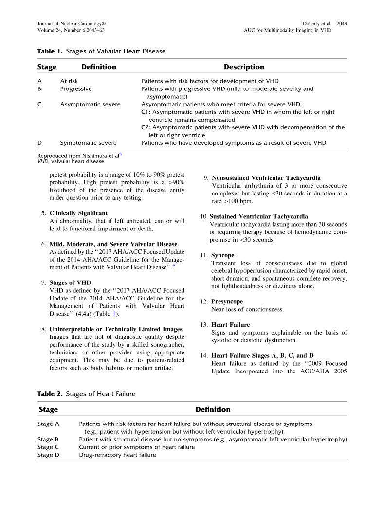
7. Stages of VHDVHD as defined by the ‘‘2017 AHA/ACC Focused
Update of the 2014 AHA/ACC Guideline for the
Management of Patients with Valvular Heart
Disease’’ (4,4a) (Table 1).
8. Uninterpretable or Technically Limited ImagesImages that are not of diagnostic quality despite
performance of the study by a skilled sonographer,
technician, or other provider using appropriate
equipment. This may be due to patient-related
factors such as body habitus or motion artifact.
9. Nonsustained Ventricular TachycardiaVentricular arrhythmia of 3 or more consecutive
complexes but lasting\30 seconds in duration at a
rate[100 bpm.
10 Sustained Ventricular TachycardiaVentricular tachycardia lasting more than 30 seconds
or requiring therapy because of hemodynamic com-
promise in\30 seconds.
11. SyncopeTransient loss of consciousness due to global
cerebral hypoperfusion characterized by rapid onset,
short duration, and spontaneous complete recovery,
not lightheadedness or dizziness alone.
12. PresyncopeNear loss of consciousness.
13. Heart FailureSigns and symptoms explainable on the basis of
systolic or diastolic dysfunction.
14. Heart Failure Stages A, B, C, and DHeart failure as defined by the ‘‘2009 Focused
Update Incorporated into the ACC/AHA 2005
Table 1. Stages of Valvular Heart Disease
Stage Definition Description
A At risk Patients with risk factors for development of VHD
B Progressive Patients with progressive VHD (mild-to-moderate severity and
asymptomatic)
C Asymptomatic severe Asymptomatic patients who meet criteria for severe VHD:
C1: Asymptomatic patients with severe VHD in whom the left or right
ventricle remains compensated
C2: Asymptomatic patients with severe VHD with decompensation of the
left or right ventricle
D Symptomatic severe Patients who have developed symptoms as a result of severe VHD
Reproduced from Nishimura et al5
VHD, valvular heart disease
Table 2. Stages of Heart Failure
Stage Definition
Stage A Patients with risk factors for heart failure but without structural disease or symptoms
(e.g., patient with hypertension but without left ventricular hypertrophy).
Stage B Patient with structural disease but no symptoms (e.g., asymptomatic left ventricular hypertrophy)
Stage C Current or prior symptoms of heart failure
Stage D Drug-refractory heart failure
Journal of Nuclear Cardiology� Doherty et al 2049
Volume 24, Number 6;2043–63 AUC for Multimodality Imaging in VHD
Guidelines for the Diagnosis and Management of
Heart Failure in Adults’’6 (Table 2).
15. IndicationSynonymous with scenario. A set of patient-specific
conditions defines ‘‘indication.’’ The term clinical
indication does not necessarily imply that testing is
warranted. In other words, for some clinical indi-
cations, all modalities may be rated as Rarely
Appropriate.
16. Low-Flow, Low-Gradient Valvular Aortic Steno-sisSevere aortic stenosis (AS) by valve area in the
presence of a low transaortic volume flow rate due
to either left ventricular (LV) systolic dysfunction
with a low LV ejection fraction (stage D2) or to a
small hypertrophied LV with a low stroke volume
(stage D3, also known as paradoxical low-flow AS).
17. Primary Mitral RegurgitationMitral regurgitation (MR) related to pathology of at
least 1 of the components of the valve (leaflets,
chordae tendineae, papillary muscles, or annulus)
resulting in valve incompetence.
18. Secondary MRMR in the presence of a relatively normal mitral
valve, related to LV dysfunction caused by coronary
artery disease, myocardial infarction (ischemic
chronic secondary MR), or idiopathic myocardial
disease (nonischemic chronic secondary MR). The
abnormal and dilated LV causes papillary muscle
displacement, which in turn results in leaflet
tethering and/or associated annular dilation that
prevents coaptation.
ABBREVIATIONS
AS = aortic stenosis
AUC = appropriate use criteria
CCT = cardiac computed tomography
LV = left ventricle/left ventricular
MR = mitral regurgitation
TAVR = transcatheter aortic valve replacement
TEE = transesophageal echocardiography
TTE = transthoracic echocardiography
VHD = valvular heart disease
MULTIMODALITY IMAGING IN VHD:APPROPRIATE USE CRITERIA (BY INDICATION)
Section 1: Initial Evaluation for VHD
See Tables 3 and 4.
Section 2: Prior Testing
See Tables 5, 6, 7, and 8.
Section 3: Transcatheter Intervention forVHD
See Tables 9, 10, 11, 12, 13, and 14.
Table 3. Initial Evaluation of an Asymptomatic Patient
3D, 3-dimensional; A, appropriate; CCT, cardiac computed tomography; CMR, cardiovascular magnetic resonance imaging; M,may be appropriate; R, rarely appropriate; TEE, transesophageal echocardiography; TTE, transthoracic echocardiography; VHD,valvular heart disease
2050 Doherty et al Journal of Nuclear Cardiology�AUC for Multimodality Imaging in VHD November/December 2017
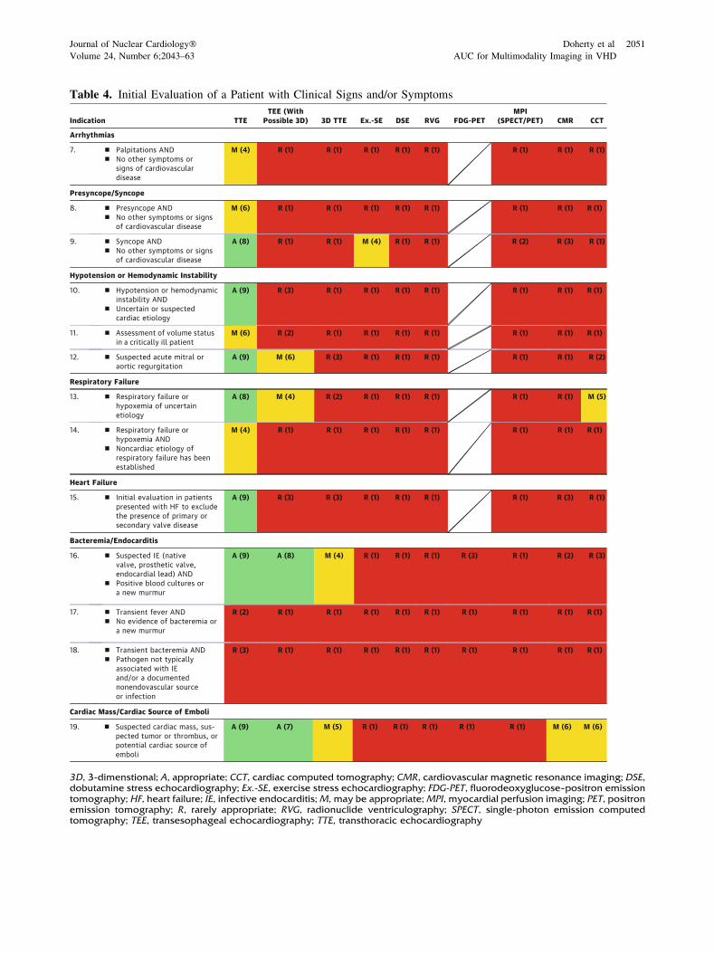
Table 4. Initial Evaluation of a Patient with Clinical Signs and/or Symptoms
3D, 3-dimenstional; A, appropriate; CCT, cardiac computed tomography; CMR, cardiovascular magnetic resonance imaging; DSE,dobutamine stress echocardiography; Ex.-SE, exercise stress echocardiography; FDG-PET, fluorodeoxyglucose–positron emissiontomography; HF, heart failure; IE, infective endocarditis; M, may be appropriate; MPI, myocardial perfusion imaging; PET, positronemission tomography; R, rarely appropriate; RVG, radionuclide ventriculography; SPECT, single-photon emission computedtomography; TEE, transesophageal echocardiography; TTE, transthoracic echocardiography
Journal of Nuclear Cardiology� Doherty et al 2051
Volume 24, Number 6;2043–63 AUC for Multimodality Imaging in VHD
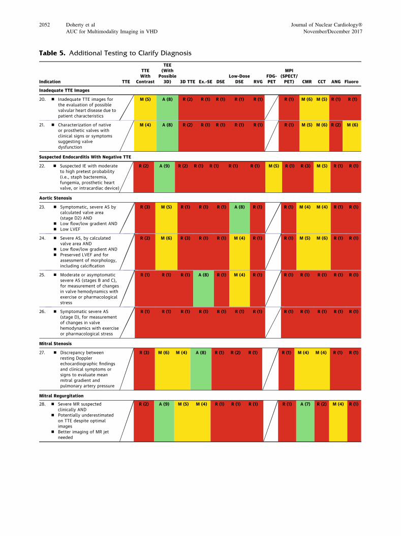
Table 5. Additional Testing to Clarify Diagnosis
2052 Doherty et al Journal of Nuclear Cardiology�AUC for Multimodality Imaging in VHD November/December 2017
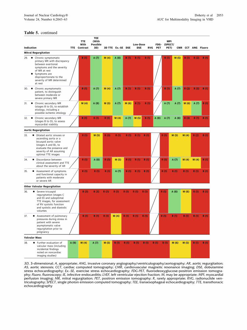
Table 5. continued
3D, 3-dimensional; A, appropriate; ANG, invasive coronary angiography/ventriculography/aortography; AR, aortic regurgitation;AS, aortic stenosis; CCT, cardiac computed tomography; CMR, cardiovascular magnetic resonance imaging; DSE, dobutaminestress echocardiography; Ex.-SE, exercise stress echocardiography; FDG-PET, fluorodeoxyglucose-positron emission tomogra-phy; Fluoro, fluoroscopy; IE, infective endocarditis; LVEF, left ventricular ejection fraction; M, may be appropriate; MPI, myocardialperfusion imaging; MR, mitral regurgitation; PET, positron emission tomography; R, rarely appropriate; RVG, radionuclide ven-triculography; SPECT, single photon emission computed tomography; TEE, transesophageal echocardiography; TTE, transthoracicechocardiography
Journal of Nuclear Cardiology� Doherty et al 2053
Volume 24, Number 6;2043–63 AUC for Multimodality Imaging in VHD
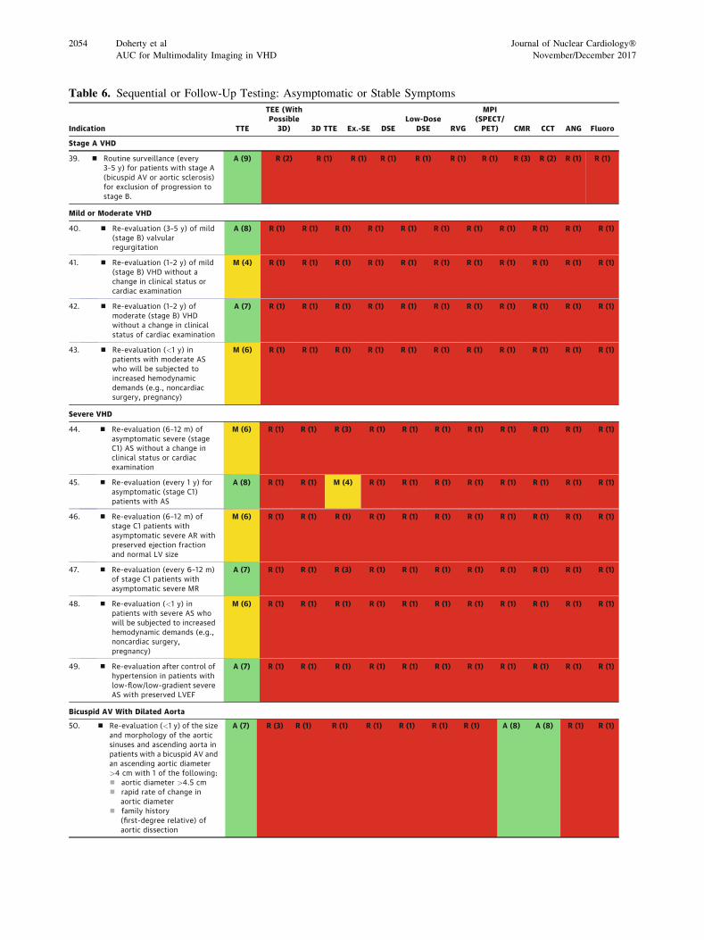
Table 6. Sequential or Follow-Up Testing: Asymptomatic or Stable Symptoms
2054 Doherty et al Journal of Nuclear Cardiology�AUC for Multimodality Imaging in VHD November/December 2017
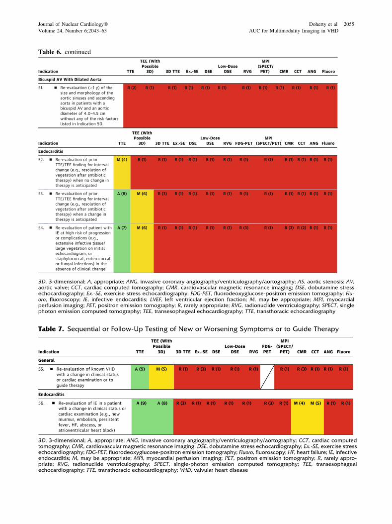
Table 7. Sequential or Follow-Up Testing of New or Worsening Symptoms or to Guide Therapy
3D, 3-dimensional; A, appropriate; ANG, invasive coronary angiography/ventriculography/aortography; CCT, cardiac computedtomography; CMR, cardiovascular magnetic resonance imaging; DSE, dobutamine stress echocardiography; Ex.-SE, exercise stressechocardiography; FDG-PET, fluorodeoxyglucose-positron emission tomography; Fluoro, fluoroscopy; HF, heart failure; IE, infectiveendocarditis; M, may be appropriate; MPI, myocardial perfusion imaging; PET, positron emission tomography; R, rarely appro-priate; RVG, radionuclide ventriculography; SPECT, single-photon emission computed tomography; TEE, transesophagealechocardiography; TTE, transthoracic echocardiography; VHD, valvular heart disease
Table 6. continued
3D, 3-dimensional; A, appropriate; ANG, invasive coronary angiography/ventriculography/aortography; AS, aortic stenosis; AV,aortic valve; CCT, cardiac computed tomography; CMR, cardiovascular magnetic resonance imaging; DSE, dobutamine stressechocardiography; Ex.-SE, exercise stress echocardiography; FDG-PET, fluorodeoxyglucose-positron emission tomography; Flu-oro, fluoroscopy; IE, infective endocarditis; LVEF, left ventricular ejection fraction; M, may be appropriate; MPI, myocardialperfusion imaging; PET, positron emission tomography; R, rarely appropriate; RVG, radionuclide ventriculography; SPECT, singlephoton emission computed tomography; TEE, transesophageal echocardiography; TTE, transthoracic echocardiography
Journal of Nuclear Cardiology� Doherty et al 2055
Volume 24, Number 6;2043–63 AUC for Multimodality Imaging in VHD
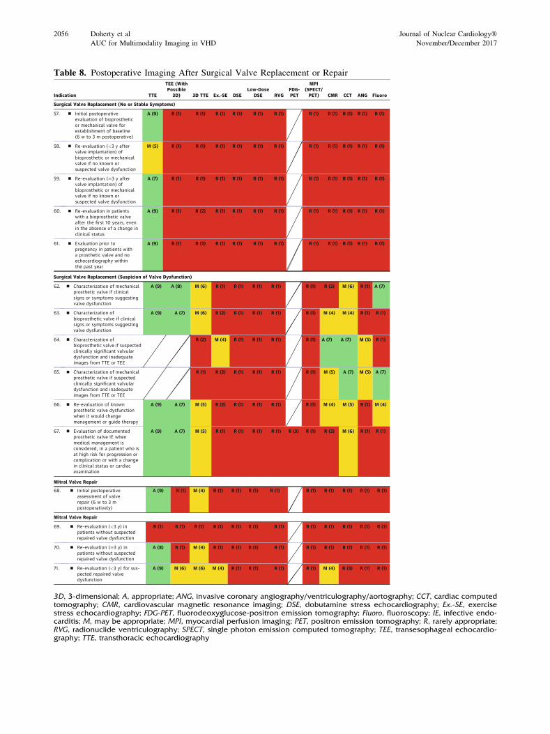
Table 8. Postoperative Imaging After Surgical Valve Replacement or Repair
3D, 3-dimensional; A, appropriate; ANG, invasive coronary angiography/ventriculography/aortography; CCT, cardiac computedtomography; CMR, cardiovascular magnetic resonance imaging; DSE, dobutamine stress echocardiography; Ex.-SE, exercisestress echocardiography; FDG-PET, fluorodeoxyglucose-positron emission tomography; Fluoro, fluoroscopy; IE, infective endo-carditis; M, may be appropriate; MPI, myocardial perfusion imaging; PET, positron emission tomography; R, rarely appropriate;RVG, radionuclide ventriculography; SPECT, single photon emission computed tomography; TEE, transesophageal echocardio-graphy; TTE, transthoracic echocardiography
2056 Doherty et al Journal of Nuclear Cardiology�AUC for Multimodality Imaging in VHD November/December 2017
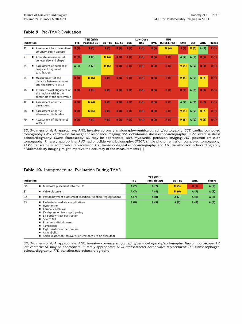
Table 9. Pre-TAVR Evaluation
3D, 3-dimensional; A, appropriate; ANG, invasive coronary angiography/ventriculography/aortography; CCT, cardiac computedtomography; CMR, cardiovascular magnetic resonance imaging; DSE, dobutamine stress echocardiography; Ex.-SE, exercise stressechocardiography; Fluoro, fluoroscopy; M, may be appropriate; MPI, myocardial perfusion imaging; PET, positron emissiontomography; R, rarely appropriate; RVG, radionuclide ventriculography; SPECT, single photon emission computed tomography;TAVR, transcatheter aortic valve replacement; TEE, transesophageal echocardiography; and TTE, transthoracic echocardiography*Multimodality imaging might improve the accuracy of the measurements (1)
Table 10. Intraprocedural Evaluation During TAVR
3D, 3-dimensional; A, appropriate; ANG, invasive coronary angiography/ventriculography/aortography; Fluoro, fluoroscopy; LV,left ventricle; M, may be appropriate; R, rarely appropriate; TAVR, transcatheter aortic valve replacement; TEE, transesophagealechocardiography; TTE, transthoracic echocardiography
Journal of Nuclear Cardiology� Doherty et al 2057
Volume 24, Number 6;2043–63 AUC for Multimodality Imaging in VHD
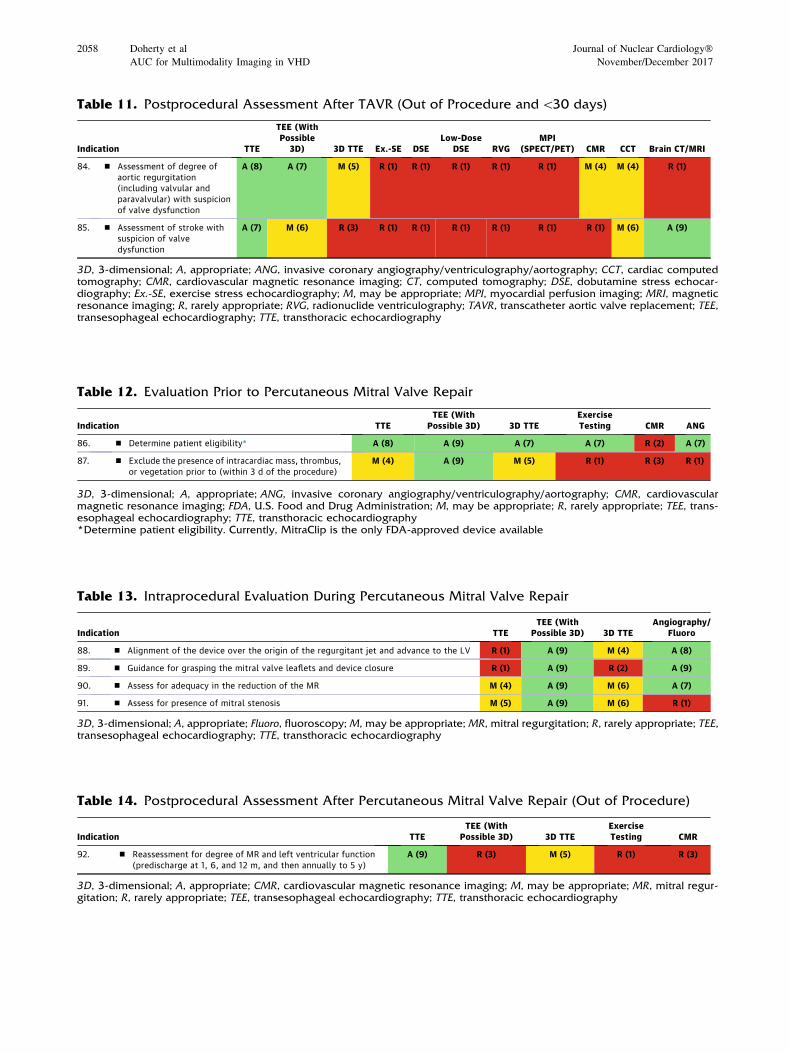
Table 11. Postprocedural Assessment After TAVR (Out of Procedure and\30 days)
3D, 3-dimensional; A, appropriate; ANG, invasive coronary angiography/ventriculography/aortography; CCT, cardiac computedtomography; CMR, cardiovascular magnetic resonance imaging; CT, computed tomography; DSE, dobutamine stress echocar-diography; Ex.-SE, exercise stress echocardiography; M, may be appropriate; MPI, myocardial perfusion imaging; MRI, magneticresonance imaging; R, rarely appropriate; RVG, radionuclide ventriculography; TAVR, transcatheter aortic valve replacement; TEE,transesophageal echocardiography; TTE, transthoracic echocardiography
Table 12. Evaluation Prior to Percutaneous Mitral Valve Repair
3D, 3-dimensional; A, appropriate; ANG, invasive coronary angiography/ventriculography/aortography; CMR, cardiovascularmagnetic resonance imaging; FDA, U.S. Food and Drug Administration; M, may be appropriate; R, rarely appropriate; TEE, trans-esophageal echocardiography; TTE, transthoracic echocardiography*Determine patient eligibility. Currently, MitraClip is the only FDA-approved device available
Table 13. Intraprocedural Evaluation During Percutaneous Mitral Valve Repair
3D, 3-dimensional; A, appropriate; Fluoro, fluoroscopy; M, may be appropriate; MR, mitral regurgitation; R, rarely appropriate; TEE,transesophageal echocardiography; TTE, transthoracic echocardiography
Table 14. Postprocedural Assessment After Percutaneous Mitral Valve Repair (Out of Procedure)
3D, 3-dimensional; A, appropriate; CMR, cardiovascular magnetic resonance imaging; M, may be appropriate; MR, mitral regur-gitation; R, rarely appropriate; TEE, transesophageal echocardiography; TTE, transthoracic echocardiography
2058 Doherty et al Journal of Nuclear Cardiology�AUC for Multimodality Imaging in VHD November/December 2017
DISCUSSION
AUC are intended to inform clinicians, patients, and
health policy makers about the reasonable use of tech-
nologies to help improve patient symptoms and health
outcomes. Since 2005, the ACC, along with its profes-
sional partners, has worked to provide criteria for both
invasive and noninvasive testing and selected treat-
ments, with the intention of further expanding the AUC
portfolio.1,2,7,11–14
The ‘‘2017 Appropriate Use Criteria for Multi-
modality Imaging in Valvular Heart Disease’’ is the
culmination of the analysis of various modalities used in
the evaluation and treatment of patients with VHD. This
document signals a shift from documents evaluating a
single modality in various disease states to documents
evaluating multiple imaging modalities and focusing on
evidence and clinical experience within a given category
of disease. We believe that this approach better reflects
clinical decision making in real-world scenarios and
offers the diagnostic choices available to the clinician.
Because a given modality may address diverse dis-
ease states, indications previously compiled in a single
document may be spread over several AUC documents.
The previous VHD–related indications that the current
paper supplants are contained in the echocardiography,14
radionuclide imaging,13 and computed tomogra-
phy/magnetic resonance imaging11,12 AUC documents.
Other indications in these documents remain in force until
these scenarios are evaluated in subsequent documents.
The tables in this paper are organized to reflect the
spectrum of patients with VHD—from patients with no
symptoms suspected of having VHD to patients with signs
and symptoms ranging from mild to severe. The first 2
tables are for initial evaluation when no prior imaging has
been done. As is noted, the diagnostic choices vary
between the tables and reflect the options that would be
considered in the initial evaluation by most clinicians. If a
diagnostic test would seldom or never be considered, it
was not included as an option for the rating panel.
In the asymptomatic patient either who is at risk of
developing VHD or in whom VHD was clinically sus-
pected, TTE was rated Appropriate for these indications.
Three-dimensional (3D) TTE was rated May Be
Appropriate for indications 2 and 3. All other modalities
(computed tomography, magnetic resonance imaging,
and TEE) were rated Rarely Appropriate. These are new
indications, so there are no prior ratings in older docu-
ments for comparison.
Table 4 evaluates the symptomatic patient. This
table adds exercise stress echocardiography, dobutamine
stress echocardiography, radionuclide ventriculography,
fluorodeoxyglucose–positron emission tomography, and
myocardial perfusion imaging/single-photon emission
computed tomography/positron emission tomography.
In general, echocardiography was the preferred option
for initial testing in such patients. The ratings correlate
well with those in the prior echocardiography AUC,14
with the exception of the evaluation of presyncope,
which was rated May Be Appropriate here and Inap-
propriate (‘‘I’’ in the old nomenclature) in the prior
document. This difference is minor and is attributable to
the fact that the symptom of lightheadedness was
included with presyncope in the older document, which
may have prompted the rating panel to apply a lower
rating to echocardiography. All other ratings in this
table are either in line with prior rankings or are new
scenarios not included in prior documents.
Table 5 evaluates the use of subsequent imaging in
scenarios in which prior imaging—presumably using
TTE—did not yield a clear diagnosis. The diagnostic
options are the same as in Table 4, with the exclusion of
TTE. The table is further subdivided into inadequate
TTE images, suspected endocarditis, various types of
VHD, and valvular mass.
In Table 5, TEE is rated Appropriate and TTE with
contrast as May Be Appropriate in evaluating native and
prosthetic valves with inadequate images.21,22 TEE is
also rated Appropriate and fluorodeoxyglucose–positron
emission tomography as May Be Appropriate in the
diagnosis of endocarditis in patients with a negative
TTE. Scenarios 23 to 25 identify the role of low-dose
dobutamine stress echocardiography in patients with
low-flow, low-gradient severe aortic stenosis (with low
ejection fraction as Appropriate and preserved ejection
fraction as May Be Appropriate).23–25 Exercise stress
echocardiography and dobutamine stress echocardiog-
raphy were rated Rarely Appropriate in patients with
severe, symptomatic AS. The common conundrum of
evaluating the severity of MR—examined in scenarios
28 to 32—particularly distinguishing moderate from
severe MR, elucidating the discrepancy between symp-
toms and severity, and evaluating an ischemic etiology
of MR, demonstrates the role of various modalities in
these very specific but very common scenarios.26 These
indications are new and are not included in prior
documents.
Table 6 evaluates sequential or follow-up imaging
in various stages of VHD and incorporates the newer
VHD classification4 where TTE ratings are in line with
the prior echocardiography AUC14 and reflect the pri-
macy of TTE at appropriate intervals in following
patients with VHD. Time intervals shorten with the
severity of VHD, and the role of exercise stress
echocardiography—rated May Be Appropriate—in
evaluating patients with severe and asymptomatic AS to
aid in clinical decision making is highlighted. TTE in
patients with moderate or severe AS imaged with a\1-
Journal of Nuclear Cardiology� Doherty et al 2059
Volume 24, Number 6;2043–63 AUC for Multimodality Imaging in VHD
year time interval when subjected to increased hemo-
dynamic demands is rated May Be Appropriate and can
be considered on a case-by-case basis. The utility of
cardiac computed tomography (CCT) or cardiovascular
magnetic resonance imaging in evaluating the ascending
aorta in patients with a bicuspid aortic valve is defined in
indications 49 to 51.
Table 7 evaluates new or worsening symptoms. In
the general scenarios, TTE is rated Appropriate and TEE
is rated May Be Appropriate. In the specific endocarditis
scenario, both TTE and TEE are rated Appropriate.
Table 8 evaluates postoperative imaging in patients
undergoing surgical valve replacement and/or mitral
repair. In patients with no symptoms (indications 57 to
61), the interval of follow-up (which is limited to TTE)
aligns well with the prior document, with the exception
of the evaluation of a mechanical or bioprosthetic valve
with TTE in\3 years—indication 58.14 In the current
document, it is rated May Be Appropriate. In the prior
AUC, it was rated Inappropriate (old nomenclature).
Reasons for this difference are not apparent, but may be
related to rating panel composition, which can account
for small differences. The authors suggest that there are
cases in which follow-up imaging may be done in a
shorter time frame, such as small prosthesis size and an
elevated transvalvular gradient by Doppler.
Whereas TTE is the modality of choice in the
asymptomatic patient, TEE is considered Appropriate,
and 3D TTE May Be Appropriate and useful in the
evaluation of patients with suspected prosthetic valve
dysfunction.
Section 6.3. (Tables 9, 10, 11, 12, 13, 14) evaluates
the dynamic field of structural valve interventions.
Tables 9, 10, 11 cover preprocedural, intraprocedural,
and postprocedural imaging for transcatheter aortic
valve replacement (TAVR) for AS.27,28 Table 9 cata-
logues all of the necessary measurements in the pre-
TAVR evaluation. It is worth noting that this table cov-
ers the imaging support needed and not whether the
procedure should be done. The latter is being evaluated
in an AUC document for severe AS, which is currently
under development. It is in the AS AUC that CCT and
cardiovascular magnetic resonance imaging, as
advanced imaging techniques, establish themselves as
essential technologies for planning these procedures.
Likewise, assessment for concomitant coronary artery
disease is accomplished through CCT, myocardial per-
fusion imaging/single-photon emission computed
tomography/positron emission tomography, and
angiography.
Intraprocedural evaluation (Table 10) is accom-
plished with TTE, TEE, angiography, and fluoroscopy.
Because TAVR procedures are increasingly being per-
formed with conscious sedation, TTE29 is being
increasingly used in lieu of TEE. Both modalities are
rated Appropriate.
Postprocedural assessment (Table 11) for valve
dysfunction can be accomplished with TTE or TEE
rated as Appropriate tests, with the additional use of 3D
TTE rated as May Be Appropriate. CCT or cardiovas-
cular magnetic resonance imaging are both rated May
Be Appropriate. For assessment of stroke, TTE is rated
Appropriate, whereas TEE and CCT are rated May Be
Appropriate. Brain imaging with computed tomography
or magnetic resonance imaging is rated Appropriate.
For percutaneous mitral valve repair (Table 12, 13,
14), there is only 1 U.S. Food and Drug Administration–
approved device and imaging support, especially in
follow-up, hence, the U.S. Food and Drug Administra-
tion–directed protocol.30 Patient eligibility (including
assessment for concomitant coronary artery disease) is
assessed with TTE, TEE, 3D TTE, exercise testing of
various types, and coronary angiography, all of which
are rated Appropriate. If there is concern regarding an
intracardiac mass, thrombus, or vegetation, this is
assessed with TEE, as Appropriate, whereas TTE is
rated as May Be Appropriate, as is 3D TTE.
Intraprocedural assessment is accomplished with
TEE as Appropriate and angiography/fluoroscopy as
Appropriate for all measures except for the presence of
mitral stenosis, which is assessed with TEE as Appro-
priate. TTE and 3D TTE are also useful for some
determinations during the procedure as May Be
Appropriate, but TEE offers a more comprehensive
examination and is rated Appropriate.
The postprocedure assessment is currently deter-
mined by U.S. Food and Drug Administration
regulations and involves echocardiography predischarge
at 1, 6, and 12 months and annually up to 5 years. TTE is
rated Appropriate and 3D TTE is rated May Be
Appropriate.
CONCLUSIONS
This document assesses a wide array of imaging
modalities available to the clinician in the evaluation
of patients with VHD. Presented here is a broad
spectrum of clinical scenarios in such patients. Some
of these scenarios replicate those of prior documents,
but many are new, specifically, structural valve
interventions, which were not in the armamentarium
of clinicians when prior, single-modality documents
were published. Where comparisons can be made,
the ratings are remarkably consistent with prior
documents.
We believe the multimodality approach more clo-
sely replicates clinical decision making and will be
useful. Future documents will not provide single-source
2060 Doherty et al Journal of Nuclear Cardiology�AUC for Multimodality Imaging in VHD November/December 2017
guidance for appropriateness in all disease states.
Echocardiography indications, for example, will be
spread across complimentary documents such as multi-
modality stable ischemic heart disease AUC,
multimodality structural heart disease AUC, the current
document, and multimodality preoperative evaluation
AUC, which is under development.
A few clinical scenarios, describing evaluation
of symptoms that could be secondary to valvular or
structural heart disease, can be found in both doc-
uments (e.g., the evaluation of pre-syncope/syncope
in Table 4). Although these scenarios were devel-
oped against a background of both valvular and
structural heart disease, they were rated separately
in the context of other clinical scenarios focused on
either valvular or structural heart disease. The
writing group and its representatives have placed
particular emphasis on this issue during all stages of
the development of the AUC document to avoid
discordant recommendations for these scenarios.
As with all prior documents, the evaluation is a
product of current guidelines, where available, and
expert consensus. The modalities are not to be
considered in a rank order and may be used relative
to individual patient circumstances and risk versus
benefit. Accordingly, a study rated May Be Appro-
priate should not be denied reimbursement in lieu of
one rated Appropriate. There will be individual
circumstances when a study ranked Rarely Appro-
priate may be clinically useful if properly
documented.
ACC PRESIDENT AND STAFF
Mary Norine Walsh, MD, FACC, President
Shalom Jacobovitz, Chief Executive Officer
William J. Oetgen, MD, FACC, Executive Vice Presi-
dent, Science, Education, Quality, and Publishing
Joseph M. Allen, MA, Team Leader, Clinical Policy and
Pathways
Leah White, MPH, CCRP, Team Leader, Appropriate
Use Criteria
Marıa Velasquez, Senior Research Specialist, Appro-
priate Use Criteria
Amelia Scholtz, PhD, Publications Manager, Science,
Education, Quality, and Publishing
RATING PANEL MEMBERS
Greg J. Dehmer, MD, MACC, MSCAI, FACP,
FAHA, Moderator*, John U. Doherty, MD, FACC,
FAHA, Writing Group Liaison*, Paul Schoenhagen,
MD, FAHA, Writing Group Liaison§, Zahid Amin, MD,
FSCAI, FAHA�, Thomas M. Bashore, MD, FACC*,
Andrew Boyle, MD*, Dennis A. Calnon, MD, FACC,
FASE, MASNC, FSCCTk, Blase Carabello, MD,
FACC*, Manuel D. Cerqueira, MD, FACC, MASNC*,
John Conte, MD}, Milind Desai, MD, FACC*, Daniel
Edmundowicz, MD, FACC*, Victor A. Ferrari, MD,
FACC#, Brian Ghoshhajra MD, MBA§, Praveen
Mehrotra, MD, FACC*, Saman Nazarian, MD, PHD**,
T. Brett Reece, MD��, Balaji Tamarappoo, MD, PHD*,
Wendy S. Tzou, MD, FACC, FHRS��, John B. Wong,
MD. }Society of Thoracic Surgeons Representative.#Society for Cardiovascular Magnetic Resonance Rep-
resentative. **Heart Rhythm Society Representative.��American Association for Thoracic Surgery Repre-
sentative. ��American Heart Association Representative.
APPROPRIATE USE CRITERIA TASK FORCE
John U. Doherty, MD, FACC, FACP, FAHA, Co-
Chair, Gregory J. Dehmer, MD, MACC, MSCAI, FACP,
FAHA, Co-Chair, Steven R. Bailey, MD, FACC,
MSCAI, FAHA, Nicole M. Bhave, MD, FACC, Alan S.
Brown, MD, FACC§§, Stacie L. Daugherty, MD, FACC,
Larry S. Dean, MD, FACC, MSCAI, Milind Y. Desai,
MBBS, FACC, Claire S. Duvernoy, MD, FACC§§,
Linda D. Gillam, MD, FACC, Robert C. Hendel, MD,
FACC, FAHA§§, Christopher M. Kramer, MD, FACC,
FAHAkk, Bruce D. Lindsay, MD, FACC§§, Warren J.
Manning, MD, FACC, Praveen Mehrotra, MD, FACC,
FASE, Manesh R. Patel, MD, FACC, FSCAI, FAHA}},
Ritu Sachdeva, MBBS, FACC, L. Samuel Wann, MD,
MACC§§, David E. Winchester, MD, FACC, Michael J.
Wolk, MD, MACC§§, Joseph M. Allen, MA.§§Former
Task Force member; current member during writing
effort. kkFormer Task Force co-chair; current co-chair
during writing effort. }}Former Task Force chair; cur-
rent chair during writing effort.
References
1. Patel MR, Spertus JA, Brindis RG, et al. ACCF proposed method
for evaluating the appropriateness of cardiovascular imaging. J
Am Coll Cardiol. 2005;46:1606–13.
2. Hendel RC, Patel MR, Allen JM, et al. Appropriate use of car-
diovascular technology: 2013 ACCF appropriate use criteria
methodology update: A report of the American College of Car-
diology Foundation Appropriate Use Criteria Task Force. J Am
Coll Cardiol. 2013;61:1305–17.
3. Fitch K, Bernstein SJ, Aguilar MD, et al. The RAND/UCLA
Appropriateness Method User’s Manual. Arlington, VA: RAND;
2001.
4. Nishimura RA, Otto CM, Bonow RO, et al. 2017 AHA/ACC
focused update of the 2014 AHA/ACC Guideline for the Man-
agement of Patients With Valvular Heart Disease. J Am Coll
Cardiol. 2017;70:252–89.
5. Nishimura RA, Otto CM, Bonow RO, et al. 2014 AHA/ACC
guideline for the management of patients with valvular heart
Journal of Nuclear Cardiology� Doherty et al 2061
Volume 24, Number 6;2043–63 AUC for Multimodality Imaging in VHD
disease: A report of the American College of Cardiology/Ameri-
can Heart Association Task Force on Practice Guidelines. J Am
Coll Cardiol. 2014;63:e57–185.
6. Hunt SA, Abraham WT, Chin MH, et al. 2009 focused update
incorporated into the ACC/AHA 2005 Guidelines for the Diag-
nosis and Management of Heart Failure in Adults: A report of the
American College of Cardiology Foundation/American Heart
Association Task Force on Practice Guidelines. J Am Coll Cardiol.
2009;53:e1–90.
7. Wolk MJ, Bailey SR, Doherty JU, et al. 2013 ACCF/AHA/ASE/
ASNC/HFSA/HRS/SCAI/SCCT/SCMR/STS multimodality
appropriate use criteria for the detection and risk assessment of
stable ischemic heart disease: A report of the American College of
Cardiology Foundation Appropriate Use Criteria Task Force,
American Heart Association, American Society of Echocardiogra-
phy, American Society of Nuclear Cardiology, Heart Failure Society
of America, Heart Rhythm Society, Society for Cardiovascular
Angiography and Interventions, Society of Cardiovascular Computed
Tomography, Society for Cardiovascular Magnetic Resonance, and
Society of Thoracic Surgeons. J Am Coll Cardiol. 2014;63:380–406.
8. Fihn SD, Blankenship JC, Alexander KP, et al. 2014 ACC/AHA/
AATS/PCNA/SCAI/STS focused update of the guideline for the
diagnosis and management of patients with stable ischemic heart
disease: A report of the American College of Cardiology/American
Heart Association Task Force on Practice Guidelines, and the
American Association for Thoracic Surgery, Preventive Cardio-
vascular Nurses Association, Society for Cardiovascular
Angiography and Interventions, and Society of Thoracic Surgeons.
J Am Coll Cardiol. 2014;64:1929–49.
9. Warnes CA, Williams RG, Bashore TM, et al. 2008 ACCF/AHA
guidelines for the management of adults with congenital heart
disease: A report of the American College of Cardiology Foun-
dation/American Heart Association Task Force on Practice
Guidelines (Writing Committee to Develop Guidelines on the
Management of Adults With Congenital Heart Disease). J Am
Coll Cardiol. 2008;52:e1–121.
10. Hiratzka LF, Bakris GL, Beckman JA, et al. 2010 ACCF/AHA/
AATS/ACR/ASA/SCA/SCAI/SIR/STS/SVM guidelines for the
diagnosis and management of patients with thoracic aortic disease:
A report of the American College of Cardiology Foundation/
American Heart Association Task Force on Practice Guidelines,
American Association for Thoracic Surgery, American College of
Radiology, American Stroke Association, Society of Cardiovas-
cular Anesthesiologists, Society for Cardiovascular Angiography
and Interventions, Society of Interventional Radiology, Society of
Thoracic Surgeons, and Society for Vascular Medicine. J Am Coll
Cardiol. 2010;55:e27–129.
11. Hendel RC, Patel MR, Kramer CM, et al. 2006 ACCF/ACR/
SCCT/SCMR/ASNC/NASCI/SCAI/SIR appropriateness criteria
for cardiac computed tomography and cardiac magnetic resonance
imaging: A report of the American College of Cardiology Foun-
dation Quality Strategic Directions Committee Appropriateness
Criteria Working Group, American College of Radiology, Society
of Cardiovascular Computed Tomography, Society for Cardio-
vascular Magnetic Resonance, American Society of Nuclear
Cardiology, North American Society for Cardiac Imaging, Society
for Cardiovascular Angiography and Interventions, and Society of
Interventional Radiology. J Am Coll Cardiol. 2006;48:1475–97.
12. Taylor AJ, Cerqueira M, Hodgson JM, et al. 2010 ACCF/SCCT/
ACR/AHA/ASE/ASNC/NASCI/SCAI/SCMR appropriate use cri-
teria for cardiac computed tomography: A report of the American
College of Cardiology Foundation Appropriate Use Criteria Task
Force, the Society of Cardiovascular Computed Tomography, the
American College of Radiology, the American Heart Association,
the American Society of Echocardiography, the American Society
of Nuclear Cardiology, the North American Society for Cardio-
vascular Imaging, the Society for Cardiovascular Angiography and
Interventions, and the Society for Cardiovascular Magnetic Res-
onance. J Am Coll Cardiol. 2010;56:1864–94.
13. Hendel RC, Berman DS, Di Carli MF, et al. 2009 ACCF/ASNC/
ACR/AHA/ASE/SCCT/SCMR/SNM appropriate use criteria for
cardiac radionuclide imaging: A report of the American College of
Cardiology Foundation Appropriate Use Criteria Task Force, the
American Society of Nuclear Cardiology, the American College of
Radiology, the American Heart Association, the American Society
of Echocardiography, the Society of Cardiovascular Computed
Tomography, the Society for Cardiovascular Magnetic Resonance,
and the Society of Nuclear Medicine. J Am Coll Cardiol.
2009;53:2201–29.
14. Douglas PS, Garcia MJ, Haines DE, et al. 2011 ACCF/ASE/AHA/
ASNC/HFSA/HRS/SCAI/SCCM/SCCT/SCMR appropriate use
criteria for echocardiography: A report of the American College of
Cardiology Foundation Appropriate Use Criteria Task Force,
American Society of Echocardiography, American Heart Associ-
ation, American Society of Nuclear Cardiology, Heart Failure
Society of America, Heart Rhythm Society, Society for Cardio-
vascular Angiography and Interventions, Society of Critical Care
Medicine, Society of Cardiovascular Computed Tomography, and
Society for Cardiovascular Magnetic Resonance. J Am Coll Car-
diol. 2011;57:1126–66.
15. Halliburton SS, Abbara S, Chen MY, et al. SCCT guidelines on
radiation dose and dose-optimization strategies in cardiovascular
CT. J Cardiovasc Comput Tomogr. 2011;5:198–224.
16. Pellikka PA, Nagueh SF, Elhendy AA, et al. American Society of
Echocardiography recommendations for performance, interpretation,
and application of stress echocardiography. J Am Soc Echocardiogr.
2007;20:1021–41.
17. Hansen CL, Goldstein RA, Akinboboye OO, et al. Myocardial
perfusion and function: Single photon emission computed
tomography. J Nucl Cardiol. 2007;14:e39–60.
18. Kramer CM, Barkhausen J, Flamm SD, et al. Standardized car-
diovascular magnetic resonance imaging (CMR) protocols,
Society For Cardiovascular Magnetic Resonance: Board of Trus-
tees Task Force On Standardized Protocols. J Cardiovasc Magn
Reson. 2008;10:35.
19. Abbara S, Blanke P, Maroules CD, et al. SCCT guidelines for the
performance and acquisition of coronary computed tomographic
angiography: A report of the Society of Cardiovascular Computed
Tomography Guidelines Committee. J Cardiovasc Comput Tomogr.
2016;10:435–49.
20. Naidu SS, Rao SV, Blankenship J, et al. Clinical expert consensus
statement on best practices in the cardiac catheterization labora-
tory: Society for Cardiovascular Angiography and Interventions.
Catheter Cardiovasc Interv. 2012;80:456–64.
21. Ward RP, Mansour IN, Lemieux N, et al. Prospective evaluation
of the clinical application of the American College of Cardiology
Foundation/American Society of Echocardiography Appropriate-
ness Criteria for Transthoracic Echocardiography. J Am Coll
Cardiol Img. 2008;1:663–71.
22. Bai AD, Steinberg M, Showler A, et al. Diagnostic accuracy of
transthoracic echocardiography for infective endocarditis findings
using transesophageal echocardiography as the reference standard:
A meta-analysis. J Am Soc Echocardiogr. 2017;30(639–46):e8.
23. Clavel MA, Burwash IG, Pibarot P. Cardiac imaging for assessing
low-gradient severe aortic stenosis. J Am Coll Cardiol Img.
2017;10:185–202.
24. Tribouilloy C, Levy F, Rusinaru D, et al. Outcome after aortic
valve replacement for low-flow/low-gradient aortic stenosis
2062 Doherty et al Journal of Nuclear Cardiology�AUC for Multimodality Imaging in VHD November/December 2017
without contractile reserve on dobutamine stress echocardiogra-
phy. J Am Coll Cardiol. 2009;53:1865–73.
25. Levy F, Laurent M, Monin JL, et al. Aortic valve replacement for
low-flow/low-gradient aortic stenosis operative risk stratification
and long-term outcome: A European multicenter study. J Am Coll
Cardiol. 2008;51:1466–72.
26. Uretsky S, Gillam L, Lang R, et al. Discordance between echocar-
diography and MRI in the assessment of mitral regurgitation severity:
A prospective multicenter trial. J Am Coll Cardiol. 2015;65:1078–88.
27. Hahn RT, Little SH, Monaghan MJ, et al. Recommendations for
comprehensive intraprocedural echocardiographic imaging during
TAVR. J Am Coll Cardiol Img. 2015;8:261–87.
28. Otto CM, Kumbhani DJ, Alexander KP, et al. 2017 ACC expert
consensus decision pathway for transcatheter aortic valve
replacement in the management of adults with aortic stenosis: A
report of the American College of Cardiology Task Force on
Clinical Expert Consensus Documents. J Am Coll Cardiol.
2017;69:1313–46.
29. Sengupta PP, Wiley BM, Basnet S, et al. Transthoracic echocar-
diography guidance for TAVR under monitored anesthesia care. J
Am Coll Cardiol Img. 2015;8:379–80.
30. Sorajja P, Mack M, Vemulapalli S, et al. Initial experience with
commercial transcatheter mitral valve repair in the United States. J
Am Coll Cardiol. 2016;67:1129–40.
Journal of Nuclear Cardiology� Doherty et al 2063
Volume 24, Number 6;2043–63 AUC for Multimodality Imaging in VHD


![arXiv:1801.07455v2 [cs.CV] 25 Jan 2018...convolutional neural networks (CNNs) to graphs of arbi-trary structures, have received increasing attention and suc-cessfully been adopted](https://img.pdfslide.us/doc/110x75/602bb3aee112322e7f55db1f/arxiv180107455v2-cscv-25-jan-convolutional-neural-networks-cnns-to-graphs.jpg)

![Applying the Nash Bargaining Solution for a Reasonable Royalty · tic [7]. This paper shows Nash’s solution with an arbi-trary bargaining weight to account for unequal bargain-ing](https://img.pdfslide.us/doc/110x75/5fd3b9e6b0408940ff539fe3/applying-the-nash-bargaining-solution-for-a-reasonable-royalty-tic-7-this-paper.jpg)