Embed Size (px)
Citation preview
32
Case Report
Appropriate planning, thorough examinations and decent
implementations for a predictable prognosis in the man-
agement of a severely worn dentition: A case report
Siti Mariam Ab Ghani*,1 and Graeme Lillywhite
2
1Head, Centre for Restorative Dentistry Studies, Faculty of Dentistry, Universiti Teknologi MARA, 40450 Shah Alam, Selangor, Malaysia 2Consultant, Restorative Department, Edinburgh Dental Institute, Edinburgh, United Kingdom Institution: Centre for Restorative Dentistry Studies, Faculty of Dentistry, Universiti Teknologi MARA, 40450 Shah Alam, Selangor, Malaysia
Abstract The management of patients with severely worn dentition is challenging due to the loss of occlu-
sal vertical dimension and tooth structure creating an uneven plane of occlusion. This case report describes the
importance of every step of the conventional and improvised methods in treating tooth wear patients. Stages
from the initial work-up of tooth wear assessment, substantial surgical crown lengthening, the controlled method
of increasing vertical dimension, the precise method of crown preparations, advanced impression techniques
till the cementation procedure of final restorations.
The whole treatment was in a reorganized approach such that the new inter-cuspal position (ICP) coincided
with the retruded axial position (RAP). When restoring worn dentition, clinician should always have a proper
planning, decent implementation for each stages thus guarantee excellent performances. However mainte-
nance and recall visits are the main keys to long term success.
Keywords: crown lengthening, locating impressions, occlusal vertical dimension, tooth wear, silver
dies
Introduction
Tooth wear is a physiological loss of tooth
structure due to prolong usage/ retain especial-
ly in the elderly. However, it can be regarded as
pathological if the teeth become excessively
worn and do not function effectively or affect
the aesthetic appearances (Van‘t Spijker et al.,
2007). The aetiology of tooth wear can be from
erosion, abrasion, attrition and abfraction. Ero-
sion is defined as loss of tooth structure due to
acidic and non-bacterial tooth wear that can be
cause by intrinsic and extrinsic factor. Extrinsic
causes are like diet (citrus and isotonic drinks),
habits (swishing soft drinks and sucking sour
foods) and occupation (swimmer in chlorinated
pool water, battery factory worker, bar tender
and wine tester).
Intrinsic causes of erosion can be related with
medical history such as gastric reflux, bullimea,
reduced salivary flow and pregnancy. Clinically,
‗cupping‘ of the incisors (perimolysis) can be
seen as the dentine has lower resistance to
wear and erosion compared to enamel, matt
surface of the enamel and dentine exposed
with continuous erosion and ‗proud‘ amalgam
restoration. In intrinsic causes, the erosion is
usually at the palatal of anterior teeth when the
acid regurgitate forcibly. In extrinsic etiology,
more surfaces are generally involved. Attrition
*Correspondence to: Dr Siti Mariam Ab Ghani, Head, Cen-tre for Restorative Dentistry Studies, Faculty of Dentistry,Universiti Teknologi MARA, 40450 Shah Alam, Selangor,MalaysiaEmail: [email protected]: +60355211955 (office)/+60122306980(mobile)
Compendium of Oral Science Volume 1|2014

33
is caused by grinding and contact of teeth to
teeth. It can be due to parafunctional such as
bruxism and clenching, loss of posterior teeth
resulting in higher occlusal load of anterior
teeth, dentition opposed with ceramic crown
and also in patient with malocclusion Class ll
Div ll. The lesions clinically appear as well-
defined wear facets on the functional surface
matching of the wear facets of opposing denti-
tion. The enamel and dentine wear at the same
level. Abrasion is defined as loss of tooth struc-
ture due to excessive tooth and foreign object
contact (Allen, 2003). The classical example is
mechanical trauma from tooth-brushing and
biting of foreign object such pipe in pipe smok-
ers, pins for hairdresser or pens. Abfraction is
loss of tooth structure at cervical area due to
occlusal load with the clinical characteristic of
wedge-shaped Class V lesion. During tensile
force, the enamel rods which are less elastic
than dentine will fracture thus creating lesion at
the cervical region. In daily practice, it is difficult
to diagnose an initial tooth wear condition as
sometimes patient does not recognize the sign
and symptom themselves or might not volun-
teer to reveal sensitive condition as eating dis-
order. Smith and Knight (1984) did publish a
tooth wear index (TWI) to help in diagnosis and
monitoring of tooth wear. Each tooth have 4
surface recorded (cervical, labial, incisal/
occlusal and lingual surfaces) with a value of 0-
4. This TWI can be used to monitor individual
patient by year recall or even produce profiles
of tooth wear distribution in a specific group of
patient. There are also other investigations that
can be done for assessment of a potential tooth
wear case prior achieving a definite diagnosis.
Medical questionnaire - investigate gastric reflux condition, vomiting frequencies, psychologi-
cal status.
Mounted study cast on the articulator - occlusal and space analysis.
Intra-oral photoraphs taken at interval time.
Imaging - Peri-apicals (PA), Dentopantograph (DPT), Cone Beam Computer Tomography CBCT)
Resting and simulated salivary flow test
Diet diary- if suspected erosion elements
Table 1:. Additional investigations in tooth wear management
When the diagnosis is confirmed, the first step
in tooth wear management is to avoid further
tooth loss by addressing the aetiology. Once
the aetiology and tooth wear is well controlled,
the definitive treatment can be started. Treat-
ment plan can be formulated after thorough
investigations and discussion with the patient.
Case Report
Mrs. MS, a 45 year old female patient, was
referred for the poor appearance of her upper
front teeth which were worn down and sensitive
to cold foods. The tooth surface loss had been
identified and highlighted by her dentist one
year previously who suggested that her high
daily intake of citrus fruits was the probable
cause. MS reported that she was generally in
good health, no known allergies to medications,
non smoker and drinks alcohol occasionally.
Examination And Findings
Extra-oral- Patient has a skeletal pattern Class I
with no adenopathy, trismus or facial asym-
metry noted. The temporo-mandibular joint
(TMJ) was not tender to palpation, no sounds
and no deviation upon opening and closing the
mouth. The amounts of maxillary teeth dis-
played during speech were very minimal with a
reverse smile line.
Compend. Oral Sci:vol1(5);2014;32-41

34
Intra-oral -
Occlusion - Class III incisal relationship -
edge to edge (Fig. 1). Group function
(canine and premolar guidance) in right and
left excursions with minimal disclusion of
posterior teeth. Mandible was easily manip-
ulated into retruded axial position (RAP)
with initial contact between the distal
ridge of the 46 and the mesio-palatal cusp
of 16
Moderate to severe tooth surface loss was
noted palatal to 13 to 23 (Fig. 2) with
substantial loss of tooth structure and
cupped appearance (perimolysis).
Maxillary and mandibular posterior teeth
had mild tooth surface loss. Slight cupping
of cusp tips of premolars, intact and proud
standing of amalgam restorations present
with no marginal breakdown or secondary
caries noted.
Investigations
Sensibility test- 13, 12, 11, 21, 22 and 23
showed positive responses to thermal and
electric pulp testing.
Radiographic investigations- Radiographic ex-
amination of Dentopantograph (DPT) and peri-
apical (PA) of 17 to 23 showed no periapical
lesion, normal alveolar bone level and good
root shape and length (Fig. 3)
Articulated study casts- Both casts were then
mounted in RAP on a Denar Combi average
value articulator with the aid of the face bow
transfer. An occlusal record was taken using
Moyco wax and Tempbond. First contact was
noted between the distal ridge of the 46 and the
mesio-palatal cusp of 16 (Fig. 4).
Diagnostic wax-up- A preliminary diagnostic
wax up was done at 2 mm increased occlusal
vertical dimension (OVD). It was noticed at this
stage that the maxillary incisors appeared
squares and in order to achieve improved aes-
thetics, the length were increased 1 mm apical-
ly (Fig. 5).
Diagnosis
The diagnosed aetiology of the tooth wear was
erosion with an element of attrition as a conse-
quence to the softened tooth structure due to
high intake of acidic food.
Restorative Management
The main clinical and investigative findings
were informed to Mrs. MS. Various treatment
options were discussed and in order to fully
address the issues and concerns raised by her,
an extensive dental reconstruction was ideally
the way forward. Her cooperation was essential
in terms of attending the multiple visits and
maintaining an excellent level of oral hygiene
throughout the treatment.
Figure 1: Short and yellowish appearance
of the anterior teeth
Figure 2: Moderate to severe erosion of the
palatal surfaces
Figure 1 and 2: Pre-operative photographs
Ab Ghani et al.

35
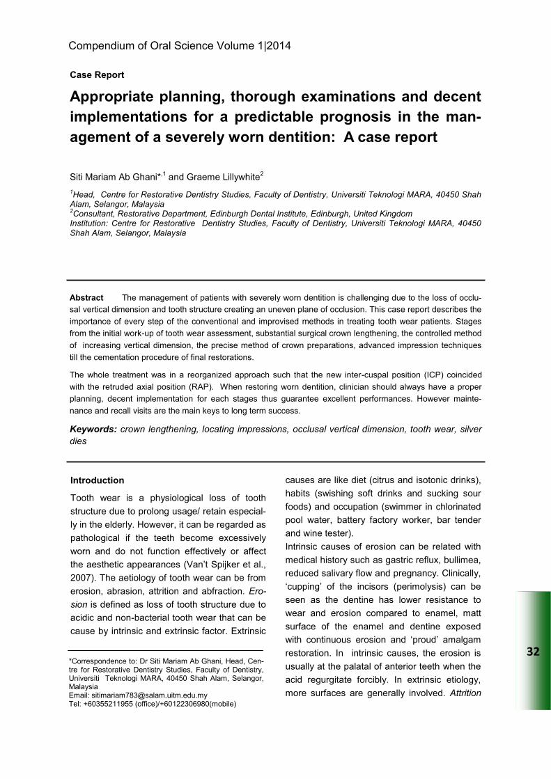
Intra-oral mock-up
Intra-oral mock-up is an effective tool for com-
munication between dentist, patient and labor-
atory personnel. Assessment of the aesthetics
and phonetics in relation with the soft tissues
can be done. A silicone putty index of the diag-
nostic wax-up was filled with bis-acryl provision-
al C&B material (ProTemp) and seated over the
maxillary teeth in order to assess the proposed
appearance of the maxillary anterior teeth
(Magne et al., 1996; Phuhong and Goldstein,
2007). Mrs. MS was very delighted with the
proposed dental appearance (Figure 6).
Stabilization
phase
Preventive advice
OHI and full mouth scaling
and polishing.
Re-evaluation Mock-up
Preliminary
restorative
phase
Replacement of amalgam
restoration/core build-up
16
Removal of crown 14 and
assessment of underlying
tooth preparation/coronal
tooth structure
Surgical crown lengthening
from 14 - 23
Re-evaluation OH, motivation and oral
condition
Definitive
restorative
treatment
Ceramic bonded crowns
for 13. 12, 11, 21, 22, 23
Composite restoration at
41 , 42, 31 and 32
Metal ceramic bridge at 16
-14
Restoration of worn teeth
24 and 25
Maintenance
phase Occlusal splint
Table 2: Key stages on management of the patient
Figure 3: Imaging (Periapicals, DPT)
Figure 4: Articulated study casts
Figure 5: Diagnostic wax-up
Compend. Oral Sci:vol1(5);2014;32-41
36
Surgical crown lengthening from 14 - 23
Using the diagnostic wax-up, a surgical stent
was fabricated to guide the amount of gingival
and bone recontouring required. 3 mm of root
surface allocated from the repositioned gingival
margin to the alveolar crest (Gargiulo et al.,
1961) Post-operatively instructions were given
to the patient and four months were allowed for
soft tissue healing before proceeding with treat-
ment (Figure 7-8).
Crown preparations and impression tech-
nique
A silicone index (Fig. 9) fabricated from the
diagnostic wax-up used as a guide to tooth
reduction for teeth 16, 14, 13, 12 11, 21, 22 and
23 (Aminian and Brunton, 2003; Mizrahi, 2004).
Individual impressions were made of the eight
preparations using heavy/ light polyvinyl-
siloxane (PVS) material. Retraction cord was
placed alternately around the preparations dur-
ing impression taking (Fig 10) and silver plated
dies and impression transfer copings (DuraLay)
were then fabricated. The impression transfer
copings were verified on the crown preparations
(13-23) intra-orally (Figure 11). These were
then splinted together with stainless steel wire
and self-curing acrylic resin for a locating im-
pression made in PVS (Figure 12).
Working cast was poured from the locating im-
pression and mounted on a semi-adjustable
articulator with the aid of face bow transfer. A
face bow taken with the tooth preparations (13-
23) and reline with TempBond for precise detail
of tooth preparation location (Figure 16-17).
Increasing the occlusal vertical dimension
(OVD)
During provisional stage, a laboratory made
maxillary anterior provisional crowns (Figure 13)
were fitted at the increased OVD. Glass-
ionomer stops were placed at 36, 37 and under
rubber dam, composite restorations were
placed on the 32-42 to level the incisal plane
and stabilize the new OVD. The occlusion
was adjusted so that intercuspal position (ICP)
Figure 6: Intra-oral mock-up
Figure 7: Incision level marked with surgical
stent from diagnostic wax-up
Figure 8: Removal of soft tissue and alveo-
lar bone recontouring
Figure 7 – 8: Procedure on crown lengthening Figure 3 – 6. Investigations for the management
of tooth wear patients
Ab Ghani et al.

37
coincides with RAP and canine guidance occlu-
sion was provided. Patient was reviewed 2
weeks later and she reported no discomfort on
the TMJ and had adapted well to the increased
OVD. Phonetics was also satisfactory.
Figure 10: Gingival retraction to refine
margin preparations
Figure 11: DuraLay resin coping try-in
Figure 12: Locating impression of six anterior
crown preparations
Figure 9 - 12. Stages in preparation for full ceramic
crowns
Figure 13: Laboratory made provisionals
Figure 9: Preparations guided by silicone index
from diagnostic wax-up
Figure 14: Trimmed individual silver dies
Compend. Oral Sci:vol1(5);2014;32-41
38
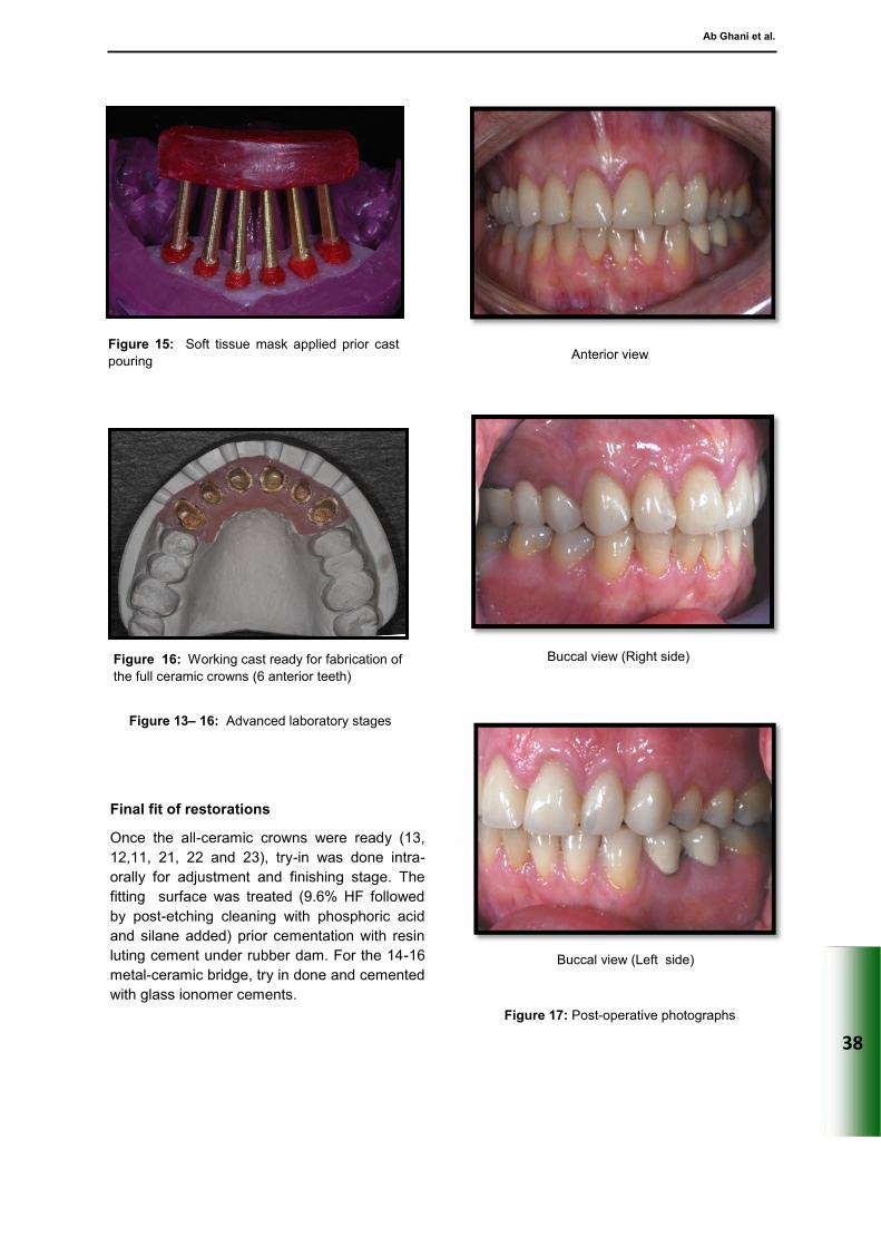
Final fit of restorations
Once the all-ceramic crowns were ready (13,
12,11, 21, 22 and 23), try-in was done intra-
orally for adjustment and finishing stage. The
fitting surface was treated (9.6% HF followed
by post-etching cleaning with phosphoric acid
and silane added) prior cementation with resin
luting cement under rubber dam. For the 14-16
metal-ceramic bridge, try in done and cemented
with glass ionomer cements.
Figure 16: Working cast ready for fabrication of
the full ceramic crowns (6 anterior teeth)
Figure 13– 16: Advanced laboratory stages
Buccal view (Right side)
Buccal view (Left side)
Figure 17: Post-operative photographs
Figure 15: Soft tissue mask applied prior cast
pouring Anterior view
Ab Ghani et al.

39
Maintenance care
After fitting the restorations, the patient was
given OHI with emphasis on the use of super-
floss beneath the bridge 14-16. At review 2
weeks later the patient demonstrated an excel-
lent level of oral hygiene and this was main-
tained. An occlusal splint was also provided to
protect the oral reconstruction and patient ar-
ranged for further review visits.
Discussion
The management of this patient‘s worn denti-
tion was done in a controlled manner of in-
creased OVD in order to create space for the
restorations, avoid pulpal stress during tooth
reduction and aligned the occlusal plane. Clini-
cians always worries that an increase in OVD
will jeopardize the muscle activity thus result in
tooth mobility, repeated restoration failure, TMJ
disorder or myofacial pain. Patient adaptation is
also a concern as there is no firm clinical rec-
ommendation available to determine if an in-
creased OVD will be well adapted. An electro-
myograph (EMG) study found that the mastica-
tory muscle can actually adapt well to an in-
creased OVD as long as the increased is ac-
companied by a stable occlusion (Ramfjord et
al., 1961). However, certain patients with OVD
raised beyond their interocclusal distance do
exhibit initial symptoms such as headache,
clenching and grinding, muscle and joint fa-
tigue, soreness of teeth, cheek biting, and prob-
lems with chewing and speech articulation for
the first 2 days. However, there was no in-
crease in the EMG of the masticatory muscle
(Carlsson et al., 1979). In this case report, the
increased OVD was done during provisional
stage after tooth preparations and 1 month was
given to assess the patient‘s adaptability.
Surgical crown lengthening (SCL) was carried
out to improve both aesthetics and retention
and resistance form of the tooth preparations.
The usage of surgical stents was very im-
portant during the surgery to ensure the dimen-
sion provided does not invade the biological
width. Further assessment should be done to
evaluate suitability of the procedure prior com-
mencing the surgery. Assessments of the perio-
dontal condition such as the bone level,
periradicular lesion and gingival biotype was
done clinically and radiographically (Ward,
1999).
Following crown preparation of the six maxillary
anterior teeth and bridge 14-16, precaution that
was taken to avoid unnecessary tooth reduction
was by having an index. An index made from
the diagnostic wax up was very beneficial be-
cause sometimes the worn areas do not require
any tooth reductions. Tooth structure was pre-
served as much as possible and the pulpal sta-
tus remained healthy.
As for the impression stage, it was difficult to
obtain accurate impressions of the 6 anterior
teeth preparations in a single impression. Thus,
locating impression was made. Other ad-
vantages from this technique were it allows for
verification of the final restorations margin with
the DuraLay copings, facilitates impression
making, minimizes gingival trauma from the use
of retraction cord in the final impression and
record gingivae in their normal position. (Fig. 13
-16)
A full ceramic (lithium disilicate) was the chosen
material for the anterior crowns for its aesthet-
ics and the capability to be bond to tooth struc-
ture. Different type of etchable ceramics when
bonded to tooth surface exhibit similar flexural
strength among them and also as in natural
tooth (Burke, 1999). Ceramic surface treatment
with hydrofluoric acid (HF) and silane applica-
tion does increase the microtensile bond
strength between ceramic and resin cements.
Factors that are essential for success when
using all-ceramic restorations have been identi-
fied by many authors such as the precise atten-
tion to detail with regard to tooth preparation,
cervical margin design and location, soft-tissue
management and impression-making. Proper
selection of materials and the ceramist also are
essential, as are correct shade matching proce-
dures and correct luting protocols ensure the
long-term success (Donovan, 2008).
The whole restorative treatment for this patient
was done in reorganized approach such that
the new ICP coincided with the RAP. Canine
guidance also known as mutually protected
Compend. Oral Sci:vol1(5);2014;32-41

40
occlusion was provided to the patient. Mutually
protected articulation is described as ―an occlu-
sal scheme in which the posterior teeth prevent
excessive contact of the anterior teeth in maxi-
mum intercuspation, and the anterior teeth dis-
engage the posterior teeth in all mandibular
excursive movements‘‘ (Glossary of Prostho-
dontics, 2008). After completing the case, the
glass-ionomer (GIC) stops on 36 and 37 (stops
that hold the new OVD) were removed. As ex-
pected, these teeth were not in occlusion but
contact re-establishment did occur during the
review visit (4 weeks post treatment). The con-
cept of relative axial tooth movement (RATM)
was applied (Poyser et al., 2005). It is a con-
cept introduced by Dahl in 1975 when they
found that tooth with filling/ restoration in high
contact display intrusion and tooth not in con-
tact display extrusion movement to be in con-
tact.
Conclusion
By applying all the described techniques in the
treatment procedure, the clinician had a good
controlled of the case, able to provide decent
execution of each procedure and reduced the
risk of complications. Patient did come for addi-
tional visits with longer treatment time and in-
creased cost, however it was emphasized that
the final results with predictable prognosis was
the ultimate treatment outcome. Mrs MS was
pleased with the treatment outcome in terms of
appearance, masticatory function, speech and
comfort (Fig. 17). She was reviewed after 2
years and reported no complications. The prog-
nosis of the restorations was considered to be
very good however maintenance and recalls
were essential. However, the result was from a
single case report, therefore further cases re-
quired before coming to the conclusion that all
the described stages is a must or highly advisa-
ble to ensure a good prognosis of the restora-
tions in the management of tooth wear.
References
1. Van‘t Spijker A, Kreulen CM and Creugers
NH. Attrition, occlusion, (dys)function and
intervention: a systematic review. Clin Oral
Implants Res 2007. 18 (Supp 13), 117-126
2. Allen PF. Use of Tooth-coloured Restora-
tions in the Management of Toothwear.
Dent Update 2003. 30, 550–556
3. Magne P, Magne M and Belser U. The di-
agnostic template: A key element to the
comprehensive esthetic treatment con-
cept. Int J Periodontics Restorative Dent
1996; 16, 560-590
4. Phuhong DD, Goldstein GR. The use of a
diagnostic matrix in the management of the
severely worn occlusion. J Prosthodont
2007. 16, 277-281
5. Gargiulo AW, Wentz F and Orban B. Di-
mensions and relations of the dentogingival
junction in humans. J Periodontol 1961.
32, 261–267
6. Aminian A, Brunton PA. A comparison of
the depths produced using three different
tooth preparation techniques. J Prosthet
Dent. 2003 Jan. 89(1), 19-22
7. Basil Mizrahi. A Technique for Simple and
Aesthetic Treatment of Anterior Toothwear.
Dent Update 2004. 31, 109–114
8. Ramfjord SP. Dysfunctional temporoman-
dibular joint and muscle pain. J Prosthet
Dent 1961. 11, 353-374
9. Carlsson GE and Kocal G. Effect of in-
creasing vertical dimension on the mastica-
tory system in subjects with natural teeth.
Journal of Prosthetic Dentistry 1979. 41,
284-289
10. Ward VJ. Tooth surface loss. Surgical
crown lengthening. Br Dent J. 1999 Jul 10.
187(1), 21-24
11. Burke FJ. Maximising the fracture re-
sistance of dentine-bonded all-
ceramic crowns. J Dent. 1999 Mar. 27(3),
169-73
12. Donovan TE. Factors Essential for Suc-
cessful All-Ceramic Restorations. J Am
Dent Assoc 2008. 139, 14S-18S
Ab Ghani et al.

41
13. The Glossary of Prosthodontic Terms. 8th
Edition. J Prosthet Dent 2005. 94, 10-92
14. Poyser NJ, Porter RW, Briggs PF, Chana
HS, Kelleher M and Patel M. The Dahl Con-
cept: past, present and future. Br Dent J
2005.198, 669–676
15. Dahl BL, Krogstad O and Karlsen K. An
alternative treatment in cases with ad-
vanced attrition. J Oral Rehabil 1975. 2,
209–214
Compend. Oral Sci:vol1(5);2014;32-41


![[CATEGORIZED] in other words, into grammatical categories ... · Academic Vocabulary A thorough survey of various textbooks, assignments, content area standards, and examinations](https://img.pdfslide.us/doc/110x75/5f622b9f659b18476627861c/categorized-in-other-words-into-grammatical-categories-academic-vocabulary.jpg)