Embed Size (px)
Citation preview
Approach to Hemolytic Anemia
Catherine MoltzanMD FRCPC
University of Manitoba
Objectives
• List the laboratory indications of hemolysis, and distinguish between intravascular and extravascular hemolysis.
• List the differential diagnosis of hemolytic anemia.
• Diagnose the various types of hemolytic anemia according to the appearance of the peripheral blood film, and ancillary laboratory tests.
Objectives- cont’d
• Distinguish hereditary spherocytosis from acquired spherocytic anemia using appropriate tests, and list the possible causes of the latter.
• Distinguish the types and mechanisms of microangiopathic hemolytic anemia using appropriate tests.
What Is Hemolytic Anemia?
• Anemia due to shortened survival of red cells in the circulation
• Normal RBC lifespan is 120 days, therefore it is useful to think of hemolytic anemia as representing RBC survival of less than 100 days
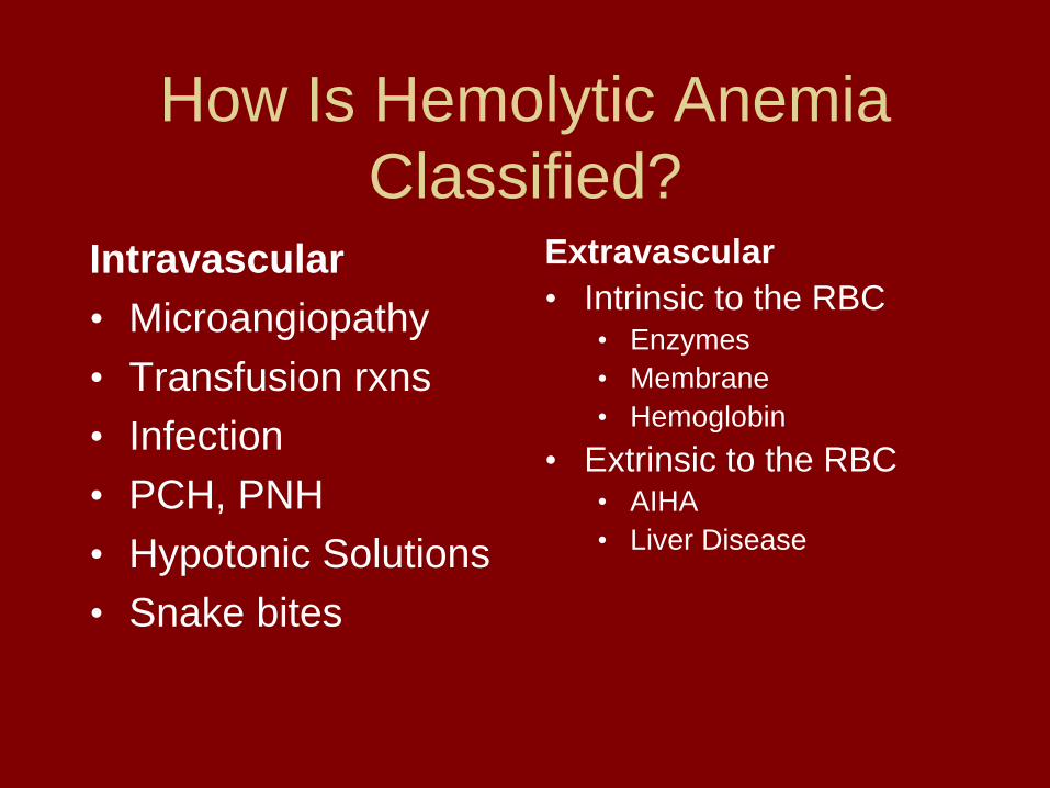
How Is Hemolytic Anemia Classified?
Intravascular• Microangiopathy• Transfusion rxns• Infection• PCH, PNH• Hypotonic Solutions• Snake bites
Extravascular• Intrinsic to the RBC
• Enzymes• Membrane• Hemoglobin
• Extrinsic to the RBC• AIHA• Liver Disease
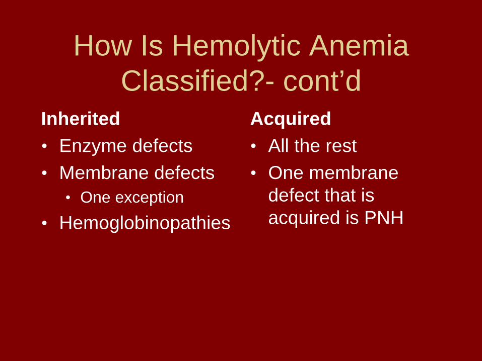
How Is Hemolytic Anemia Classified?- cont’d
Inherited• Enzyme defects• Membrane defects
• One exception• Hemoglobinopathies
Acquired• All the rest• One membrane
defect that is acquired is PNH
How Do Patients With Hemolytic Anemia Present?
• New onset of pallor and anemia• Jaundice• Gallstones• Splenomegaly
How is Hemolytic Anemia Diagnosed?
• Two main principles– One is to confirm that it is hemolysis– Two is to determine the etiology
Lab Tests To Confirm Hemolysis
• Elevated absolute reticulocyte count• Elevated LDH (note elevation is more
pronounced in intravascular hemolysis)• Elevated Indirect Bilirubin• Decreased Haptoglobin• Urine Hemosiderin- useful in the
diagnosis of intravascular hemolysis
Lab Tests To Confirm Hemolysis
• the combination of LDH and haptoglobin is 83 percent sensitive and 96 percent specific in the diagnosis of hemolytic anemia
Atypical Presentations
• Hemolysis without anemia• Compensated hereditary spherocytosis
• Hemolytic anemia without reticulocytosis• AIHA with autoantibodies against erythroid
precursors• Ineffective erythropoiesis
• Megaloblastic anemia• Myelodysplastic syndrome• Leukoerythroblastic reaction
The key to the etiology of hemolytic anemia is…
• The history AND• The peripheral blood film
Patient History
• Lifelong or family history• Medication/Drug precipitants
– G6PD– AIHA
• Acute versus chronic• Concomitant medical illnesses• Clinical presentation
Peripheral Blood Film
• Morphology can help guide further investigation
• Therefore it is important that the blood film is reviewed
Case 1
• 68-year-old male• African descent• Seen by a physician for bladder discharge • Urinalysis showed numerous WBC and
positive leukocyte esterase• Diagnosis of UTI made• CBC normal• Patient started on TMP/SMX I ds BID
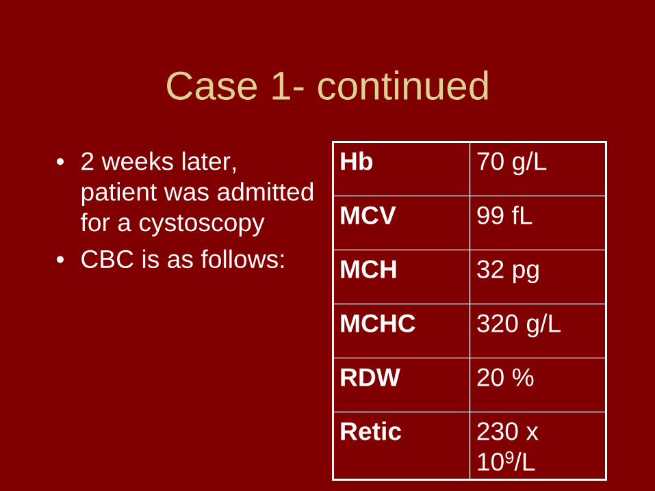
Case 1- continued
• 2 weeks later, patient was admitted for a cystoscopy
• CBC is as follows:
Hb 70 g/L
MCV 99 fL
MCH 32 pg
MCHC 320 g/L
RDW 20 %
Retic 230 x 109/L
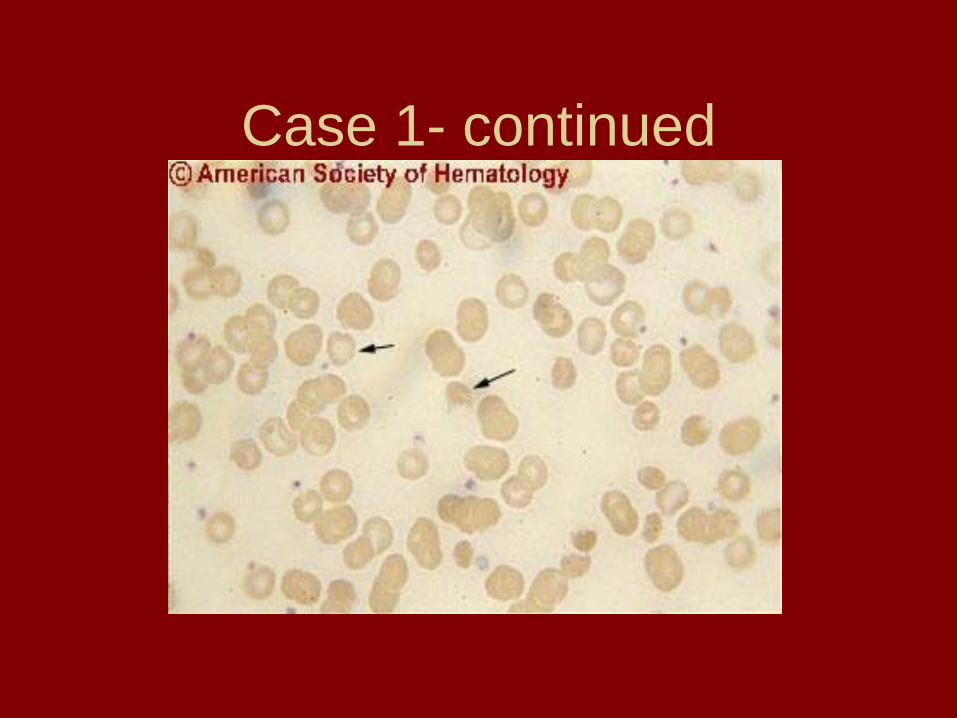
Case 1- continued
Case 1-cont’d
• LDH 600 U/L (normal up to 200)• Total bilirubin 45 umol/L• Direct bilirubin 4 umol/L• Haptoglobin < 0.1 g/L

• What is the diagnosis?• How do you prove it?
Case 1- continued
• The most likely cause of the anemia is hemolysis secondary to G6PD deficiency– Hemolysis picture- low hemoglobin, high
retic, high-normal MCV– G6PD deficiency- typical history, patient is
male and of an at-risk ethnic background
Case 1- continued
• What other diagnostic tests are needed?– Tests to confirm G6PD
• Heinz body prep• G6PD level- can NOT do this test right now,
however it should be done once the hemoglobin normalizes to confirm diagnosis
Case 1- continued
Case 1- continued
• G6PD deficiency– One of the most common causes of hemolysis– X-linked = disease in males– Usually persons of African or Mediterranean
descent– Usually history of getting an offending drug
• Antibiotics, antimalarials, etc
– Management is supportive– G6PD protects RBC against oxidative damage
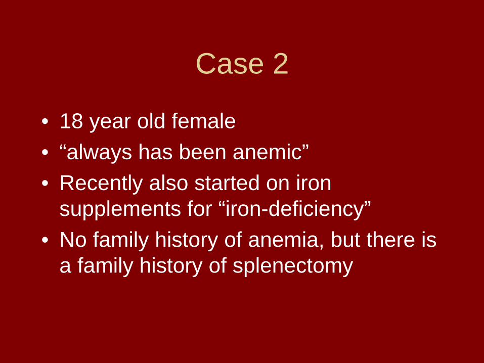
Case 2
• 18 year old female• “always has been anemic”• Recently also started on iron
supplements for “iron-deficiency”• No family history of anemia, but there is
a family history of splenectomy
Case 2- cont’d
• On exam– Vitals stable– scleral icterus– Spleen tip is palpable– Rest of the exam is unremarkable
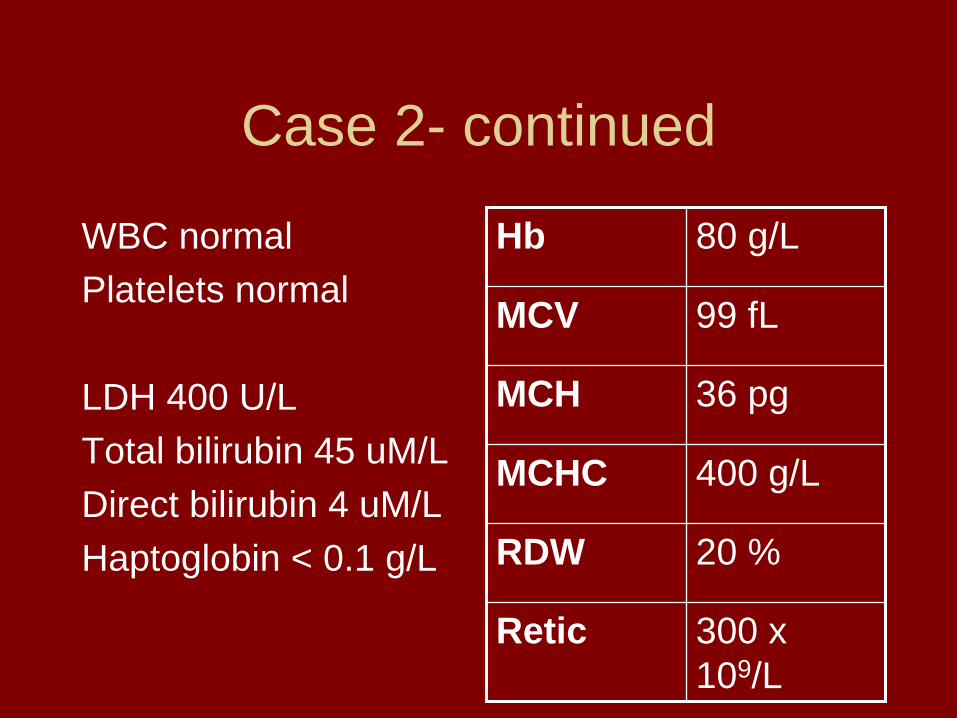
Case 2- continued
WBC normalPlatelets normal
LDH 400 U/LTotal bilirubin 45 uM/LDirect bilirubin 4 uM/LHaptoglobin < 0.1 g/L
Hb 80 g/L
MCV 99 fL
MCH 36 pg
MCHC 400 g/L
RDW 20 %
Retic 300 x 109/L
.

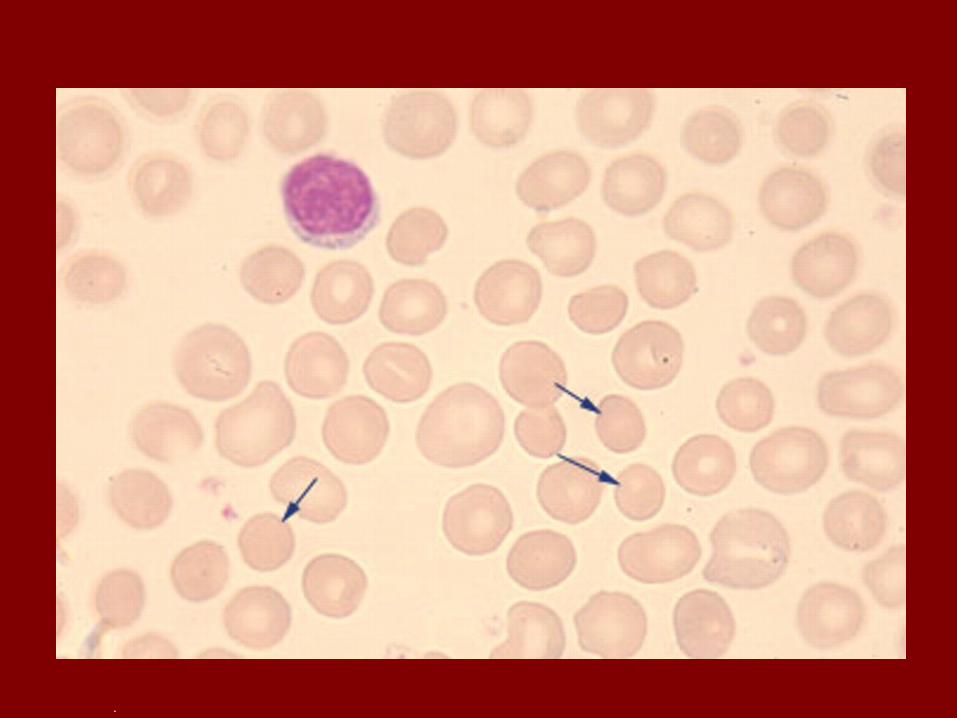
• What is the diagnosis?• How do you prove it?
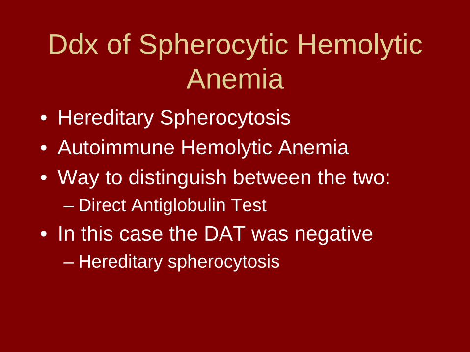
Ddx of Spherocytic Hemolytic Anemia
• Hereditary Spherocytosis• Autoimmune Hemolytic Anemia• Way to distinguish between the two:
– Direct Antiglobulin Test• In this case the DAT was negative
– Hereditary spherocytosis
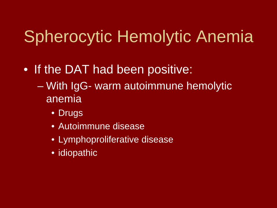
Spherocytic Hemolytic Anemia
• If the DAT had been positive:– With IgG- warm autoimmune hemolytic
anemia• Drugs• Autoimmune disease• Lymphoproliferative disease• idiopathic
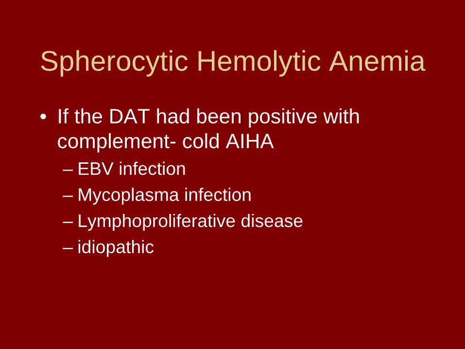
Spherocytic Hemolytic Anemia
• If the DAT had been positive with complement- cold AIHA– EBV infection– Mycoplasma infection– Lymphoproliferative disease– idiopathic
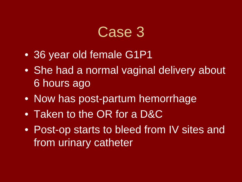
Case 3• 36 year old female G1P1• She had a normal vaginal delivery about
6 hours ago• Now has post-partum hemorrhage• Taken to the OR for a D&C• Post-op starts to bleed from IV sites and
from urinary catheter
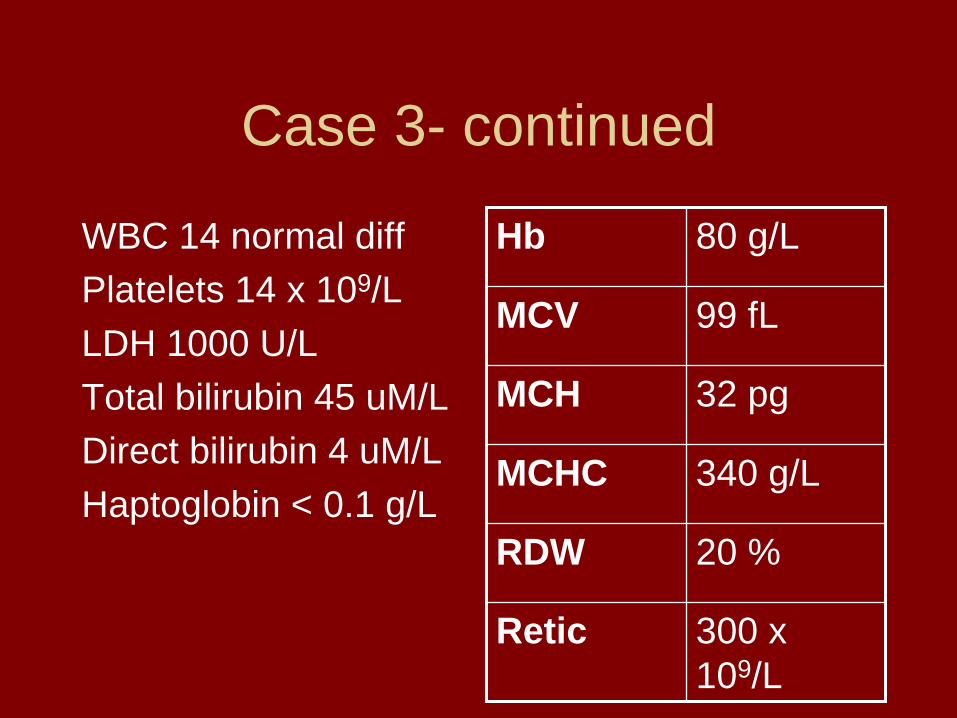
Case 3- continued
WBC 14 normal diffPlatelets 14 x 109/LLDH 1000 U/LTotal bilirubin 45 uM/LDirect bilirubin 4 uM/LHaptoglobin < 0.1 g/L
Hb 80 g/L
MCV 99 fL
MCH 32 pg
MCHC 340 g/L
RDW 20 %
Retic 300 x 109/L
• What is the diagnosis?• How do you prove it?
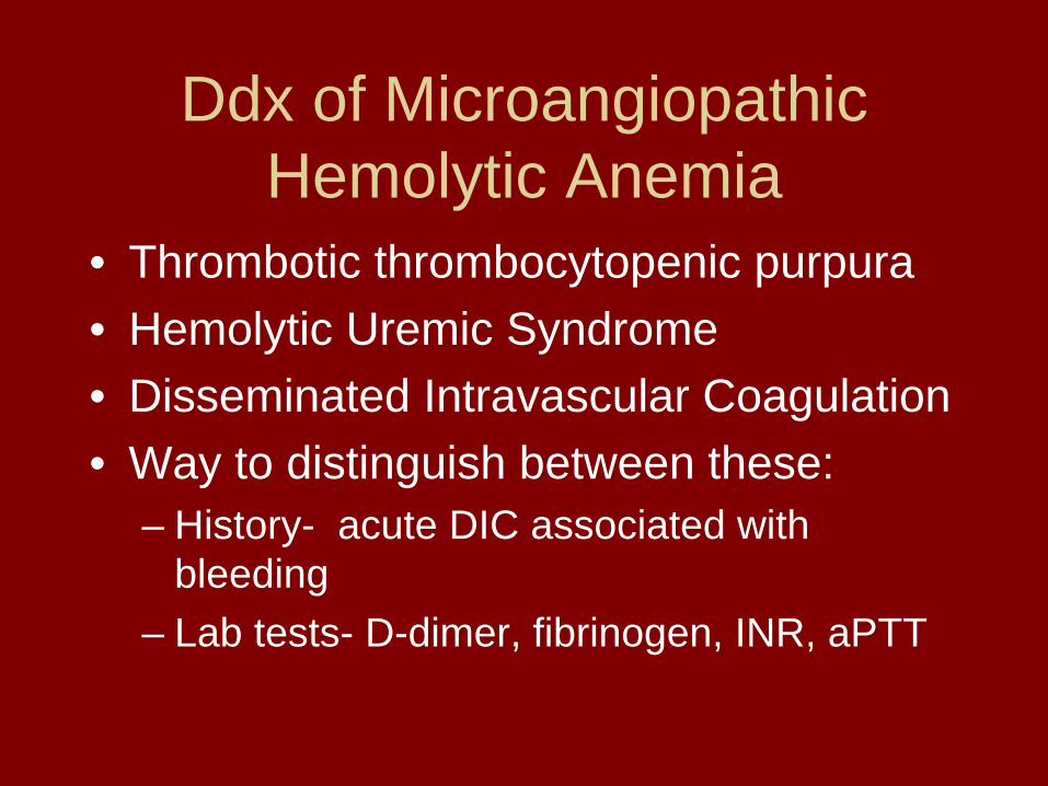
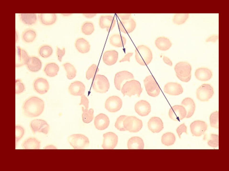
Ddx of Microangiopathic Hemolytic Anemia
• Thrombotic thrombocytopenic purpura• Hemolytic Uremic Syndrome• Disseminated Intravascular Coagulation• Way to distinguish between these:
– History- acute DIC associated with bleeding
– Lab tests- D-dimer, fibrinogen, INR, aPTT
Case 3-continued
• In this case, the diagnosis was DIC secondary to an amniotic fluid embolus
• Patient improved with blood products and supportive care over 24 hours
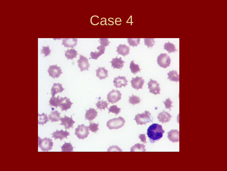
Case 4
Case 4- cont’d
• What is the diagnosis?– Liver disease
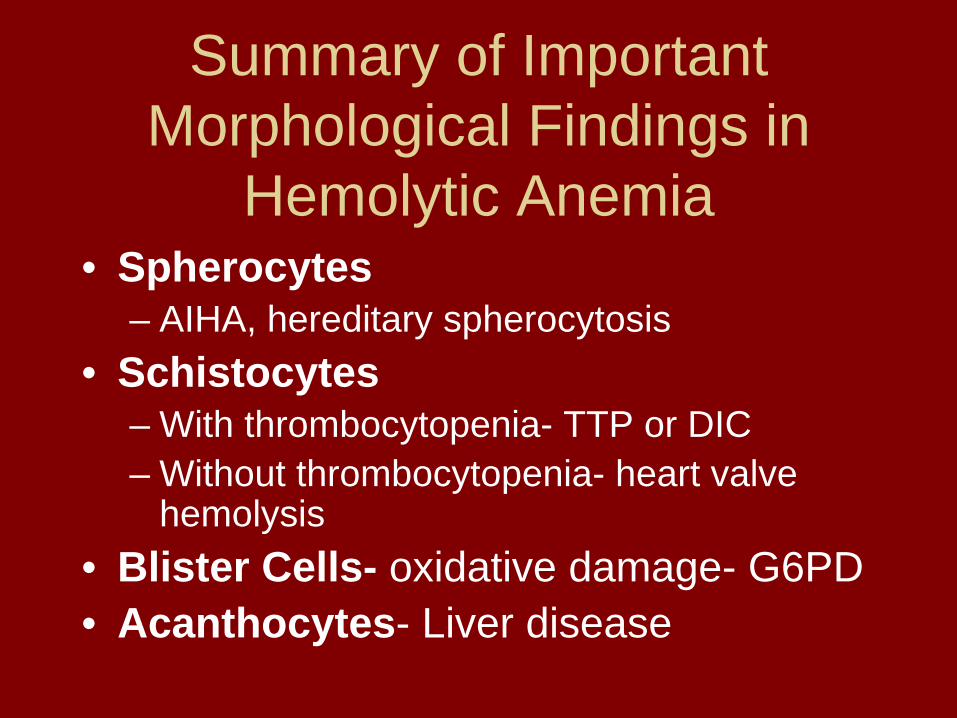
Summary of Important Morphological Findings in
Hemolytic Anemia• Spherocytes
– AIHA, hereditary spherocytosis• Schistocytes
– With thrombocytopenia- TTP or DIC– Without thrombocytopenia- heart valve
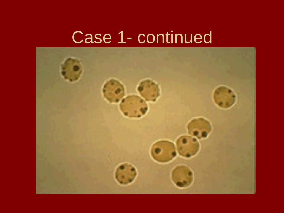
hemolysis• Blister Cells- oxidative damage- G6PD• Acanthocytes- Liver disease
Conclusions
• Hemolytic anemia can be recognized by the clinical picture– History and physical– Lab tests to confim hemolysis– Peripheral blood film to guide further tests