Embed Size (px)
Citation preview

Applied PsychoacousticsLecture 1: Anatomy and Physiology of the human auditory system
Jonas Braasch
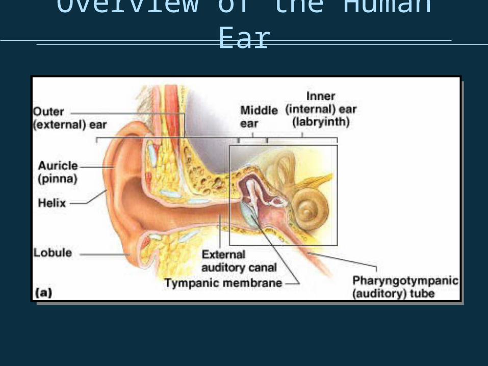
Overview of the Human Ear
Outer Ear
• Pinna: External cartiledge– Provides direction dependent frequency cues
for sound localization through spectral filtering– Position can be actively controlled by some
mammals (e.g., cat)
• Meatus (Auditory Canal) – Pathway to the middle ear, approx. 7mm
diameter, 27mm length– Amplifies sounds in the range of 2000 to 5000
Hz through resonance (approx. 10 – 15 dB)

Simulation of the sound pressure wave in the ear
canal1 2
3 1 frontal, 2.7 kHz 2 lateral, 10 kHz3 rear, 2.7 kHz
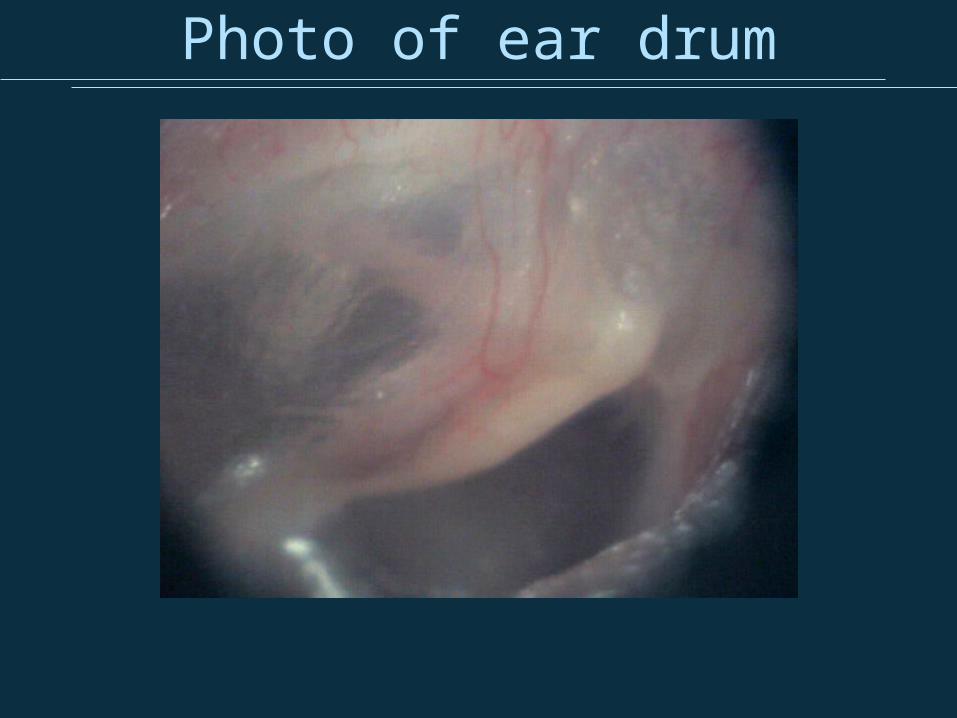
Photo of ear drum

Middle Ear
http://www.bioon.com/book/biology/whole/image/11/11-5.jpg

Middle Ear
Middle Ear
• Tympanic Membrane– Sound pressure vibration is trancduced into
mechanical oscillation and passed on to the malleus– protects ear (e.g, water, wind)
• Ossicles– Malleus, incus, stapes (hammer, anvil, and stirrup)– are the Smallest bones in human body
• Muscles– Stapedius muscle (connected to the stapes)– Tensor tympani muscle (connected to the malleus)– are the smallest muscles in the human body
• Oval Window– connection to the cochlea
• Eustachian Tube– connects the middle ear to the throat for pressure
relief
Function of the middle Ear
• Is an impedance transformer• Without it difference in densities of air
and the cochlear liquid would result in lossy energy transfer
• Pressure increase the pressure between the oval window and the ear drum by nearly a factor of 30– Amplitude ratio ear (drum/stapes) ~1.3:1 – Area ratio (ear drum /oval window):
~20:1
Acoustic Reflex
• Transmission can be attenuated in the middle ear by stiffening the Stapedius muscle and the tensor tympani muscle to protect the inner ear
• Is controlled by the auditory system and react to loud sound exposure
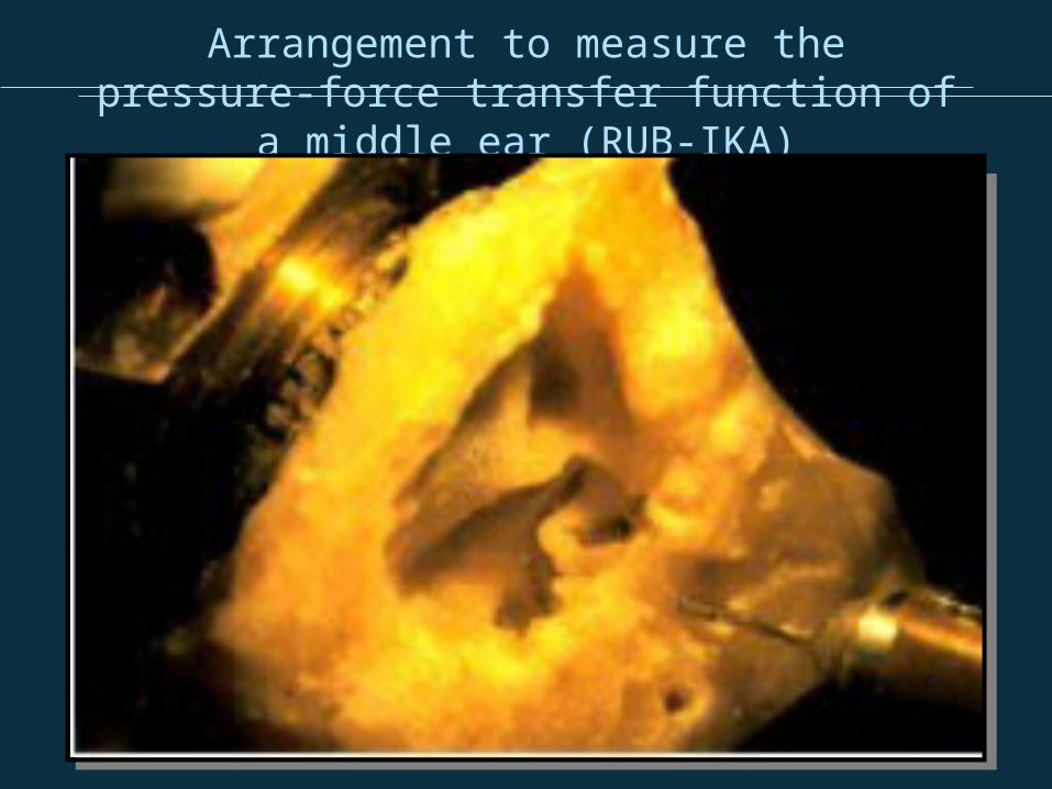
Arrangement to measure the pressure-force transfer function of a middle ear
(RUB-IKA)

Vibration of the ossicular chain
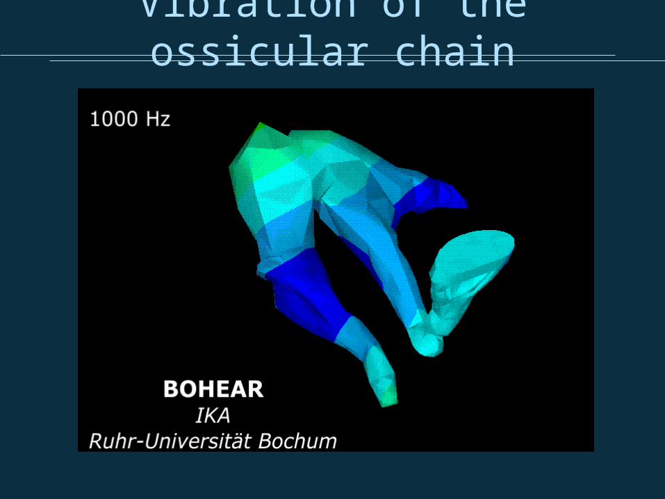
Vibration of the ossicular chain

Vibration of the ossicular chain
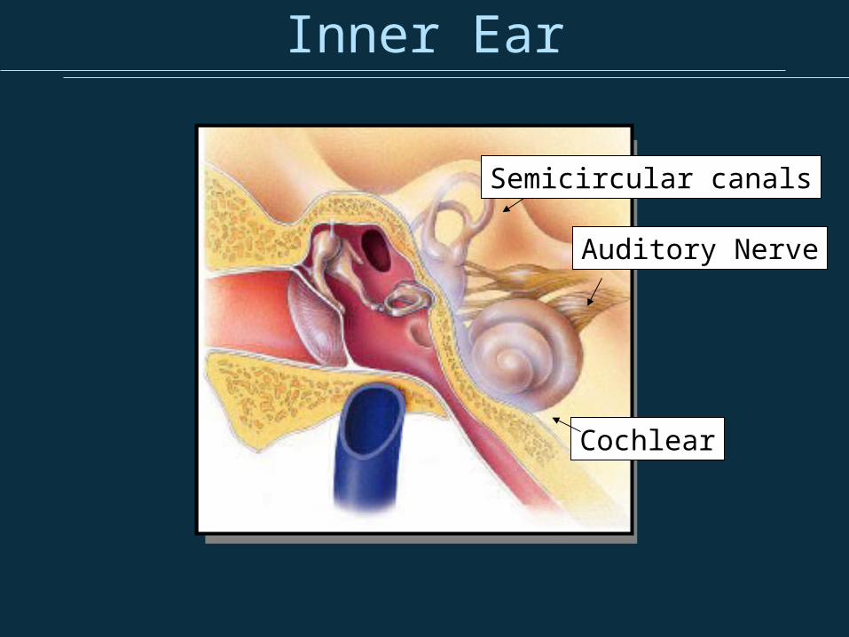
Inner Ear
Semicircular canals
Cochlear
Auditory Nerve

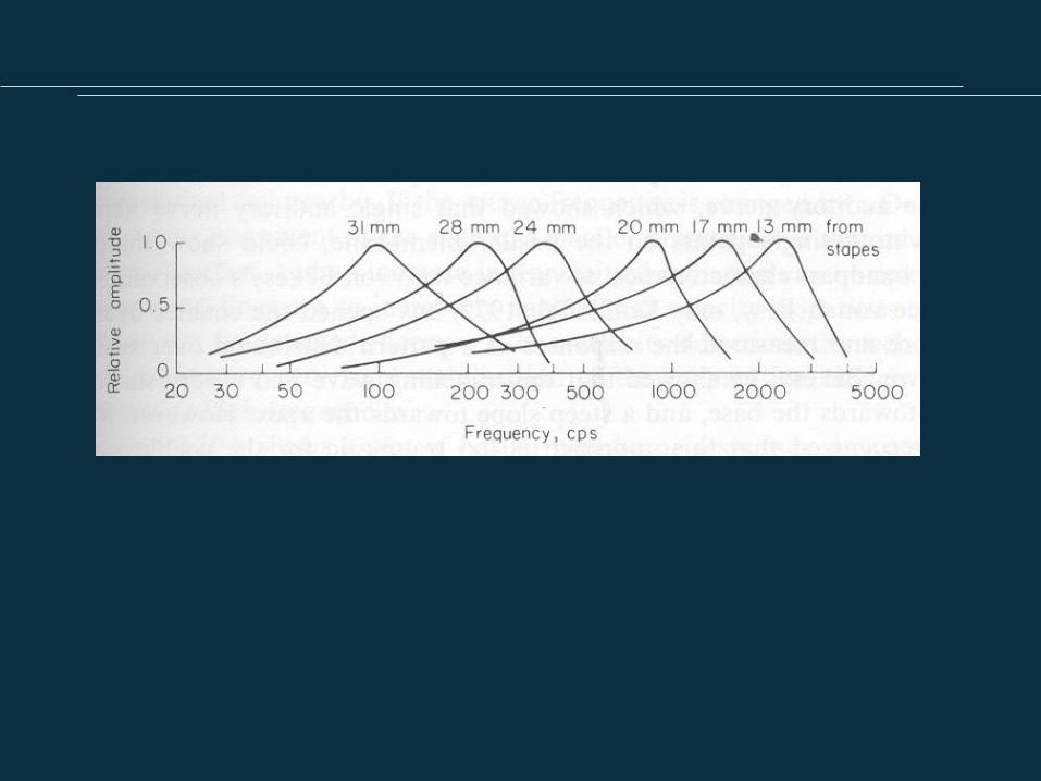
Basilar Membrane

The Traveling Wave in the Basilar Membrane

Frequency Mapping on the BM
Logarithmic Frequency Mapping
http://www.bioon.com/book/biology/whole/image/11/11-10.jpg
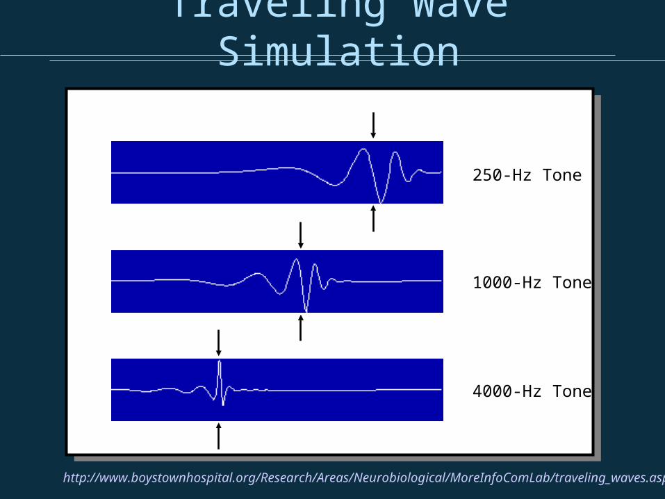
Traveling Wave Simulation
http://www.boystownhospital.org/Research/Areas/Neurobiological/MoreInfoComLab/traveling_waves.asp
250-Hz Tone
1000-Hz Tone
4000-Hz Tone

Neurotransmitters
• are chemicals that enable communication between two neurons
• are released from one neuron at its presynaptic nerve terminal and cross the synapse, a small gap, to the receptor of the second neuron

Connecting the ear to the auditory pathway
•95% of auditory nerve fibres (Type-I fibres: large diameter, myelinated) innervate IHCs (20- 30 to a single IHC) sending information to the CNS•5% (Type-II fibres: thin, unmyelinated) innervate OHCs (each fibre innervating 50-100 OHCs)
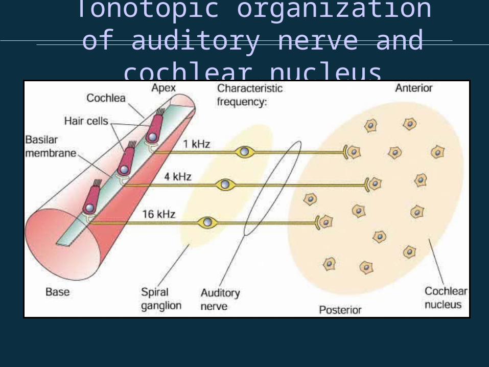
Tonotopic organization of auditory nerve and cochlear
nucleus

Definition Tonotopy
(from greek tono- and topos = place: the place of tones) is the spatial arrangement of where sound is perceived, transmitted, or received. It refers to the fact that tones close to each other in terms of frequency are represented in topologically neighbouring neurons in the brain.
from Wikipedia
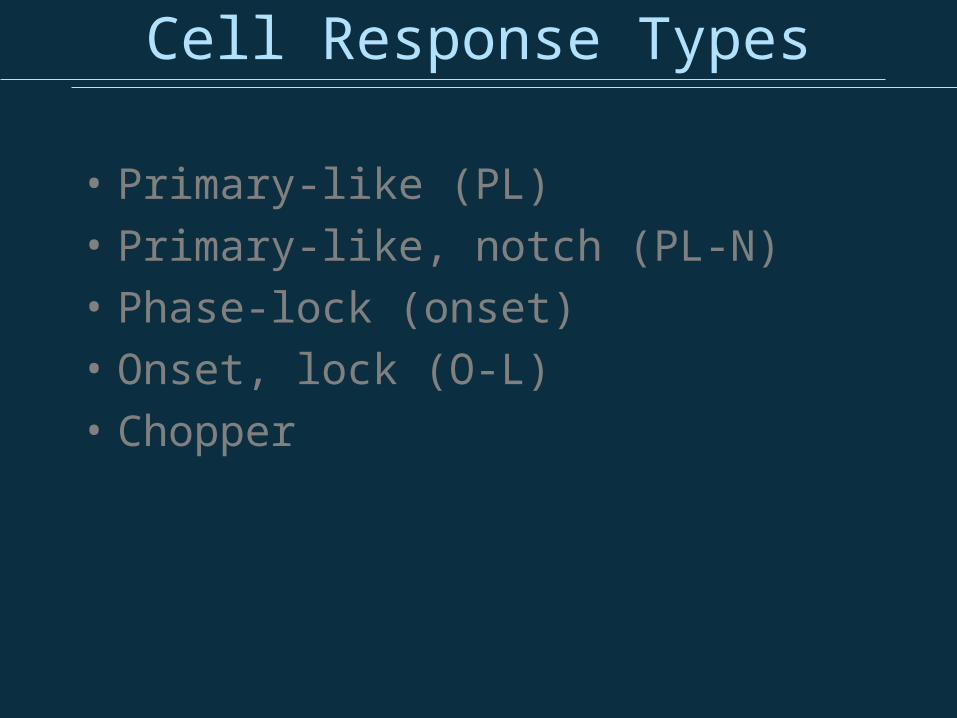
Cell Response Types
• Primary-like (PL)• Primary-like, notch (PL-N)• Phase-lock (onset)• Onset, lock (O-L)• Chopper
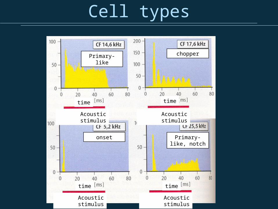
Cell types
time time
time time
Acoustic stimulus Acoustic stimulus
Acoustic stimulus Acoustic stimulus
Primary-like chopper
onset Primary-like, notch

Tuning Curves
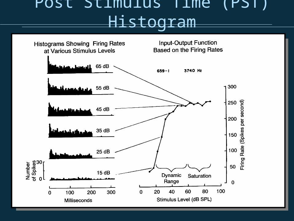
Post Stimulus Time (PST) Histogram

Phase Locking
sound pressure
action potential

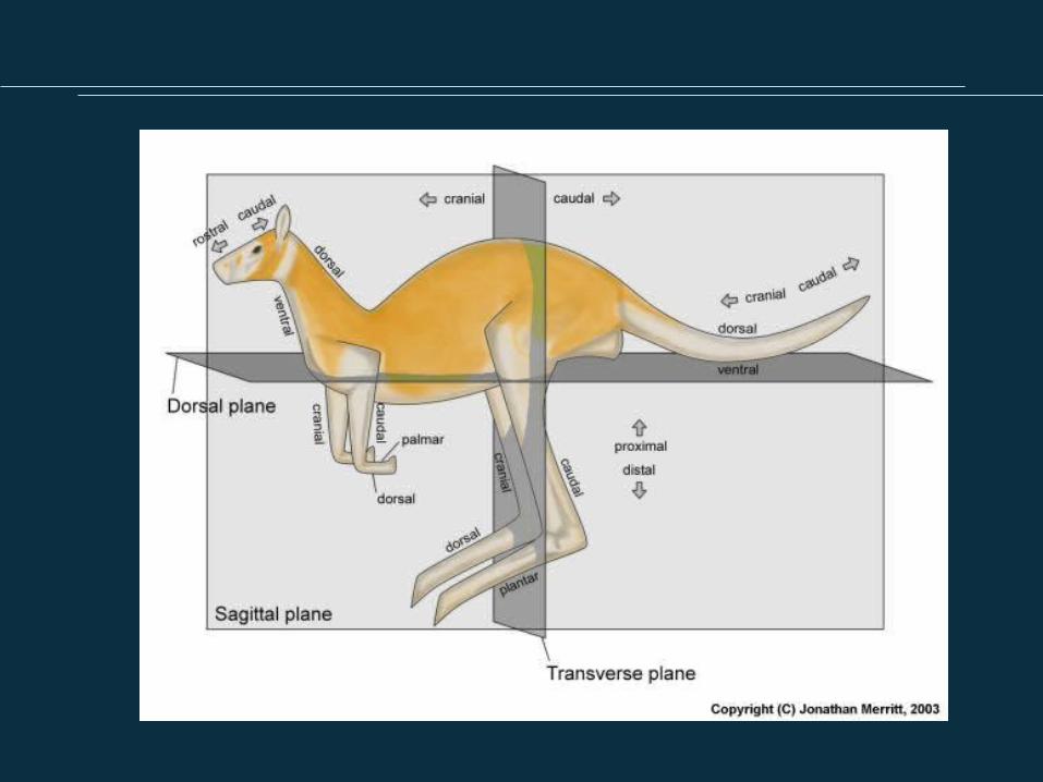
Physiological Coordinate System
Direction Description
Lateral Away from the midline
Medial Toward the midline
Bilateral On both sides of the body or head
Ipsilateral On the same side of the body or
head
Contralateral
On the opposite side of the body or head