Embed Size (px)
Citation preview
Pertanika J. Sci. & Technol. 23 (1): 1 - 12 (2015)
SCIENCE & TECHNOLOGYJournal homepage: http://www.pertanika.upm.edu.my/
ISSN: 0128-7680 © 2015 Universiti Putra Malaysia Press.
Review Article
Applications of 18(F) FDG PET/CT in Oncology
AS Fathinul Fikri1*, AJ Nordin1, YK Cheah2 and FN Ahmad Saad3
1Centre for Diagnostic Nuclear Imaging, Universiti Putra Malaysia, 43100 Selangor, Malaysia2Department of Biomedical Science, Universiti Putra Malaysia, 43100 Selangor, Malaysia3Faculty of Engineering, University Teknologi MARA, Pulau Pinang, Malaysia
ABSTRACT
The escalating costs of conventional diagnostic technology in oncology have yet to obviate futile surgery intervention and the spiralling treatment cost. The evolution in engineering technology which looks at the correlation of the anatomy and the function of tumours i.e. Positron Emission Tomography-Computed Tomography (PET-CT) have impacted on the improved diagnostic accuracy and treatment in oncology. Clinical data have demonstrated that the information provided by PET/CT often changes patient management. This review addresses the value of PET-CT as a surrogate molecular marker in tumours and to discuss some issues in adopting PET/CT in routine daily practice as supported by the numbers of literature reviews of its application in oncology since it was first commercialized in 2001. The description of the technology used in multimodality imaging has gained encouraging interest among physicians, policy makers and insurance companies on the importance of the PET-CT, for which roles are not limited to the staging, disease prognostication and treatment monitoring with potential impact on treatment cost and justification of radiation safety for the patient. PET/CT is a useful tool in cancer investigation as evidenced by its role as a surrogate marker in underpinning the cellular reprogramming of different pathological entities.
Keywords: Casemix, PET/CT, oncology, image fusion, technology
Article history:Received: 6 August 2012Accepted: 21 May 2014
E-mail addresses: [email protected] (AS Fathinul Fikri), [email protected] (AJ Nordin), [email protected] (YK Cheah), [email protected] (FN Ahmad Saad)*Corresponding Author
INTRODUCTION
Most radiologic procedures i.e. Computed Tomography (CT) or Magnetic Resonance Imaging (MRI) map the morphology of tumours with little or no information about their metabolism. Positron Emission Tomography (PET) employing 2-(fluorine-18)
AS Fathinul Fikri, AJ Nordin, YK Cheah and FN Ahmad Saad
2 Pertanika J. Sci. & Technol. 23 (1): 1 - 12 (2015)
fluoro-2-deoxy-D-glucose (FDG) is being gradually received as an important tool in providing qualitative and quantitative metabolic informations that is critical in influencing diagnosis and follow-up (Lau et al., 2006; Czernin, 2007). Radio-labelling of the PET tracer, the Flourine-18 (18(F)) with FDG (glucose analogue) provides an accurate localisation of a cancerous biological target via signalling the intracellular glycolysis obtained by the co-registration of the PET and CT images (Antti, 2010). The value of combining the FDG-PET and the CT has improved the diagnostic accuracy in cancer at large (Fathinul, 2013a; Fathinul et al., 2013b; Nordin, 2012; Pfannenberg, 2007; Niikura et al., 2011). For the purpose of this review, we address the utility of FDG-PET CT as a useful imaging tool by highlighting its use in the field of oncology imaging.
Scanning Procedure
PET/CT equipped with a crystal detector arranged in a ring around the patient covering an extended 50cm to 70cm per field of view is the common prototype system in commercial use. Prior to undergoing a PET/CT examination, patients are required to fast for at least 6 hours and to avoid strenuous physical activities. After validating the desired venous glucose level (< 7.0mmol/L), patients are injected with approximately 10mCi 18F-FDG and are instructed to lie completely still in the first 60 minutes in a designated injection room. A CT scan is performed for the purpose of an attenuation correction to rescale the 511kV PET data. The PET emission acquisition is performed at approximately 3.0 minutes per bed position. PET, CT and fusion PET/CT images are displayed on a dedicated PET-CT display system for qualitative and semi-quantitative analysis (Ronald et al., 2010).
Flurodeoxyglucose (Fdg) as a Signaling Probe for PET/CT
One of the primary metabolic changes associated with proliferating tumour cells is induction of aerobic glycolysis. Glucose is a critical nutrient for proliferating cells (Lee et al., 2009). Malignant cells have increased facilitated glucose transport and up regulation of hexokinase activity; hence, tumours can be identified by regions of increased glucose utilisation (Gatenby & Gillies, 2004). FDG is used to signal altered glucose metabolism in patients with malignancy. The focal area of abnormally increased FDG uptake is considered suspicious for malignant disease, particularly as metabolic changes which often precede the morphological changes are associated with disease (Wahl, 1991). Whole-body PET/CT has become the standard of care for cancer staging because of its high diagnostic accuracy and ability to provide a rapid survey for both regional and distant forms of metastatic disease (Czernin, 2007).
Qualitative and Semi-quantification of FDG Uptake
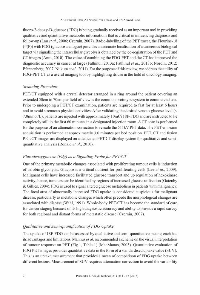
The uptake of 18F-FDG can be assessed by qualitative and semi-quantitative means; each has its advantages and limitations. Mannus et al. recommended a scheme on the visual interpretation of tumour response on PET (Fig.1, Table 1) (MacManus, 2003). Quantitative evaluation of FDG PET images provides quantitative data in the form of a standardised uptake value (SUV). This is an uptake measurement that provides a mean of comparison of FDG uptake between different lesions. Measurement of SUV requires attenuation correction to avoid the variability
Applications of 18(F) FDG PET/CT in Oncology
3Pertanika J. Sci. & Technol. 23 (1): 1 - 12 (2015)
in FDG uptake due to the differences in tumour habitus within the body. This value normalises the tumor FDG uptake with the FDG injected activity and the body weight (Kim, 1994)
TABLE 1: Scheme Defining the Different Qualitative Assessment of the Disease Response as Evaluated on PET-CT (Macmanus, 2003)
Type of response Description
Complete metabolic response (CMR) -return of 18F-FDG uptake in previously documented lesions to a level of equivalent to or lower than the activity in normal tissue
Partial metabolic response (PMR) (Fig. 1) -a significant visual reduction in 18F-FDG PET uptake in tumour sites on the visual analysis of a tumour in question but residual abnormalities suggesting malignancy
Progressive metabolic disease (PMD) -an increase in the extent of metabolic abnormality favouring tumour growth or evidence of new sites of disease
Stable metabolic disease (SMD) - no change
PET-CT and the Recist Criteria
The standalone structural imaging modality assessment of a small cancer lesion is devoid of information on the functional changes. The actual metabolic activity of a cancer is thence deemed as under evaluated in important parameters that influence successful treatment as cancer healing is confounded by area or fibrosis which mimic the viable cancer tissue. The use
Fig.1: PET/CT treatment monitoring of a 60-year-old patient with left basal non-small lung carcinoma (NSCLC). The left panel: A coronal baseline PET image showing a 3.0 mm left FDG-avid lesion (SUV max: 7.56). The right panel: A coronal post-treatment PET image showing partial metabolic response of the left NSCLC lesion (SUV max: 3.17)
AS Fathinul Fikri, AJ Nordin, YK Cheah and FN Ahmad Saad
4 Pertanika J. Sci. & Technol. 23 (1): 1 - 12 (2015)
of Response Evaluation Criteria in Solid Tumours (RECIST 1.1) to criteria in the assessment of a lesion does not represent the actual intracellular changes which lead many limitations in preparing a patient for an appropriate treatment strategy (Eisenhauera et al., 2009). An instance of this is a lymph node of size larger than 1 cm denoted as containing a tumour on the structural imaging, which always misleads the treating physician on the nodal staging on the TNM AJCC 6th edition (Yan-Ping et al., 2009). The most favourable index of the 18F-FDG PET/CT is that it is capable of exhibiting more rapid change in cellular metabolism than in tumour size (Stroobants et al., 2003). Functional information derived from PET is complementary to the high resolution structural imaging data available from such modalities as CT and MRI. Because of the limitations of CT scanning, PET/CT scanning may also have a role in response assessment after induction therapy prior to surgery, particularly for stage IIIA NSCLC. Choi et al. found that the residual metabolic rate of glucose (MRglc) as measured using FDG-PET was strongly correlated with response to preoperative chemoradiotherapy in locally advanced NSCLC as assessed by a pathological examination of a tumour obtained from a thoracotomy (Choi et al., 2002). Currently, the American College of Radiology Imaging Network 6668/RTOG 0235 trial is prospectively evaluating whether the primary tumour 18F-FDG SUVmax shortly after definitive chemoradiation can predict long-term survival in inoperable stage II or III NSCLC (Greene et al., 2002). In GIST, Choi et al. confirmed their previous observation that RECIST significantly underestimated tumour response. They suggested that a more than 10% decrease in one dimension of a cancer lesion on CT at 2 months after treatment is adequate to identify good responders to FDG-PET, and predicts a longer long-term prognosis (Haesun et al., 2007). The use of contrast-enhanced CT has enabled demonstration of tumour characteristics i.e. tumour density, enhancing tumour nodules and tumour vessels, in addition to tumour size. The additional information on the tumour enhancement on CT connotes that the outside dimensions of a tumour mass may not accurately reflect how active the tumour is and the decrease in tumour density of the responding tumours on CT is correlated with the development of tumour necrosis or cystic myxoid degeneration.
An evolving new guideline looking at the metabolic changes as a yardstick for post treatment evaluation of a solid tumour has been suggested i.e. PET assessment evaluation of a solid tumour (PERSIST). However, a lot more work on the factors that confound the parameters used before these new criteria are to be accepted given assessment on the metabolic changes require more parameters that need standardization (Richard et al., 2009). PERSIST, however, offers the potential to characterise the nature of tumour cells on the understanding of the alteration of their normal biochemical and biologic features. Thus, the information obtained is basically different from that alluded by anatomic imaging.
FDG PET/CT in Tumor Staging
Poor sensitivity of standalone CT, MRI and PET may lead to inaccurate staging of a tumour. Recent data, with regards to tumour staging, have shown that integrated PET/CT images are superior to PET images alone and PET and CT images viewed side by side (Kim, 1994). Contrasted CT technique used for the evaluation of equivocal PET results promises higher achievable diagnostic results in many tumours (Nordin, 2012), for instance, the prevalence
Applications of 18(F) FDG PET/CT in Oncology
5Pertanika J. Sci. & Technol. 23 (1): 1 - 12 (2015)
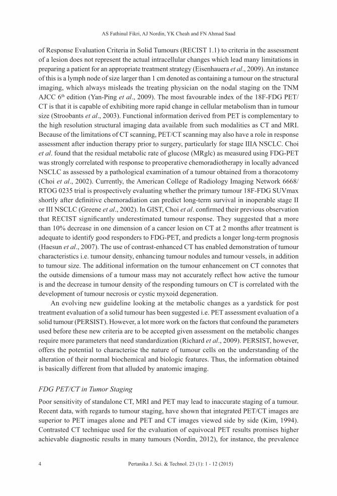
of brown fat FDG-accumulation in patient neuroendocrine tumour. The contrasted CT has a greater sensitivity in distinguishing an occult lesion given the raised lesion-to-background noise ratio (Yon, 2006; Pottgen, 2006). The impact of PET in detecting diffuse involvement of other organ systems as part of the metastatic spread or delineation of the subcentimetre focus of FDG-avidity has averted futile surgery and unnecessary treatment costs (Pottgen, 2006). In this regards, we observed the change in the patient management as a result of the up-staging of tumour by the PET-CT modality (Fathinul, 2011). In addition, the metabolic information on the PET image would facilitate the biopsy localisation of a lesion. This is supported by many published data on the improved diagnostic yield of biopsy employing PET/CT as compared to the conventional approach (Fathinul, 2011; Caroline, 2008). Subcentimeter metastatic lesions are better visualised on the PET/CT as compared to CT given the benefit of signalling glycolytic metabolism that has improved lesion detection at large (Fig.2) (Yoon, 2011).
Fig.2: Example of discordant positron emission tomography (PET) and computed tomography (CT) results. CT (left) images of a mediastinal lymph node showing a subcentimeter lymph node. The corresponding PET image (right) exhibited an FDG-avid lymph node denoting the altered glucose metabolism suspicious for pathological lymph node. [Images courtesy of Pusat Pengimejan Diagnostik Nuklear]
The value of using PET/CT for patients undergoing restaging after treatment is equally apparent. It is now possible for this modality to distinguish between malignancy and post-therapeutic change (Van et al., 2002). According to Selzner et al., combined PET/CT over scored standalone CT and PET in restaging tumours after years of disease-free survival, when the distorted anatomy may not be easily distinguished from the site of a tumour recurrence (Selzner et al., 2004). PET/CT has proven to be a very sensitive non-invasive staging technique and may even determine the exact location of a solitary lymph node particularly in evaluating the stage of non-small cell lung cancer (NSCLC), thus concluding the precise classification as N1 or N2 lymph node station based on classification by the American Joint Committee of Cancer (AJCC) (Asamura, 2000). Unsuspected extra thoracic soft tissue or skeletal metastases also may be revealed by PET/CT in cases where other imaging methods fail to demonstrate distant metastasis (Schoder, 2007).
AS Fathinul Fikri, AJ Nordin, YK Cheah and FN Ahmad Saad
6 Pertanika J. Sci. & Technol. 23 (1): 1 - 12 (2015)
FDG PET/CT Predicts Tumor Aggressiveness
In addition, the degree of metabolic defect via semi-quantitative analysis, SUV could predict tumour aggressiveness and overall patient survival as high SUV values correlate with poor disease prognosis (Yamada et al., 1992). We reported 23 patients with recurrent phaechromocytoma/paraganglioma, with regards to SUVmax evaluation on tumour aggressiveness; nine patients had local controls (34.1%) with mean progression-free survival (PFS) of 19.35±3.34 months with a significant number of patients with metastatic disease who had SUV > 9.2 as compared to the local disease group (p<0.05) (Fathinul et al., 2014). The prediction of tumour aggressiveness is important for tailoring a management plan obviating the risk of unnecessary treatment toxicity and to reduce the cost burden for patients. This is in line with many studies which demonstrated that the decrease of FDG uptake after a single infusion of chemotherapy was a predictor of eventual response to this regimen (Kostakoglu et al., 2002). On the other hand, no decrease of tumour FDG uptake after the first infusion was a predictor of non-response.
FDG PET/CT Alters the Management Plan
PET/CT changed the primary diagnosis in approximately 16% of cases, whereas PET/CT resulted in a change in staging and treatment plan in approximately 28% to 32% of the cases, respectively, and thus enabled the establishment of an appropriate scheme for disease response to treatment (Pottgen, 2006). Current procedures to monitor therapy using anatomical imaging modalities, such as CT, have a major setback given that functional changes often precede anatomical changes. A significant metabolic change can be established by comparing the standardised uptake values (SUV) from pre- and post-treatment scans, although such comparisons can only be made accurately on attenuation-corrected, quantitative PET images (Wahl, 1991). In our experience, we found that in a cohort of 23 patients with head and neck tumours, there were changes in the management plan in 58% of the patients being evaluated on the PET/CT as compared to CT-based staging (Fathinul, 2013a).
PET-CT in Radiation Oncology
The new approach of radiation therapy and intensity modulated radiation therapy necessitates more precise target volume definitions for dose-sparing of normal tissues. Traditionally, CT has been popular as a tool of choice for radiation therapy planning. Nevertheless, CT has been shown to have relatively low sensitivity and specificity for detecting tumour tissue (Gregoire, 2007). In a meta analysis for solid tumours, PET/CT imaging was found to have a better sensitivity of 92% and a specificity of 93% compared to 85% and 88% for PET and 64% and 83% for CT alone, respectively, and hence for the radiation treatment (Antoch, 2004). Tumour biology has been identified as an essential factor for effective dose delivery (Ling et al., 2000). With PET/ CT imaging the biological tumour volume allows the radiation dose to be modulated according to the distribution of the PET signal intensity within the tumour volume specifically (Ling et al., 2000; Schwartz et al., 2005). In addition, the PET-CT provides a single reproducible session for highly precise patient positioning during the imaging and
Applications of 18(F) FDG PET/CT in Oncology
7Pertanika J. Sci. & Technol. 23 (1): 1 - 12 (2015)
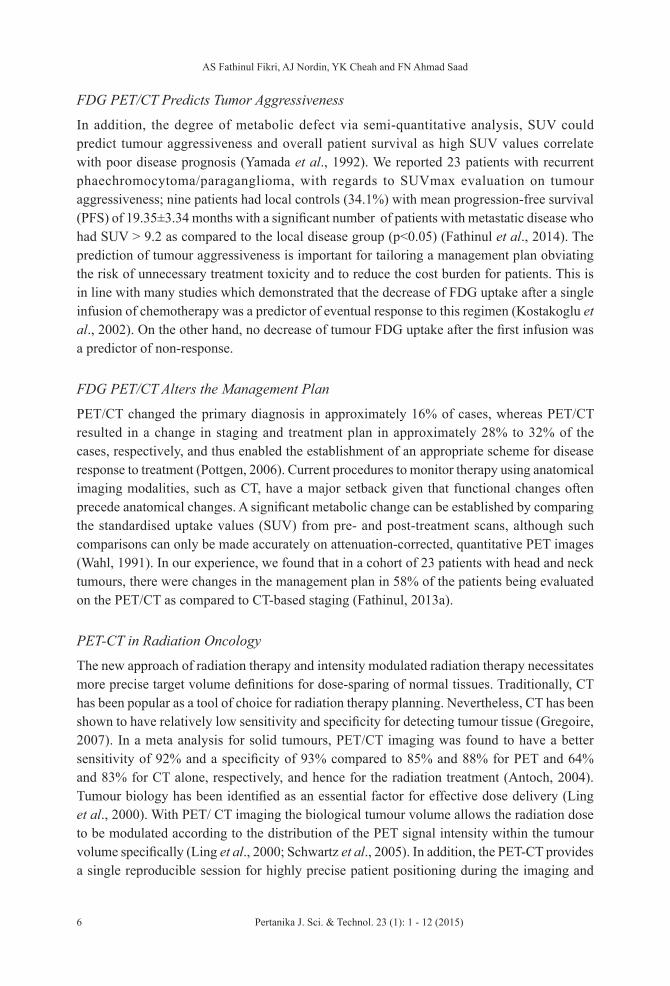
treatment sessions rendering an accurate tumour delineation and effective dose delivery over standalone PET and CT systems (Gilman, 2007). The proliferating tissue has increased FDG avidity although the radiation inflammatory effect may at times be misinterpreted as a true positive lesion (Figure 3). A recent review of PET/CT utilisation for radiotherapy planning in lung cancer showed differences in the range of 30% to 60% between PET-derived contours versus CT-only target volumes (Greco, 2007).
Fig.3: CT and PET coronal images; A 54-year-old man had completed radiotheraphy of the right lung for recurrent NSCLC. The images show typical dome-shape consolidative changes with increased FDG accumulation in radiation pneumonitis. (Images courtesy of Peter Mac Callum Centre)
PET/CT and the Radiation Risk
Most commonly, PET utilises 18F-FDG as a radiotracer; the short half-life of 110 min reduces radiation exposure compared with other commonly used radionuclides such as 99mTechnetium (6 hours) or 201Thallium (72 hours). It carries a low absorbed dose to the patient estimated at approximately 7 mSv. The radiation (X-rays) from our diagnostic CT protocol ranged from 8mSv to 16 mSv for a two-dedicated low tube current dose (35mAs) and the CARE Dose 4D (120kV). The 64-multislice CT technique was equipped with the dual focal spot that ensured more image yields without increase in the total radiation dose and the CARE dose 4D is capable of modulating tube current adaptation tailoring to the patient’s size, for which features render an efficient effective patient dose of up to 20% as compared to the lower 16 multislice of the same kind (Zito, 2009). In general, the benefits of subjecting patients for the PET-CT outweigh the radiation risk as most information required for the staging of a tumour is available in a single session study. In a study by Hishar et al., the dose minimisation strategy on PET was adequate to yield a good PET-CT image without significant compromise (Hishar, 2014).
AS Fathinul Fikri, AJ Nordin, YK Cheah and FN Ahmad Saad
8 Pertanika J. Sci. & Technol. 23 (1): 1 - 12 (2015)
PET-CT and Economy
Despite increasing evidence supporting the accuracy of standalone imaging i.e. CT, MRI or PET, high cost and limited cost-effectiveness data have militated against funding for routine clinical use in many countries (Gambhir, 1996). There is now substantial evidence that PET/CT is an exceedingly accurate multimodality imaging in detecting malignant tissue and provides a higher specificity than conventional imaging (Michael et al., 2011; Ell, 2006). Precision in staging may avert futile surgery for which PET-CT has changed the management plan whilst providing additional clinical benefits to patients (Ell, 2006). Considering the median length of pre- and post-operative hospital stay, the cost of surgical resections and biopsies evaluated on PET-CT was found overall to be cost beneficial (Rohren, 2004). In Heinrich et al.’s trial on patients with pancreatic cancers who underwent PET-CT for disease staging, metastases were found in 16 patients with cancer initially deemed resectable, leading to different management and cost savings of USD 1,066 per patient (Heinrich et al., 2005). In the United States, the use of FDG PET scanning has been shown to be a cost-effective alternative to conventional imaging methods in the evaluation of non-small-cell lung cancer, and this led MEDICARE, an insurance provider, to reimburse patients for those indications (Dewan, 1995). In our initial experience in working with patients with cancers referred for staging, a remarkable change in management strategy reflected a potential reduction in the total costs incurred to be borne by the patients.
PET-CT Potential Pitfalls
As PET and CT are operated on a sequential basis, where co-registration of data obtained from both methods is deemed to some degradation artefacts. The use of contrasted CT in PET/CT; among other known artefacts the following were seen: banana lesion caused by diaphragmatic movement in the lower lung; breathing artefacts; patient motion; incidental metal device on the patient torso or FDG-RBC microembolus; leakage of FDG at the injection site or from the contaminated patient’s gown after urine voiding; and pooling of intravenous contrast as source of false positive FDG-avidity were also observed as an attribute to the overestimation of the attenuation correction (Fathinul & Lau, 2009a; Fathinul & Lau, 2009b; William, 2007). Therefore, a careful instruction for PET/CT preparation and adoption of improved techniques during PET-CT i.e. gated respiration and improvement of the PET detector to facilitate image enhancement are urgently required.
CONCLUSION
A combined PET and CT scanner is a practical and effective approach in acquiring co-registered anatomical and functional images in a single scanning session. It denotes the new era in molecular imaging whereby advancement in science and technology has impacted the way physicians personalise treatment plans with more effective strategy and a cost effective package for the patient. Combined contrasted PET/CT imaging facilitates the separation of normal physiologic uptake from pathological tissue with a more favourable accuracy and, hence, helps reduce the incidence of false-positive and false-negative incidence.
Applications of 18(F) FDG PET/CT in Oncology
9Pertanika J. Sci. & Technol. 23 (1): 1 - 12 (2015)
ACKNOWLEDGEMENTS
This work was supported in part by the Research University Grant Scheme (RUGS), University Putra Malaysia.
REFERENCESAntoch, G., Saoudi, N., Kuehl, H., Dahmen, G., Mueller, S. P., Beyer, T., Freudenberg, L. S. (2004).
Accuracy of whole-body dual-modality fluorine-18-2-fluoro-2-deoxy-D-glucose positron emission tomography and computed tomography (FDG-PET/CT) for tumor staging in solid tumors: comparison with CT and PET. J Clin Oncol, 22(21), 4357-4368.
Antti, S., Heikki, U., & Sami, K. (2010). Integrated anatomy and viability assessment PET/CT. Euro Intervention Supplement, 6(G), 132-137.
Asamura, H., Suzuki, K., Kondo, & H., Tsuchiya, R. (2000). Where is the boundary between N1 and N2 stations in lung cancer? Ann Thorac Surg., 70, 1839–1845.
Boellaard, R., O’Doherty, M. J., Weber, W. A., Mottaghy, F. M., Lonsdale, M. N., Stroobants, S. G., & Krause, B. J. (2010). FDG PET and PET/CT: EANM procedure guidelines for tumour PET imaging: version 1.0. European Journal of Nuclear Medicine and Molecular Imaging, 37(1), 181-200. doi: 10.1007/s00259-009-1297-4
Caroline, B. M., Françoise, K. B., Philippe, M. L., Campion, B. D., & Steven, L. G. (2008). Investigation of FDG-PET/CT imaging to guide biopsies in the detection of histological transformation of indolent lymphoma. Haematol, 3(93), 471-472.
Choi, H., Charnsangavej, C., Faria, S. C., Macapinlac, H. A., Burgess, M. A., Patel, S. R., Benjamin, R. S. (2007). Correlation of computed tomography and positron emission tomography in patients with metastatic gastrointestinal stromal tumor treated at a single institution with imatinib mesylate: proposal of new computed tomography response criteria. J Clin Oncol, 25(13), 1753-1759.
Choi, N. C., Fischman, A. J., Niemierko, A., Ryu, J. S., Lynch, T., Wain, J., Mathisen, D. (2002). Dose-response relationship between probability of pathologic tumor control and glucose metabolic rate measured with FDG PET after preoperative chemoradiotherapy in locally advanced non-small-cell lung cancer. Int J Radiat Oncol Biol Phys, 54(4), 1024-1035.
Czernin, J., Allen-Auerbach, M., & Schelbert, H. R. (2007). Improvements in cancer staging with PET/CT: Literature-based evidence as of September 2006. J Nucl Med., 48(1), 78–88.
Dewan, N. A., Reeb, S. D., & Gupta, N. (1995). PET-FDG imaging and transthoracic needle lung aspiration biopsy in evaluation of pulmonary lesions: A comparative risk-benefit analysis. Chest, 108, 441-446.
Eisenhauera, E. A., Therasseb, P., Bogaerts, P., Schwartz, L. H., & Verweij, J. (2009). New response evaluation criteria in solid tumors: Revised RECIST guideline (version 1.1). Eur J Cancer, 45, 228–247.
Ell, P. J. (2006). The contribution of PET/CT to improved patient management. Br J Radiol., 79, 32–36.
Fathinul Fikri, A. S, Nordin, A. J., & Eddie Lau, E. F. (2013a). 18(F) FDG-PET/CT is a useful molecular marker in evaluating tumor aggressiveness; A revised understanding of an in-vivo FDGPET imaging that alludes the alteration of cancer biology. Cell Biochemistry and Biophysic, 66, 37-43. DOI: 10.1007/s12013-012-9395-5.
AS Fathinul Fikri, AJ Nordin, YK Cheah and FN Ahmad Saad
10 Pertanika J. Sci. & Technol. 23 (1): 1 - 12 (2015)
Fathinul Fikri, A. S., Nordin, A. J., Mohtarrudin, N., Hemalata, & Lau, W. F. E. (2011). 18[F] FDG-PET/CT is a useful molecular marker in evaluating thymoma aggressiveness. European Journal of Radiology Extra, 78(2), e89-e92. doi: http://dx.doi.org/10.1016/j.ejrex.2011.02.006
Fathinul, F., & Lau, W. F. E. (2009a). An intense 18F- FDG pulmonary microfocus on PET without detectable abnormality on CT: A manifestation of an iatrogenic FDG pulmonary embolus. Biomed Imaging Interv J, 6(4):e37.
Fathinul, F., & Lau, W. F. E. (2009b). Avid 18F-FDG uptake of pectoralis major muscle: An equivocal sequela of strenuous physical exercise. Biomed Imaging Interv J, 5(2), e7.
Fikri, A. S., Kroiss, A., Ahmad, A. Z., Zanariah, H., Lau, W. F., Uprimny, C., Virgolini, I. J. (2014). Localization and prediction of malignant potential in recurrent pheochromocytoma/paraganglioma (PCC/PGL) using 18F-FDG PET/CT. Acta Radiol, 55(5), 631-640.
Gambhir, S. S., Hoh, C. K., & Phelps, M. E. 1996. Decision tree sensitivity analysis for cost-effectiveness of FDG-PET in the staging and management of non-small-cell lung carcinoma. J Nucl Med, 37, 1428-1436
Gatenby, R. A., & Gillies, R. J. (2004). Why do cancers have high aerobic glycolysis? Nat Rev Cancer, 4, 891-899.
Gilman, M. D., Fischman, A. J., Krishnasetty V, Halpern EF, Aquino SL. 2007. Hybrid PET/CT of the thorax: when is computer registration necessary?. J Comput Assist Tomogr, 31, 395-401.
Greco, C., Rosenzweig, K, Cascini, G. L., & Tamburrini, O. 2007. Current status of PET/CT for tumor volume definition in radiotherapy treatment planning for non-small cell lung cancer (NSCLC). Lung Cancer, 57, 125–34.
Greene, F. L., Page, D. L., Fleming, I. D. et al. (Eds.) (2002). Lung. In: AJCC cancer staging manual, 6th ed. New York, NY: Springer.
Gregoire, V., Haustermans, K., Geets, X., Roels, S., & Lonneux, M. (2007). PET based treatment planning in radiotherapy: A new standard? J Nucl Med, 48(Suppl 1), 68S-77S.
Heinrich, S., Goerres, G. W., Schafer, M., Sagmeister, M., Bauerfeind, P., Pestalozzi, B. C., . . . Clavien, P. A. (2005). Positron emission tomography/computed tomography influences on the management of resectable pancreatic cancer and its cost-effectiveness. Ann Surg, 242(2), 235-243.
Herbrik, M., Treffert, J., Geiger, B., Riegger, C., Hartung, V., Rosenbaum-Krumme, S. J., & Heusner, T. A. (2011). Diagnostic accuracy of virtual 18F-FDG PET/CT bronchoscopy for the detection of lymph node metastases in non-small cell lung cancer patients. J Nucl Med, 52(10), 1520-1525.
Hishar, H., Fathinul Fikri, A. S., Salasiah, M., Noramaliza Mohd, N., & Abdul Jalil, N. (2014). Investigation on the influence of dose minimisation management on the PET image quality. Radiography, 20(1), 65-69. doi: http://dx.doi.org/10.1016/j.radi.2013.10.005
Kim, C. K., Gupta, N. C., Chandramouli, B., & Alavi, A. (1994). Standardized uptake values of FDG: body surface area correction is preferable to body weight correction. J Nucl Med, 35(1), 164-167.
Kostakoglu, L., Coleman, M., Leonard, J. P., Kuji, I., Zoe, H., & Goldsmith, S. J. (2002). PET predicts prognosis after 1 cycle of chemotherapy in aggressive lymphoma and Hodgkin’s disease. J Nucl Med, 43(8), 1018-1027.
Applications of 18(F) FDG PET/CT in Oncology
11Pertanika J. Sci. & Technol. 23 (1): 1 - 12 (2015)
Kubota, R., Yamada, S., Kubota, K., Ishiwata, K., Tamahashi, N., & Ido, T. (1992). Intratumoral distribution of fluorine-18-fluorodeoxyglucose in vivo: high accumulation in macrophages and granulation tissues studied by microautoradiography. J Nucl Med, 33(11), 1972-1980.
Lau, W. F., Binns, D. S., Ware, R. E., Ramdave, S., Cachin, F., Pitman, A. G., & Hicks, R. J. (2005). Clinical experience with the first combined positron emission tomography/computed tomography scanner in Australia. Med J Aust, 182(4), 172-176.
Lee, T. S., Ahn, S. H., Moon, B. S., Chun, K. S., Kang, J. H., Cheon, G. J., & Lim, S. M. Comparison of 18F-FDG, 18F-FET and 18F-FLT for differentiation between tumor and inflammation in rats. Nuclear Medicine and Biology, 36(6), 681-686. doi: 10.1016/j.nucmedbio.2009.03.009
Ling, C. C., Humm, J., Larson, S., Amols, H., Fuks, Z., Leibel, S., & Koutcher, J. A. (2000). Towards multidimensional radiotherapy (MD-CRT): biological imaging and biological conformality. Int J Radiat Oncol Biol Phys, 47(3), 551-560.
Mac Manus, M. P., Hicks, R. J., Matthews, J. P., McKenzie, A., Rischin, D., Salminen, E. K., & Ball, D. L. (2003). Positron Emission Tomography Is Superior to Computed Tomography Scanning for Response-Assessment After Radical Radiotherapy or Chemoradiotherapy in Patients With Non–Small-Cell Lung Cancer. Journal of Clinical Oncology, 21(7), 1285-1292. doi: 10.1200/jco.2003.07.054
Mao, Y. P., Xie, F. Y., Liu, L. Z., Sun, Y., Li, L., Tang, L. L., & Ma, J. (2009). Re-evaluation of 6th edition of AJCC staging system for nasopharyngeal carcinoma and proposed improvement based on magnetic resonance imaging. Int J Radiat Oncol Biol Phys, 73(5), 1326-1334.
Moses, W. W. (2007). Recent Advances and Future Advances in Time-of-Flight PET. Nuclear Instruments & Methods in Physics Research. Section A, Accelerators, Spectrometers, Detectors and Associated Equipment, 580(2), 919–924. doi:10.1016/j.nima.2007.06.038
Niikura, N., Costelloe, C. M., Madewell, J. E., Hayashi, N., Yu, T. K., Liu, J., & Ueno, N. T. (2011). FDG-PET/CT compared with conventional imaging in the detection of distant metastases of primary breast cancer. Oncologist, 16(8), 1111-1119.
Nordin, A. J., Noraini, I., Fathinul, F., Ahmad, Z. F. (2012). The role of contrast Enhanced CT in integrated PET/CT study CT Imaging Book. Computed Tomography- Clinical applications. InTech, 293-312.
Pfannenberg, A. C., Aschoff, P., & Brechtel, K. (2007). Value of contrast-enhanced multiphase CT in combined PET/CT protocols for oncological imaging. (2007). The British Journal of Radiology, 80(954), 437-445. doi: doi:10.1259/bjr/34082277
Pottgen, C., Levegrun, S., Theegarten, D., Marnitz, S., Grehl, S., Pink, R., & Stuschke, M. (2006). Value of 18F-fluoro-2-deoxy-D-glucose-positron emission tomography/computed tomography in non-small-cell lung cancer for prediction of pathologic response and times to relapse after neoadjuvant chemoradiotherapy. Clin Cancer Res, 12(1), 97-106.
Rohren, E. M., Turkington, T. G., & Coleman, R. E. (2004). Clinical Applications of PET in Oncology. Radiology, 231(2), 305-332. doi: 10.1148/radiol.2312021185
Schoder, H., & Gonen, M. (2007). Screening for cancer with PET and PET/CT: potential and limitations. J Nucl Med, 48(1), 4S-18S.
Schwartz, D. L., Ford, E. C., Rajendran, J., Yueh, B., Coltrera, M. D., Virgin, J., & Laramore, G. E. (2005). FDG-PET/CT-guided intensity modulated head and neck radiotherapy: a pilot investigation. Head Neck, 27(6), 478-487.
AS Fathinul Fikri, AJ Nordin, YK Cheah and FN Ahmad Saad
12 Pertanika J. Sci. & Technol. 23 (1): 1 - 12 (2015)
Selzner, M., Hany, T. F., Wildbrett, P., McCormack, L., Kadry, Z., & Clavien, P. A. (2004). Does the novel PET/CT imaging modality impact on the treatment of patients with metastatic colorectal cancer of the liver? Ann Surg, 240(6), 1027-1034.
Stroobants, S., Goeminne, J., Seegers, M., Dimitrijevic, S., Dupont, P., Nuyts, J., van Oosterom, A. (2003). 18FDG-Positron emission tomography for the early prediction of response in advanced soft tissue sarcoma treated with imatinib mesylate (Glivec). Eur J Cancer, 39(14), 2012-2020.
Sung, Y. M., Lee, K. S., Kim, B. T., Choi, J. Y., Shim, Y. M., & Yi, C. A. (2006). 18F-FDG PET/CT of thymic epithelial tumors: usefulness for distinguishing and staging tumor subgroups. J Nucl Med, 47(10), 1628-1634.
Wahl, R. L., Hutchins, G. D., Buchsbaum, D. J., Liebert, M., Grossman, H. B., & Fisher, S. (1991). 18F-2-deoxy-2-fluoro-D-glucose uptake into human tumor xenografts. Feasibility studies for cancer imaging with positron-emission tomography. Cancer, 67(6), 1544-1550.
Wahl, R. L., Jacene, H., Kasamon, Y., & Lodge, M. A. (2009). From RECIST to PERCIST: Evolving Considerations for PET response criteria in solid tumors. J Nucl Med, 50(S1), 122S-50S.
van Tinteren, H., Hoekstra, O. S., Smit, E. F., van den Bergh, J. H., Schreurs, A. J., Stallaert, R. A., .Teule, G. J. (2002). Effectiveness of positron emission tomography in the preoperative assessment of patients with suspected non-small-cell lung cancer: the PLUS multicentre randomised trial. Lancet, 359(9315), 1388-1393.
Yoon, H. J., Lee, J. J., Kim, Y. K., & Kim, S. E. (2011). FDG-PET/CT Is Superior to Enhanced CT in Detecting Recurrent Subcentimeter Lesions in the Abdominopelvic Cavity in Colorectal Cancer. Nucl Med Mol Imaging, 45(2), 132-138.
Zito, F., Zappa, L., Canzi, C., Leonardi, L., Re, G., Tosi, G., & Gerundini, P. (2009). Radiation exposure during PET-CT transmission imaging with 6 and 64-slice-CT scanners. Journal of Nuclear Medicine, 50(supplement 2), 1485.
![FDG-PET in Large Vessel Vasculitis...FDG-PET in Large Vessel Vasculitis 61 5. [18 F]FDG-PET and [18 F]FDG-PET/CT [18 F]FDG-PET is an operator-independent, non- invasive imaging modality](https://img.pdfslide.us/doc/110x75/5f6c13132f0609183b646bce/fdg-pet-in-large-vessel-vasculitis-fdg-pet-in-large-vessel-vasculitis-61-5.jpg)