Embed Size (px)
Citation preview
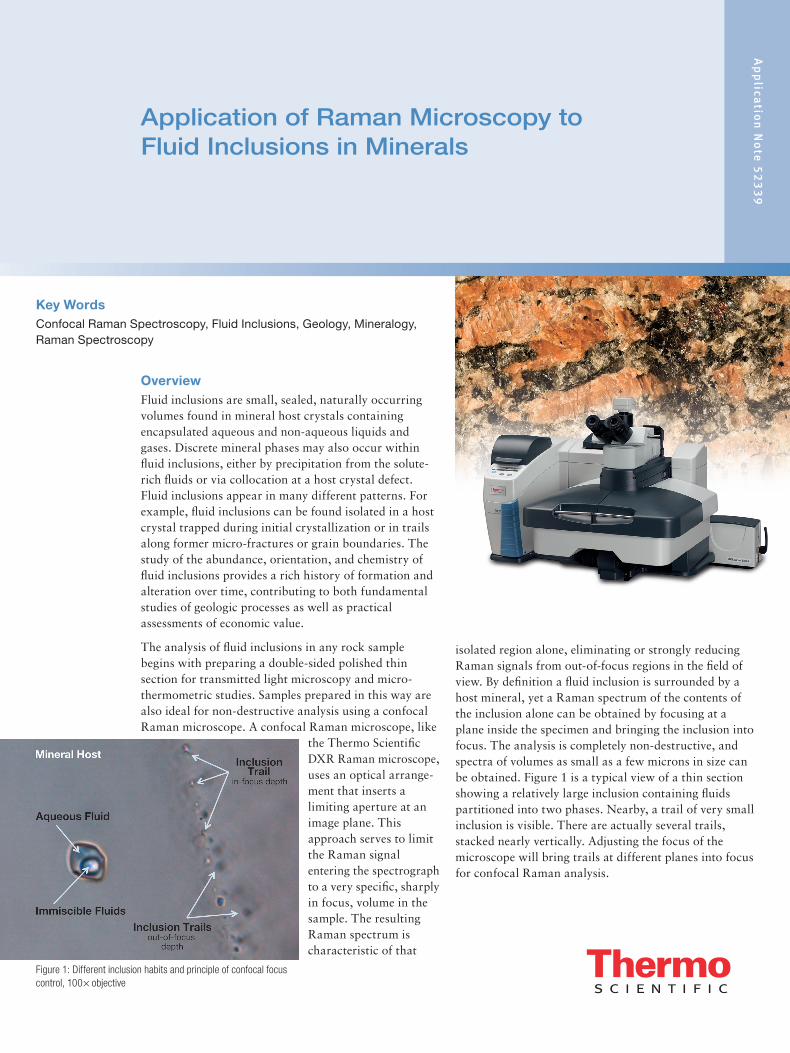
Application of Raman Microscopy to Fluid Inclusions in Minerals
Ap
plica
tion
No
te 5
23
39
Key WordsConfocal Raman Spectroscopy, Fluid Inclusions, Geology, Mineralogy, Raman Spectroscopy
OverviewFluid inclusions are small, sealed, naturally occurring volumes found in mineral host crystals containing encapsulated aqueous and non-aqueous liquids and gases. Discrete mineral phases may also occur within fluid inclusions, either by precipitation from the solute-rich fluids or via collocation at a host crystal defect. Fluid inclusions appear in many different patterns. For example, fluid inclusions can be found isolated in a host crystal trapped during initial crystallization or in trails along former micro-fractures or grain boundaries. The study of the abundance, orientation, and chemistry of fluid inclusions provides a rich history of formation and alteration over time, contributing to both fundamental studies of geologic processes as well as practical assessments of economic value.
The analysis of fluid inclusions in any rock sample begins with preparing a double-sided polished thin section for transmitted light microscopy and micro-thermometric studies. Samples prepared in this way are also ideal for non-destructive analysis using a confocal Raman microscope. A confocal Raman microscope, like
the Thermo Scientific DXR Raman microscope, uses an optical arrange-ment that inserts a limiting aperture at an image plane. This approach serves to limit the Raman signal entering the spectrograph to a very specific, sharply in focus, volume in the sample. The resulting Raman spectrum is characteristic of that
isolated region alone, eliminating or strongly reducing Raman signals from out-of-focus regions in the field of view. By definition a fluid inclusion is surrounded by a host mineral, yet a Raman spectrum of the contents of the inclusion alone can be obtained by focusing at a plane inside the specimen and bringing the inclusion into focus. The analysis is completely non-destructive, and spectra of volumes as small as a few microns in size can be obtained. Figure 1 is a typical view of a thin section showing a relatively large inclusion containing fluids partitioned into two phases. Nearby, a trail of very small inclusion is visible. There are actually several trails, stacked nearly vertically. Adjusting the focus of the microscope will bring trails at different planes into focus for confocal Raman analysis.
Figure 1: Different inclusion habits and principle of confocal focus control, 100× objective
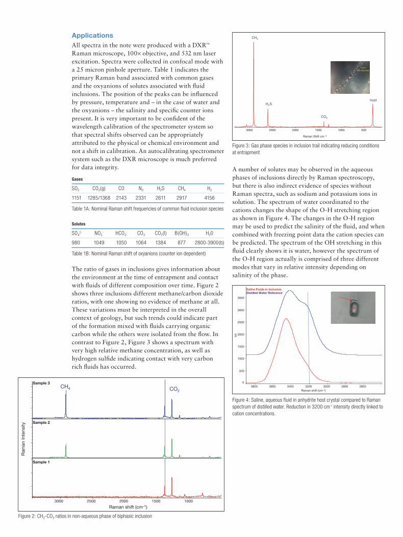
ApplicationsAll spectra in the note were produced with a DXR™ Raman microscope, 100× objective, and 532 nm laser excitation. Spectra were collected in confocal mode with a 25 micron pinhole aperture. Table 1 indicates the primary Raman band associated with common gases and the oxyanions of solutes associated with fluid inclusions. The position of the peaks can be influenced by pressure, temperature and – in the case of water and the oxyanions – the salinity and specific counter ions present. It is very important to be confident of the wavelength calibration of the spectrometer system so that spectral shifts observed can be appropriately attributed to the physical or chemical environment and not a shift in calibration. An autocalibrating spectrometer system such as the DXR microscope is much preferred for data integrity.
The ratio of gases in inclusions gives information about the environment at the time of entrapment and contact with fluids of different composition over time. Figure 2 shows three inclusions different methane/carbon dioxide ratios, with one showing no evidence of methane at all. These variations must be interpreted in the overall context of geology, but such trends could indicate part of the formation mixed with fluids carrying organic carbon while the others were isolated from the flow. In contrast to Figure 2, Figure 3 shows a spectrum with very high relative methane concentration, as well as hydrogen sulfide indicating contact with very carbon rich fluids has occurred.
A number of solutes may be observed in the aqueous phases of inclusions directly by Raman spectroscopy, but there is also indirect evidence of species without Raman spectra, such as sodium and potassium ions in solution. The spectrum of water coordinated to the cations changes the shape of the O-H stretching region as shown in Figure 4. The changes in the O-H region may be used to predict the salinity of the fluid, and when combined with freezing point data the cation species can be predicted. The spectrum of the OH stretching in this fluid clearly shows it is water, however the spectrum of the O-H region actually is comprised of three different modes that vary in relative intensity depending on salinity of the phase.
Gases
SO2 CO2(g) CO N2 H2S CH4 H2
1151 1285/1368 2143 2331 2611 2917 4156
Table 1A: Nominal Raman shift frequencies of common fluid inclusion species
Solutes
SO42- NO3
- HCO3- CO3 CO2(l) B(OH)3 H20
980 1049 1050 1064 1384 877 2800-3900(b)
Table 1B: Nominal Raman shift of oxyanions (counter ion dependent)
Figure 2: CH3-CO2 ratios in non-aqueous phase of biphasic inclusion
Figure 3: Gas phase species in inclusion trail indicating reducing conditions at entrapment
Figure 4: Saline, aqueous fluid in anhydrite host crystal compared to Raman spectrum of distilled water. Reduction in 3200 cm-1 intensity directly linked to cation concentrations.
20 microns
CH4
H2S
CO2
500 1000 1500 2000 2500 3000
Raman Shift cm-1
host
Ap
plica
tion
No
te 5
23
39
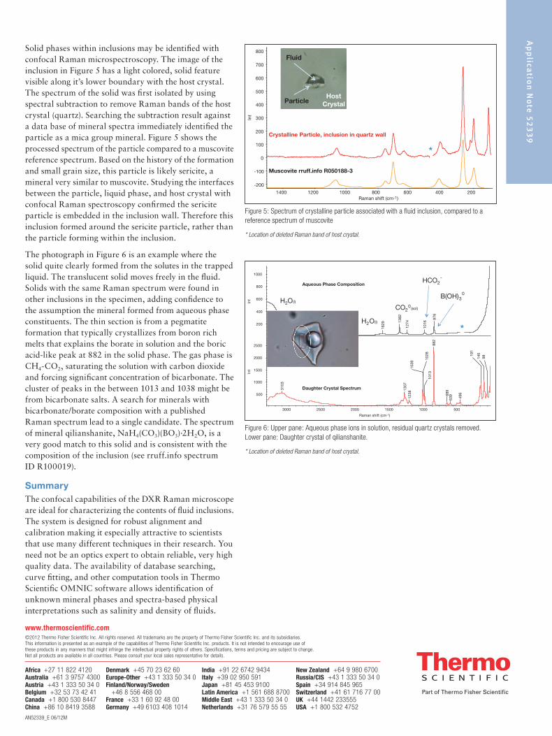
Solid phases within inclusions may be identified with confocal Raman microspectroscopy. The image of the inclusion in Figure 5 has a light colored, solid feature visible along it’s lower boundary with the host crystal. The spectrum of the solid was first isolated by using spectral subtraction to remove Raman bands of the host crystal (quartz). Searching the subtraction result against a data base of mineral spectra immediately identified the particle as a mica group mineral. Figure 5 shows the processed spectrum of the particle compared to a muscovite reference spectrum. Based on the history of the formation and small grain size, this particle is likely sericite, a mineral very similar to muscovite. Studying the interfaces between the particle, liquid phase, and host crystal with confocal Raman spectroscopy confirmed the sericite particle is embedded in the inclusion wall. Therefore this inclusion formed around the sericite particle, rather than the particle forming within the inclusion.
The photograph in Figure 6 is an example where the solid quite clearly formed from the solutes in the trapped liquid. The translucent solid moves freely in the fluid. Solids with the same Raman spectrum were found in other inclusions in the specimen, adding confidence to the assumption the mineral formed from aqueous phase constituents. The thin section is from a pegmatite formation that typically crystallizes from boron rich melts that explains the borate in solution and the boric acid-like peak at 882 in the solid phase. The gas phase is CH4-CO2, saturating the solution with carbon dioxide and forcing significant concentration of bicarbonate. The cluster of peaks in the between 1013 and 1038 might be from bicarbonate salts. A search for minerals with bicarbonate/borate composition with a published Raman spectrum lead to a single candidate. The spectrum of mineral qilianshanite, NaH4(CO3)(BO3)·2H2O, is a very good match to this solid and is consistent with the composition of the inclusion (see rruff.info spectrum ID R100019).
SummaryThe confocal capabilities of the DXR Raman microscope are ideal for characterizing the contents of fluid inclusions. The system is designed for robust alignment and calibration making it especially attractive to scientists that use many different techniques in their research. You need not be an optics expert to obtain reliable, very high quality data. The availability of database searching, curve fitting, and other computation tools in Thermo Scientific OMNIC software allows identification of unknown mineral phases and spectra-based physical interpretations such as salinity and density of fluids.
Figure 5: Spectrum of crystalline particle associated with a fluid inclusion, compared to a reference spectrum of muscovite
* Location of deleted Raman band of host crystal.
Figure 6: Upper pane: Aqueous phase ions in solution, residual quartz crystals removed. Lower pane: Daughter crystal of qilianshanite.
* Location of deleted Raman band of host crystal.
*
*
AN52339_E 06/12M
Africa +27 11 822 4120Australia +61 3 9757 4300Austria +43 1 333 50 34 0Belgium +32 53 73 42 41Canada +1 800 530 8447China +86 10 8419 3588
Denmark +45 70 23 62 60Europe-Other +43 1 333 50 34 0Finland/Norway/Sweden +46 8 556 468 00France +33 1 60 92 48 00Germany +49 6103 408 1014
India +91 22 6742 9434Italy +39 02 950 591Japan +81 45 453 9100Latin America +1 561 688 8700Middle East +43 1 333 50 34 0Netherlands +31 76 579 55 55
New Zealand +64 9 980 6700Russia/CIS +43 1 333 50 34 0Spain +34 914 845 965Switzerland +41 61 716 77 00UK +44 1442 233555USA +1 800 532 4752
www.thermoscientific.com©2012 Thermo Fisher Scientific Inc. All rights reserved. All trademarks are the property of Thermo Fisher Scientific Inc. and its subsidiaries. This information is presented as an example of the capabilities of Thermo Fisher Scientific Inc. products. It is not intended to encourage use of these products in any manners that might infringe the intellectual property rights of others. Specifications, terms and pricing are subject to change. Not all products are available in all countries. Please consult your local sales representative for details.

![Case Study 10: Measuring Fluid Inclusions in Geochemical ... · Case Study 10: Measuring Fluid Inclusions in Geochemical Samples Summary “Fluids play a key [geochemical] role in](https://img.pdfslide.us/doc/110x75/5b8218317f8b9a2b6f8dc50b/case-study-10-measuring-fluid-inclusions-in-geochemical-case-study-10.jpg)