Embed Size (px)
Citation preview
Application of Light and Scanning Electron Microscopy in the
Identification of Herbal Medicines
R. Serrano, G. da Silva and O. Silva
iMed.UL, Faculty of Pharmacy University of Lisbon, Av. Prof. Gama Pinto, 1649-019 Lisboa, Portugal
Herbal drug technology includes all the necessary steps for the conversion of botanical materials into medicines, where
standardization and quality control are essential analytical tools to assure the correct identification of drugs. Advances in
microscope technology and improvements in light and scanning electron microscopes have increased the accuracy and
capabilities of microscopy as a mean of botanical identification. The objective of this chapter is to present a set of the most
relevant histological characters by which some selected herbal drugs must be identified, taking into account the
corresponding plant part used, namely, bark, root and rhizome, leaf, flower, fruit and seed. The botanical control by
microscopic examinations, using histological identification, can be used as a rapid and inexpensive identification
technique, but requires highly trained individuals and a limited standard references libraries are available for comparison.
This approach aims at the establishment of botanical biomarkers based on the major microscopic features observed in the
studied drugs.
Keywords Herbal drugs; Herbal medicines; Light microscopy (LM); Medicinal plants; Scanning electron microscopy
(SEM)
1. Introduction
The use of medicinal plants has become very popular in recent years. Herbal drug technology includes all the necessary
steps for the conversion of botanical materials into medicines, where standardization and quality control are essential
analytical tools to assure the correct identification of drugs. Microscopy allows the identification of herbal drugs and the
detection of individual components of a mixture. It is highly important to ensure quality and purity of herbal medicines
in order to maximize the efficacy and minimize adverse side effects. For these reasons, microscopic analysis of the
powdered herbal drugs is a mandatory technique on the official Pharmacopeia monographs. Sometimes, the
characterization of histological sections of each herbal drug is very useful in the determination of the most relevant
characteristics of each powder.
Adulteration and misidentification of herbal drug can cause serious health problems to consumers, as well as
publicity and legal headaches for the pharmaceutical industry. The past decade has witnessed the introduction and
institution of new Good Manufacturing Practices (GMP) in quality control of raw materials, intermediates and finished
products of botanical origin [1]. The first step in quality control of medicinal plants is ensuring the authenticity of the desired species for the intended
use. The quality control can be conducted via a variety of techniques, namely macro- and microscopic identification and
chemical analysis. Microanatomy sometimes provides information that gross anatomy does not. Hence the importance
of the description of microscopic botanical aspects in order to help determine definitively the proper species of plant
material being collected, harvested or processed, i.e., while the plant material is still in its non-extracted form.
Microscopical descriptions can include the characterisation of the histological structures, cells and cell contents
visible only via light microscopy (LM) and scanning electron microscopy (SEM). The observation of cellular-level
morphology or anatomy is a major aid for the authentication of drugs. These characters are especially important for
identification of out broken or powdered drugs, because in these cases most of the morphological diagnostic features are
lost.
The powdered crude drugs can be identified based on the form, the presence or absence of different cell types based
on their cytomorphological characters, e.g. parenchyma, collenchyma, fibers, stone cells, vessels, trichomes, secretory
cells, epidermal cells. Cell inclusion characteristics are of considerable value in the identification of some unorganized
crude drugs, e.g. ergastic contents as aleurone grains, cluster crystals and prisms of calcium oxalate, silica and starch
granules [2]. For some well-known medicinal plants, data for the quality control were providing on official pharmacopoeias.
However, for most of vast number of plants used worldwide, such criteria are not found in these books [3]. For convenience, herbal medicines can be arranged into morphological groups as bark, root, leaf, seed, etc. Some of
them constitute more than one morphological part, for example, whole herb and commercial root, which may consist of
both rhizome and root. When herbal drug used is the whole plant or any part or parts, it is fairly easy to identify the
drug botanically, but when the drug is reduced to powder, a greater degree of expertise is required on part of
pharmacognosists.
Microscopy: Science, Technology, Applications and Education A. Méndez-Vilas and J. Díaz (Eds.)
182 ©FORMATEX 2010
______________________________________________
2. Identification of herbal medicines by microscopy
For hundreds of years, the magnifying glass and the microscope were the only possible method for the analytical
evaluation of herbal drugs. Advances in microscope technology and improvements in light and scanning electron
microscopes have improved the accuracy and capabilities of microscopy as a mean of botanical identification. In
addition, the microscopic identification involves the comparative analysis of broken and powdered herbal products.
Universally accepted standards and specifications for herbal products are useful for industry quality control and
standard-setting bodies.
Several types of microscopes can be used for the pharmacognostic studies, including LM, polarizing microscopy,
phase contrast microscopy, SEM. In our current methodology we applied LM and SEM for the identification of herbal
medicines. SEM produces a higher resolution compared to that possible using a light microscope, and the images
obtained are three-dimensional and consequently this technique has been extensively used to investigate the surface
topology of a wide variety of plant materials and can play a vital role in authentication of entire botanicals and those in
fragmentized or in powder form.
2.1 Light microscopy
Sampling procedures are required for the quality control of herbal drugs. The reliability of the microscopic analysis
depends if the samples are truly representative, i.e., the samples must represent the whole batch. Very specific sample
techniques are described in current pharmacopoeias.
After representative samples of herbal drugs are selected, dried materials often require softening before preparation
for microscopic studies. It may be done by exposing the sample in moist condition (for leaves and flowers) or by boiling
in water (for roots and barks).
The observation of micro morphological features by light microscope includes the preparation of slides temporary
mounts with thin sections of plant parts and wet mounts of powdered material. For the anatomical analysis by LM,
tangential and longitudinal hand sections of the dried plant material were prepared by conventional micro techniques.
Clearing agents, mountants, and stains are commonly used and a cover glass must always be applied.
Samples can be prepared in different mounting media. However the chloral hydrate solution is particularly useful as a
clearing reagent.
For the analysis of powdered material an aliquot of the plant material was transferred to a slide containing a drop of
water or other mounting medium or a specific reagent solution, and cover with a coverslip.
When it is necessary permanent mounts can be used, with thin tissue sections, the plant material is fixed, dehydrated,
and embedded in paraffin or other embedding media. This is used as a solid support matrix during tissue sectioning in a
microtome to cut sections of about 10 µm thickness. After sectioning and mounting, staining of the specimen is
frequently performed to aid in the differentiation of certain structures. Canada balsam, diluted with a small portion of
xylene, can be used as an adhesive. Other mountants are also commercially available. Upon drying of the mountant, the
slide can then be examined under a microscope.
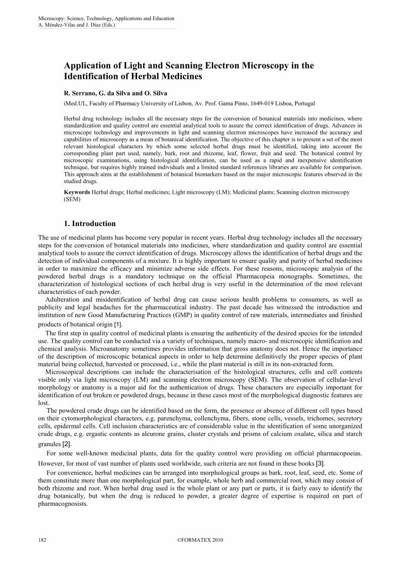
Fig. 1 shows a thin transverse section of Passiflora incarnata L. stem, a plant part integrant of the passiflorae herba
(passionflower herb), traditionally used as a sedative and anxiolytic drug. This section was obtained with a paraffin
microtome and prepared for permanent mounts [4]. The photomicrographs were obtained on an Olympus CX40 upright
microscope coupled with an Olympus ColorView IIIu camera. Image analysis was performed with Cell D 2006
Olympus Software.
Fig. 1 LM micrograph of stem transverse section of Passiflora incarnata, showing
collateral bundles with external phloem and internal xylem and calcium oxalate cluster
crystals on the surroundings of the vascular cambium. Scale bar = 200 µm.
Microscopy: Science, Technology, Applications and Education A. Méndez-Vilas and J. Díaz (Eds.)
©FORMATEX 2010 183
______________________________________________
2.2 Scanning electron microscopy
In contrast to LM, which uses visible light as a source of illumination and optical (glass) lenses to magnify specimens in
the range between approximately 10 to 1,000 times their original size, electron microscopy is operated in the vacuum
and focuses the electron beam and magnifies images with the help of electromagnetic lenses [5].
SEM is a powerful method for the investigation of surface structures of herbal medicines namely leaves, pollen
grains and seeds.
This technique provides a large depth of field, which means, the area of the sample that can be viewed in focus at the
same time is actually quite large [5]. SEM has also the advantage that the range of magnification is relatively wide
allowing the investigator to easily focus in on an area of interest on a specimen that was initially scanned at a lower
magnification. Furthermore, the tridimensional view images may be to an investigator it easier to interpret SEM images.
The basic steps involved in SEM sample preparation include surface cleaning, stabilizing the sample with a fixative,
rinsing, dehydrating, drying, mounting the specimen on a metal holder, and coating the sample with a layer of a material
that is electrically conductive [6].
In general, SEM not only produces images that are analogous to those from an optical microscope, but it also can
produce images whose contrast is based on compositional variations of specimens. However, SEM analysis is much
more expensive when compared to the LM.
In our observations for SEM, the dried material was mounted directly on stubs using double-side adhesive tape, and
sputtered with a thin layer of gold in a JEOL JSM-1200 Fine Coater. The electron micrographs were obtained in a JEOL
JSM-T220 scanning electron microscope at 15 kV, with an integrated digital image acquisition system.
3. Major morphological groups
Lack of information concerning some species as Artemisia annua L. (Asteraceae), Digitalis thapsi L.
(Scrophulareaceae), Frangula azorica V. Grubow (Rhamnaceae), Guiera senegalensis J. F. Gmel. (Combretaceae),
Hypericum androsaemum L. (Clusiaceae), Hypericum foliosum Aiton. (Clusiaceae), Hypoxis hemerocallidea Fisch. &
C.A. Mey (Hypoxidaceae), Jateorhiza palmata (Lam.) Miers (Menispermaceae), Maytenus heterophylla Eckl. & Zeyh.
N. Robson (Celastraceae) and Maytenus senegalensis (Lam.) Exell (Celastraceae) is a major obstacle to the
establishment of quality control and certification of these medicinal plants. The selection of these plants was based on
our investigation experience, making an attempt to fulfill important factual data from current ongoing work at the
Pharmacognosy Laboratory of the Faculty of Pharmacy, University of Lisbon.
The objective of this chapter is to present a set of the most relevant histological characters by which each herbal
drugs may be identified, particularly within the morphological group to which it belongs, namely bark, root and
rhizome, leaf, flower, fruit and seed. The presence of certain botanical features of the various plant tissues and stomata
types, epidermal trichomes, papillae, as well as the size, shape of cells and cell contents like calcium oxalate crystals is
extremely relevant for the botanical diagnosis.
The evaluation of the microscopic characters consists of anatomical details of drugs pertaining to arrangement of
different cells and tissues, their dimensions, details of abnormal cells or tissues arrangements and cell inclusions.
3.1 Subterranean organs
3.1.1 Root
The primary root shows the following structures: a parenchymatous cortex, endodermis, and a vascular cylinder with
vascular tissues enclosed in a single or many-layered pericycle. The secondary root usually contains cork and vascular
tissues in varying amounts. Secondary xylem is formed by the vascular cambium, a single ring of cells that produce
layers of xylem toward the inside and secondary phloem toward the outside. Chlorenchyma, palisade tissue, and
aleurone grains are not visible in this plant part.
As example of an herbal drug constituted by root we refer calumba, the common name of Jateorhiza palmate root
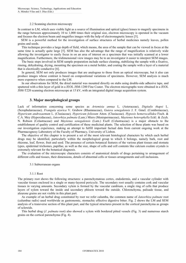
(calumbae radix) used worldwide as gastronomic, stomachic effective digestive bitter. Fig. 2 shows the LM and SEM
analysis of a transverse section of this plant part, and the typical structures present in the cortical parenchyma as groups
of sclereids.
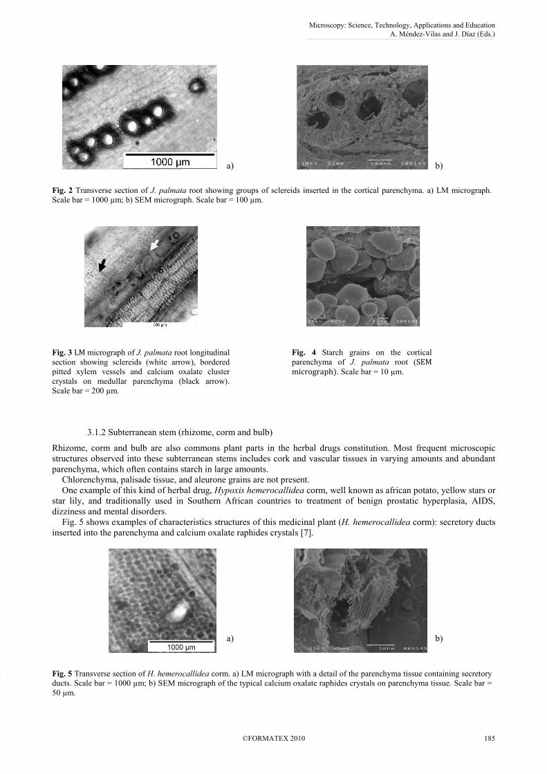
This herbal drug (J. palmata root) also showed a xylem with bordered pitted vessels (Fig. 3) and numerous starch
grains on the cortical parenchyma (Fig. 4).
Microscopy: Science, Technology, Applications and Education A. Méndez-Vilas and J. Díaz (Eds.)
184 ©FORMATEX 2010
______________________________________________
a)
b)
Fig. 2 Transverse section of J. palmata root showing groups of sclereids inserted in the cortical parenchyma. a) LM micrograph.
Scale bar = 1000 µm; b) SEM micrograph. Scale bar = 100 µm.
Fig. 3 LM micrograph of J. palmata root longitudinal
section showing sclereids (white arrow), bordered
pitted xylem vessels and calcium oxalate cluster
crystals on medullar parenchyma (black arrow).
Scale bar = 200 µm.
Fig. 4 Starch grains on the cortical
parenchyma of J. palmata root (SEM
micrograph). Scale bar = 10 µm.
3.1.2 Subterranean stem (rhizome, corm and bulb)
Rhizome, corm and bulb are also commons plant parts in the herbal drugs constitution. Most frequent microscopic
structures observed into these subterranean stems includes cork and vascular tissues in varying amounts and abundant
parenchyma, which often contains starch in large amounts.
Chlorenchyma, palisade tissue, and aleurone grains are not present.
One example of this kind of herbal drug, Hypoxis hemerocallidea corm, well known as african potato, yellow stars or
star lily, and traditionally used in Southern African countries to treatment of benign prostatic hyperplasia, AIDS,
dizziness and mental disorders.
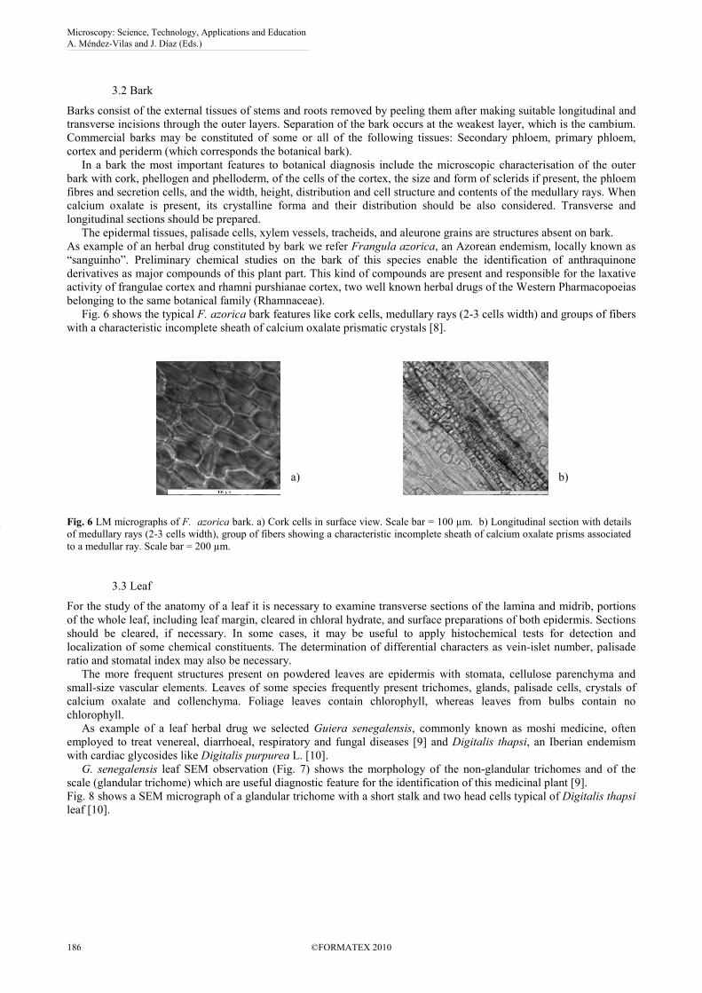
Fig. 5 shows examples of characteristics structures of this medicinal plant (H. hemerocallidea corm): secretory ducts
inserted into the parenchyma and calcium oxalate raphides crystals [7].
a)
b)
Fig. 5 Transverse section of H. hemerocallidea corm. a) LM micrograph with a detail of the parenchyma tissue containing secretory
ducts. Scale bar = 1000 µm; b) SEM micrograph of the typical calcium oxalate raphides crystals on parenchyma tissue. Scale bar =
50 µm.
Microscopy: Science, Technology, Applications and Education A. Méndez-Vilas and J. Díaz (Eds.)
©FORMATEX 2010 185
______________________________________________
3.2 Bark
Barks consist of the external tissues of stems and roots removed by peeling them after making suitable longitudinal and
transverse incisions through the outer layers. Separation of the bark occurs at the weakest layer, which is the cambium.
Commercial barks may be constituted of some or all of the following tissues: Secondary phloem, primary phloem,
cortex and periderm (which corresponds the botanical bark).
In a bark the most important features to botanical diagnosis include the microscopic characterisation of the outer
bark with cork, phellogen and phelloderm, of the cells of the cortex, the size and form of sclerids if present, the phloem
fibres and secretion cells, and the width, height, distribution and cell structure and contents of the medullary rays. When
calcium oxalate is present, its crystalline forma and their distribution should be also considered. Transverse and
longitudinal sections should be prepared.
The epidermal tissues, palisade cells, xylem vessels, tracheids, and aleurone grains are structures absent on bark.
As example of an herbal drug constituted by bark we refer Frangula azorica, an Azorean endemism, locally known as
“sanguinho”. Preliminary chemical studies on the bark of this species enable the identification of anthraquinone
derivatives as major compounds of this plant part. This kind of compounds are present and responsible for the laxative
activity of frangulae cortex and rhamni purshianae cortex, two well known herbal drugs of the Western Pharmacopoeias
belonging to the same botanical family (Rhamnaceae).
Fig. 6 shows the typical F. azorica bark features like cork cells, medullary rays (2-3 cells width) and groups of fibers
with a characteristic incomplete sheath of calcium oxalate prismatic crystals [8].
a)
b)
Fig. 6 LM micrographs of F. azorica bark. a) Cork cells in surface view. Scale bar = 100 µm. b) Longitudinal section with details
of medullary rays (2-3 cells width), group of fibers showing a characteristic incomplete sheath of calcium oxalate prisms associated
to a medullar ray. Scale bar = 200 µm.
3.3 Leaf
For the study of the anatomy of a leaf it is necessary to examine transverse sections of the lamina and midrib, portions
of the whole leaf, including leaf margin, cleared in chloral hydrate, and surface preparations of both epidermis. Sections
should be cleared, if necessary. In some cases, it may be useful to apply histochemical tests for detection and
localization of some chemical constituents. The determination of differential characters as vein-islet number, palisade
ratio and stomatal index may also be necessary.
The more frequent structures present on powdered leaves are epidermis with stomata, cellulose parenchyma and
small-size vascular elements. Leaves of some species frequently present trichomes, glands, palisade cells, crystals of
calcium oxalate and collenchyma. Foliage leaves contain chlorophyll, whereas leaves from bulbs contain no
chlorophyll.
As example of a leaf herbal drug we selected Guiera senegalensis, commonly known as moshi medicine, often
employed to treat venereal, diarrhoeal, respiratory and fungal diseases [9] and Digitalis thapsi, an Iberian endemism
with cardiac glycosides like Digitalis purpurea L. [10].
G. senegalensis leaf SEM observation (Fig. 7) shows the morphology of the non-glandular trichomes and of the
scale (glandular trichome) which are useful diagnostic feature for the identification of this medicinal plant [9].
Fig. 8 shows a SEM micrograph of a glandular trichome with a short stalk and two head cells typical of Digitalis thapsi
leaf [10].
Microscopy: Science, Technology, Applications and Education A. Méndez-Vilas and J. Díaz (Eds.)
186 ©FORMATEX 2010
______________________________________________
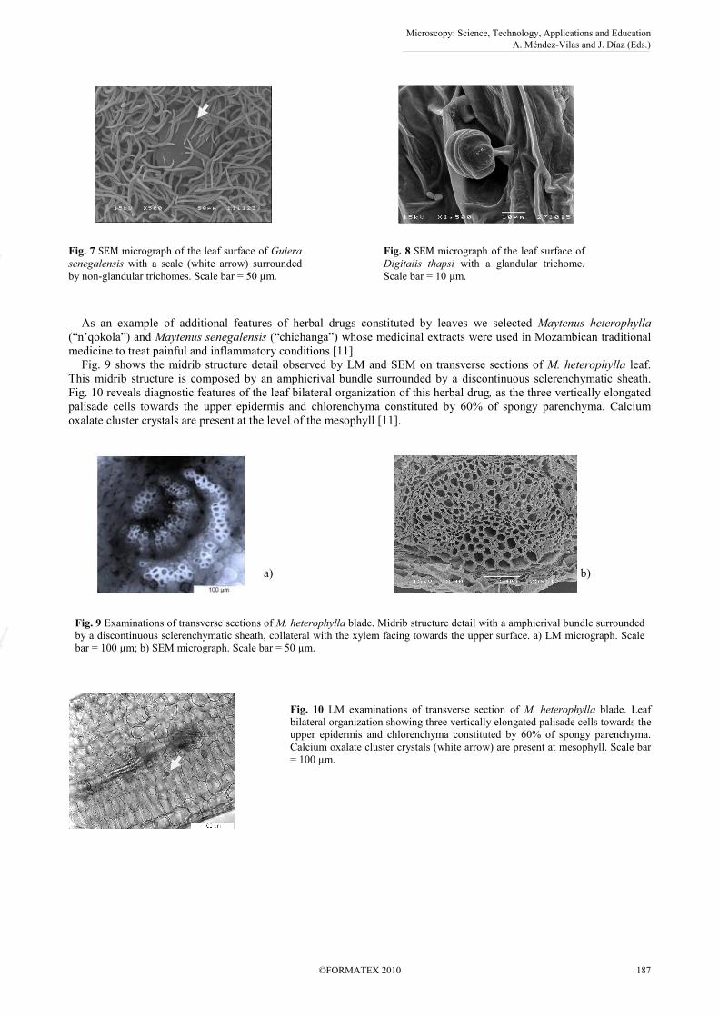
Fig. 7 SEM micrograph of the leaf surface of Guiera
senegalensis with a scale (white arrow) surrounded
by non-glandular trichomes. Scale bar = 50 µm.
Fig. 8 SEM micrograph of the leaf surface of
Digitalis thapsi with a glandular trichome.
Scale bar = 10 µm.
As an example of additional features of herbal drugs constituted by leaves we selected Maytenus heterophylla
(“n’qokola”) and Maytenus senegalensis (“chichanga”) whose medicinal extracts were used in Mozambican traditional
medicine to treat painful and inflammatory conditions [11].
Fig. 9 shows the midrib structure detail observed by LM and SEM on transverse sections of M. heterophylla leaf.
This midrib structure is composed by an amphicrival bundle surrounded by a discontinuous sclerenchymatic sheath.
Fig. 10 reveals diagnostic features of the leaf bilateral organization of this herbal drug, as the three vertically elongated
palisade cells towards the upper epidermis and chlorenchyma constituted by 60% of spongy parenchyma. Calcium
oxalate cluster crystals are present at the level of the mesophyll [11].
a)
b)
Fig. 9 Examinations of transverse sections of M. heterophylla blade. Midrib structure detail with a amphicrival bundle surrounded
by a discontinuous sclerenchymatic sheath, collateral with the xylem facing towards the upper surface. a) LM micrograph. Scale
bar = 100 µm; b) SEM micrograph. Scale bar = 50 µm.
Fig. 10 LM examinations of transverse section of M. heterophylla blade. Leaf
bilateral organization showing three vertically elongated palisade cells towards the
upper epidermis and chlorenchyma constituted by 60% of spongy parenchyma.
Calcium oxalate cluster crystals (white arrow) are present at mesophyll. Scale bar
= 100 µm.
Microscopy: Science, Technology, Applications and Education A. Méndez-Vilas and J. Díaz (Eds.)
©FORMATEX 2010 187
______________________________________________
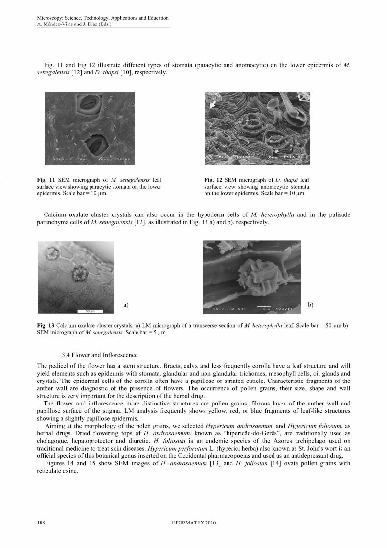
Fig. 11 and Fig 12 illustrate different types of stomata (paracytic and anomocytic) on the lower epidermis of M.
senegalensis [12] and D. thapsi [10], respectively.
Fig. 11 SEM micrograph of M. senegalensis leaf
surface view showing paracytic stomata on the lower
epidermis. Scale bar = 10 µm.
Fig. 12 SEM micrograph of D. thapsi leaf
surface view showing anomocytic stomata
on the lower epidermis. Scale bar = 10 µm.
Calcium oxalate cluster crystals can also occur in the hypoderm cells of M. heterophylla and in the palisade
parenchyma cells of M. senegalensis [12], as illustrated in Fig. 13 a) and b), respectively.
a)
b)
Fig. 13 Calcium oxalate cluster crystals. a) LM micrograph of a transverse section of M. heterophylla leaf. Scale bar = 50 µm b)
SEM micrograph of M. senegalensis. Scale bar = 5 µm.
3.4 Flower and Inflorescence
The pedicel of the flower has a stem structure. Bracts, calyx and less frequently corolla have a leaf structure and will
yield elements such as epidermis with stomata, glandular and non-glandular trichomes, mesophyll cells, oil glands and
crystals. The epidermal cells of the corolla often have a papillose or striated cuticle. Characteristic fragments of the
anther wall are diagnostic of the presence of flowers. The occurrence of pollen grains, their size, shape and wall
structure is very important for the description of the herbal drug.
The flower and inflorescence more distinctive structures are pollen grains, fibrous layer of the anther wall and
papillose surface of the stigma. LM analysis frequently shows yellow, red, or blue fragments of leaf-like structures
showing a slightly papillose epidermis.
Aiming at the morphology of the polen grains, we selected Hypericum androsaemum and Hypericum foliosum, as
herbal drugs. Dried flowering tops of H. androsaemum, known as “hipericão-do-Gerês”, are traditionally used as
cholagogue, hepatoprotector and diuretic. H. foliosum is an endemic species of the Azores archipelago used on
traditional medicine to treat skin diseases. Hypericum perforatum L. (hyperici herba) also known as St. John's wort is an
official species of this botanical genus inserted on the Occidental pharmacopoeias and used as an antidepressant drug.
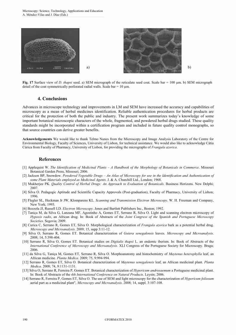
Figures 14 and 15 show SEM images of H. androsaemum [13] and H. foliosum [14] ovate pollen grains with
reticulate exine.
Microscopy: Science, Technology, Applications and Education A. Méndez-Vilas and J. Díaz (Eds.)
188 ©FORMATEX 2010
______________________________________________
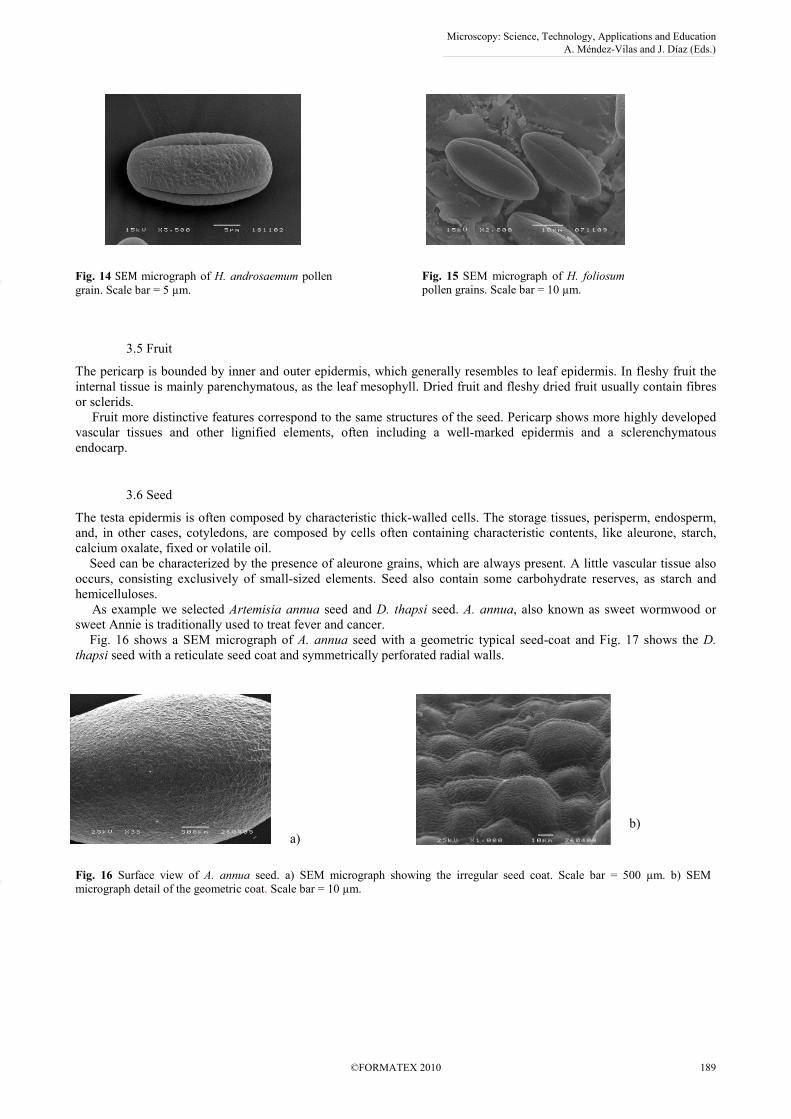
Fig. 14 SEM micrograph of H. androsaemum pollen
grain. Scale bar = 5 µm.
Fig. 15 SEM micrograph of H. foliosum
pollen grains. Scale bar = 10 µm.
3.5 Fruit
The pericarp is bounded by inner and outer epidermis, which generally resembles to leaf epidermis. In fleshy fruit the
internal tissue is mainly parenchymatous, as the leaf mesophyll. Dried fruit and fleshy dried fruit usually contain fibres
or sclerids.
Fruit more distinctive features correspond to the same structures of the seed. Pericarp shows more highly developed
vascular tissues and other lignified elements, often including a well-marked epidermis and a sclerenchymatous
endocarp.
3.6 Seed
The testa epidermis is often composed by characteristic thick-walled cells. The storage tissues, perisperm, endosperm,
and, in other cases, cotyledons, are composed by cells often containing characteristic contents, like aleurone, starch,
calcium oxalate, fixed or volatile oil.
Seed can be characterized by the presence of aleurone grains, which are always present. A little vascular tissue also
occurs, consisting exclusively of small-sized elements. Seed also contain some carbohydrate reserves, as starch and
hemicelluloses.
As example we selected Artemisia annua seed and D. thapsi seed. A. annua, also known as sweet wormwood or
sweet Annie is traditionally used to treat fever and cancer.
Fig. 16 shows a SEM micrograph of A. annua seed with a geometric typical seed-coat and Fig. 17 shows the D.
thapsi seed with a reticulate seed coat and symmetrically perforated radial walls.
a)
b)
Fig. 16 Surface view of A. annua seed. a) SEM micrograph showing the irregular seed coat. Scale bar = 500 µm. b) SEM
micrograph detail of the geometric coat. Scale bar = 10 µm.
Microscopy: Science, Technology, Applications and Education A. Méndez-Vilas and J. Díaz (Eds.)
©FORMATEX 2010 189
______________________________________________
a)
b)
Fig. 17 Surface view of D. thapsi seed. a) SEM micrograph of the reticulate seed coat. Scale bar = 100 µm. b) SEM micrograph
detail of the coat symmetrically perforated radial walls. Scale bar = 10 µm.
4. Conclusions
Advances in microscope technology and improvements in LM and SEM have increased the accuracy and capabilities of
microscopy as a mean of herbal medicines identification. Reliable authentication procedures for herbal products are
critical for the protection of both the public and industry. The present work summarizes today’s knowledge of some
important botanical microscopic characters of the whole, fragmented, and powdered herbal drugs studied. These quality
standards might be incorporated within a certification program and included in future quality control monographs, so
that source countries can derive greater benefits.
Acknowledgements We would like to thank Telmo Nunes from the Microscopy and Image Analysis Laboratory of the Centre for
Environmental Biology, Faculty of Sciences, University of Lisbon, for technical assistance. We would also like to acknowledge Cátia
Cúrica from Faculty of Pharmacy, University of Lisbon, for providing the micrographs of Frangula azorica.
References
[1] Applequist W. The Identification of Medicinal Plants – A Handbook of the Morphology of Botanicals in Commerce. Missouri
Botanical Garden Press, Missouri; 2006.
[2] Jackson BP, Snowdow. Powdered Vegetable Drugs – An Atlas of Microscopy for use in the Identification and Authentication of
some Plant Materials employed as Medicinal Agents. J. & A. Churchill Ltd., London; 1968.
[3] Mukherjee PK. Quality Control of Herbal Drugs: An Approach to Evaluation of Botanicals. Business Horizons. New Delphi;
2007.
[4] Silva O. Pedagogic Aptitude and Scientific Capacity Approvals (Post-graduation), Faculty of Pharmacy, University of Lisbon;
1996.
[5] Flegler SL, Heckman Jr JW, Klomparens KL. Scanning and Transmission Electron Microscopy. W. H. Freeman and Company,
New York; 1993.
[6] Bozzola JJ, Russell LD. Electron Microscopy. Jones and Bartlett Publishers Inc., Boston; 1992.
[7] Taniça M, da Silva G, Lanzana MF, Agostinho A, Gomes ET, Serrano R, Silva O. Light and scanning electron microscopy of
Hypoxis radix, an African drug. In: Book of Abstracts of the Joint Congress of the Spanish and Portuguese Microscopy
Societies. Segovia; 2009.
[8] Curica C, Serrano R, Gomes ET, Silva O. Morphological characterization of Frangula azorica bark as a potential herbal drug.
Microscopy and Microanalysis. 2009; 15, supp 3:11-12.
[9] Silva O, Serrano R, Gomes ET. Botanical characterization of Guiera senegalensis leaves. Microscopy and Microanalysis.
2008; 14, 5:398-404.
[10] Serrano R, Silva O, Gomes ET. Botanical studies on Digitalis thapsi L. an endemic iberism. In: Book of Abstracts of the
International Conference of Microscopy and Microanalysis. XLI Congress of the Portuguese Society for Microscopy. Braga;
2006.
[11] da Silva G, Taniça M, Gomes ET, Serrano R, Silva O. Morphoanatomy and histochemistry of Maytenus heterophylla leaf, an
African medicine. Planta Medica. 2009; 75, 9:994-994.
[12] Serrano R, Gomes ET, Silva O. Botanical characterization of Maytenus senegalensis leaf, an African medicinal plant. Planta
Medica. 2008; 74, 9:1131-1131.
[13] Silva O, Serrano R, Ferreira P, Gomes ET. Botanical characterization of Hypericum androsaemum a Portuguese medicinal plant.
In: Book of Abstracts of the 4th International Conference on Natural Products. Leysin; 2006.
[14] Serrano R, Ferreira P, Gomes ET, Silva O. The use of SEM and light microscopy for the characterization of Hypericum foliosum
aerial part as a medicinal plant", Microscopy and Microanalysis. 2008; 14, suppl. 3:107-108.
Microscopy: Science, Technology, Applications and Education A. Méndez-Vilas and J. Díaz (Eds.)
190 ©FORMATEX 2010
______________________________________________


![Ultrafast transmission electron microscopy using a laser ...transmission electron microscopy [4], scanning electron microscopy [5], x-ray diffraction [6], scanning tunneling and atomic](https://img.pdfslide.us/doc/110x75/607eb1335ce8082131294459/ultrafast-transmission-electron-microscopy-using-a-laser-transmission-electron.jpg)