Embed Size (px)
Citation preview
Send Orders for Reprints to [email protected]
The Open Conference Proceedings Journal, 2013, 4, 11-22 11
2210-2892/13 2013 Bentham Open
Open Access
Application of In Silico Methods to Support Experimental Data: Interactions of Antidepressants with Nicotinic Acetylcholine Receptors
Katarzyna M. Targowska-Duda1,*, Hugo R. Arias2 and Krzysztof Jozwiak1
1Department of Chemistry, Laboratory of Medicinal Chemistry and Neuroengineering, Medical University of Lublin, 20-093 Lublin, Poland 2Department of Medical Education, California Northstate University College of Medicine, Elk Grove, CA 95757, USA
Abstract: Over the last decades, several computational (in silico) methods have been developed and applied to test pharmacological hypotheses. An important hypothesis is that the therapeutic activity of many commonly used antidepressants may be partially mediated through the inhibition of different nicotinic acetylcholine receptors (nAChRs). This is based on pathologic conditions where the activity of the cholinergic system is exacerbated compared to the adrenergic system. Different in silico methods, including comparative/homology modeling, molecular docking, and molecular dynamics simulations, have been employed to study the interactions between several classes of antidepressants with distinct nAChR subtypes. More specifically, these methods were used to structurally characterize the antidepressant binding sites and to better understand their inhibitory mechanisms. This review focuses on computational methods that were found important in explaining and supporting several experimental results concerning the interaction of antidepressants with different nAChR subtypes. Among the studied antidepressants are norepinephrine selective reuptake inhibitors [e.g., (-)-reboxetine] as well as less selective antidepressants such as dopamine/norepinephrine reuptake inhibitors [e.g., (±)-bupropion and its derivatives], tricyclic antidepressants (e.g., imipramine), and (±)-mecamylamine and its enantiomers.
Keywords: Antidepressants, in silico, molecular docking, molecular modeling, molecular dynamics, nicotinic acetylcholine receptors, noncompetitive antagonists.
INTRODUCTION
Nicotinic acetylcholine receptors (nAChRs) are membrane embedded proteins member of the Cys-loop ligand-gated ion channel (LGIC) superfamily that includes the type 3 serotonin (5-HT), type A and C γ-aminobutyric acid, and glycine receptors (reviewed in [1-3]). nAChRs are pentameric assemblies of separate subunits oriented around a centrally located pore permeable to cations. In vertebrates, seventeen nAChR subunits have been identified (α1–α10, β1–β4, γ, δ, and ε) which can co-assemble to generate diverse nAChR subtypes. All these subunits are found in humans and other mammalian species, except α8 which has been identified only in avian species [4]. These subunits can form homomeric (i.e., containing only the α7, α8, or α9 subunit) or heteromeric pentamers (i.e., containing different subunits such as α3β4, α4β2, and α1β1γδ). These receptors are heterogeneously distributed in the central nervous system (CNS), while in the periphery, they mediate synaptic transmission at the neuromuscular junction and ganglia [4, 5]. Presynaptic nAChRs regulate the release of several neurotransmitters, such as acetylcholine (ACh), serotonin (5-hydroxytryptamine; 5-HT), norepinephrine (NE), dopamine (DA), glutamate, and γ-aminobutyric acid (GABA) [6]. In
*Address correspondence to this author at the Laboratory of Medicinal Chemistry and Neuroengineering. Medical University of Lublin, W. Chodzki 4a Street, 20-093 Lublin, Poland; Tel: (+48) 756 48 63; E-mail: [email protected]
this regards, nAChRs are involved in several physiological functions, including cognition and memory process, muscle contraction, and pain (reviewed in [5]). nAChRs are also found in non-neuronal cells (e.g., keratinocytes, epithelia, and macrophages), where they modulate the anti-inflammatory cholinergic pathway [5] and angiogenesis [7]. Based on their important functional activities, nAChRs are promising pharmaceutical targets for CNS disorders such as schizophrenia, attention deficit hyperactivity disorder, anxiety and depression, Tourette´s syndrome, nicotine and drug addiction, and Alzheimer’s and Parkinson’s disease [8, 9].
MOLECULAR STRUCTURE OF THE NACHR AND POTENTIAL LIGAND BINDING SITES
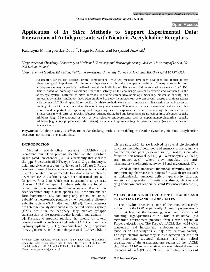
The nAChR structure is one of the most extensively studied from the LGIC superfamily. A very important reason for it, at least at the beginning, was the possibility of obtaining large quantities of nAChRs in its native lipid membrane environment prepared from electric organs of Torpedo electric rays. The Torpedo nAChR (i.e., α1β1γδ) is structurally and functionally analogous to the human muscular nAChR subtype (i.e., α1β1δγ/ε, embryonic/adult). The cryo-electron microscopy studies detected for the first time important structural domains and the helical organization of the transmembrane region of the nAChR [10]. The nAChR molecular structure was refined down to a resolution of ~4 Å (PDB id: 2BG9). Each subunit consists of
12 The Open Conference Proccedings Journal, 2013, Volume 4 Targowska-Duda et al.
three domains (Fig. 1): the N-terminal extracellular domain where β-sheet folding arrangements dominate, the membrane domain which consists of four transmembrane α-helices (M1–M4), and the intracellular domain, with lengths that depend on the subunit type. This model was used to build the homology models of neuronal and muscular nAChR subtypes presented in the current review. Interestingly, a recently published paper presents the conformational changes underlying the receptor activation by ACh using ACh-sprayed and freeze-trapped postsynaptic membranes [11]. Based on these results, the closed (PDB id: 4AQ5) and open (PDB id: 4AQ9) Torpedo nAChRs were obtained at a resolution of ~6.2 Å. Among the transmembrane segments, M2 is relatively more hydrophilic and thus, it tends to face the center of the ion channel. Considering that each subunit has one M2 segment, the ion channel is formed by five M2 segments. The amino acid sequences in M2 are highly conserved among different subunits and species, forming a series of amino acid rings exposed to the center of the pore and distributed along the axis of the channel (see Table 2). For example, in the Torpedo nAChR, the amino acid rings are named: outer or extracellular (position 20’), nonpolar (position 17’), valine (position 13’), leucine (position 9’), serine (position 6’), threonine (position 2’), intermediate (position -2’), and cytoplasmic or inner (position -5’). Although the ion channel is very much conserved among species, differences are also apparent among nAChR subunit sequences (see Table 1), producing variations in the nAChR ion channel structure. For instance, the α7 nAChR has the polar rings (positions 2’ and 6’) switched: the serine ring occurs at position 2’ and the threonine ring at position 6’ (see Table 2). On the other hand, the α4 subunit carries an
important change: several Phe residues are located at position 13’, becoming the valine/phenylalanine ring. This single difference dramatically changes the overall binding properties of the channel in docking simulations. Thus, the pattern of interactions between the channel and the ligand depends on the amino acid sequence of each subunit in the M2 region. The extracellular portion of the nAChR carries the binding sites for the endogenous neurotransmitter ACh (i.e., orthosteric sites) and other agonists (e.g., nicotine, epibatidine, and cytisine) as well as for competitive antagonists [1, 9, 12, 13]. Agonists trigger a series of conformational changes in the receptor, including local structural changes, the gating process comprising selected domains from the extracellular portion, and finally the opening of the intrinsic cation channel. This activation, in turn, causes membrane depolarization that finally produces the respective physiologic function (e.g., neurotransmitter release, muscle contraction, etc.). There are many different types of ligands that can modulate the pharmacological activity mediated by agonists. Competitive antagonists (e.g., α- and κ-bungarotoxins, methyllycaconitine, dihydro-β-erythroidine, and α-conotoxins) inhibit the activity of agonists by a simple mechanism where the ligand directly occupies the agonist binding sites [9, 14]. Allosteric ligands, on the other hand, can modulate the activity of agonists not by interacting with the orthosteric sites but with allosteric loci. Several types of modulators have been described based on their pharmacological mechanisms. Noncompetitive antagonists (NCAs) are represented by a large number of structurally different compounds that inhibit nAChR function by binding to allosteric sites located in general in
Fig. (1). The molecular structure of the Torpedo nAChR obtained by cryo-electron microscopy at 4 Å resolution (modified from [10]). (A) Side view of the receptor showing the extracellular (EXD), transmembrane (TMD), and intracellular (IND) domains, where two α1 subunits are depicted in black and the other three (i.e., β1, γ, and δ) are shown in grey. (B) Top view of the nAChR showing the EXD containing the agonist binding sites at the α1/γ and α1/δ subunit interfaces (see arrows). The ion pore is visible in the center of the structure.
Molecular Modeling Interactions between Antidepressant and nAChRs The Open Conference Proccedings Journal, 2013, Volume 4 13
Table 1. Amino Acid Sequence Alignment of the M2 Segments from Several nAChR Subunits
Subunit -5’ -4’ -3’ -2’ -1’ 1’ 2’ 3’ 4’ 5’ 6’ 7’ 8’ 9’ 10’ 11’ 12’ 13’ 14’ 15’ 16’ 17’ 18’ 19’ 20’
hα3 D C G E K V T L C I S V L L S L T V F L L V I T E
hα4 E C G E K I T L C I S V L L S L T V F L L L I T E
hα7 D S G E K I S L G I T V L L S L T V F M L L V A E
hα1 D S G E K M T L S I S V L L S L T V F L L V I V E
Tα1 D S G E K M T L S I S V L L S L T V F L L V I V E
hβ2 D C G E K M T L C I S V L L A L T V F L L L I S K
hβ4 D C G E K M T L C I S V L L A L T F F L L L I S K
hβ1 D A G E K M G L S I F A L L T L T V F L L L L A D
Tβ D A G E K M S L S I S A L L A L T V F L L L L A D
Tδ E S G(G) E L M S T A I C V L L A Q A V F L L L T S Q
hδ D S G E K T S V A I S V L L A Q S V F L L L I S K
Tγ Q A G Q K C T L S I S V L L A Q T I F L F L I A Q
hγ K A G(G) Q K C T V A I N V L L A Q T V F L F L V A K
hε Q A G(G) Q K C T V S I N V L L A Q T V F L F L I A Q
The M2 segments are helical, thus every 3rd–4th residue is exposed to the center of the channel forming amino acid rings. The position of the amino acid rings (in gray) are labeled using the prime nomenclature corresponding to the sequence from Asp238 (-5’) to Glu262 (20’) in the Torpedo nAChR α1-subunit. h, human; T, Torpedo.
Table 2. Inhibitory Potency of Structurally Different Antidepressants at Different Human nAChR Subtypes
nAChR Subtype Antidepressants Method IC50 a µM Reference
Amitriptyline Ca2+ influx fluorimetry 2.2±0.6 [21]
Imipramine Ca2+ influx fluorimetry 5.4±1.2 [21]
Doxepin Ca2+ influx fluorimetry 6.8±1.6 [21]
(±)-Mecamylamine Ca2+ influx fluorimetry
Electrophysiology
3.0±0.7
2.5±0.6
[21]
[27]
(-)-Reboxetine Ca2+ influx fluorimetry 16.0±1.0 [28]
hα4β2
(±)-Bupropion 86Rb+ efflux 12.0±1.1 [23]
hα4β4 (±)-Bupropion 86Rb+ efflux 14.0±1.1 [23]
Imipramine Ca2+ influx fluorimetry 7.8±1.4 [29] hα7
(±)-Mecamylamine Electrophysiology 6.9±1.6 [27]
Amitriptyline Ca2+ influx fluorimetry 1.8±0.4 [22]
Imipramine Ca2+ influx fluorimetry 2.3±1.2 [22]
Doxepin Ca2+ influx fluorimetry 6.8±1.6 [22]
(±)-Mecamylamine Ca2+ influx fluorimetry
Electrophysiology
3.0±0.7
0.64±0.09
[22]
[27]
hα3β4
(±)-Bupropion 86Rb+ efflux 1.51±0.32b [30]
hα3β4* (±)-Bupropion Ca2+ influx fluorimetry
86Rb+ efflux
0.82±0.10b
1.8±1.1
[31]
[23]
14 The Open Conference Proccedings Journal, 2013, Volume 4 Targowska-Duda et al.
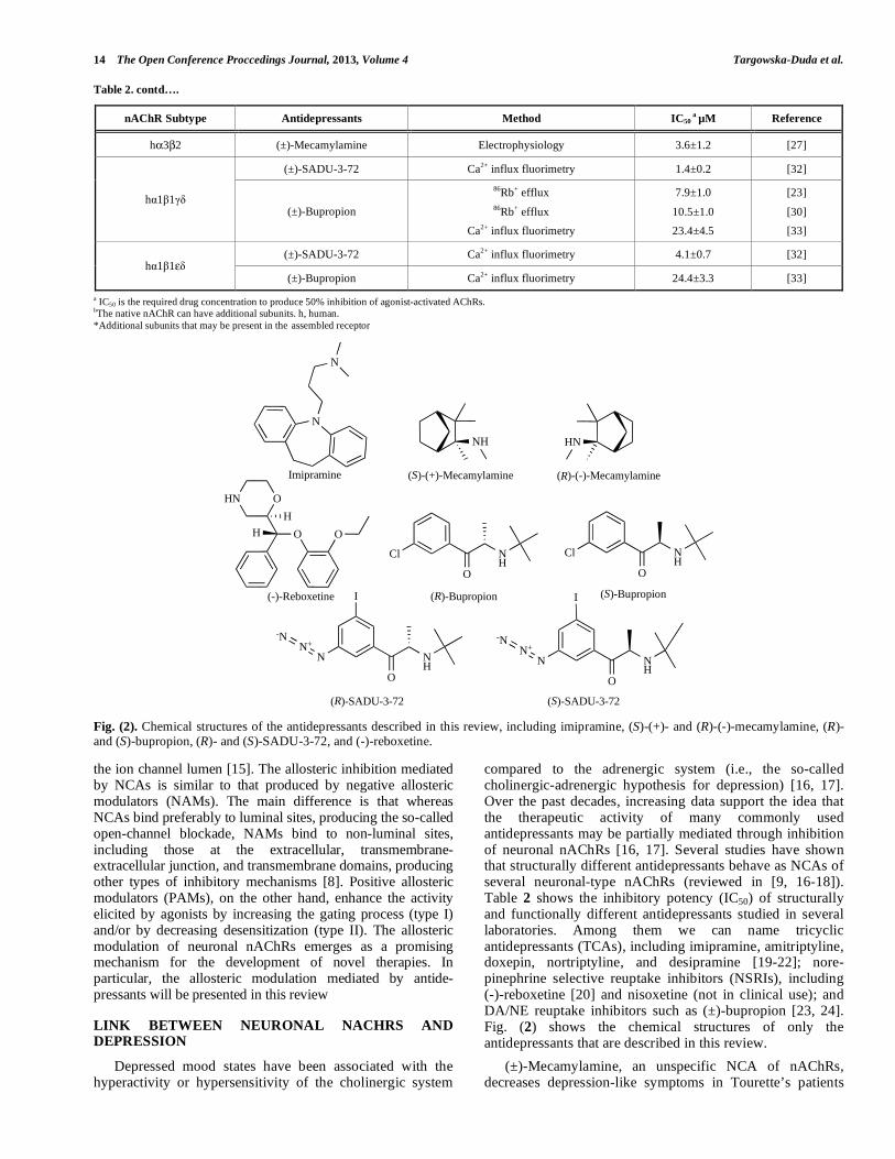
Table 2. contd….
nAChR Subtype Antidepressants Method IC50 a µM Reference
hα3β2 (±)-Mecamylamine Electrophysiology 3.6±1.2 [27]
(±)-SADU-3-72 Ca2+ influx fluorimetry 1.4±0.2 [32]
hα1β1γδ (±)-Bupropion
86Rb+ efflux 86Rb+ efflux
Ca2+ influx fluorimetry
7.9±1.0
10.5±1.0 23.4±4.5
[23]
[30] [33]
(±)-SADU-3-72 Ca2+ influx fluorimetry 4.1±0.7 [32] hα1β1εδ
(±)-Bupropion Ca2+ influx fluorimetry 24.4±3.3 [33] a IC50 is the required drug concentration to produce 50% inhibition of agonist-activated AChRs. bThe native nAChR can have additional subunits. h, human. *Additional subunits that may be present in the assembled receptor
N
N
NH HN
( )-(-)-Mecamylamine( )-(+)-Mecamylamine Imipramine
NH
O
Cl
( )-Bupropion
( )-SADU-3-72
O
HN O
OHH
(-)-Reboxetine
Cl NH
O
NN+
-N
NH
O
I
NH
I
O
NN+
-N
( )-Bupropion
( )-SADU-3-72
RS
R
R
S
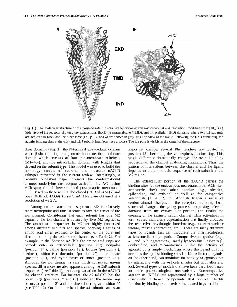
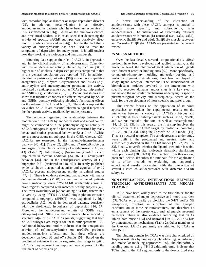
S Fig. (2). Chemical structures of the antidepressants described in this review, including imipramine, (S)-(+)- and (R)-(-)-mecamylamine, (R)- and (S)-bupropion, (R)- and (S)-SADU-3-72, and (-)-reboxetine.
the ion channel lumen [15]. The allosteric inhibition mediated by NCAs is similar to that produced by negative allosteric modulators (NAMs). The main difference is that whereas NCAs bind preferably to luminal sites, producing the so-called open-channel blockade, NAMs bind to non-luminal sites, including those at the extracellular, transmembrane-extracellular junction, and transmembrane domains, producing other types of inhibitory mechanisms [8]. Positive allosteric modulators (PAMs), on the other hand, enhance the activity elicited by agonists by increasing the gating process (type I) and/or by decreasing desensitization (type II). The allosteric modulation of neuronal nAChRs emerges as a promising mechanism for the development of novel therapies. In particular, the allosteric modulation mediated by antide-pressants will be presented in this review
LINK BETWEEN NEURONAL NACHRS AND DEPRESSION
Depressed mood states have been associated with the hyperactivity or hypersensitivity of the cholinergic system
compared to the adrenergic system (i.e., the so-called cholinergic-adrenergic hypothesis for depression) [16, 17]. Over the past decades, increasing data support the idea that the therapeutic activity of many commonly used antidepressants may be partially mediated through inhibition of neuronal nAChRs [16, 17]. Several studies have shown that structurally different antidepressants behave as NCAs of several neuronal-type nAChRs (reviewed in [9, 16-18]). Table 2 shows the inhibitory potency (IC50) of structurally and functionally different antidepressants studied in several laboratories. Among them we can name tricyclic antidepressants (TCAs), including imipramine, amitriptyline, doxepin, nortriptyline, and desipramine [19-22]; nore-pinephrine selective reuptake inhibitors (NSRIs), including (-)-reboxetine [20] and nisoxetine (not in clinical use); and DA/NE reuptake inhibitors such as (±)-bupropion [23, 24]. Fig. (2) shows the chemical structures of only the antidepressants that are described in this review. (±)-Mecamylamine, an unspecific NCA of nAChRs, decreases depression-like symptoms in Tourette’s patients
Molecular Modeling Interactions between Antidepressant and nAChRs The Open Conference Proccedings Journal, 2013, Volume 4 15
with comorbid bipolar disorder or major depressive disorder [25]. In addition, mecamylamine is an effective antidepressant in patients who have been unresponsive to SSRIs (reviewed in [26]). Based on the numerous clinical and preclinical studies, it is established that decreasing the activity of specific nAChR subtypes can positively affect mood disorders (reviewed in [26]). Despite the fact that a variety of antidepressants has been used to treat the symptoms of depression for many years, it is still unclear how they work at the molecular and neuronal levels. Mounting data support the role of nAChRs in depression and in the clinical activity of antidepressants. Coincident with the antidepressant activity elicited by nicotine [34], a higher rate of smokers in depressed patients compared to that in the general population was reported [35]. In addition, nicotinic agonists (e.g., nicotine [36]) as well as competitive antagonists (e.g., dihydro-β-erythroidine) and NCAs (e.g., amitriptyline and mecamylamine) potentiate the activity mediated by antidepressants such as TCAs (e.g., imipramine) and SSRIs (e.g., citalopram) [37, 38]. Behavioral studies also show that nicotine enhances the effects mediated by SSRIs and NSRIs, possibly reflecting nicotine's facilitating effects on the release of 5-HT and NE [39]. These data support the view that nAChRs are essential pharmacological targets for the therapeutic activity mediated by antidepressants.
The evidence regarding the relationship between the modulation of nAChRs by antidepressants and mood control might be connected with the pathophysiological activity of nAChR subtypes in specific brain areas confirmed by many behavioral studies presented below. α4β2 and α7 nAChRs are the most abundant subtypes in the CNS, whereas α3β4 nAChRs are expressed in the habenulo-interpeduncular pathway [40, 41]. The α4β2, α3β4, and α7 nAChR subtypes are targets for the clinical activity of antidepressants [18, 42, 43] (Table 2). Interestingly, β4-containing nAChRs are implicated in nicotine addiction [34, 36, 39], anxiety-like behavior [44], and in the antidepressant activity of (±)-bupropion [45], (reviewed in [18, 46]). Recently published evidence shows that partial agonists and agonists of α4β2 nAChRs present antidepressant activity in animal studies [47, 48]. There is evidence showing that subjects with major depressive disorder (MDD) as well as recovered patients have significantly lower β2*-nAChR availability across all brain regions compared with matched healthy subjects [49]. The lower availability of β2-containing nAChRs, determined in vivo by using [125I]5-I-A-85380 single photon emission computed tomography (SPECT), was explained by high extracellular ACh levels in depressed patients, consistent with the cholinergic hypothesis of depression [16, 17]. Moreover, the antidepressant activity of SSRIs (e.g., citalopram) and SNRIs (e.g., reboxetine) can be enhanced by selective α4β2 or α7 nAChR agonists, suggesting that both nAChR subtypes are targets for these antidepressants [50]. Additional behavioral studies indicate that the antagonistic activity of (±)-mecamylamine on nAChRs produces antidepressant-like effects, and that these effects are dependent on both β2 and α7 subunits [51]. Based on the preclinical evidence it can be suggested that drugs targeting nAChRs may represent an important new approach to the treatment of depression [20].
A better understanding of the interaction of antidepressants with these nAChR subtypes is crucial to develop more specific, and subsequently, safer antidepressants. The interactions of structurally different antidepressants with human (h) neuronal (i.e., α3β4, α4β2), embryonic (hα1β1γδ) and adult (hα1β1εδ) muscle subtypes, and Torpedo (Tα1β1γδ) nAChRs are presented in the current review.
IN SILICO METHODS
Over the last decade, several computational (in silico) methods have been developed and applied to study, at the molecular level, the pharmacological interaction of ligands with different receptor binding sites [52]. More specifically, comparative/homology modeling, molecular docking, and molecular dynamics simulations, have been employed to study ligand–receptor interactions. The identification of biomolecular moieties involved in the interaction with specific receptor domains and/or sites is a key step to understand the molecular mechanisms underlying its specific pharmacological activity and subsequently, serves as the basis for the development of more specific and safer drugs. This review focuses on the application of in silico approaches to explain the experimental results on the interaction between a variety of nAChRs subtypes and structurally different antidepressants such as TCAs, NSRIs, and DA/NE reuptake inhibitors, as well as mecamylamine [14, 15, 28, 33]. In this regard, the first step involves the construction of the nAChR target by molecular homology [21, 22, 28, 31-33], using the Torpedo nAChR model (Fig. 1) as a structural template. The antidepressants under study were built in the protonated and neutral state, and subsequently docked in the nAChR model [21, 22, 28, 31-33]. Finally, to verify whether the ligand orientation is stable within each binding site, molecular dynamics simulations were performed [28, 31-33]. Each section from this review, presented below, describes the rationale for the application of in silico methods to explaining and supporting experimental results concerning with the interaction of several classes of antidepressants with different nAChR subtypes.
NON-OVERLAPPING INTERACTIONS BETWEEN TRICYCLIC ANTIDEPRESSANTS AND MECAM-YLAMINE
TCAs have been widely used as the first choice for the clinical treatment of major depressive and mood disorders [53]. TCAs act primarily by blocking the 5-HT and/or NE transporters, resulting in elevation of the synaptic concentration of these neurotransmitters, and therefore an enhancement of the serotonergic and adrenergic neuronal pathways. There is also evidence indicating that TCAs inhibit both muscle [54] and neuronal [19, 21, 22] nAChRs by noncompetitive mechanisms (Table 2). Other members of the Cys-loop LGIC superfamily are inhibited by TCAs as well [55]. The binding domain for TCAs was first characterized on Torpedo nAChRs by employing photolabeling, radioligand, and molecular modeling approaches [56]. The photoaffinity labeling studies using [3H] 2-azidoimipramie indicate that TCAs bind to the M2 segment only in the desensitized state
16 The Open Conference Proccedings Journal, 2013, Volume 4 Targowska-Duda et al.
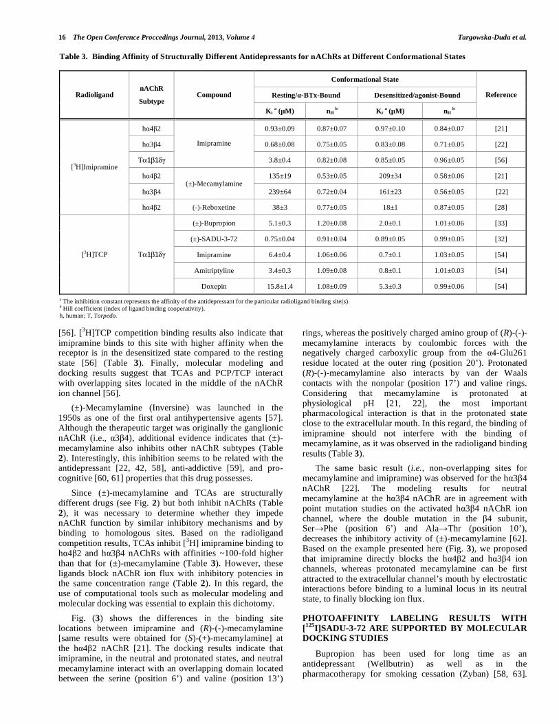
[56]. [3H]TCP competition binding results also indicate that imipramine binds to this site with higher affinity when the receptor is in the desensitized state compared to the resting state [56] (Table 3). Finally, molecular modeling and docking results suggest that TCAs and PCP/TCP interact with overlapping sites located in the middle of the nAChR ion channel [56]. (±)-Mecamylamine (Inversine) was launched in the 1950s as one of the first oral antihypertensive agents [57]. Although the therapeutic target was originally the ganglionic nAChR (i.e., α3β4), additional evidence indicates that (±)-mecamylamine also inhibits other nAChR subtypes (Table 2). Interestingly, this inhibition seems to be related with the antidepressant [22, 42, 58], anti-addictive [59], and pro-cognitive [60, 61] properties that this drug possesses. Since (±)-mecamylamine and TCAs are structurally different drugs (see Fig. 2) but both inhibit nAChRs (Table 2), it was necessary to determine whether they impede nAChR function by similar inhibitory mechanisms and by binding to homologous sites. Based on the radioligand competition results, TCAs inhibit [3H] imipramine binding to hα4β2 and hα3β4 nAChRs with affinities ~100-fold higher than that for (±)-mecamylamine (Table 3). However, these ligands block nAChR ion flux with inhibitory potencies in the same concentration range (Table 2). In this regard, the use of computational tools such as molecular modeling and molecular docking was essential to explain this dichotomy. Fig. (3) shows the differences in the binding site locations between imipramine and (R)-(-)-mecamylamine [same results were obtained for (S)-(+)-mecamylamine] at the hα4β2 nAChR [21]. The docking results indicate that imipramine, in the neutral and protonated states, and neutral mecamylamine interact with an overlapping domain located between the serine (position 6’) and valine (position 13’)
rings, whereas the positively charged amino group of (R)-(-)-mecamylamine interacts by coulombic forces with the negatively charged carboxylic group from the α4-Glu261 residue located at the outer ring (position 20’). Protonated (R)-(-)-mecamylamine also interacts by van der Waals contacts with the nonpolar (position 17’) and valine rings. Considering that mecamylamine is protonated at physiological pH [21, 22], the most important pharmacological interaction is that in the protonated state close to the extracellular mouth. In this regard, the binding of imipramine should not interfere with the binding of mecamylamine, as it was observed in the radioligand binding results (Table 3). The same basic result (i.e., non-overlapping sites for mecamylamine and imipramine) was observed for the hα3β4 nAChR [22]. The modeling results for neutral mecamylamine at the hα3β4 nAChR are in agreement with point mutation studies on the activated hα3β4 nAChR ion channel, where the double mutation in the β4 subunit, Ser→Phe (position 6’) and Ala→Thr (position 10’), decreases the inhibitory activity of (±)-mecamylamine [62]. Based on the example presented here (Fig. 3), we proposed that imipramine directly blocks the hα4β2 and hα3β4 ion channels, whereas protonated mecamylamine can be first attracted to the extracellular channel’s mouth by electrostatic interactions before binding to a luminal locus in its neutral state, to finally blocking ion flux.
PHOTOAFFINITY LABELING RESULTS WITH [125I]SADU-3-72 ARE SUPPORTED BY MOLECULAR DOCKING STUDIES
Bupropion has been used for long time as an antidepressant (Wellbutrin) as well as in the pharmacotherapy for smoking cessation (Zyban) [58, 63].
Table 3. Binding Affinity of Structurally Different Antidepressants for nAChRs at Different Conformational States
Conformational State
Resting/α-BTx-Bound Desensitized/agonist-Bound Radioligand nAChR
Subtype Compound
Ki a (µM) nH b Ki a (µM) nH
b
Reference
hα4β2 0.93±0.09 0.87±0.07 0.97±0.10 0.84±0.07 [21]
hα3β4 0.68±0.08 0.75±0.05 0.83±0.08 0.71±0.05 [22]
Τα1β1δγ
Imipramine
3.8±0.4 0.82±0.08 0.85±0.05 0.96±0.05 [56]
hα4β2 135±19 0.53±0.05 209±34 0.58±0.06 [21]
hα3β4 (±)-Mecamylamine
239±64 0.72±0.04 161±23 0.56±0.05 [22]
[3H]Imipramine
hα4β2 (-)-Reboxetine 38±3 0.77±0.05 18±1 0.87±0.05 [28]
(±)-Bupropion 5.1±0.3 1.20±0.08 2.0±0.1 1.01±0.06 [33]
(±)-SADU-3-72 0.75±0.04 0.91±0.04 0.89±0.05 0.99±0.05 [32]
Imipramine 6.4±0.4 1.06±0.06 0.7±0.1 1.03±0.05 [54]
Amitriptyline 3.4±0.3 1.09±0.08 0.8±0.1 1.01±0.03 [54]
[3H]TCP Τα1β1δγ
Doxepin 15.8±1.4 1.08±0.09 5.3±0.3 0.99±0.06 [54] a The inhibition constant represents the affinity of the antidepressant for the particular radioligand binding site(s). b Hill coefficient (index of ligand binding cooperativity). h, human; T, Torpedo.
Molecular Modeling Interactions between Antidepressant and nAChRs The Open Conference Proccedings Journal, 2013, Volume 4 17
Bupropion can also be used for the treatment of atypical depression, which is associated with interpersonal deficits (i.e., rejection sensitivity and social avoidance) [64]. The pharmacological [e.g., inhibitory potency (Table 2) and binding affinity (Table 3)] and behavioral (e.g., antidepressant, locomotor, and anti-addictive) activities of bupropion, its natural metabolites and isomers, as well as a large variety of synthetic bupropion derivatives have been studied by in vitro and in vivo approaches [23, 32, 65, 66]. These studies open the possibility of developing novel bupropion derivatives with improved clinical profiles important for their antidepressant and anti-smoking activities (reviewed in [46]). Although bupropion is classified as a dual DA/NE reuptake inhibitor, it also inhibits several neuronal nAChR subtypes (Table 2). The aim of this particular work was to determine the pharmacological properties of (±)-SADU-3-72, a photoreactive derivative of (±)-bupropion, in muscle-type nAChRs, with the intention of using this compound as a photoaffinity probe to characterize the bupropion binding sites in neuronal nAChRs [32]. In this regard, the docking of bupropion enantiomers and its analog SADU-3-72 with the ion channel of different muscle nAChR subtypes was performed to determine their binding sites and to support additional experimental results (i.e., Ca2+ influx, receptor state-dependent radioligand binding, and photoaffinity labeling study) [32]. Ca2+ influx results indicate that (±)-SADU-3-72 is 17- and 6-fold more potent than (±)-bupropion in inhibiting hα1β1γδ and hα1β1εδ nAChRs, respectively (Table 2). From the [3H]imipramine competition binding results it was evident that (±)-bupropion displays higher affinity for the desensitized nAChR compared to that for the resting nAChR [33], whereas (±)-SADU-3-72 has high affinity [even higher than (±)-bupropion] for both conformational states [32] (Table 3). This evidence suggests that although (±)-bupropion discriminates between the
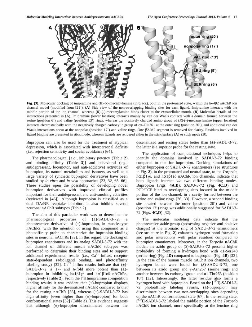
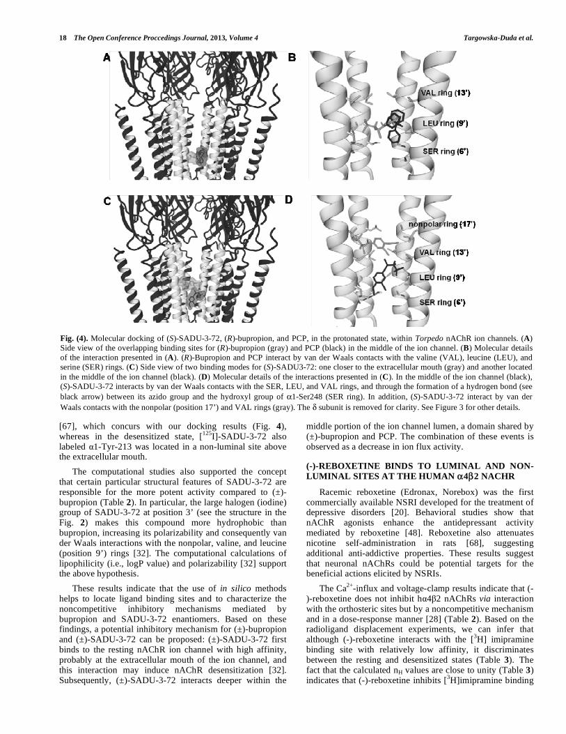
desensitized and resting states better than (±)-SADU-3-72, the latter is a superior probe for the resting state. The application of computational techniques helps to identify the domains involved in SADU-3-72 binding compared to that for bupropion. Docking simulations of either bupropion or SADU-3-72 enantiomers (see structures in Fig. 2), in the protonated and neutral state, to the Torpedo, hα1β1γδ, and hα1β1εδ nAChR ion channels, indicate that both ligands interact via two different binding modes. Bupropion (Figs. 4A,B), SADU-3-72 (Fig. 4C,D) and PCP/TCP bind to overlapping sites located in the middle portion of the ion channel, in a cavity formed between the serine and valine rings [26, 33]. However, a second binding site located between the outer (position 20’) and valine (position 13’) rings was additionally suggested for SADU-3-72 (Figs. 4C,D) [32]. The molecular modeling data indicate that the photoreactive azide group (possessing negative and positive charges) at the aromatic ring of SADU-3-72 enantiomers (see structure in Fig. 2) enhances hydrogen bond formation and polar interactions with polar residues compared to bupropion enantiomers. Moreover, in the Torpedo nAChR model, the azido group of (S)-SADU-3-72 presents higher probability of forming a hydrogen bond with α1-Ser248 (serine ring) (Fig. 4D) compared to bupropion (Fig. 4B) [33]. In the case of the human muscle nAChR ion channels, two hydrogen bonds were found for (S)-SADU-3-72, one between its azido group and γ-Asn257 (serine ring) and another between its carbonyl group and α1-Thr263 (position 10’) [32]. Interestingly, the latter residue also forms a hydrogen bond with bupropion. Based on the [125I]-SADU-3-72 photoaffinity labeling results, (±)-bupropion may discriminate between two non-overlapping sites depending on the nAChR conformational state [67]. In the resting state, [125I]-SADU-3-72 labeled the middle portion of the Torpedo nAChR ion channel, more specifically at the leucine ring
Fig. (3). Molecular docking of imipramine and (R)-(-)-mecamylamine (in black), both in the protonated state, within the hα4β2 nAChR ion channel model (modified from [21]). (A) Side view of the non-overlapping binding sites for each ligand. Imipramine interacts with the middle portion of the ion channel, whereas (R)-(-)-mecamylamine binds closer to the extracellular mouth. (B) Molecular details of the interactions presented in (A). Imipramine (lower location) interacts mainly by van der Waals contacts with a domain formed between the serine (position 6’) and valine (position 13’) rings, whereas the positively charged amino group of (R)-(-)-mecamylamine (upper location) interacts electrostatically with the negatively charged carboxylic group of α4-Glu261 at the outer ring (position 20’), and additional van der Waals interactions occur at the nonpolar (position 17’) and valine rings. One β2 -M2 segment is removed for clarity. Residues involved in ligand binding are presented in stick mode, whereas ligands are rendered either in the stick/surface (A) or stick mode (B).
18 The Open Conference Proccedings Journal, 2013, Volume 4 Targowska-Duda et al.
[67], which concurs with our docking results (Fig. 4), whereas in the desensitized state, [125I]-SADU-3-72 also labeled α1-Tyr-213 was located in a non-luminal site above the extracellular mouth. The computational studies also supported the concept that certain particular structural features of SADU-3-72 are responsible for the more potent activity compared to (±)-bupropion (Table 2). In particular, the large halogen (iodine) group of SADU-3-72 at position 3’ (see the structure in the Fig. 2) makes this compound more hydrophobic than bupropion, increasing its polarizability and consequently van der Waals interactions with the nonpolar, valine, and leucine (position 9’) rings [32]. The computational calculations of lipophilicity (i.e., logP value) and polarizability [32] support the above hypothesis. These results indicate that the use of in silico methods helps to locate ligand binding sites and to characterize the noncompetitive inhibitory mechanisms mediated by bupropion and SADU-3-72 enantiomers. Based on these findings, a potential inhibitory mechanism for (±)-bupropion and (±)-SADU-3-72 can be proposed: (±)-SADU-3-72 first binds to the resting nAChR ion channel with high affinity, probably at the extracellular mouth of the ion channel, and this interaction may induce nAChR desensitization [32]. Subsequently, (±)-SADU-3-72 interacts deeper within the
middle portion of the ion channel lumen, a domain shared by (±)-bupropion and PCP. The combination of these events is observed as a decrease in ion flux activity.
(-)-REBOXETINE BINDS TO LUMINAL AND NON-LUMINAL SITES AT THE HUMAN α4β2 NACHR
Racemic reboxetine (Edronax, Norebox) was the first commercially available NSRI developed for the treatment of depressive disorders [20]. Behavioral studies show that nAChR agonists enhance the antidepressant activity mediated by reboxetine [48]. Reboxetine also attenuates nicotine self-administration in rats [68], suggesting additional anti-addictive properties. These results suggest that neuronal nAChRs could be potential targets for the beneficial actions elicited by NSRIs. The Ca2+-influx and voltage-clamp results indicate that (-)-reboxetine does not inhibit hα4β2 nAChRs via interaction with the orthosteric sites but by a noncompetitive mechanism and in a dose-response manner [28] (Table 2). Based on the radioligand displacement experiments, we can infer that although (-)-reboxetine interacts with the [3H] imipramine binding site with relatively low affinity, it discriminates between the resting and desensitized states (Table 3). The fact that the calculated nH values are close to unity (Table 3) indicates that (-)-reboxetine inhibits [3H]imipramine binding
Fig. (4). Molecular docking of (S)-SADU-3-72, (R)-bupropion, and PCP, in the protonated state, within Torpedo nAChR ion channels. (A) Side view of the overlapping binding sites for (R)-bupropion (gray) and PCP (black) in the middle of the ion channel. (B) Molecular details of the interaction presented in (A). (R)-Bupropion and PCP interact by van der Waals contacts with the valine (VAL), leucine (LEU), and serine (SER) rings. (C) Side view of two binding modes for (S)-SADU3-72: one closer to the extracellular mouth (gray) and another located in the middle of the ion channel (black). (D) Molecular details of the interactions presented in (C). In the middle of the ion channel (black), (S)-SADU-3-72 interacts by van der Waals contacts with the SER, LEU, and VAL rings, and through the formation of a hydrogen bond (see black arrow) between its azido group and the hydroxyl group of α1-Ser248 (SER ring). In addition, (S)-SADU-3-72 interact by van der Waals contacts with the nonpolar (position 17’) and VAL rings (gray). The δ subunit is removed for clarity. See Figure 3 for other details.
Molecular Modeling Interactions between Antidepressant and nAChRs The Open Conference Proccedings Journal, 2013, Volume 4 19
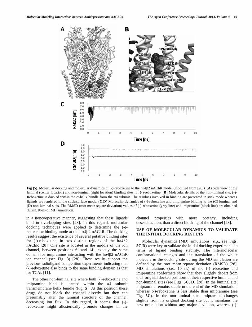
in a noncooperative manner, suggesting that these ligands bind to overlapping sites [28]. In this regard, molecular docking techniques were applied to determine the (-)-reboxetine binding mode at the hα4β2 nAChR. The docking results suggest the existence of several putative binding sites for (-)-reboxetine, in two distinct regions of the hα4β2 nAChR [28]. One site is located in the middle of the ion channel, between positions 6’ and 14’, exactly the same domain for imipramine interacting with the hα4β2 nAChR ion channel (see Fig. 3) [28]. These results support the previous radioligand competition experiments indicating that (-)-reboxetine also binds to the same binding domain as that for TCAs [11]. The other non-luminal site where both (-)-reboxetine and imipramine bind is located within the α4 subunit transmembrane helix bundle (Fig. 5). At this position these drugs do not block the channel directly but they can presumably alter the luminal structure of the channel, decreasing ion flux. In this regard, it seems that (-)-reboxetine might allosterically promote changes in the
channel properties with more potency, including desensitization, than a direct blocking of the channel [28].
USE OF MOLECULAR DYNAMICS TO VALIDATE THE INITIAL DOCKING RESULTS
Molecular dynamics (MD) simulations (e.g., see Figs. 5C,D) were key to validate the initial docking experiments in terms of ligand binding stability. The intermolecular conformational changes and the translation of the whole molecule in the docking site during the MD simulation are defined by the root mean square deviation (RMSD) [28]. MD simulations (i.e., 10 ns) of the (-)-reboxetine and imipramine conformers show that they slightly depart from their original docked positions at their respective luminal and non-luminal sites (see Figs. 5C, D) [28]. In the luminal site, imipramine remains stable to the end of the MD simulation, whereas (-)-reboxetine is less stable than imipramine (see Fig. 5C). In the non-luminal site, imipramine changes slightly from its original docking site but it maintains the new orientation without any major deviation, whereas (-)-
Fig (5). Molecular docking and molecular dynamics of (-)-reboxetine to the hα4β2 nAChR model (modified from [28]). (A) Side view of the luminal (center location) and non-luminal (right location) binding sites for (-)-reboxetine. (B) Molecular details of the non-luminal site. (-)-Reboxetine is docked within the α-helix bundle from the α4 subunit. The residues involved in binding are presented in stick mode whereas ligands are rendered in the stick/surface mode. (C,D) Molecular dynamics of (-)-reboxetine and imipramine binding to the (C) luminal and (D) non-luminal sites. The RMSD (root mean square deviation) values of (-)-reboxetine (grey line) and imipramine (black line) are obtained during 10-ns of MD simulation.
20 The Open Conference Proccedings Journal, 2013, Volume 4 Targowska-Duda et al.
reboxetine deviates more from the original docking site but it remains there in a more stable way that in the ion channel lumen (see Fig. 5D). In this regard, it seems that (-)-reboxetine might allosterically promote changes in the channel properties with more potency, including desensitization, than a direct blocking of the channel. The MD results on bupropion and SADU-3-72 enantiomers indicate that the molecules slightly change positions compared to the starting pose (i.e., original docked positions) but the complexes were stable during the simulations. In the case of human muscle and Torpedo nAChR ion channels, the interaction of each SADU-3-72 enantiomer with nearby pore-lining residues (between the valine and serine rings) is stable during the 15-ns simulation. In the case of Torpedo nAChRs, the interaction of (S)- and (R)-SADU-3-72 with the nonpolar ring (position 17’) also remained stable, suggesting that this ring is also important for SADU-3-72 binding at this receptor [32].
CONCLUSIONS
The application of in silico methods (i.e., comparative/homology modeling, molecular docking, and molecular dynamics simulations) in combination with experimental techniques (e.g. receptor state-dependent radioligand binding, and Ca2+-influx and electrophysiological experiments) facilitated a better understanding of the molecular interactions of each presented antidepressant with several nAChRs subtypes. Furthermore, these results expanded the current knowledge on the noncompetitive inhibitory mechanisms elicited by antidepressants on nAChRs that might be significant to further studies on the Cys-loop LGIC superfamily. Finally, the combination of functional and structural studies will facilitate the development of novel drugs for the therapy of depression.
CONFLICT OF INTEREST
The authors confirm that this article content has no conflicts of interest.
ACKNOWLEDGEMENTS
This work was supported by the TEAM research subsidy from the Foundation for Polish Science (to K.J.). The molecular modeling experiments were developed using the equipment purchased within the Project "The equipment of innovative laboratories doing research on new medicines used in the therapy of civilization and neoplastic diseases" within the Operational Program Development of Eastern Poland 2007-2013, Priority Axis I Modern Economy (to K.J.).
ABBREVIATIONS
Ach = acetylcholine
α-BTx = α-bungarotoxin
CCh = carbamylcholine
CNS = central nervous system
DA = dopamine
EXD = extracellular domain
FST = forced swim test
GABA = γ-aminobutyric acid
serotonin = 5-hydroxytryptamine (5-HT)
IC50 = ligand concentration that inhibits 50% binding
Ki = inhibition constant
LGIC = ligand-gated ion channel
MD = molecular dynamics
MDD = major depressive disorder
nAChR = nicotinic acetylcholine receptor
NAMs = negative allosteric modulators
NCAs = noncompetitive antagonists
NE = norepinephrine
NSRIs = norepinephrine selective reuptake inhibitors
PAMs = positive allosteric modulators
PCP = phencyclidine
SADU-3-72 = 2-(N-tert-butylamino)-3’-iodo-4’-azidopropiophenone
SSRIs = serotonin selective reuptake inhibitors
TCAs = tricyclic antidepressants
[3H]TCP = [piperidyl-3,4-3H(N)]-(N-(1-(2-thienyl)cyclohexyl)-3,4-piperidine)
TMD = transmembrane domain
REFERENCES [1] Sine, S.M.; Engel, A.G. Recent advances in Cys-loop receptor
structure and function. Nature, 2006, 440(7083), 448-455. [2] Albuquerque, E.X.; Pereira, E.F.; Alkondon, M.; Rogers, S.W.
Mammalian nicotinic acetylcholine receptors: from structure to function. Physiol. Rev., 2009, 89(1), 73-120.
[3] Arias, H.R. Biological and biophysical aspects of ligand-gated ion channel receptor superfamilies; In: Ligand-gated ion channel receptor superfamilies, Arias, H.R. Ed., Research Signpost, Kerala, India, 2006, pp.1-25.
[4] Millar, N.S.; Gotti, C. Diversity of vertebrate nicotinic acetylcholine receptors. Neuropharmacology, 2009, 56(1), 237-246.
[5] Kalamida, D.; Poulas, K.; Avramopoulou, V.; Fostieri, E.; Lagoumintzis, G.; Lazaridis, K.; Sideri, A.; Zouridakis, M.; Tzartos, S.J. Muscle and neuronal nicotinic acetylcholine receptors. Structure, function and pathogenicity. FEBS J., 2007, 274(15), 3799-3845.
[6] Wonnacott, S. Presynaptic nicotinic ACh receptors. Trends Neurosci., 1997, 20(2), 92-98.
[7] Mousa, S.A.; Arias, H.R. Angiogenesis modulation by nicotine and nicotinic ligands. J. Pediatr. Biochem., 2010, 191-104.
[8] Gotti, C.; Clementi, F.; Fornari, A.; Gaimarri, A.; Guiducci, S.; Manfredi, I.; Moretti, M.; Pedrazzi, P.; Pucci, L.; Zoli, M. Structural and functional diversity of native brain neuronal nicotinic receptors. Biochem. Pharmacol., 2009, 78(7), 703-711.
[9] Arias, H.R. Positive and negative modulation of nicotinic receptors. Adv. Protein Chem. Struct. Biol., 2010, 80, 153-203.
Molecular Modeling Interactions between Antidepressant and nAChRs The Open Conference Proccedings Journal, 2013, Volume 4 21
[10] Unwin, N. Refined structure of the nicotinic acetylcholine receptor at 4 Å resolution. J. Mol. Biol., 2005, 346(4), 967-989.
[11] Unwin, N.; Fujiyoshi, Y. Gating movement of acetylcholine receptor caught by plunge-freezing. J. Mol. Biol., 2012, 422(5), 617-634.
[12] Bartos, M.; Corradi, J.; Bouzat, C. Structural basis of activation of cys-loop receptors: the extracellular-transmembrane interface as a coupling region. Mol. Neurobiol., 2009, 40(3), 236-252.
[13] Arias, H.R. Molecular interactions between ligands and nicotinic acetylcholine receptors revealed by studies with acetylcholine binding proteins. J. Thermodynam. Catal., 2012, 3(4), 116.
[14] Baldessarini, R.J. Drugs and the treatment of psychiatric disorders. In: Goodman & Gilman´s The pharmacological basis of therapeutics, 10th ed.; Hardman, J.G., Limbird, L.E., Eds., McGraw-Hill: New York, 2001, pp. 447-483.
[15] Arias, H.R.; Bhumireddy, P.; Bouzat, C. Molecular mechanisms and binding site locations for noncompetitive antagonists of nicotinic acetylcholine receptors. Int. J. Biochem Cell. Biol., 2006, 38(8), 1254-1276.
[16] Shytle, R.D.; Silver, A.A.; Lukas, R.J.; Newman, M.B.; Sheehan, D.V.; Sanberg, P.R. Nicotinic acetylcholine receptors as targets for antidepressants. Mol. Psychiatry, 2002, 7(6), 525-535.
[17] Shytle, R.D.; Sheehan, D.V.; Sanberg, P.R.; Arias, H.R. Neuronal nicotinic receptors as therapeutic targets for mood disorders. In: Pharmacology of nicotinic acetylcholine receptors from the basic and therapeutic perspectives. Arias, H.R. Ed. Vol. Chapter 9, Research Signpost, Kerala, India, 2011, pp.187-198.
[18] Arias, H.R. Is the inhibition of nicotinic acetylcholine receptors by bupropion involved in its clinical actions? Int. J. Biochem. Cell Biol., 2009, 41(11), 2098-2108.
[19] Rana, B.; McMorn, S.O.; Reeve, H.L.; Wyatt, C.N.; Vaughan, P.F.; Peers, C. Inhibition of neuronal nicotinic acetylcholine receptors by imipramine and desipramine. Eur. J. Pharmacol., 1993, 250(2), 247-251.
[20] Philip, N.S.; Carpenter, L.L.; Tyrka, A.R.; Price, L.H. Nicotinic acetylcholine receptors and depression: a review of the preclinical and clinical literature. Psychopharmacology (Berl), 2010, 212(1), 1-12.
[21] Arias, H.R.; Rosenberg, A.; Targowska-Duda, K.M.; Feuerbach, D.; Jozwiak, K.; Moaddel, R.; Wainer, I.W. Tricyclic antidepressants and mecamylamine bind to different sites in the human α4β2 nicotinic receptor ion channel. Int. J. Biochem. Cell Biol., 2010, 42(6), 1007-1018.
[22] Arias, H.R.; Targowska-Duda, K.M.; Feuerbach, D.; Sullivan, C.J.; Maciejewski, R.; Jozwiak, K. Different interaction between tricyclic antidepressants and mecamylamine with the human α3β4 nicotinic acetylcholine receptor ion channel. Neurochem. Int., 2010, 56(4), 642-649.
[23] Damaj, M.I.; Carroll, F.I.; Eaton, J.B.; Navarro, H.A.; Blough, B.E.; Mirza, S.; Lukas, R.J.; Martin, B.R. Enantioselective effects of hydroxy metabolites of bupropion on behavior and on function of monoamine transporters and nicotinic receptors. Mol. Pharmacol., 2004, 66(3), 675-682.
[24] Garcia-Colunga, J.; Godoy-Garcia, U.; Vazquez-Gomez, E. Interaction of bupropion and zinc with neuronal nicotinic acetylcholine receptors. Neuropharmacology, 2011, 61(8), 1202-1209.
[25] Shytle, R.D.; Silver, A.A.; Sanberg, P.R. Comorbid bipolar disorder in Tourette's syndrome responds to the nicotinic receptor antagonist mecamylamine (Inversine). Biol. Psychiatry, 2000, 48(10), 1028-1031.
[26] Mineur, Y.S.; Picciotto, M.R. Nicotine receptors and depression: revisiting and revising the cholinergic hypothesis. Trends Pharmacol. Sci., 2010, 31(12), 580-586.
[27] Papke, R.L.; Sanberg, P.R.; Shytle, R.D. Analysis of mecamylamine stereoisomers on human nicotinic receptor subtypes. J. Pharmacol. Exp. Ther., 2001, 297(2), 646-656.
[28] Arias, H.R.; Fedorov, N.B.; Benson, L.C.; Lippiello, P.M.; Gatto, G.J.; Feuerbach, D.; Ortells, M.O. Functional and structural interaction of (-)-reboxetine with the human α4β2 nicotinic acetylcholine receptor. J. Pharmacol. Exp. Ther., 2013, 344(1), 113-123.
[29] Feuerbach, D.; Lingenhohl, K.; Dobbins, P.; Mosbacher, J.; Corbett, N.; Nozulak, J.; Hoyer, D. Coupling of human nicotinic acetylcholine receptors α7 to calcium channels in GH3 cells. Neuropharmacology, 2005, 48(2), 215-227.
[30] Fryer, J.D.; Lukas, R.J. Noncompetitive functional inhibition at diverse, human nicotinic acetylcholine receptor subtypes by bupropion, phencyclidine, and ibogaine. J. Pharmacol. Exp. Ther., 1999, 288(1), 88-92.
[31] Sidhpura, N.; Redfern, P.; Wonnacott, S. Comparison of the effects of bupropion on nicotinic receptor-evoked [3H]dopamine release from rat striatal synaptosomes and slices. Eur. J. Pharmacol., 2007, 567(1-2), 102-109.
[32] Arias, H.R.; Feuerbachb, D.; Targowska-Duda, K.M.; Aggarwald S.; Lapinsky D.J.; Jozwiak, K. Structural and functional interaction of (±)-2-(N-tert-butylamino)-3’-iodo-4’-azidopropiophenone, a photoreactive bupropion derivative, with nicotinic acetylcholine receptors. Neurochem. Int., 2012, 61(8), 1433-1441.
[33] Arias, H.R.; Gumilar, F.; Rosenberg, A.; Targowska-Duda, K.M.; Feuerbach, D.; Jozwiak, K.; Moaddel, R.; Wainer, I.W.; Bouzat, C. Interaction of bupropion with muscle-type nicotinic acetylcholine receptors in different conformational states. Biochemistry, 2009, 48(21), 4506-4518.
[34] Popik, P.; Krawczyk, M.; Kos, T.; Nalepa, I.; Kowalska, M.; Witarski, T.; Antkiewicz-Michaluk, L.; Vetulani, J. Nicotine produces antidepressant-like actions: behavioral and neurochemical evidence. Eur. J. Pharmacol., 2005, 515(1-3), 128-133.
[35] Picciotto, M.R.; Brunzell, D.H.; Caldarone, B.J. Effect of nicotine and nicotinic receptors on anxiety and depression. Neuroreport, 2002, 13(9), 1097-1106.
[36] Salas, R.; Cook, K.D.; Bassetto, L.; De Biasi, M. The α3 and β4 nicotinic acetylcholine receptor subunits are necessary for nicotine-induced seizures and hypolocomotion in mice. Neuropharmacology, 2004, 47(3), 401-407.
[37] Caldarone, B.J.; Harrist, A.; Cleary, M.A.; Beech, R.D.; King, S.L.; Picciotto, M.R. High-affinity nicotinic acetylcholine receptors are required for antidepressant effects of amitriptyline on behavior and hippocampal cell proliferation. Biol. Psychiatry, 2004, 56(9), 657-664.
[38] Popik, P.; Kozela, E.; Krawczyk, M. Nicotine and nicotinic receptor antagonists potentiate the antidepressant-like effects of imipramine and citalopram. Br. J. Pharmacol., 2003, 139(6), 1196-1202.
[39] Baldwin, P.R.; Alanis, R.; Salas, R. The Role of the Habenula in Nicotine Addiction. J. Addict. Res. Ther., 2011, S1(2). pii: 002.
[40] Quick, M.W.; Ceballos, R.M.; Kasten, M.; McIntosh, J.M.; Lester, R.A. α3β4 subunit-containing nicotinic receptors dominate function in rat medial habenula neurons. Neuropharmacology, 1999, 38(6), 769-783.
[41] Grady, S.R.; Moretti, M.; Zoli, M.; Marks, M.J.; Zanardi, A.; Pucci, L.; Clementi, F.; Gotti, C. Rodent habenulo-interpeduncular pathway expresses a large variety of uncommon nAChR subtypes, but only the α3β4* and α3β3β4* subtypes mediate acetylcholine release. J. Neurosci., 2009, 29(7), 2272-2282.
[42] Azam, L.; Winzer-Serhan, U.H.; Chen, Y.; Leslie, F.M. Expression of neuronal nicotinic acetylcholine receptor subunit mRNAs within midbrain dopamine neurons. J. Comp. Neurol., 2002, 444(3), 260-274.
[43] Fedorov, N.B.; Benson, L.C.; Graef, J.; Lippiello, P.M.; Bencherif, M. Differential pharmacologies of mecamylamine enantiomers: positive allosteric modulation and noncompetitive inhibition. J. Pharmacol. Exp. Ther., 2009, 328(2), 525-532.
[44] Salas, R.; Pieri, F.; Fung, B.; Dani, J.A.; De Biasi, M. Altered anxiety-related responses in mutant mice lacking the β4 subunit of the nicotinic receptor. J. Neurosci., 2003, 23(15), 6255-6263.
[45] Radhakrishnan, R.; Escobar, L.; Santamaría, A.; Arias, H.R. The β4 nicotinic receptor subunit modulates the chronic antidepressant activity mediated by bupropion. Neurosci. Lett., 2013, [submitted].
[46] Arias, H.R.; Santamaria, A.; Ali, S.F. Pharmacological and neurotoxicological actions mediated by bupropion and diethylpropion. Int. Rev. Neurobiol., 2009, 88, 223-255.
[47] Yu, L.F.; Eaton, J.B.; Fedolak, A.; Zhang, H.K.; Hanania, T.; Brunner, D.; Lukas, R.J.; Kozikowski, A.P. Discovery of highly potent and selective α4β2-nicotinic acetylcholine receptor (nAChR) partial agonists containing an isoxazolylpyridine ether scaffold that demonstrate antidepressant-like activity. Part II. J. Med. Chem., 2012, 55(22), 9998-10009.
[48] Yu, L.F.; Tuckmantel, W.; Eaton, J.B.; Caldarone, B.; Fedolak, A.; Hanania, T.; Brunner, D.; Lukas, R.J.; Kozikowski, A.P. Identification of novel α4β2-nicotinic acetylcholine receptor (nAChR) agonists based on an isoxazole ether scaffold that
22 The Open Conference Proccedings Journal, 2013, Volume 4 Targowska-Duda et al.
demonstrate antidepressant-like activity. J. Med. Chem., 2012, 55(2), 812-823.
[49] Saricicek, A.; Esterlis, I.; Maloney, K.H.; Mineur, Y.S.; Ruf, B.M.; Muralidharan, A.; Chen, J.I.; Cosgrove, K.P.; Kerestes, R., Ghose, S., Tamminga, C.A., Pittman, B., Bois, F., Tamagnan, G.; Seibyl, J.; Picciotto, M.R.; Staley, J.K.; Bhagwagar, Z. Persistent β2*-nicotinic acetylcholinergic receptor dysfunction in major depressive disorder. Am. J. Psychiatry., 2012, 169(8), 851-859.
[50] Andreasen, J.T.; Nielsen, E.O.; Christensen, J.K.; Olsen, G.M., Peters, D.; Mirza, N.R.; Redrobe, J.P. Subtype-selective nicotinic acetylcholine receptor agonists enhance the responsiveness to citalopram and reboxetine in the mouse forced swim test. J. Psychopharmacol., 2011, 25(10), 1347-1356.
[51] Rabenstein, R.L.; Caldarone, B.J.; Picciotto, M.R. The nicotinic antagonist mecamylamine has antidepressant-like effects in wild-type but not β2- or α7-nicotinic acetylcholine receptor subunit knockout mice. Psychopharmacology (Berl), 2006, 189(3), 395-401.
[52] Ekins, S.; Mestres, J.; Testa, B. In silico pharmacology for drug discovery: methods for virtual ligand screening and profiling. Br. J. Pharmacol., 2007, 152(1), 9-20.
[53] Mitchell, P.B.; Mitchell, M.S. The management of depression. Part 2. The place of the new antidepressants. Aust. Fam. Physician., 1994, 23(9), 1771-1773, 1776-1781.
[54] Gumilar, F.; Arias, H.R.; Spitzmaul, G.; Bouzat, C. Molecular mechanisms of inhibition of nicotinic acetylcholine receptors by tricyclic antidepressants. Neuropharmacology, 2003, 45(7), 964-976.
[55] Gumilar, F.; Bouzat, C. Tricyclic antidepressants inhibit homomeric Cys-loop receptors by acting at different conformational states. Eur. J. Pharmacol., 2008, 584, 30-39.
[56] Sanghvi, M.; Hamouda, A.K.; Jozwiak, K.; Blanton, M.P.; Trudell, J.R.; Arias, H.R. Identifying the binding site(s) for antidepressants on the Torpedo nicotinic acetylcholine receptor: [3H]2-azidoimipramine photolabeling and molecular dynamics studies. Biochim. Biophys. Acta., 2008, 1778(12), 2690-2699.
[57] Bacher, I.; Wu, B.; Shytle, D.R.; George, T.P. Mecamylamine - a nicotinic acetylcholine receptor antagonist with potential for the treatment of neuropsychiatric disorders. Expert Opin. Pharmacother., 2009, 10(16), 2709-2721.
[58] Wilkes, S. Bupropion. Drugs Today (Barc)., 2006, 42(10), 671-681.
[59] George, T.P.; Sacco, K.A.; Vessicchio, J.C.; Weinberger, A.H.; Shytle, R.D. Nicotinic antagonist augmentation of selective serotonin reuptake inhibitor-refractory major depressive disorder: a preliminary study. J. Clin. Psychopharmacol., 2008, 28(3), 340-344.
[60] Terry, A.V.; Buccafusco, J.J.; Prendergast, M.A. Dose-specific improvements in memory-related task performance by rats and aged monkeys administered the nicotinic-cholinergic antagonist mecamylamine. Drug Dev. Res., 1999, (47), 127-136.
[61] Levin, E.D.; Castonguay, M.; Ellison, G.D. Effects of the nicotinic receptor blocker mecamylamine on radial-arm maze performance in rats. Behav. Neural. Biol., 1987, 48(2), 206-212.
[62] Webster, J.C.; Francis, M.M.; Porter, J.K.; Robinson, G.; Stokes, C.; Horenstein, B.; Papke, R.L. Antagonist activities of mecamylamine and nicotine show reciprocal dependence on β subunit sequence in the second transmembrane domain. Br. J. Pharmacol., 1999, 127(6), 1337-1348.
[63] Dwoskin, L.P.; Rauhut, A.S.; King-Pospisil, K.A.; Bardo, M.T. Review of the pharmacology and clinical profile of bupropion, an antidepressant and tobacco use cessation agent. CNS Drug Rev., 2006, 12(3-4), 178-207.
[64] Levitan, R. Atypical major depression - Past, present, and future. Curr. Psychiatry Rev., 2007, (3), 259-264.
[65] Santamaria, A.; Arias, H.R. Neurochemical and behavioral effects elicited by bupropion and diethylpropion in rats. Behav. Brain Res., 2010, 211(1), 132-139.
[66] Damaj, M.I.; Grabus, S.D.; Navarro, H.A.; Vann, R.E.; Warner, J.A.; King, L.S.; Wiley, J.L.; Blough, B.E.; Lukas, R.J.; Carroll, F.I. Effects of hydroxymetabolites of bupropion on nicotine dependence behavior in mice. J. Pharmacol. Exp. Ther., 2010, 334(3), 1087-1095.
[67] Pandhare, A.; Hamouda, A.K.; Staggs, B.; Aggarwal, S.; Duddempudi, P.K.; Lever, J.R.; Lapinsky, D.J.; Jansen, M.; Cohen, J.B.; Blanton, M.P. Bupropion binds to two sites in the Torpedo nicotinic acetylcholine receptor transmembrane domain: a photoaffinity labeling study with the bupropion analogue [125I]-SADU-3-72. Biochemistry, 2012, 51(12), 2425-2435.
[68] Miller, D.K.; Wong, E.H.; Chesnut, M.D.; Dwoskin, L.P. Reboxetine: functional inhibition of monoamine transporters and nicotinic acetylcholine receptors. J. Pharmacol. Exp. Ther., 2002, 302(2), 687-695.
Received: March 20, 2013 Revised: July 25, 2013 Accepted: July 30, 2013
© Targowska-Duda et al.; Licensee Bentham Open.
This is an open access article licensed under the terms of the Creative Commons Attribution Non-Commercial License (http://creativecommons.org/licenses/by-nc/3.0/), which permits unrestricted, non-commercial use, distribution and reproduction in any medium, provided the work is properly cited.