Embed Size (px)
Citation preview
International Journal on
“Technical and Physical Problems of Engineering”
(IJTPE)
Published by International Organization of IOTPE
ISSN 2077-3528
IJTPE Journal
www.iotpe.com
December 2012 Issue 13 Volume 4 Number 4 Pages 133-138
133
APPLICATION OF CONTENT BASED IMAGE RETRIEVAL IN DIAGNOSIS BRAIN DISEASE
Y. Fanid Fathabad 1 M.A. Balafar 2
1. Computer Engineering Department, Shabestar Branch, Islamic Azad University, Shabestar, Iran, [email protected]
2. Department of Computer, Faculty of Engineering, University of Tabriz, Tabriz, Iran, [email protected]
Abstract- Content Based Image Retrieval (CBIR) systems retrieve brain images from that database which are similar to the query image. CBIR is the application of computer vision. That has been one on the most vivid research areas in the field of computer vision over the last 10 years. Instead of text based searching, CBIR efficiently retrieves images that are visually similar to query image. In CBIR query is given in the form of image. This paper aims to provide an efficient medical image data Retrieval in Diagnosis Brain Disease. Keywords: Content Based Image Retrieval, CBIR, Imaging Informatics, Information Storage and Retrieval, Image Segmentation, Feature Extraction.
I. INTRODUCTION CBIR in the medical field presents a growing trend in
publications [1].The use of CBIR in medical diagnostics is the hardest but it is the most important application for image retrieval in the medical domain [2]. For the clinical decision-making process, it is important to find similar images in various modalities acquired in various stages of the disease progression. Content based image retrieval has been one of the most active areas in computer science in the last decade as the number of digital images available keeps growing. One of the fields that may benefit more from CBIR is medicine, where the efficiency of digital images is huge. Image retrieval can be very rich to a big variety of companies [3].
Teaching and research in the healthcare domain may benefit significantly by the use of CBIR as visually interesting images are found in the existing large repositories. Content Based Image Retrieval technology has seen proposed to benefit not only the management of increasingly large image collections, but also to aid clinical medicine, research, and education relying on visual content in the data [4]. As a result of advances in the internet and various imaging technologies, the volume of images produced from different sources increases drastically [5].
CBIR including its key components: image feature extraction, similarity comparison, indexing scheme, and interactive query interface; followed by a short review of the major image visual features, such as color, texture,
shape, and spatial relationships .Content-based image retrieval is becoming an important field with the advance of multimedia and imaging technology ever increasingly. It makes use of image features, such as color, shape and texture, to index images with minimal human intervention [6]. Content based indexing and retrieval of images exploits automatic extraction of these features for managing information on large-scale image databases. Digital image processing consists of five stages: acquisition, preprocessing, segmentation, representation and description, and recognizing and interpretation [7] but commonly used in medical grayscale image for recognizing. The fundamental content based image retrieval system consists of two major parts, feature extraction and classification (Figure 1).
Figure 1. A scheme of a typical M-CBIR system
One of the important application domains in medical imaging is the MR brain imaging. Although early systems existed already in the beginning of the 1980s [48], the majority would recall IBMs Query by Image Content (QBIC) as the start of content based image retrieval. Table 1 provides a more complete list of the major CBIR systems along with the citations.

IInternational
II.
Image Enquality of a image with typical brain
Figure 2. Algo
I
Feature einherent featshape transdetermining by automatanatomical preliminary saccurate segma correct diagimaging and(MRI) can pgood soft tisprocessing description o
l Journal on “
Table 1.
System NamBlobworld PicHunter PicToSeek
NeTra WIBIIS
PICASSO MUVIS
WALRUS Viper
I-Browse medGIFT *
* The proposed imMedical applicatio
MRI IMAGEnhancement idigitally storsoftware. FigMRI image e
orithm for automacontrast enhanc
III. FEATURextraction is tures in imagsform-based. role in many
ting or facstructures.
stage of inclumentation of bgnosis by thed diagnosis, provide volumssue contrast operation w
of anatomically
“Technical an
CBIR Systems
me System NaQBIC Virage
SIMPLIciMARS
COMPASMediaNePicSOMCortinaUCID
Kingfishe
mage retrieval systeon known as “medG
E ENHANCEs the processred image bygure 2 show
enhancement.
ated detection andcing MR brain tu
RE EXTRACa process to
ges, such as cFeature ext
y medical imacilitating thefeature extr
usion in diagnbrain MRI imse tools [9]. IMagnetic Re
metric images - segmentati
which abstry relevant stru
nd Physical Pr
ame
ity
SS et
M a
er
em for Gift”[8]
EMENT of improving
y manipulatingws a scheme
d feature extractioumors
TION compute a s
color, texturetraction playaging applica
e delineationraction is anosis tools and
mages is crucian the human besonance Ima
of the brain ion is then a racts quantituctures [10].
roblems of En
134
g the g the of a
on of
et of e and ys a ations n of at a d the al for brain aging with post
tative
FimagimplThismultOfflthenimagsysteinterprocthat are v
Tsearmethannodataglobcombecoretrithe Conusedobjefiner(subcomextra
Tco-otexturelatmos[15]wheperiobrainretrisyste A. C
Oconsvisujust undeusedtech
IcomdomspacmakimagThiscategto th
ngineering” (
Firstly, the vge database arlement then frs content constidimensional line feature exn stored in a fges, the user em, and the ernal feature vcess. In a broaare text based
visual (color, tText based imch on the intehods are fastotated, they abases [12]. Tbal or local. If
mplete image,ome global feieval (CBIR) global featur
ntent based imd for quantitatects. In order r resolution,
bareas or hommputed from
action. The feature exoccurrence maure analysis ted to secondt relevant fea is given in T
en gray level odic form. An images is ieval of conteems:
Color Based ROut of the mansidered as th
ual feature [17to that sh
erstanding. Ind feature andhniques, featurIn color based
mpare with thminant color rce is describeke a resumege is sector ins color informgories in colo
he same catego
IJTPE), Iss. 1
visual contentre extracted. F
from these segsists of a set o
feature vecxtraction procfeature metadsubmits a qu
example imagvector via anad sense, featud (keywords, texture, shape
mage retrieval ernet web browt and reliablcannot searc
Therewith, a vf the feature e, the isolateatures. Most omethods takeres of the im
mage retrieval tive and qualitto generate mthe image is
mogeneous reeach part, a
xtraction techatrix (GLCM)
methods, esd order statistatures that areTable1. Energ
distribution hA wavelet-baintroduced b
ent based ima
Retrieval ny feature exthe most dom7]. Shape hashape is ver
n image retrievd often veryre sets included retrieval, thehe second imregion applieded using 25 cinto a color
nto areas matcmation imports
or space and ory.
13, Vol. 4, No
ts for each iFirstly image gmented regioof distinguishinctor) precompcess. The featdata repositoryuery example e is then conv
n online featuures may incluannotations) a
e, spatial relatisystem is pre
wsers. Althoule when imach in unannovisual feature extraction is aed content fof the content
e the overall amages into abased on loc
tative analysismore selectives often dividgions) beforeand this is
hniques are [1[1], one of thestimates imagtics. The dese widely usedgy reaches its has either a csed retrieval y Traina et
age involves t
traction techniminant and ds low level imry importantval, the color y simple. In e color histogre color contenmage with red in the entircolor categorlook up tabl
ching to their mapping all pgrouping pix
o. 4, Dec. 201
image in thesegmentationns color [11].ng features (aputed via anture vector isy. To retrieveimage to the
verted into anure extractionude both thoseand those thationships). evalent in the
ugh text-basedges are wellotated imagecan be either
applied on thefeatures thent-based imageappearance oraccount [13].cal features iss of containede features at aed into parts
e features arelocal feature
4] Gray levele most knownge propertiesscription of 4d in literaturehighest valueconstant or a
solution foral. [16]. Thethe following
iques, color isdistinguishingmage featurest to humanis commonlycolor based
ram. nt of an imageespect to there RGB colorries, which isle. The inputrealize color.
pixels to theirels belonging
2
e n . a n s e e n n e t
e d l e r e n e r . s d a s e e
l n s 4 e e a r e g
s g s n y d
e e r s t . r g
International Journal on “Technical and Physical Problems of Engineering” (IJTPE), Iss. 13, Vol. 4, No. 4, Dec. 2012
135
A color from the color look-up table that is very near to the image pixel color is then selected and it will be stored as new color pixel in the image. These operations will be done using the Euclidean distance formula.
2 21 1 2 2
2
1
( , ) ( ) ( ) ... ( )
( )
n n
n
i ii
d p q q p q p q p
q p=
= − + − + + − =
= −∑ (1)
Region marking is done using 8 Connected Neighboring Region Growing method [18]. But for brain images, usually three tissue classes are considered: gray matter, white matter, and cerebrospinal fluid [9]. The basic standard behind color indexing an image database is, given a query image, to find and return all database images whose color composition and content are very similar to that of the query image. The following subsections will explore some of the most popular and effective methods of color-based image retrieval, addressing both their potential strengths as well as their associated weaknesses. B. Color Histogram Based CBIR Methods in MRI
Comparing the color content of images is an obvious, and consequently popular, choice for performing image retrieval duties. One of the most important features that make the recognition of images possible by humans is color. Color is a property that depends on the reflection of light to the eye and the processing of that information in the brain. Color histogram distances should include some measure of how similar two different colors are for example QBIC system defines its color histogram distance as:
( , ) ( ( ) ( )) ( ( ) ( ))Thistd I Q h I h Q A h I h Q= − − (2)
Usually colors are defined in three dimensional color spaces. These could be RGB (Red, Green, and Blue), This space is rarely used for indexing and querying because it does not correspond well to the human color perception. Other color spaces such as hue, saturation, value or HSV or HSB (Hue, Saturation, and Brightness). To being a measurement of the overall color content in an image, histograms have certain characteristics which make them well suited for image retrieval tasks to retrieve images based on their color histograms, some similarity or distance measure must first be defined.
C. Texture Based Retrieval
Texture is one of the most important defining features of an image. This similarity is more complex than color similarity. It is characterized by the spatial distribution of gray levels in a neighborhood [19, 20]. Performing image retrieval based on texture features in many ways resembles the basic methods of color-based CBIR. Due to the imprecise understanding and definition of texture, the researches in texture based features have larger variety than color-based features. Hence, texture is an important feature in defining high-level semantics for image retrieval purpose [21]. Texture features commonly used in image retrieval systems include spectral features, such as features obtained using Gabor filtering [22].
In texture based techniques, feature sets normally include co-occurrence matrices and Gabor filters, region based approaches use various kinds of segmentation schemes. We can be using to feature extraction with Gabor filter. Gabor filter is used as the texture feature representation in the implemented system. Gabor filter can be represented by the following equation in the spatial domain:
, , ( , ) ( , ), exp[2 ( cos sin )]G x y g x y j x yσ ϕ θ σ π ϕ θ θ= + (3) where
2 2
2 21 ( )exp
2 2n x ygσ πσ σ
⎛ ⎞+= ⎜ ⎟⎜ ⎟
⎝ ⎠ (4)
In the conventional Gabor filter design approaches, the best filter parameters are commonly selected so that the corresponding energy is a maximum for each specific texture. Two-dimensional Gabor functions are given by:
2 2
2 21 1( , ) exp 2
2 2x y x y
x yg x y jWxππδ δ δ δ
⎛ ⎞⎛ ⎞⎛ ⎞⎜ ⎟⎜ ⎟= − + +⎜ ⎟⎜ ⎟ ⎜ ⎟⎜ ⎟⎝ ⎠ ⎝ ⎠⎝ ⎠
(5)
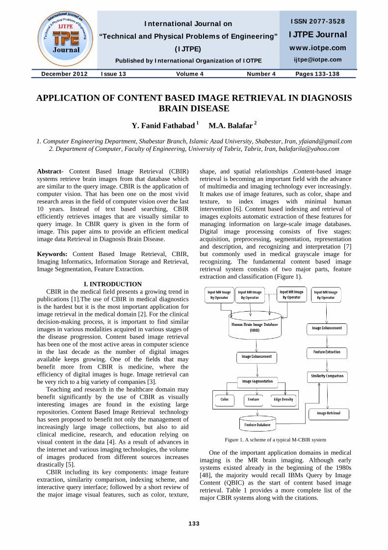
D. Shape Based CBIR Retrieval
In shape-based techniques, feature sets normally include edges, corners, and curvature scale space and chain codes. Unlike color and texture, shapes and objects are not global attributes of an image. In the color and texture realm, distance measures are used to establish if a given image has a specified color or texture, and whether or not it exists in the same approximate position as the query image. Shape representations can be generally divided into two categories [19]: 1. Boundary based; 2. Region based.
Figure 3. Boundary based shape representation
Boundary based shape representation only uses the outer boundary of the shape as shown in Figure 3. This is done by describing the considered region using its external characteristics; i.e., the pixels along the object boundary. For a sequence of pixels, one classical kind of matting uses Fourier descriptors to compare two shapes. In separate case, the shape is represented by a sequence of N points. From this sequence of points, a sequence of unit vector:
1
1
k kk
k k
v vv
v v+
+
−=
− (6)
International Journal on “Technical and Physical Problems of Engineering” (IJTPE), Iss. 13, Vol. 4, No. 4, Dec. 2012
136
E. Boundary Matching with Fourier Descriptor So the dimensions of the Fourier descriptors used for
indexing shapes are reduced much. s(t), t = 0, 1, …, L, complacent it is normalized to N points in the sampling stage, the discrete Fourier transform of s(t) is given by below function:
1
0
1 2( )exp , 0,1,..., 1N
nt
j ntu s t n NN N
π−
=
−⎛ ⎞= = −⎜ ⎟⎝ ⎠
∑ (7)
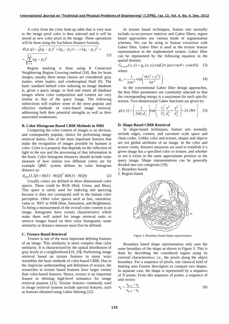
Region based shape representation uses the entire shape region by describing the considered region using its internal characteristics; i.e., the pixels contained in that region [13]. Querying a database using shape features can allow physicians to identify malformations or tumors that otherwise might be missed [23]. To identify a shape, we must find where its edges, that is, are where a big change in the gray level intensities occurs (Figure 4).
(a) (b)
Figure 4. Example of (a) an original grey level image and (b) its segmented form
IV. SIMILARITY MEASURES
One of the biggest challenges in any CBIR system is how to define an appropriate measure assessing the similarity to be used for database indexing and/or similarity-based ranking of the retrieved images with respect to the query [24]. A common and rather straightforward method is to employ vector distances in a high dimensional normal vector space, commonly a Euclidean space, in which each image is represented with a point corresponding to its image descriptor/feature vector [25]. Intuitively, shorter distances correspond to higher similarity. The choice of metric depends on the type of image features/descriptors as well as on their representation.
V. IMAGE RETRIEVAL APPLICATION FOR MAGNETIC RESONANCE (MR) BRAIN IMAGES
The goal of diagnostic medical image retrieval is to provide diagnostic support by displaying relevant past cases, along with proven pathologies as ground truth [26]. fMRI (functional Magnetic Resonance Imaging) [27] is a technique used to “monitor” brain activities. Many of the proposed retrieval systems in the area of medical domain are adopted from general image retrieval schemes which perform satisfactorily with databases consisting of heterogeneous images of different modalities and anatomical regions.
These systems use imprecise segmentation and feature extraction techniques which are not suitable for precise matching required for the retrieval of same 2D brain images (slices) in 3D volumes for diagnostic support. In one research paper [28] has been reported to solve 2D slice retrieval problem. In 2D form, for each pixel in an image, a binary code is produced by thresholding its value with the value of a center pixel. A histogram is then generated to calculate the occurrences of different binary patterns [29].
VI. MEDICAL APPLICATIONS OF CONTENT BASED IMAGE RETRIEVAL
Image registration is the process of overlaying two or more images of the same scene taken at different times, from different viewpoints, and/or by different sensors. Image registration is a crucial step in all image analysis tasks in which the final information is gained from the combination of various data sources, like in image fusion, change detection, and multichannel image restoration [30].
Table 2 lists several image retrieval systems proposed for various medical departments. Although content-based image retrieval has frequently been proposed for use in medical image management, only a few content-based retrieval systems have been developed specifically for medical images [31].
Table 2. Various image types and respective retrieval systems
Name/Feature Imaging Modality Domain QBISM / intensity-based MRI/PET Brain FICBDS / Physiological
information based Functional PET Brain
MIMS / ontology based All All MIRAGE / 3D texture based MR Brain
Knowledge based All All ILive modality based All All organs
2D Texture based MR Heart 3D PET / lesion based PET Brain
Predefined semantic based CT Brain
A. ASSERT ASSERT or Automatic Search and Selection Engine
with Retrieval Tools were developed by Indiana University in USA. The ASSERT system extracts 255 features: textures, shape, edges, and gray scale properties in pathology bearing regions. B. 3D PET/CT
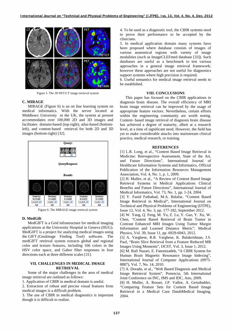
3D PET/CT Fusion supports the effective interpretation with whole body FDG oncology studies and real-time interaction with PET, CT and fused volumes. It enables radiologists to accurately and efficiently blend PET and CT studies to combine anatomical and functional images for rapid lesion analysis and characterization. 3D allows you to separately license the advanced visualization and analysis tools you need on a routine basis (Figure 5).
International Journal on “Technical and Physical Problems of Engineering” (IJTPE), Iss. 13, Vol. 4, No. 4, Dec. 2012
137
Figure 5. The 3D PET/CT image retrieval system C. MIRAGE
MIRAGE (Figure 6) is an on line learning system on medical informatics. With the server located at Middlesex University in the UK, the system at present accommodates over 100,000 2D and 3D images and facilitates domain-based (top-right), atlas-based (bottom-left), and content-based retrieval for both 2D and 3D images (bottom-right) [32].
Figure 6. The MIRAGE image retrieval system D. MedGift
MedGIFT is a Grid infrastructure for medical imaging applications at the University Hospital in Geneva (HUG). MedGIFT is a project for analyzing medical images using the GIFT (GnuImage Finding Tool) software. The medGIFT retrieval system extracts global and regional color and texture features, including 166 colors in the HSV color space, and Gabor filter responses in four directions each at three different scales [31].
VII. CHALLENGES IN MEDICAL IMAGE RETRIEVAL
Some of the major challenges in the area of medical image retrieval are outlined as follows: 1. Application of CBIR in medical domain is useful. 2. Extraction of robust and precise visual features from medical images is a difficult problem. 3. The use of CBIR in medical diagnostics is important though it is difficult to realize.
4. To be used as a diagnostic tool, the CBIR systems need to prove their performance to be accepted by the clinicians. 5. In medical application domain many systems have been proposed where database consists of images of various anatomical regions with variety of image modalities (such as ImageCLEFmed database [33]). Such databases are useful as a benchmark to test various approaches in a general image retrieval framework; however these approaches are not useful for diagnostics support systems where high precision is required. 6. Useful semantics for medical image retrieval needs to be established.
VIII. CONCLUSIONS This paper has focused on the CBIR applications in
diagnosis brain disease. The overall efficiency of MRI brain image retrieval can be improved by the usage of appropriate feature vectors. Nevertheless, certain efforts within the engineering community are worth noting. Content- based image retrieval of diagnosis brain disease has achieved a degree of maturity, albeit at a research level, at a time of significant need. However, the field has yet to make considerable attacks into mainstream clinical practice, medical research, or training.
REFERENCES [1] L.R. Long, et al., “Content Based Image Retrieval in Medicine: Retrospective Assessment, State of the Art, and Future Directions”, International Journal of Healthcare Information Systems and Informatics, Official Publication of the Information Resources Management Association, Vol. 4, No. 1, p. 1, 2009. [2] H. Muller, et al., “A Review of Content Based Image Retrieval Systems in Medical Applications Clinical Benefits and Future Directions”, International Journal of Medical Informatics, Vol. 73, No. 1, pp. 1-24, 2004. [3] Y. Fanid Fathabad, M.A. Balafar, “Content Based Image Retrieval in Medical”, International Journal on Technical and Physical Problems of Engineering (IJTPE), Issue 12, Vol. 4, No. 3, pp. 177-182, September 2012. [4] W. Yang, Q. Feng, M. Yu, Z. Lu, Y. Gao, Y. Xu, W. Chen, “Content Based Retrieval of Brain Tumor in Contrast Enhanced MRI Images Using Tumor Margin Information and Learned Distance Metric”, Medical Physics, Vol. 39, Issue 11, pp. 6929-6943, 2012. [5] A. Varghese, R.R. Varghese, K. Balakrishnan, J.S. Paul, “Brain Slice Retrieval from a Feature Reduced MR Images Using Moments”, IJCST, Vol. 3, Issue 1, 2012. [6] M. Rafi Nazari, E. Fatemizadeh, “A CBIR System for Human Brain Magnetic Resonance Image Indexing”, International Journal of Computer Applications (0975- 8887), Vol. 7, No. 14, 2010. [7] A. Dorado, et al., “Web Based Diagnosis and Medical Image Retrieval System”, Ponencia, 5th International Joint Conference on INC, IMS and IDC, Ano, 2009.
[8] H. Muller, A. Rosset, J.P. Vallee, A. Geissbuhler, “Comparing Feature Sets for Content Based Image Retrieval in a Medical Case DatabMedical Imaging, 2004.

International Journal on “Technical and Physical Problems of Engineering” (IJTPE), Iss. 13, Vol. 4, No. 4, Dec. 2012
138
[9] M.A. Balafar, “Gaussian Mixture Model Based Segmentation Methods for Brain MRI Images”, Artificial Intelligence Review, pp. 1-11, 2012. [10] H.B. Kekre, T.K. Sarode, S. Gharge, “Detection and Demarcation of Tumor Using Vector Quantization in MRI Images”, International Journal of Engineering Science and Technology (IJEST), Vol. 2, No. 2, pp. 59-66, 2009. [11] J. Laaksonen, E. Oja, M. Koskela, S. Brandt, “Analyzing Low Level Visual Features Using Content Based Image Retrieval”, International Conference Neural Information Processing, Taejon, pp. 14-18, 2000. [12] K.H. Hwang, H. Lee, D. Choi, “Medical Image Retrieval: Past and Present”, Healthcare Informatics Research, Vol. 18, No. 1, pp. 3-9, 2012. [13] M.R. Siadat, et al., “Content Based Image Database System for Epilepsy”, Computer Methods and Programs in Biomedicine, Vol. 79, No. 3, pp. 209-226, 2005. [14] A.P. Killedar, V.P. Patil, M.S. Borse, “Content Based Image Retrieval Approach to Tumor Detection in Human Brain Using Magnetic Resonance Image”, 1st International Conference on Recent Trends in Engineering and Technology, pp. 211-214, March 2012. [15] R.C. Gonzalez, R.E. Wood, “Digital Image Processing”, Prentice Hall, 2008. [16] A.J.M. Traina, C.A.B. Castanon, C. Traina, “MultiWaveMed: A System for Medical Image Retrieval through Wavelets Transformations”, IEEE Symp. Comput. Based Med. Syst., New York, USA, pp. 150-155, 2003. [17] K. Jain, S. Singh, “A Survey on: Content Based Image Retrieval Systems Using Clustering Techniques for Large Data Sets”, International Journal of Managing Information Technology, Vol. 3, No. 4, Nov. 2011. [18] R. Adams, L. Bischof, “Seeded Region Growing”, IEEE Transactions on Pattern Analysis and Machine Intelligence, Vol. 16, No. 6, pp. 641-647, 1994. [19] A.A.A. Youssif, A.A. Darwish, R.A. Mohamed, “Content Based Medical Image Retrieval Based on Pyramid Structure Wavelet”, International Journal of Computer Science and Network Security, Vol. 10, No. 3, 2010. [20] W. Shengjiu, “A Robust CBIR Approach Using Local Color Histograms”, Tech. Rep. TR 01-13, 2001. [21] Y. Liu, D. Zhang, G. Lu, W.Y. Ma, “A Survey of Content Based Image Retrieval with High Level Semantics”, Pattern Recog., Vol. 40, pp. 262-282, 2007. [22] A.C. Bovik, M. Clark, W. Geisler, “Multichannel Texture Analysis Using Localized Spatial Filters”, IEEE Transactions on Pattern Analysis and Machine Intelligence, Vol. 12, pp. 55-73, 1990. [23] P. Korn, N. Sidiropoulos, C. Faloutsos, E. Siegel, Z. Protopapas, “Fast and Effective Retrieval of Medical Tumor Shapes”, IEEE Trans. on Knowledge and Data Engineering, Vol. 10, pp. 889-904, 1998. [24] C.B. Akgul, et al., “Content Based Image Retrieval in Radiology: Current Status and Future Directions”, Journal of Digital Imaging, Vol. 24, No. 2, pp. 208-222, 2011.
[25] D. Pokrajac, et al., “Applying Spatial Distribution Analysis Techniques to Classification of 3D Medical Images”, Artificial Intelligence in Medicine, Vol. 33, No. 3, pp. 261-280, 2005. [26] H.D. Tagare, C.C. Jaffe, J. Duncan, “Medical Image Databases a Content Based Retrieval Approach”, Journal of the American Medical Informatics Association, Vol. 4, No. 3, pp. 184-198, 1997. [27] R.S.J. Frackowiak, K.J. Friston, C.D. Frith, R.J. Dolan, C.J. Price, S. Zeki, J. Ashburner, W. Penny, “Human Brain Function”, 2nd Edition, Elsevier Academic Press, 2004. [28] D. Unay, A. Ekin, R. Jasinschi. “Medical Image Search and Retrieval Using Local Binary Patterns and KLT Feature Points”, 15th IEEE International Conference on Image Processing, ICIP, 2008. [29] Y. Qian, et al., “Content Based Retrieval of 3D Medical Images”, 3rd International Conference on eHealth, Telemedicine, and Social Medicine, eTELEMED, 2011. [30] B. Zitova, J. Flusser, “Image Registration Methods: A Survey”, Image and Vision Computing, Vol. 21, pp. 977-1000, 2003. [31] C.H. Wei, C.T. Li, R. Wilson, “A Content Based Approach to Medical Image Database Retrieval”, Database Modeling for Industrial Data Management: Emerging Technologies and Applications, Ed. by Z. Ma, Idea Group Publishing, 2005. [32] X.W. Gao, Y. Qian, R. Hui, “Suppl 1: The State of the Art of Medical Imaging Technology: From Creation to Archive and Back”, The Open Medical Informatics Journal, Vol. 5, p. 73, 2011. [33] W. Hersh, J. Kalpathy Cramer, H. Muller, “The ImageCLEFmed Medical Image Retrieval Task Test Collection”, J. Digit. Imaging, Vol. 22, pp. 648-655, 2009. [34] A.U. Jawadekar, G.M. Dhole, S.R. Paraskar, M.A. Beg, “Novel Wavelet ANN Technique to Classify Bearing Faults in Three Phase Induction Motor”, International Journal on Technical and Physical Problems of Engineering (IJTPE), Issue 8, Vol. 3, No. 3, pp. 48-54 September 2011.
BIOGRAPHIES
Younes Fanid Fathabad was born in Tabriz, Iran, 1974. He received the B.Sc. and M.Sc. degrees from Shabestar Branch, Islamic Azad University, Shabestar, Iran. His research interests are in artificial intelligence and image processing. He has published 3 books about computer
science.
Mohammad Ali Balafar was born in Tabriz, Iran, in June 1975. He received the Ph.D. degree in IT in 2010. Currently, he is an Assistant Professor. His research interests are in artificial intelligence and image processing. He has published 9 journal papers and 4 book chapters.