Embed Size (px)
Citation preview
Reviews�KEYNOTEREVIEW
Drug Discovery Today � Volume 00, Number 00 �April 2016 REVIEWS
Bioprinting is a powerful technology in the fabrication of living tissues and organs for tissueengineering and regenerative medicine, transplantation and clinics, pharmaceutics and
high-throughput screening, as well as cancer research.
Application areas of 3D bioprintingIbrahim T. Ozbolat1,2, Weijie Peng1,2,3 and Veli Ozbolat4
1 Engineering Science and Mechanics Department, The Pennsylvania State University, State College, PA 16802, USA2 The Huck Institutes of the Life Sciences, The Pennsylvania State University, State College, PA 16802, USA3Department of Pharmacology, Nanchang University, Nanchang, JX 330006, China4Mechanical Engineering Department, Ceyhan Engineering Faculty, Cukurova University, 01330 Adana, Turkey
Three dimensional (3D) bioprinting has been a powerful tool in patterning
and precisely placing biologics, including living cells, nucleic acids, drug
particles, proteins and growth factors, to recapitulate tissue anatomy,
biology and physiology. Since the first time of cytoscribing cells
demonstrated in 1986, bioprinting has made a substantial leap forward,
particularly in the past 10 years, and it has been widely used in fabrication
of living tissues for various application areas. The technology has been
recently commercialized by several emerging businesses, and bioprinters
and bioprinted tissues have gained significant interest in medicine and
pharmaceutics. This Keynote review presents the bioprinting technology
and covers a first-time comprehensive overview of its application areas
from tissue engineering and regenerative medicine to pharmaceutics and
cancer research.
IntroductionBioprinting is a growing field that makes a revolutionary impact on medical and pharmaceutical
sciences, and has gained significant attention worldwide [1]. Bioprinting can be defined as the
simultaneous writing of living cells and biomaterials with a prescribed layer-by-layer stacking
organization using a computer-aided transfer process for fabrication of bioengineered constructs
[2]. It offers great precision on spatial placement of cells, proteins, DNA, drug particles, growth
factors and biologically active particles to guide tissue generation and formation better. This
powerful technology appears to be more promising for advancing tissue fabrication toward
physiologically relevant tissue constructs, tissue models, tissues and organs and organs-on-a-chip
models for medicine and pharmaceutics.
Bioprinting technology has a broad utility in various application areas such as tissue engineer-
ing and regenerative medicine [3,4], transplantation and clinics [1], drug screening and high-
throughput assays [5] and cancer research [6], as depicted in Fig. 1. Bioprinting for tissue
engineering and regenerative medicine fields has been around for more than a decade, and
Ibrahim Tarik Ozbolat
is an Associate Professor in
the Engineering Science
and Mechanics
Department and the Huck
Institutes of the Life
Sciences at Penn State
University. He received his
PhD from the University at
Buffalo, New York, and
dual BS degrees in mechanical and industrial
engineering from Middle East Technical University,
Turkey. Dr Ozbolat’s major research effort is in the
area of 3D bioprinting. He has published over 80
journal and conference articles, and his research has
been featured in local, national and international
media. He is the recipient of several prestigious
international awards.
Weijie Peng received his
PhD degree from the
Department of
Pharmacology, Central
South University, China, in
2005. He is an Associate
Professor in the
Department of
Pharmacology, Nanchang
University, China. He has
recently joined the Ozbolat Lab at Penn State
University as a visiting scholar supported by the
Chinese Scholarship Committee. His major research
work is on estrogen receptor, estrogenic substances
and bioprinting for pharmaceutical research.
Veli Ozbolat received his
BS degree in mechanical
engineering from the
Middle East Technical
University, Ankara,
Turkey, in 2008; and his MS
degree in industrial
engineering from the
University at Buffalo, New
York, USA, in 2010. He
obtained his PhD in mechanical engineering from
Cukurova University, Adana, Turkey, in 2015. He will
join Penn State University as a postdoctoral scholar in
Summer 2016. His research mainly focuses on
biomedical engineering, heat transfer and fluid
mechanics.
Please cite this article in press as: Ozbolat, I.T. et al. Application areas of 3D bioprinting, Drug Discov Today (2016), http://dx.doi.org/10.1016/j.drudis.2016.04.006
Corresponding author: Ozbolat, I.T. ([email protected])
1359-6446/� 2016 Elsevier Ltd. All rights reserved.
http://dx.doi.org/10.1016/j.drudis.2016.04.006 www.drugdiscoverytoday.com 1
DRUDIS-1788; No of Pages 15
Review
s�K
EYNOTEREVIEW
REVIEWS Drug Discovery Today � Volume 00, Number 00 �April 2016
Drug screening and high-throughput assays
Transplantation andclinics
Tissue engineering andregenerative medicine
Cancer research
Drug Discovery Today
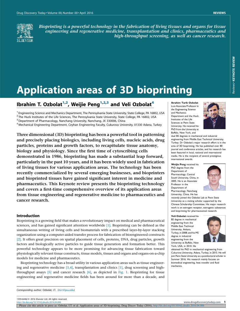
FIGURE 1
Application areas of bioprinting technology including tissue engineering and
regenerative medicine, transplantation and clinics, drug screening and high-
throughput assays, and cancer research (image courtesy of ChristopherBarnatt from http://www.explaningthefuture.com).
anatomically correct cell-laden constructs and scaffolds have been
fabricated for various tissue types from connective and epithelial
tissues to muscle and nervous tissues. With its great advantage in
patterning and precisely positioning multiple cell types, bioprint-
ing has circumvented one of the major shortcomings of traditional
scaffold fabrication techniques and has enabled fabrication of
native-like tissues with a heterocellular microenvironment. Al-
though the vast majority of the effort has been geared toward
the fundamental science behind major bioprinting techniques
such as laser-, droplet- and extrusion-based bioprinting, a substan-
tial focus has recently been given to bioprinting for functional
tissue fabrication [1]. Particularly, considerable work has been
dedicated to bioprinting for transplantation, where bioprinted
tissues have been implanted to various associated sites in vivo.
As further progress takes place in biomaterials, cell and transplan-
tation technologies, bioprinting will translate from bench to
bedside when approved for human use and has a myriad of
advantages in operating rooms in the near future. Before transi-
tioning into clinical practice, bioprinting has already made a great
leap in pharmaceutical use because it does not entail any regula-
tory approvals and there is currently an emerging bioprinting
market for tissue fabrication for drug testing and high-throughput
assays. With the inclusion of multiple cell types and facilitating a
complex heterocellular physiological environment, bioprinted
tissue models (e.g. liver) have been used in drug screening. In
addition, bioprinting has recently been used in cancer research to
investigate cancer pathology, growth and metastasis in a physio-
logically relevant microenvironment [7]. Here, we present a criti-
cal and comprehensive review of the application areas of
bioprinting technology and provide an in-depth discussion on
successfully bioprinted tissue types in tissue engineering and
regenerative medicine, transplantation and clinics, drug screening
and high-throughput assays, and cancer research. For each
Please cite this article in press as: Ozbolat, I.T. et al. Application areas of 3D bioprinting, D
2 www.drugdiscoverytoday.com
application area, limitations of existing technologies are discussed
and future prospects are provided to the reader.
Tissue engineering and regenerative medicineBioprinting of functional organs at clinically relevant dimensions
remains elusive because there are several challenges such as but
not limited to integration of the vascular network from arteries
and veins down to capillaries, incorporation of various cell types to
recapitulate complex organ biology and limited structural and
mechanical integrity and long-term functionality [8]. Despite
these difficulties, a wide variety of tissues have been successfully
bioprinted such as thin or hollow tissues, for example blood vessel
[9], or tissues that do not need vascularization such as cartilage [1].
Bone tissueBone tissue engineering has been widely studied using bioprinting
because bioprinting has the ability to fabricate anatomically cor-
rect patient-specific tissue constructs. In a recent study [10], Gao
et al. used a thermal inkjet bioprinter to fabricate poly(ethylene
glycol) dimethacrylate (PEGDMA) scaffolds. Bone-marrow-derived
human mesenchymal stem cells (hMSCs) were co-printed with
nanoparticles of bioactive glass and hydroxyapatite (HA) under
simultaneous polymerization. Bioprinting in that study enabled
uniform distribution of hMSCs compared with manually pipetted
hMSCs, which accumulated at the bottom of the scaffold because
of gravity (Fig. 2a). The bioprinted constructs encapsulating
hMSCs and HA demonstrated the highest cell viability, collagen
production and alkaline phosphate activity with increased com-
pressive modulus after 21-day culture in vitro. Bioprinting HA
particles were also performed for in situ bioprinting purposes,
where a laser-based bioprinting system was used to deposit HA
nanoparticles into mouse calvarial defects in a framework study
[11], as detailed below.
In another study, Fedorovich et al. bioprinted heterocellular
tissue constructs made of MatrigelTM and alginate hydrogels [12].
Endothelial progenitor and multipotent stromal cells were bio-
printed in a spatially controlled manner and the bioprinted con-
structs were subcutaneously implanted into immune-deficient
mice. By incorporating osteoinductive biphasic calcium phos-
phate microparticles, multipotent stromal cells were differentiated
into an osteogenic lineage and facilitated bone formation in 6
weeks. In addition to osteoinductive materials, incorporation of
growth factors is also crucial in stem cell differentiation in bone
tissue engineering. Phillippi et al. demonstrated the effect of bone
morphogenetic protein (BMP)-2 on stem cell fate [13]. Using inkjet
bioprinting of patterned BMP-2 on fibrin-coated coverslips, pri-
mary-muscle-derived stem cells were differentiated toward osteo-
genic lineage on the pattern even if they were treated with
myogenic differentiation conditions.
Cardiac tissueCardiac tissue engineering has been a growing interest because
heart failure is a devastating disease [14]. Although myocardium
tissue has a limited regeneration capability because myocyte pro-
liferation rapidly ceases after birth [15], tissue engineering of such
a structurally and functionally complicated organ is essential. In
the literature, limited attempts have been made in bioprinting of
cardiac tissue models. Jakab et al. demonstrated extrusion-based
rug Discov Today (2016), http://dx.doi.org/10.1016/j.drudis.2016.04.006
Drug Discovery Today � Volume 00, Number 00 �April 2016 REVIEWS
DRUDIS-1788; No of Pages 15
Reviews�KEYNOTEREVIEW
bioprinting of tissue spheroids comprising human vascular endo-
thelial cells (HUVECs) and cardiac cells isolated from myocardial
tubes of chicken embryos [16]. Tissue spheroids that were adhesive
and scaffold-free, and possessed rapid self-assembly capabilities,
were bioprinted next to each other on collagen type-I biopaper in a
single-layer grid pattern. Upon bioprinting, tissue spheroids were
fused together in approximately 70 h and formed a single cardiac
tissue patch that could synchronously beat. In addition to the
scaffold-free approach undertaken in the above work, scaffold-
based bioprinting has been investigated in a few remarkable
studies. Xu et al. bioprinted a cardiac tissue construct in a half-
heart shape with connected ventricles using inkjet-based bioprint-
ing (Fig. 2b1,b2) [17]. In their study, primary feline adult and H1
cardiomyocytes were encapsulated in alginate–gelation composite
hydrogels and the crosslinker (calcium chloride solution) was
selectively sprayed layer by layer. The resulting tissue construct
with connected ventricles was electrically stimulated and func-
tional excitation–contraction coupling was successfully demon-
strated. In addition to these studies, patterning of cells was also
applied in cardiac tissue engineering. Gaebel et al. utilized laser-
induced forward-transfer (LIFT) to pattern HUVECs and hMSCs in
a geometrically defined pattern on polyester urethane urea and the
fabricated samples were transplanted to the infarcted zone of rat
hearts after LAD ligation [18]. In 8 weeks post-transplantation,
samples with LIFT-derived patterns facilitated increased vessel
formation compared with randomly bioprinted cells as control
groups and the resulted myocardium patch provided significant
functional improvement. Besides primary cells, human cardiac-
derived cardiomyocyte progenitor cells (hCMPCs) were also bio-
printed in a mesh pattern [19]. Bioprinted hCMPCs demonstrated
phenotypic properties of cardiac lineage with enhanced expres-
sion of early cardiac transcription factors Nkx2.5, Gata-4 and Mef-
2c.
Cartilage tissueCurrent tissue engineering techniques for cartilage regeneration
cannot produce cartilage tissue that is indistinguishable from
native tissue [20]. Owing to its great potential for precise spatial
and temporal deposition of cells and biomaterials with sophisti-
cated patterns, bioprinting has recently gained increasing atten-
tion for engineering cartilage tissues that can imitate native tissues
with zonally differentiated cells and extracellular matrix (ECM)
composition. Laser-based bioprinting of stem-cell-differentiated
chondrocytes was attempted by Gruene et al., in which a comput-
er-aided biofabrication technique was used with the assistance of
LIFT [21]. They successfully bioprinted porcine-bone-marrow-de-
rived mesenchymal stem cells (MSCs) with high viability, and the
cells maintained their functionality and differentiation ability
into osteogenic and chondrogenic lineages.
Inkjet-based bioprinting has also been used in cartilage tissue
engineering as well as for cartilage defect repair. Cui et al. modified
an HP desktop printer, where they were able to bioprint human
chondrocytes loaded in PEGDMA hydrogel [22]. The bioprinted
cartilage construct had mechanical properties and biochemical
composition close to native cartilage. Also, by implanting bio-
printed cartilage constructs into articular cartilage defects, inte-
gration with the native tissue was observed with enhanced
interface strength, which improved the quality of the repaired
Please cite this article in press as: Ozbolat, I.T. et al. Application areas of 3D bioprinting, Dr
cartilage significantly. In another study using the above experi-
mental setup, the same group fabricated PEG scaffolds (Fig. 2c1)
and investigated the effect of combined transforming growth
factor (TGF)-b1 and fibroblast growth factor (FGF)-2 on cell pro-
liferation and differentiation capability, and demonstrated that
samples treated with TGF-b1 and FGF-2 facilitated the highest
glycosaminoglycan (GAG) content [23] and samples without
growth factor treatment did not secrete GAG even in 4-week
culture (Fig. 2c2,c5). Most recently, Xu et al. created a hybrid
bioprinting method to fabricate mechanically improved cartilage
tissue constructs by combining 3D bioprinting and electrospin-
ning techniques [24]. In that study, electrospinning of polycapro-
lactone (PCL) fibers together with inkjet bioprinting of rabbit
elastic chondrocytes in fibrin–collagen hydrogel was demonstrat-
ed. After printing, cell viability was well maintained and fabricated
constructs formed cartilage tissues, with improved mechanical
properties, in vitro and in vivo.
In addition to hydrogels used in the abovementioned studies,
sodium alginate has been widely used for cartilage tissue bioprint-
ing. Ozbolat et al. demonstrated hybrid bioprinting of chondro-
cytes loaded in printed alginate filaments in tandem with
bioprinting of chondrocyte spheroids to increase the cell density
[25]. A multi-arm bioprinter was used to facilitate such a complex
hybrid architecture. Using sodium alginate and silver nanoparti-
cles, McAlpine’s group [26] successfully printed a bionic ear model,
which was composed of chondrocyte-loaded alginate in an ear-
shape and a conductive coil with the ability to translate sound
waves into digital data. Recently, Markstedt et al. demonstrated
mixing alginate with nanocellulose, which has outstanding shear-
thinning properties that enabled fabrication of anatomically cor-
rect ear and meniscus constructs [27].
Despite the great progress in bioprinting for cartilage tissue
regeneration, bioprinting of zonally stratified articular cartilage
tissues with different structural, biomechanical and biological
properties is still a challenge and further progress is needed to
achieve articular cartilage tissue constructs with zonal differentia-
tion including more horizontal and thinner collagen fibers with
high cell density in the superficial zone, and relatively vertical and
thicker collagen fibers with less cell density in the deeper zones.
Heart valvesIn addition to cardiac tissue engineering, engineering heart valves
is also important because heart valves do not possess regeneration
capability and dysfunctional heart valves, if the damage or disease
is detrimental, need to be replaced by mechanical or biological
prosthetic counterparts [28]. Such replacement valves, however,
are limited by thrombogenicity and calcification [29]. Despite its
crucial role in the cardiovascular system, only a limited amount of
work has been carried out in the bioprinting of heart valves.
Butcher’s group demonstrated the first-time bioprinting of a heart
valve [30] using a dual-head bioprinter modified from a Fab@-
Home printer [31]. In that work, a dual crosslinking mechanism,
consisting of ionic and physical crosslinking, was used to print
PEG-diacrylate (PEGDA) mixed with sodium alginate. After print-
ing, porcine aortic valve interstitial cells (PAVICs) were seeded and
cultured for up to 21 days. In their study, anatomically accurate
axisymmetric aortic valve geometries, composed of a root wall and
tri-leaflets, were demonstrated. Although the first-time presented
ug Discov Today (2016), http://dx.doi.org/10.1016/j.drudis.2016.04.006
www.drugdiscoverytoday.com 3
REVIEWS Drug Discovery Today � Volume 00, Number 00 �April 2016
DRUDIS-1788; No of Pages 15
Please cite this article in press as: Ozbolat, I.T. et al. Application areas of 3D bioprinting, Drug Discov Today (2016), http://dx.doi.org/10.1016/j.drudis.2016.04.006
(a)Top
Bioprinted sample
Uniformdistribution
Accumulation ofcells due to
gravity
SE 22-Nov-04 WD24.8mm 5. 00kV x500 100um100 μm 100 μm
50 μm
20 μm
200μm
Control (manual deposition)
Bottom
(c)1 (d)1
(f)2
(c)2 Week 3
Week 4
agaroserod
nervegraft
isle
ts in
bul
kal
gina
te/g
elat
in
Day 0 Day 4
Day 11Day 0
Day 7
biop
rinte
d is
lets
in a
lgin
ate/
gela
tin
BMSCpellet
BMSC &Schwan cell pellet
(c)3
(c)4
(g)1
(g)3
(j)1 (j)2
(g)2 (h)
(c)5
(b)1 (b)2
(d)2 αSMAVimentinNuclear
(e)1 (e)2 (f)1
(f)3
(g)4
(i)1 (i)2
Drug Discovery Today
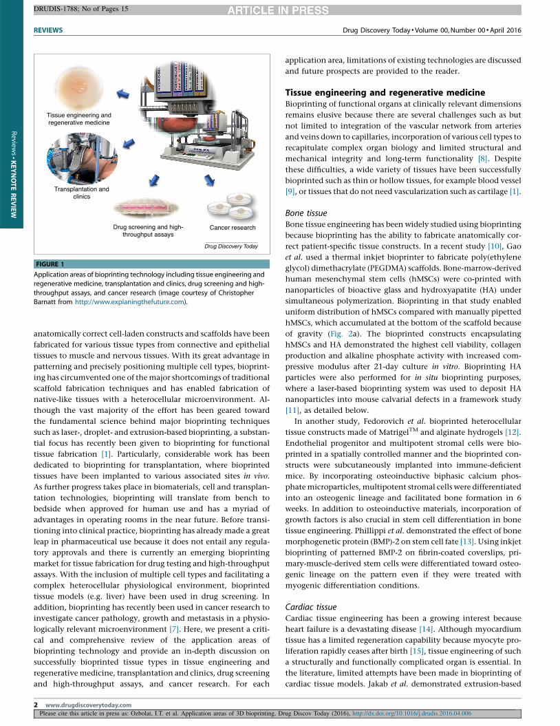
FIGURE 2
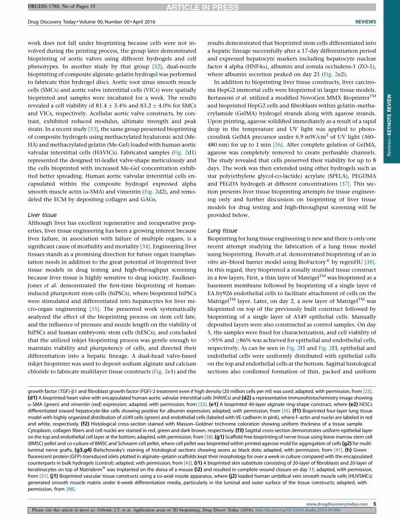
Bioprinted tissue constructs for tissue engineering and regenerative medicine. (a) Inkjet bioprinting of human mesenchymal stem cells (hMSCs) in hydrogels for
bone tissue engineering, where bioprinting resulted in uniform distribution of hMSCs opposed to accumulated hMSCs at the bottom of the scaffold as a result of
gravity when hMSCs were manually pipetted; adapted, with permission, from [10]. (b1) Bioprinting of cardiac tissue constructs with connected ventricles using a
modified-HP printer, (b2) SEM image of the cross-section of the scaffold showing loaded cells; adapted, with permission, from [17]. (c1) Bioprinting of a 4 mmpoly(ethylene glycol) (PEG) cartilage tissue construct with (c2–c5) Safranin-O staining with limited glycosaminoglycan (GAG) deposition without transforming
4 www.drugdiscoverytoday.com
Review
s�K
EYNOTEREVIEW
Drug Discovery Today � Volume 00, Number 00 �April 2016 REVIEWS
DRUDIS-1788; No of Pages 15
Reviews�KEYNOTEREVIEW
work does not fall under bioprinting because cells were not in-
volved during the printing process, the group later demonstrated
bioprinting of aortic valves using different hydrogels and cell
phenotypes. In another study by that group [32], dual-nozzle
bioprinting of composite alginate–gelatin hydrogel was performed
to fabricate thin hydrogel discs. Aortic root sinus smooth muscle
cells (SMCs) and aortic valve interstitial cells (VICs) were spatially
bioprinted and samples were incubated for a week. The results
revealed a cell viability of 81.4 � 3.4% and 83.2 � 4.0% for SMCs
and VICs, respectively. Acellular aortic valve constructs, by con-
trast, exhibited reduced modulus, ultimate strength and peak
strain. In a recent study [33], the same group presented bioprinting
of composite hydrogels using methacrylated hyaluronic acid (Me-
HA) and methacrylated gelatin (Me-Gel) loaded with human aortic
valvular interstitial cells (HAVICs). Fabricated samples (Fig. 2d1)
represented the designed tri-leaflet valve-shape meticulously and
the cells bioprinted with increased Me-Gel concentration exhib-
ited better spreading. Human aortic valvular interstitial cells en-
capsulated within the composite hydrogel expressed alpha
smooth muscle actin (a-SMA) and vimentin (Fig. 2d2), and remo-
deled the ECM by depositing collagen and GAGs.
Liver tissueAlthough liver has excellent regenerative and recuperative prop-
erties, liver tissue engineering has been a growing interest because
liver failure, in association with failure of multiple organs, is a
significant cause of morbidity and mortality [34]. Engineering liver
tissues stands as a promising direction for future organ transplan-
tation needs in addition to the great potential of bioprinted liver
tissue models in drug testing and high-throughput screening
because liver tissue is highly sensitive to drug toxicity. Faulkner-
Jones et al. demonstrated the first-time bioprinting of human-
induced pluripotent stem cells (hiPSCs), where bioprinted hiPSCs
were stimulated and differentiated into hepatocytes for liver mi-
cro-organ engineering [35]. The presented work systematically
analyzed the effect of the bioprinting process on stem cell fate,
and the influence of pressure and nozzle length on the viability of
hiPSCs and human embryonic stem cells (hESCs), and concluded
that the utilized inkjet bioprinting process was gentle enough to
maintain viability and pluripotency of cells, and directed their
differentiation into a hepatic lineage. A dual-head valve-based
inkjet bioprinter was used to deposit sodium alginate and calcium
chloride to fabricate multilayer tissue constructs (Fig. 2e1) and the
Please cite this article in press as: Ozbolat, I.T. et al. Application areas of 3D bioprinting, Dr
growth factor (TGF)-b1 and fibroblast growth factor (FGF)-2 treatment even if high d
(d1) A bioprinted heart valve with encapsulated human aortic valvular interstitial ce
a-SMA (green) and vimentin (red) expression; adapted, with permission, from [33]differentiated toward hepatocyte-like cells showing positive for albumin expressio
model with highly organized distribution of a549 cells (green) and endothelial cells
and white, respectively. (f2) Histological cross-section stained with Masson–Goldn
Cytoplasm, collagen fibers and cell nuclei are stained in red, green and dark brown,on the top and endothelial cell layer at the bottom; adapted, with permission, from [
(BMSC) pellet and co-culture of BMSC and Schwann cell pellet, where cell pellet was
luminal nerve grafts, (g3,g4) Bielschowsky’s staining of histological sections show
fluorescent protein (GFP)-transduced islets plotted in alginate–gelatin scaffolds keptcounterparts in bulk hydrogels (control); adapted, with permission, from [43]. (i1) A
keratinocytes on top of MatridermW was implanted on the dorsa of a mouse (i2) a
from [51]. (j1) Bioprinted vascular tissue constructs using a co-axial nozzle apparat
generated smooth muscle matrix under 6-week differentiation media, particularlypermission, from [98].
results demonstrated that bioprinted stem cells differentiated into
a hepatic lineage successfully after a 17-day differentiation period
and expressed hepatocyte markers including hepatocyte nuclear
factor 4 alpha (HNF4a), albumin and zonula occludens-1 (ZO-1),
where albumin secretion peaked on day 21 (Fig. 2e2).
In addition to bioprinting liver tissue constructs, liver carcino-
ma HepG2 immortal cells were bioprinted in larger tissue models.
Bertassoni et al. utilized a modified NovoGen MMX BioprinterTM
and bioprinted HepG2 cells and fibroblasts within gelatin–metha-
crylamide (GelMA) hydrogel strands along with agarose strands.
Upon printing, agarose solidified immediately as a result of a rapid
drop in the temperature and UV light was applied to photo-
crosslink GelMA precursor under 6.9 mW/cm2 of UV light (360–
480 nm) for up to 1 min [36]. After complete gelation of GelMA,
agarose was completely removed to create perfusable channels.
The study revealed that cells preserved their viability for up to 8
days. The work was then extended using other hydrogels such as
star poly(ethylene glycol-co-lactide) acrylate (SPELA), PEGDMA
and PEGDA hydrogels at different concentrations [37]. This sec-
tion presents liver tissue bioprinting attempts for tissue engineer-
ing only and further discussion on bioprinting of liver tissue
models for drug testing and high-throughput screening will be
provided below.
Lung tissueBioprinting for lung tissue engineering is new and there is only one
recent attempt studying the fabrication of a lung tissue model
using bioprinting. Hovath et al. demonstrated bioprinting of an in
vitro air–blood barrier model using BioFactory1 by regenHU [38].
In this regard, they bioprinted a zonally stratified tissue construct
in a few layers. First, a thin layer of MatrigelTM was bioprinted as a
basement membrane followed by bioprinting of a single layer of
EA.hy926 endothelial cells to facilitate attachment of cells on the
MatrigelTM layer. Later, on day 2, a new layer of MatrigelTM was
bioprinted on top of the previously built construct followed by
bioprinting of a single layer of A549 epithelial cells. Manually
deposited layers were also constructed as control samples. On day
5, the samples were fixed for characterization, and cell viability of
>95% and �86% was achieved for epithelial and endothelial cells,
respectively. As can be seen in Fig. 2f1 and Fig. 2f3, epithelial and
endothelial cells were uniformly distributed with epithelial cells
on the top and endothelial cells at the bottom. Sagittal histological
sections also confirmed formation of thin, packed and uniform
ug Discov Today (2016), http://dx.doi.org/10.1016/j.drudis.2016.04.006
ensity (20 million cells per ml) was used; adapted, with permission, from [23].
lls (HAVICs) and (d2) a representative immunohistochemistry image showing
. (e1) A bioprinted 40-layer alginate ring-shape construct, where (e2) hESCsn; adapted, with permission, from [35]. (f1) Bioprinted four-layer lung tissue
(labeled with VE-cadherin in pink), where F-actin and nuclei are labeled in red
er trichrome coloration showing uniform thickness of a tissue sample.
respectively. (f3) Sagittal cross-section demonstrates uniform epithelial layer38]. (g1) Scaffold-free bioprinting of nerve tissue using bone marrow stem cell
bioprinted within printed agarose mold for aggregation of cells (g2) for multi-
ing axons as black dots; adapted, with permission, from [41]. (h) Green
their morphology for over a week in culture compared with the encapsulated bioprinted skin substitute consisting of 20-layer of fibroblasts and 20-layer of
nd resulted in complete wound closure on day 11; adapted, with permission,
us, where (j2) loaded human umbilical vein smooth muscle cells (HUVSMCs)
in the luminal and outer surface of the tissue constructs; adapted, with
www.drugdiscoverytoday.com 5
REVIEWS Drug Discovery Today � Volume 00, Number 00 �April 2016
DRUDIS-1788; No of Pages 15
Review
s�K
EYNOTEREVIEW
tissue layers (Fig. 2f2) compared with manually pipetted control
samples. The barrier quality, such as the tightness of the con-
structs, was investigated through measuring the translocation of
blue dextran molecules from the apical to the basolateral com-
partment of the samples after 3 days of culture and results revealed
that the tightness of the bioprinted samples was better than that of
the manually pipetted samples.
Neural tissueEngineering nervous system tissues offers tremendous promise to
replace diseased, aged or injured components of nervous system;
however, there is limited work done in the context of bioprinting
for neural tissue fabrication. Lee et al. studied the effect of vascular
endothelial growth factor (VEGF) release on proliferation and
migration of murine neural stem cells (C17.2) [39]. In their study,
C17.2 cells were bioprinted on a collagen layer next to a fibrin disk
loaded with VEGF. The study showed that neural cells migrated
toward VEGF-releasing fibrin gel and proliferated successfully in
contrast to the cells that could not proliferate within the collagen
matrix. Recently, Hsieh et al. demonstrated bioprinting of a ther-
moresponsive polyurethane hydrogel with tunable stiffness and
gelation ability at 378C without the need for a crosslinker [40].
They showed the effectiveness of the bioink by loading it with
neural stem cells and injecting it into a zebrafish embryo neural
injury model. The results revealed that the injected gel rescued the
function of the impaired nervous system in 6 days.
Owens et al. used a scaffold-free approach, where a pellet of
Schwann cells (SCs) and bone marrow stem cells (BMSCs) were
extruded within a 3D-printed agarose mold [41] (Fig. 2g1 for the
schematic of the process). Cells in the agarose mold aggregated
and formed a nerve tissue graft with three lumina in each as shown
in Fig. 2g2. The fabricated grafts were then implanted into mice,
and their histology (Fig. 2g3,g4) and functionality was evaluated
10 months after implantation, and compared to the functionality
of autologous grafts and hollow collagen grafts. Although the
number of samples were not enough to draw a definitive conclu-
sion about the performance of the 3D bioprinted grafts with
respect to commercially available collagen grafts, the presented
case demonstrated a proof-of-concept for bioprinting nerve grafts.
Pancreas tissueBecause primary pancreatic b-cells do not easily survive in vitro and
only a very few attempts have taken place to differentiate b-cells
from human stem cells, regeneration of pancreas tissue is primarily
embodied to the extent that b-cells from mouse lines or insulin-
oma cells have been used to fabricate pancreatic islets [42]. A
limited amount of work has been done in the context of bioprint-
ing for pancreatic tissue fabrication. Recently, Marchioli et al.
encapsulated human and mouse islets as well as rat insulinoma
INS1E b-cells within alginate or alginate–gelatin hydrogels and
bioprinted them in dual-layer scaffolds [43]. The scaffolds were
later implanted in diabetic mice and explanted 7 days thereafter.
Although the viability and morphology of islets was not impaired
by encapsulation and bioprinting processes in alginate and algi-
nate–gelatin hydrogels (Fig. 2h), bioprinted islets and INS1E b-cells
lost their functionality in 7 days because they were not responsive
to the change in glucose level. This can be attributed to the high
level of calcium (Ca2+) ions within crosslinked alginate because
Please cite this article in press as: Ozbolat, I.T. et al. Application areas of 3D bioprinting, D
6 www.drugdiscoverytoday.com
transmembrane calcium ion-gradient, mediated by voltage-gated
calcium channels, stimulated higher insulin secretion at low
glucose level [44]. In addition, recent work demonstrated the
microfabrication of scaffold-free tissue strands (with strong ex-
pression of insulin) for extrusion-based bioprinting [45], where
tissue strands were made of rat fibroblasts and mouse insulinoma
TC-3 b-cells in the core and shell, respectively. The authors envi-
sioned to use the demonstrated tissue strands for scale-up tissue
bioprinting purposes.
Skin tissueA myriad of tissue engineering approaches have been applied in
skin tissue fabrication and tissue substitutes including autologous
split-thickness skin graft (gold standard) [46], allografts [47], acel-
lular dermal substitutes and cellularized graft-like commercial
products [47] [i.e. Dermagraft1 and Apligraf1 (Organogenesis)]
[48]. Recently, bioprinting technology has been adopted for skin
tissue fabrication as well. Lee et al. presented bioprinting of skin
tissue using an eight-channel valve-based bioprinter, where a 13-
layer-tissue construct was bioprinted using collagen hydrogel [49].
Keratinocytes were bioprinted on top of alternating layers of
human foreskin fibroblasts and acellular collagen layers, and
the resulted constructs demonstrated densely packed cells in
epidermis layers as opposed to the dermis with low density of
cells and less ECM deposition. In addition to droplet-based bio-
printing, laser-based bioprinting has also been used for biofabrica-
tion of skin tissue substitutes [50]. Cells from a human
immortalized keratinocyte cell line and NIH 3T3 fibroblasts were
bioprinted in collagen matrix in alternating layers on a sheet of
MatridermTM. Histological results demonstrated high density of
keratinocytes and fibroblasts with expression of laminin protein,
which is a major component of basement membrane in skin. The
same group extended their work [51] and demonstrated implan-
tation of the tissue constructs on dorsa of mice as shown in Fig. 2i1.
Results revealed that the bioprinted tissues were engrafted with the
hosts in 11 days (Fig. 2i2) with stratified epidermis with early signs of
differentiation and formation of the stratum corneum as well as some
blood vessels. Control samples at the air–liquid interface in in vitro
culture, by contrast, demonstrated proliferation of cells with limited
differentiation. In a recent study [52], Boland’s group demonstrated
the effect of bioprinting endothelial cells within skin substitutes on
formation of macrovasculature during new tissue remodeling. In
this regard, they encapsulated neonatal human dermal fibroblasts
and epidermal keratinocytes (NHEKs) in collagen, and laid down the
dermis layer followed by patterning human dermal microvascular
endothelial cells (HMVECs) on the dermis layer of the construct by
selectively bioprinting thrombin-laden HMVECs on the manually
deposited fibrinogen layer. The process was completed by covering
the fibrin layer with collagen-laden NHEKs. Then, the fabricated
skin substitutes were implanted on the dorsa of mice and compared
with commercially available skin substitutes (control). The results
revealed that bioprinted HMVECs formed microvessels and the
implanted constructs barely generated contraction compared with
the control groups. In the abovementioned studies, tissue con-
structs were bioprinted in vitro and implanted to a host; however,
Skardal et al. [53] demonstrated in situ bioprinting of stratified skin
substitutes by alternating layers of fibrinogen–collagen and throm-
bin loaded with amniotic-fluid-derived stem (AFS) cells owing to
rug Discov Today (2016), http://dx.doi.org/10.1016/j.drudis.2016.04.006
Drug Discovery Today � Volume 00, Number 00 �April 2016 REVIEWS
DRUDIS-1788; No of Pages 15
Reviews�KEYNOTEREVIEW
their lack of immunogenicity. With in situ bioprinting, skin sub-
stitutes were 3D bioprinted directly onto full thickness wounds on
pigs and recapitulated the native skin more closely than control
groups including the bioink loaded with MSCs and acellular hydro-
gels. Despite the efforts in skin tissue bioprinting, biofabrication of
skin substitutes that virtually mimic native skin is still a challenge
because integrating sweat glands and hair follicles has remained
elusive [54].
Vascular tissueBioprinting of scale-up tissues and organs vitally depends on vascu-
larization because integration of vascular network will essentially
provide oxygen and media supply to cells for their survival and
function [2]. Vascular tissue fabrication has been performed by
various bioprinting modalities including extrusion- [55–58], drop-
let- [59,60] and laser-based bioprinting [61]. In extrusion-based
bioprinting, a wide variety of extrusion techniques have been uti-
lized. Ozbolat and co-workers used coaxial-nozzle extrusion, where
hydrogels including sodium alginate and chitosan were bioprinted
directly in tubular form with encapsulated cells [55,57]. During co-
axial (core-shell) flow, ejected crosslinker (flowing through the core)
contacted the precursor hydrogel solution (flowing through the
shell), and facilitated rapid gelation and formation of tubular con-
structs (Fig. 2j1). Six-week cultured HUMSMC-laden samples dem-
onstrated deposition of smooth muscle matrix (Fig. 2j2). That
approach enabled direct bioprinting of vascular constructs in a
practical manner. In addition, there are other direct vascular tissue
bioprinting approaches, such as bioprinting droplets of cell-laden
hydrogels layer-by-layer using inkjet-based bioprinting performed
by Nakamura and his co-workers [62], Huang’s group [59] and
Blaeser et al. [60]. Utilizing bottom-up construction, inkjet-based
bioprinting enabled branched tubes built in horizontal and vertical
directions. A similar approach was also performed using laser-based
bioprinting demonstrated by Huang’s group [61]. In the abovemen-
tioned studies, scaffold-based approaches were utilized; however,
Please cite this article in press as: Ozbolat, I.T. et al. Application areas of 3D bioprinting, Dr
(a)1 (a)2 (b)1 1
(b)2
(a)3
(a)4
1 mm Bioink 1: Hydrogel, hMSCs, T Bioink 2: Hydrogel, hMSCs, B
1 mm
1 mm
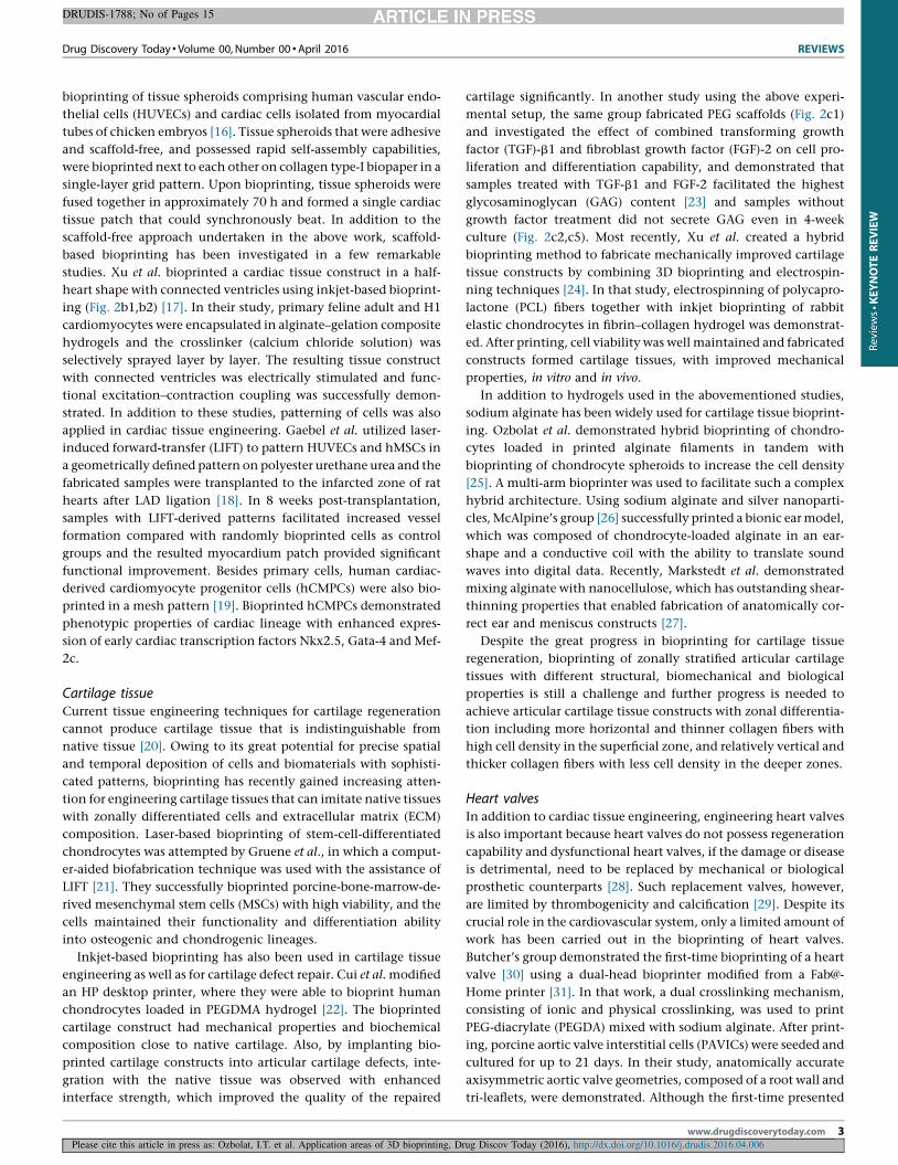
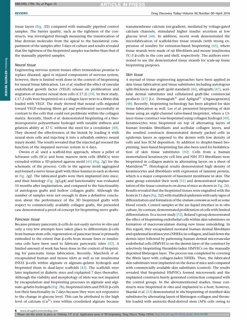
FIGURE 3
Bioprinted composite tissue constructs. (a1) A muscle-tendon unit frame made o
magnification view of (a2) printed PU filaments, (a3) filaments at the interface a
Bioprinted fibrocartilage samples (b2,b3) patterned by selectively depositing humdroplets loaded with either transforming growth factor (TGF)-b1 or bone morpho
crosslinked and droplets were demonstrated using Rhodamine B (red) and Dextr
Forgacs and co-workers followed a scaffold-free approach in bio-
printing vascular tissues, where tissue spheroids were bioprinted one
by one and self-assembled into larger tissue units [58]. Because
agarose is inert to cell adhesion, the printed agarose mold facilitated
rapid fusion of tissue spheroids and maturation of the tissue. In
addition to direct bioprinting of tubular vascular tissues, indirect
bioprinting of perfusable tissue constructs has been performed using
various hydrogels including fibrin [63], collagen [64] and GelMA
[36]. In an indirect approach, a fugitive-ink – that is dissolvable or
reversibly crosslinkable such as agarose [37], sugar [65], Pluronic1
[66] or gelatin [64] – was used to create open channels. Upon
removing the fugitive ink, endothelial cells were perfused and glued
to create endothelium within open channels [65]. This approach
enables bioprinting of highly complex vascular constructs that can
be perfused over a long time depending on the degradation profile of
the matrix. Although both approaches can be utilized, the former is
more appropriate in generating vascular grafts for transplantation
and the second is more appropriate for fabrication of perfusable
channels for in vitro tissue engineering applications [1].
Composite tissuesIn addition to single tissue types, efforts have been geared toward
bioprinting composite tissues to recapitulate the complex biology,
anatomy and functionality of organ-level structures. Merceron
et al. recently demonstrated fabrication of muscle-tendon units
by bioprinting hybrid constructs using a multi-head nozzle assem-
bly [67]. In this regard, PCL and polyurethane (PU) were 3D
printed to construct a frame to support cellular constructs, where
half of the unit was printed using PCL and the other half was
printed using PU (Fig. 3a1–a4). A composite hydrogel-based
bioink, comprising 3 mg/ml hyaluronic acid, 35 mg/ml gelatin
and 25 mg/ml fibrinogen in calcium-fee high-glucose Dulbecco’s
modified eagle medium (DMEM), was used to 3D bioprint 3T3
fibroblasts and myoblasts into the PCL and PU frames to construct
tendon and muscle units, respectively. The results revealed cell
ug Discov Today (2016), http://dx.doi.org/10.1016/j.drudis.2016.04.006
(b)4
1 mm
2
(b)3
GF-β1
MP-2
Drug Discovery Today
f PU (upper-half ) and polycaprolactone (PCL; lower-half ) at a higher
nd (a4) printed PCL filaments; adapted, with permission, from [67]. (b1)an mesenchymal stem cell (hMSC)-laden methacrylated gelatin (Me-Gel)
genetic protein (BMP)-2. (b4) Bioprinted precursor gel droplets were photo-
an-Alexa Fluor 488 (green); adapted, with permission, from [71].
www.drugdiscoverytoday.com 7
REVIEWS Drug Discovery Today � Volume 00, Number 00 �April 2016
DRUDIS-1788; No of Pages 15
Review
s�K
EYNOTEREVIEW
viability of >80% with differentiated cells at the end of a 7-day
culture. The final muscle-tendon units were elastic on the PU-
C1C12 muscle section with an elastic modulus of 0.39 � 0.05 MPa
and stiff on the PCL-3T3 tendon side with an elastic modulus of
46.47 � 2.67 MPa.
In addition to muscle-tendon units, bioprinting of osteochon-
dral models has been an interest in tissue engineering. Fedovorich
et al. demonstrated bioprinting of MSCs and chondrocytes using
alginate in a mesh pattern with two different cell types bioprinted
within two opposite ends of the scaffold [68]. Mesenchymal stem
cells were co-extruded with osteoinductive biphasic calcium phos-
phate particles, HA and b-tricalcium phosphate. The bioprinted
samples were cultured with a mixture of chondrogenic and osteo-
genic medium for 21 days. The results revealed that bioprinted
osteochondral tissue constructs demonstrated differentiated char-
acteristics of cells into osteogenic and chondrogenic lineages
along with related ECM deposition in vitro and in vivo. A similar
approach was performed by Park et al., where a systematic analysis
was performed to understand the effect of native ECM compo-
nents on the fate of osteoblasts and chondrocytes [69]. They
bioprinted osteoblasts in collagen type-I and chondrocytes in
hyaluronic acid, and compared the performance of bioprinted
osteoblasts and chondrocytes when they were bioprinted in hya-
luronic acid and collagen type-I, respectively. Fourteen-day cul-
ture in vitro suggested that osteochondral tissue regeneration could
be successfully attained when the proper hydrogel type was select-
ed. The same group demonstrated another osteochondral model
by depositing alginate hydrogel loaded with human chondrocytes
and human MG63 osteoblasts within a PCL frame [70]. Osteoblasts
and chondrocytes were supplemented with osteogenic and chon-
drogenic growth factors loaded in hydrogels for differentiation
purposes. Another approach was conducted using acoustic-based
bioprinting for the same purpose, where bioprinted nanodroplets
of MSCs in TGF-b1 and BMP-2 patterns (Fig. 3b1–b4) resulted in
localized differentiation of MSCs toward osteogenic and chondro-
genic lineages shown by the gene expression study [71].
In addition to osteochondral models, Yu et al. demonstrated
hybrid bioprinting of macrovascularized stromal tissue, where scaf-
fold-free tissue strands made of fibroblasts were assembled around a
perfusable macrovasculature loaded with smooth muscle cells ex-
truded through a co-axial nozzle unit [72]. Tissue strands quickly
fused and assembled around the macrovasculature in a week, which
can be further scaled-up by extending the macrovascular network.
Other tissue typesIn addition to the presented tissue types, there is some other work at
the early fundamental study level for bioprinting of retinal and
brain tissues. Lorber et al. presented piezoelectric inkjet bioprinting
of retinal ganglion cells (RGCs) and glia, and investigated the effect
of bioprinting parameters on the viability of cells and their growth-
promoting properties [73]. They concluded that inkjet bioprinting
did not adversely affect the cell viability and RGC neurite out-
growth, rather RGCs demonstrated further neurite growth when
bioprinted on a glial substrate. Recently, Lozano et al. presented
manual deposition of primary cortical neuron-laden gellan-gum-
RGD for brain-like tissue fabrication [74]. Three-layer constructed
tissue models, with cortical neurons encapsulated in the top and
bottom layers, demonstrated axon growth and penetration toward
Please cite this article in press as: Ozbolat, I.T. et al. Application areas of 3D bioprinting, D
8 www.drugdiscoverytoday.com
the cell-free middle layer in 5 days. Although no computer control
motion system was applied, the presented work unveiled the first
time demonstration of layer-by-layer fabrication for brain tissue
engineering.
Transplantation and clinicsBioprinting of living tissue and organ constructs has been widely
studied and performance of these constructs was assessed via
animal transplantation. Several bioprinted tissue types, including
nerve [41], cardiac [18], blood vessel [9], bone [11] and skin [52],
have been implanted into associated locations on animals to
evaluate their functionality, neovascularization and anastomosis
and engraftment with the host [1]. In addition, various tissue
constructs were bioprinted and implanted subcutaneously to as-
sess in vivo differentiation of cells and functionality of implanted
tissue constructs [68]. Despite these attempts, none of the bio-
printed tissues has been clinically used for humans because no
approval has been granted from the FDA yet. There are no regula-
tions laid down for bioprinters or bioprinted products; however,
with the increasing global interest and emerging businesses in the
growing bioprinting market, the success with the first technology
going through FDA regulations will be exemplary for preceding
technologies and products. For details of the regulatory concerns
of bioprinting, the reader is referred to our recent article [75].
Although bioprinting technology is still in its infancy in clinics,
3D-printed plastic, ceramic or metallic implants for bone tissue
replacement [76] have been successfully transplanted into humans.
In addition to permanent implants, a recent work published in New
England Journal of Medicine [77] demonstrated a unique case of
transplantation of a 3D-printed bioresorbable airway splint into
an infant. The institutional review board of the University of
Michigan consulted with the FDA and approved the use of a 3D-
printed device under the emergency-use exemption and the written
consent of the patient’s parents. No unforeseen problems have been
observed with the splint and full degradation of the device is
expected to take around 3 years. This was an exemplary case for
clinical use of 3D-printed scaffolds and hopefully will lead to similar
success with bioprinted tissues and organs. Despite the accomplish-
ments in bioprinting research, bioprinting for transplantation in a
clinical setting for humans requires further advances and transla-
tional efforts. Organs and tissues that do not need significant
vascularization (i.e. skin and cartilage) are expected to be translated
into clinical use sooner. Tissues and organs that are metabolically
highly active (i.e. heart, pancreas and liver) are immensely chal-
lenging. No bioprinting technology so far facilitated the fabrication
of a vascular hierarchical network spanning arteries and veins down
to capillaries. Because it is difficult to bioprint capillaries at the
submicron scale using the current technology, an alternative could
be to bioprint macrovasculature and then leave nature to create
capillaries by itself. Bioprinted vascular networks should be
designed and fabricated in a way that they can be easily sutured
to a blood vessel in a host, and possess certain properties such as
enough mechanical strength to satisfy burst pressure, sufficient
intactness of endothelium to prevent thrombosis and a high paten-
cy rate to support occlusion-free circulation [78].
The other approach in facilitating vascularization is in situ
bioprinting of tissue and organ constructs directly into the defect
sites in surgery settings rather than bioprinting tissue constructs,
rug Discov Today (2016), http://dx.doi.org/10.1016/j.drudis.2016.04.006
Drug Discovery Today � Volume 00, Number 00 �April 2016 REVIEWS
DRUDIS-1788; No of Pages 15
(a)
Lasersetup
1 Week 1 month 3 months3 mm 3 mm 3 mm
Calvariumdefect
Mouse
New tissueformation
Open defect(control) 1000 μm
(b)
(c)1 (c)2 (c)3
Drug Discovery Today
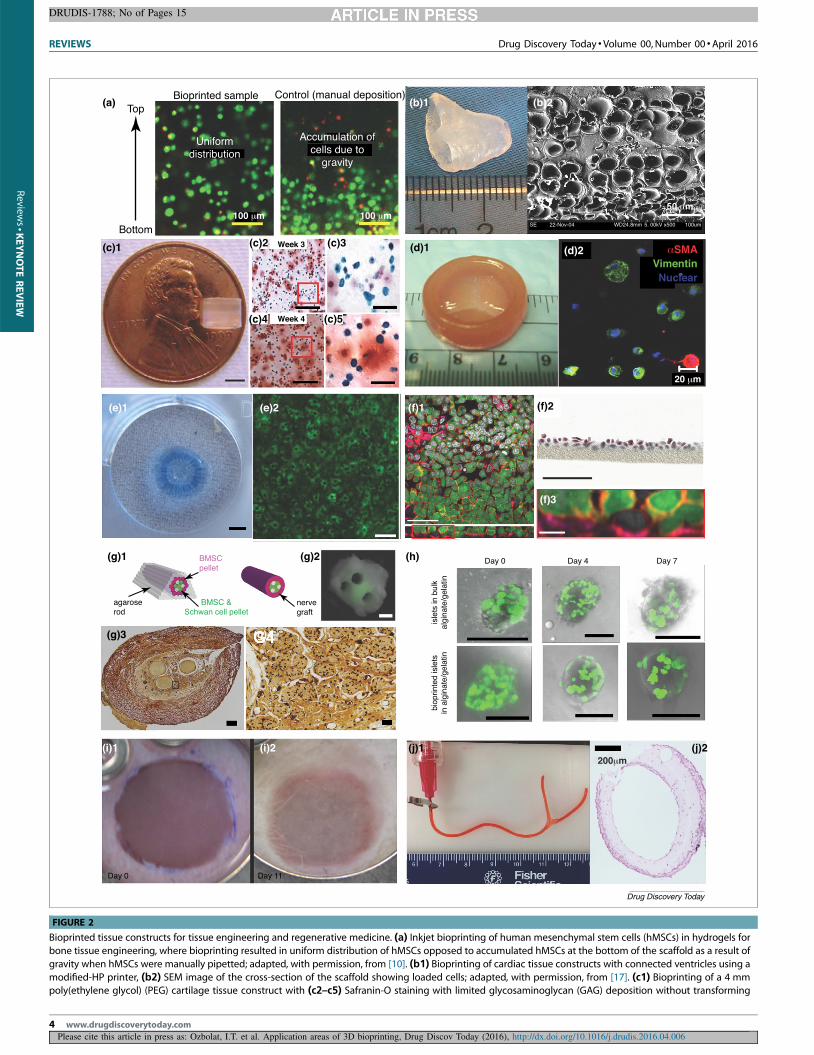
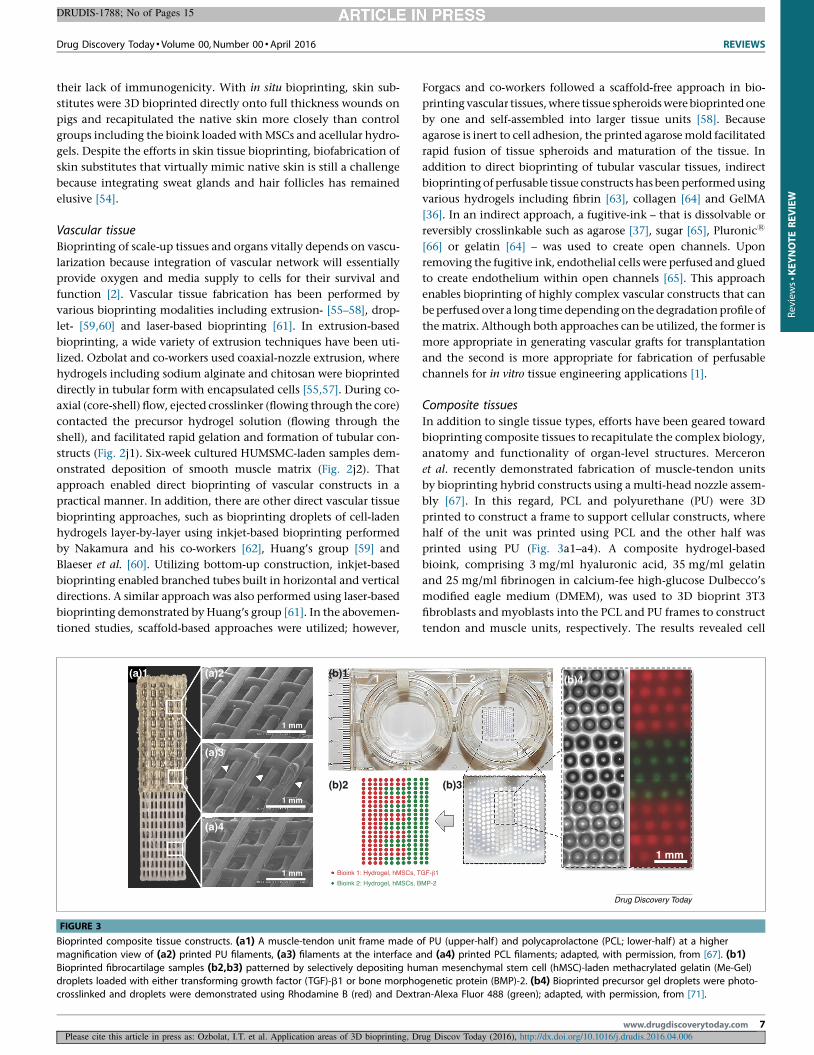
FIGURE 4
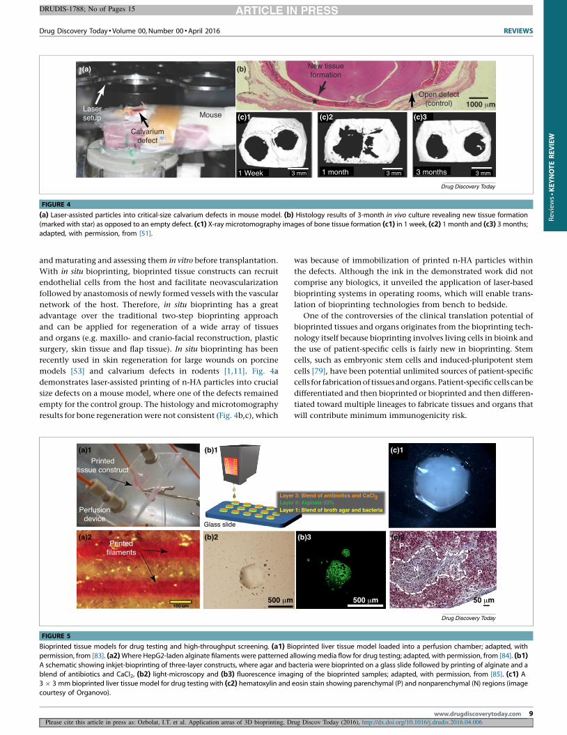
(a) Laser-assisted particles into critical-size calvarium defects in mouse model. (b) Histology results of 3-month in vivo culture revealing new tissue formation
(marked with star) as opposed to an empty defect. (c1) X-ray microtomography images of bone tissue formation (c1) in 1 week, (c2) 1 month and (c3) 3 months;
adapted, with permission, from [51].
Reviews�KEYNOTEREVIEW
and maturating and assessing them in vitro before transplantation.
With in situ bioprinting, bioprinted tissue constructs can recruit
endothelial cells from the host and facilitate neovascularization
followed by anastomosis of newly formed vessels with the vascular
network of the host. Therefore, in situ bioprinting has a great
advantage over the traditional two-step bioprinting approach
and can be applied for regeneration of a wide array of tissues
and organs (e.g. maxillo- and cranio-facial reconstruction, plastic
surgery, skin tissue and flap tissue). In situ bioprinting has been
recently used in skin regeneration for large wounds on porcine
models [53] and calvarium defects in rodents [1,11]. Fig. 4a
demonstrates laser-assisted printing of n-HA particles into crucial
size defects on a mouse model, where one of the defects remained
empty for the control group. The histology and microtomography
results for bone regeneration were not consistent (Fig. 4b,c), which
Please cite this article in press as: Ozbolat, I.T. et al. Application areas of 3D bioprinting, Dr
(a)1 (b)1
(a)2 (b)2
Glass slide
LayerLayerLayer
100 um500 μm
Printedtissue construct
Printedfilaments
Perfusiondevice
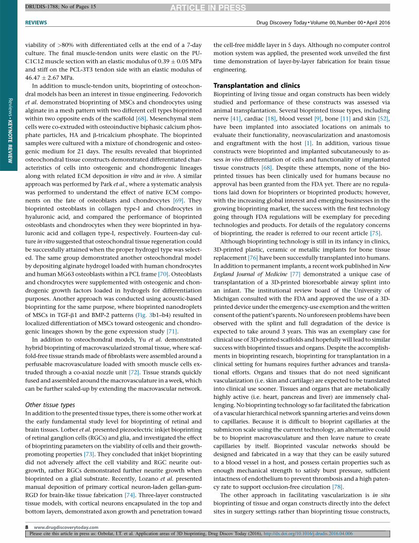
FIGURE 5
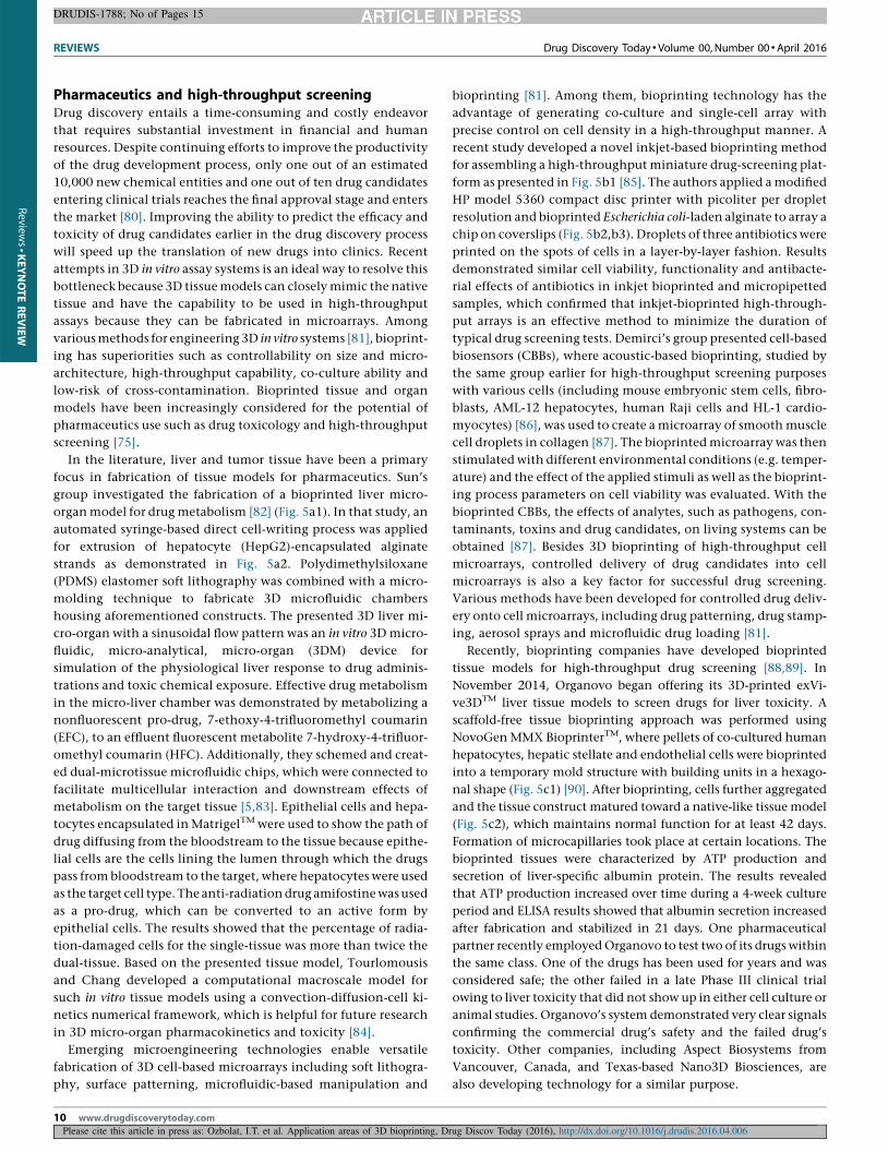
Bioprinted tissue models for drug testing and high-throughput screening. (a1) Bi
permission, from [83]. (a2) Where HepG2-laden alginate filaments were patterned a
A schematic showing inkjet-bioprinting of three-layer constructs, where agar and b
blend of antibiotics and CaCI2, (b2) light-microscopy and (b3) fluorescence imag3 � 3 mm bioprinted liver tissue model for drug testing with (c2) hematoxylin and e
courtesy of Organovo).
was because of immobilization of printed n-HA particles within
the defects. Although the ink in the demonstrated work did not
comprise any biologics, it unveiled the application of laser-based
bioprinting systems in operating rooms, which will enable trans-
lation of bioprinting technologies from bench to bedside.
One of the controversies of the clinical translation potential of
bioprinted tissues and organs originates from the bioprinting tech-
nology itself because bioprinting involves living cells in bioink and
the use of patient-specific cells is fairly new in bioprinting. Stem
cells, such as embryonic stem cells and induced-pluripotent stem
cells [79], have been potential unlimited sources of patient-specific
cells for fabrication of tissues and organs. Patient-specific cells can be
differentiated and then bioprinted or bioprinted and then differen-
tiated toward multiple lineages to fabricate tissues and organs that
will contribute minimum immunogenicity risk.
ug Discov Today (2016), http://dx.doi.org/10.1016/j.drudis.2016.04.006
3: Blend of antibiotics and CaCI2 2: Alginate 03% 1: Blend of broth agar and bacteria
50 μm500 μm
(b)3P
N P
(c)1
(c)2
Drug Discovery Today
oprinted liver tissue model loaded into a perfusion chamber; adapted, with
llowing media flow for drug testing; adapted, with permission, from [84]. (b1)acteria were bioprinted on a glass slide followed by printing of alginate and a
ing of the bioprinted samples; adapted, with permission, from [85]. (c1) Aosin stain showing parenchymal (P) and nonparenchymal (N) regions (image
www.drugdiscoverytoday.com 9
REVIEWS Drug Discovery Today � Volume 00, Number 00 �April 2016
DRUDIS-1788; No of Pages 15
Review
s�K
EYNOTEREVIEW
Pharmaceutics and high-throughput screeningDrug discovery entails a time-consuming and costly endeavor
that requires substantial investment in financial and human
resources. Despite continuing efforts to improve the productivity
of the drug development process, only one out of an estimated
10,000 new chemical entities and one out of ten drug candidates
entering clinical trials reaches the final approval stage and enters
the market [80]. Improving the ability to predict the efficacy and
toxicity of drug candidates earlier in the drug discovery process
will speed up the translation of new drugs into clinics. Recent
attempts in 3D in vitro assay systems is an ideal way to resolve this
bottleneck because 3D tissue models can closely mimic the native
tissue and have the capability to be used in high-throughput
assays because they can be fabricated in microarrays. Among
various methods for engineering 3D in vitro systems [81], bioprint-
ing has superiorities such as controllability on size and micro-
architecture, high-throughput capability, co-culture ability and
low-risk of cross-contamination. Bioprinted tissue and organ
models have been increasingly considered for the potential of
pharmaceutics use such as drug toxicology and high-throughput
screening [75].
In the literature, liver and tumor tissue have been a primary
focus in fabrication of tissue models for pharmaceutics. Sun’s
group investigated the fabrication of a bioprinted liver micro-
organ model for drug metabolism [82] (Fig. 5a1). In that study, an
automated syringe-based direct cell-writing process was applied
for extrusion of hepatocyte (HepG2)-encapsulated alginate
strands as demonstrated in Fig. 5a2. Polydimethylsiloxane
(PDMS) elastomer soft lithography was combined with a micro-
molding technique to fabricate 3D microfluidic chambers
housing aforementioned constructs. The presented 3D liver mi-
cro-organ with a sinusoidal flow pattern was an in vitro 3D micro-
fluidic, micro-analytical, micro-organ (3DM) device for
simulation of the physiological liver response to drug adminis-
trations and toxic chemical exposure. Effective drug metabolism
in the micro-liver chamber was demonstrated by metabolizing a
nonfluorescent pro-drug, 7-ethoxy-4-trifluoromethyl coumarin
(EFC), to an effluent fluorescent metabolite 7-hydroxy-4-trifluor-
omethyl coumarin (HFC). Additionally, they schemed and creat-
ed dual-microtissue microfluidic chips, which were connected to
facilitate multicellular interaction and downstream effects of
metabolism on the target tissue [5,83]. Epithelial cells and hepa-
tocytes encapsulated in MatrigelTM were used to show the path of
drug diffusing from the bloodstream to the tissue because epithe-
lial cells are the cells lining the lumen through which the drugs
pass from bloodstream to the target, where hepatocytes were used
as the target cell type. The anti-radiation drug amifostine was used
as a pro-drug, which can be converted to an active form by
epithelial cells. The results showed that the percentage of radia-
tion-damaged cells for the single-tissue was more than twice the
dual-tissue. Based on the presented tissue model, Tourlomousis
and Chang developed a computational macroscale model for
such in vitro tissue models using a convection-diffusion-cell ki-
netics numerical framework, which is helpful for future research
in 3D micro-organ pharmacokinetics and toxicity [84].
Emerging microengineering technologies enable versatile
fabrication of 3D cell-based microarrays including soft lithogra-
phy, surface patterning, microfluidic-based manipulation and
Please cite this article in press as: Ozbolat, I.T. et al. Application areas of 3D bioprinting, D
10 www.drugdiscoverytoday.com
bioprinting [81]. Among them, bioprinting technology has the
advantage of generating co-culture and single-cell array with
precise control on cell density in a high-throughput manner. A
recent study developed a novel inkjet-based bioprinting method
for assembling a high-throughput miniature drug-screening plat-
form as presented in Fig. 5b1 [85]. The authors applied a modified
HP model 5360 compact disc printer with picoliter per droplet
resolution and bioprinted Escherichia coli-laden alginate to array a
chip on coverslips (Fig. 5b2,b3). Droplets of three antibiotics were
printed on the spots of cells in a layer-by-layer fashion. Results
demonstrated similar cell viability, functionality and antibacte-
rial effects of antibiotics in inkjet bioprinted and micropipetted
samples, which confirmed that inkjet-bioprinted high-through-
put arrays is an effective method to minimize the duration of
typical drug screening tests. Demirci’s group presented cell-based
biosensors (CBBs), where acoustic-based bioprinting, studied by
the same group earlier for high-throughput screening purposes
with various cells (including mouse embryonic stem cells, fibro-
blasts, AML-12 hepatocytes, human Raji cells and HL-1 cardio-
myocytes) [86], was used to create a microarray of smooth muscle
cell droplets in collagen [87]. The bioprinted microarray was then
stimulated with different environmental conditions (e.g. temper-
ature) and the effect of the applied stimuli as well as the bioprint-
ing process parameters on cell viability was evaluated. With the
bioprinted CBBs, the effects of analytes, such as pathogens, con-
taminants, toxins and drug candidates, on living systems can be
obtained [87]. Besides 3D bioprinting of high-throughput cell
microarrays, controlled delivery of drug candidates into cell
microarrays is also a key factor for successful drug screening.
Various methods have been developed for controlled drug deliv-
ery onto cell microarrays, including drug patterning, drug stamp-
ing, aerosol sprays and microfluidic drug loading [81].
Recently, bioprinting companies have developed bioprinted
tissue models for high-throughput drug screening [88,89]. In
November 2014, Organovo began offering its 3D-printed exVi-
ve3DTM liver tissue models to screen drugs for liver toxicity. A
scaffold-free tissue bioprinting approach was performed using
NovoGen MMX BioprinterTM, where pellets of co-cultured human
hepatocytes, hepatic stellate and endothelial cells were bioprinted
into a temporary mold structure with building units in a hexago-
nal shape (Fig. 5c1) [90]. After bioprinting, cells further aggregated
and the tissue construct matured toward a native-like tissue model
(Fig. 5c2), which maintains normal function for at least 42 days.
Formation of microcapillaries took place at certain locations. The
bioprinted tissues were characterized by ATP production and
secretion of liver-specific albumin protein. The results revealed
that ATP production increased over time during a 4-week culture
period and ELISA results showed that albumin secretion increased
after fabrication and stabilized in 21 days. One pharmaceutical
partner recently employed Organovo to test two of its drugs within
the same class. One of the drugs has been used for years and was
considered safe; the other failed in a late Phase III clinical trial
owing to liver toxicity that did not show up in either cell culture or
animal studies. Organovo’s system demonstrated very clear signals
confirming the commercial drug’s safety and the failed drug’s
toxicity. Other companies, including Aspect Biosystems from
Vancouver, Canada, and Texas-based Nano3D Biosciences, are
also developing technology for a similar purpose.
rug Discov Today (2016), http://dx.doi.org/10.1016/j.drudis.2016.04.006
Drug
Disco
very
Today�Volume
00,
Number
00�A
pril
2016
REVIEWS
DR
UD
IS-1
78
8;
No
of
Pag
es 1
5
Please
cite th
is article
in p
ress as:
Ozb
olat,
I.T.
et al.
Applicatio
n areas
of
3D
bio
prin
ting,
Dru
g D
iscov
Today
(2016),
http
://dx.d
oi.o
rg/1
0.1
016/j.d
rudis.2
016.0
4.0
06
TABLE 1
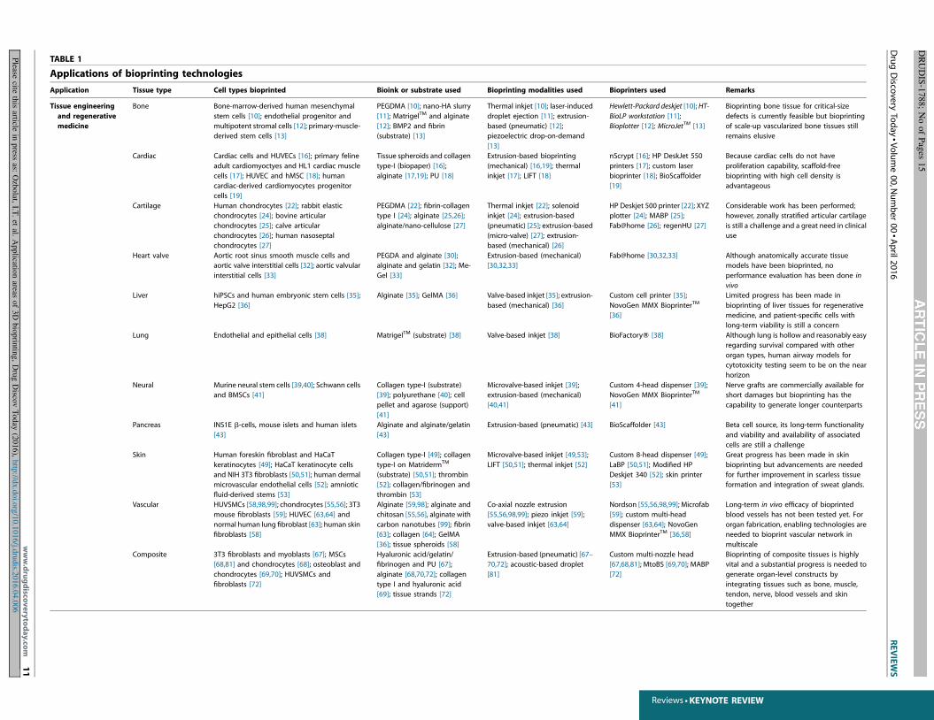
Applications of bioprinting technologies
Application Tissue type Cell types bioprinted Bioink or substrate used Bioprinting modalities used Bioprinters used Remarks
Tissue engineering
and regenerative
medicine
Bone Bone-marrow-derived human mesenchymal
stem cells [10]; endothelial progenitor and
multipotent stromal cells [12]; primary-muscle-
derived stem cells [13]
PEGDMA [10]; nano-HA slurry
[11]; MatrigelTM and alginate
[12]; BMP2 and fibrin
(substrate) [13]
Thermal inkjet [10]; laser-induced
droplet ejection [11]; extrusion-
based (pneumatic) [12];
piezoelectric drop-on-demand
[13]
Hewlett-Packard deskjet [10]; HT-
BioLP workstation [11];
Bioplotter [12]; MicroJetTM [13]
Bioprinting bone tissue for critical-size
defects is currently feasible but bioprinting
of scale-up vascularized bone tissues still
remains elusive
Cardiac Cardiac cells and HUVECs [16]; primary feline
adult cardiomyoctyes and HL1 cardiac muscle
cells [17]; HUVEC and hMSC [18]; human
cardiac-derived cardiomyocytes progenitor
cells [19]
Tissue spheroids and collagen
type-I (biopaper) [16];
alginate [17,19]; PU [18]
Extrusion-based bioprinting
(mechanical) [16,19]; thermal
inkjet [17]; LIFT [18]
nScrypt [16]; HP DeskJet 550
printers [17]; custom laser
bioprinter [18]; BioScaffolder
[19]
Because cardiac cells do not have
proliferation capability, scaffold-free
bioprinting with high cell density is
advantageous
Cartilage Human chondrocytes [22]; rabbit elastic
chondrocytes [24]; bovine articular
chondrocytes [25]; calve articular
chondrocytes [26]; human nasoseptal
chondrocytes [27]
PEGDMA [22]; fibrin-collagen
type I [24]; alginate [25,26];
alginate/nano-cellulose [27]
Thermal inkjet [22]; solenoid
inkjet [24]; extrusion-based
(pneumatic) [25]; extrusion-based
(micro-valve) [27]; extrusion-
based (mechanical) [26]
HP Deskjet 500 printer [22]; XYZ
plotter [24]; MABP [25];
Fab@home [26]; regenHU [27]
Considerable work has been performed;
however, zonally stratified articular cartilage
is still a challenge and a great need in clinical
use
Heart valve Aortic root sinus smooth muscle cells and
aortic valve interstitial cells [32]; aortic valvular
interstitial cells [33]
PEGDA and alginate [30];
alginate and gelatin [32]; Me-
Gel [33]
Extrusion-based (mechanical)
[30,32,33]
Fab@home [30,32,33] Although anatomically accurate tissue
models have been bioprinted, no
performance evaluation has been done in
vivo
Liver hiPSCs and human embryonic stem cells [35];
HepG2 [36]
Alginate [35]; GelMA [36] Valve-based inkjet [35]; extrusion-
based (mechanical) [36]
Custom cell printer [35];
NovoGen MMX BioprinterTM
[36]
Limited progress has been made in
bioprinting of liver tissues for regenerative
medicine, and patient-specific cells with
long-term viability is still a concern
Lung Endothelial and epithelial cells [38] MatrigelTM (substrate) [38] Valve-based inkjet [38] BioFactoryW [38] Although lung is hollow and reasonably easy
regarding survival compared with other
organ types, human airway models for
cytotoxicity testing seem to be on the near
horizon
Neural Murine neural stem cells [39,40]; Schwann cells
and BMSCs [41]
Collagen type-I (substrate)
[39]; polyurethane [40]; cell
pellet and agarose (support)
[41]
Microvalve-based inkjet [39];
extrusion-based (mechanical)
[40,41]
Custom 4-head dispenser [39];
NovoGen MMX BioprinterTM
[41]
Nerve grafts are commercially available for
short damages but bioprinting has the
capability to generate longer counterparts
Pancreas INS1E b-cells, mouse islets and human islets
[43]
Alginate and alginate/gelatin
[43]
Extrusion-based (pneumatic) [43] BioScaffolder [43] Beta cell source, its long-term functionality
and viability and availability of associated
cells are still a challenge
Skin Human foreskin fibroblast and HaCaT
keratinocytes [49]; HaCaT keratinocyte cells
and NIH 3T3 fibroblasts [50,51]; human dermal
microvascular endothelial cells [52]; amniotic
fluid-derived stems [53]
Collagen type-I [49]; collagen
type-I on MatridermTM
(substrate) [50,51]; thrombin
[52]; collagen/fibrinogen and
thrombin [53]
Microvalve-based inkjet [49,53];
LIFT [50,51]; thermal inkjet [52]
Custom 8-head dispenser [49];
LaBP [50,51]; Modified HP
Deskjet 340 [52]; skin printer
[53]
Great progress has been made in skin
bioprinting but advancements are needed
for further improvement in scarless tissue
formation and integration of sweat glands.
Vascular HUVSMCs [58,98,99]; chondrocytes [55,56]; 3T3
mouse fibroblasts [59]; HUVEC [63,64] and
normal human lung fibroblast [63]; human skin
fibroblasts [58]
Alginate [59,98]; alginate and
chitosan [55,56], alginate with
carbon nanotubes [99]; fibrin
[63]; collagen [64]; GelMA
[36]; tissue spheroids [58]
Co-axial nozzle extrusion
[55,56,98,99]; piezo inkjet [59];
valve-based inkjet [63,64]
Nordson [55,56,98,99]; Microfab
[59]; custom multi-head
dispenser [63,64]; NovoGen
MMX BioprinterTM [36,58]
Long-term in vivo efficacy of bioprinted
blood vessels has not been tested yet. For
organ fabrication, enabling technologies are
needed to bioprint vascular network in
multiscale
Composite 3T3 fibroblasts and myoblasts [67]; MSCs
[68,81] and chondrocytes [68]; osteoblast and
chondrocytes [69,70]; HUVSMCs and
fibroblasts [72]
Hyaluronic acid/gelatin/
fibrinogen and PU [67];
alginate [68,70,72]; collagen
type I and hyaluronic acid
[69]; tissue strands [72]
Extrusion-based (pneumatic) [67–
70,72]; acoustic-based droplet
[81]
Custom multi-nozzle head
[67,68,81]; MtoBS [69,70]; MABP
[72]
Bioprinting of composite tissues is highly
vital and a substantial progress is needed to
generate organ-level constructs by
integrating tissues such as bone, muscle,
tendon, nerve, blood vessels and skin
together
www.drugdisco
verytoday.co
m
11
Reviews �KEYNOTE REVIEW
REVIEWS Drug Discovery Today � Volume 00, Number 00 �April 2016
DRUDIS-1788; No of Pages 15
Please cite this article in press as: Ozbolat, I.T. et al. Application areas of 3D bioprinting, D
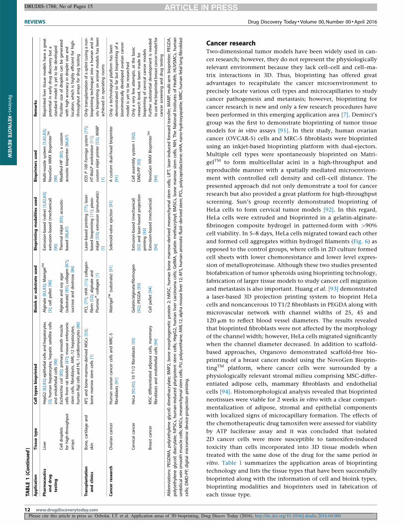
TABLE1(Continued
)
Application
Tissuetype
Celltypesbioprinted
Bioinkorsubstrate
used
Bioprintingmodalitiesused
Bioprinters
used
Remarks
Pharm
aceutics
anddrug
testing
Liver
Hep
G2[82,83];ep
ithelialcellsan
dhep
atocytes
[5];human
hep
atocytes,hep
atic
satellite
cells
andendothelialcells
[90]
Alginate[82,83];MatrigelTM
[5];cellpellet[90]
Extrusion-based
(valve)[5,82,83];
extrusion-based
(mechan
ical)
[90]
Multi-nozzle
system
[5,82,83];
Novo
Gen
MMXBioprinterTM
[90]
Bioprintedliver
tissuemodelshaveagreat
potential
inearlydrugdiscovery
buta
stan
dardmodel
isyetto
bedeveloped
Celldroplets
forhigh-throughput
arrays
Escherichia
coli[85];primarysm
ooth
muscle
cells
from
ratbladder
[87];mouse
embryonic
stem
cells,fibroblasts,AML-12hep
atocytes,
human
Rajicellsan
dHL-1cardiomyo
cytes[86]
Alginatean
dsoyag
ar
(substrate)[85];collagen
[87];
sucrose
anddextrose
[86]
Thermal
inkjet
[85];acoustic-
based
[86,87]
Modified
-HP[85];acustom
acoustic
bioprinter[86,87]
Picoliter
size
ofdroplets
canbegenerated
withhighaccuracy
indropletsize
and
location,whichishighly
efficientforhigh-
throughputarrays
fordrugtesting
Transp
lantation
andclinics
Bone,
cartilagean
d
skin
AFS
andbone-marrow-derived
MSC
s[53];
bonemarrow
stem
cells
[1]
PCL[77];nHA[11];collagen
-
fibrin[53];alginatean
d
Pluronic
W-collagen
[1]
Laser-based
printing[77];laser-
based
bioprinting[11];piezo-
inkjet
[53];extrusion(pneu
matic)
[1]
EOSP100Form
igasystem
[77];
HT-BioLP
workstation[11];
custom
inkjet
printer[53];MABP
[1]
Onlytran
splantationofasplin
t(usinganon-
bioprintingtechnique)into
ahuman
andin
situ
bioprintingonan
imal
modelshas
been
achievedin
operatingrooms
Cancerresearch
Ovarian
cancer
Human
ovarian
cancercells
andMRC-5
fibroblasts[91]
MatrigelTM(substrate)[91]
Solenoid-valve
ejection[91]
Acustom
dual-headbioprinter
[91]
Only
atechnological
platform
has
been
dem
onstratedso
farbutbioprintingofa
biomim
etically
developedovarian
cancer
model
isyetto
beresearched
Cervicalcancer
HeLa
[92,93];10T1/2
fibroblasts[93]
Gelatin/alginate/fibrinogen
[92];PEG
DA[93]
Extrusion-based
(mechan
ical)
[92]an
dlaser-based
projection
printing[93]
Cellassembly
system
I[92];
DMD-PP[93]
Only
avery
few
attempts,at
thebasic
research
level,havebeenmad
efor
bioprintingofcervical
cancermodels
Breastcancer
MSC
differentiated
adipose
cells,mam
mary
fibroblastsan
den
dothelialcells
[94]
Cellpellet[94]
Extrusion-based
(mechan
ical)
[94]
Novo
Gen
MMXBioprinterTM
[94]
Further
substan
tial
developmentisneeded
touse
thebioprintedbreastcancerm
odelfor
cancerscreen
ingan
ddrugtesting
Abbreviations:PEG
DMA,poly(ethylen
eglycol)dim
ethacrylate;BMP2,b
onemorphogen
eticprotein
2;h
MSC
,human
bonemarrow-derived
mesen
chym
alstem
cells;LIFT,laser-inducedforw
ardtran
sfer;M
ABP,multi-arm
bioprinter;PEG
DA,
poly(ethyleneglycol)diacrylate;hiPSC
s,human
-inducedpluripotentstem
cells;Hep
G2,human
liver
carcinomacells;GelMA,gelatin
methacryloyl;BMSC
s,bonemarrow
stem
cells;NIH,Th
eNational
InstitutesofHealth;HUVSM
Cs,human
umbilicalveinsm
ooth
musclecells;M
SCs,mesen
chym
alstem
cells;PU,polyurethan
e;AML12,alphamouse
liver
12;A
FS,human
amnioticfluid–d
erived
stem
;PCL,polycaprolactone;nHA,nan
o-hyd
roxyap
atite;MRC,human
fetallungfibroblast
cells;DMD-PP,digital
micromirrordevice-projectionprinting.
12 www.drugdiscoverytoday.com
Review
s�K
EYNOTEREVIEW
Cancer researchTwo-dimensional tumor models have been widely used in can-
cer research; however, they do not represent the physiologically
relevant environment because they lack cell–cell and cell–ma-
trix interactions in 3D. Thus, bioprinting has offered great
advantages to recapitulate the cancer microenvironment to
precisely locate various cell types and microcapillaries to study
cancer pathogenesis and metastasis; however, bioprinting for
cancer research is new and only a few research procedures have
been performed in this emerging application area [7]. Demirci’s
group was the first to demonstrate bioprinting of tumor tissue
models for in vitro assays [91]. In their study, human ovarian
cancer (OVCAR-5) cells and MRC-5 fibroblasts were bioprinted
using an inkjet-based bioprinting platform with dual-ejectors.
Multiple cell types were spontaneously bioprinted on Matri-
gelTM to form multicellular acini in a high-throughput and
reproducible manner with a spatially mediated microenviron-
ment with controlled cell density and cell–cell distance. The
presented approach did not only demonstrate a tool for cancer
research but also provided a great platform for high-throughput
screening. Sun’s group recently demonstrated bioprinting of
HeLa cells to form cervical tumor models [92]. In this regard,
HeLa cells were extruded and bioprinted in a gelatin-alginate-
fibrinogen composite hydrogel in patterned-form with >90%
cell viability. In 5–8 days, HeLa cells migrated toward each other
and formed cell aggregates within hydrogel filaments (Fig. 6) as
opposed to the control groups, where cells in 2D culture formed
cell sheets with lower chemoresistance and lower level expres-
sion of metalloproteinase. Although these two studies presented
biofabrication of tumor spheroids using bioprinting technology,
fabrication of larger tissue models to study cancer cell migration
and metastasis is also important. Huang et al. [93] demonstrated
a laser-based 3D projection printing system to bioprint HeLa
cells and noncancerous 10 T1/2 fibroblasts in PEGDA along with
microvascular network with channel widths of 25, 45 and
120 mm to reflect blood vessel diameters. The results revealed
that bioprinted fibroblasts were not affected by the morphology
of the channel width; however, HeLa cells migrated significantly
when the channel diameter decreased. In addition to scaffold-
based approaches, Organovo demonstrated scaffold-free bio-
printing of a breast cancer model using the NovoGen Bioprin-
tingTM platform, where cancer cells were surrounded by a
physiologically relevant stromal milieu comprising MSC-differ-
entiated adipose cells, mammary fibroblasts and endothelial
cells [94]. Histomorphological analysis revealed that bioprinted
neotissues were viable for 2 weeks in vitro with a clear compart-
mentalization of adipose, stromal and epithelial components
with localized signs of microcapillary formation. The effects of
the chemotherapeutic drug tamoxifen were assessed for viability
by ATP luciferase assay and it was concluded that isolated
2D cancer cells were more susceptible to tamoxifen-induced
toxicity than cells incorporated into 3D tissue models when
treated with the same dose of the drug for the same period in
vitro. Table 1 summarizes the application areas of bioprinting
technology and lists the tissue types that have been successfully
bioprinted along with the information of cell and bioink types,
bioprinting modalities and bioprinters used in fabrication of
each tissue type.
rug Discov Today (2016), http://dx.doi.org/10.1016/j.drudis.2016.04.006
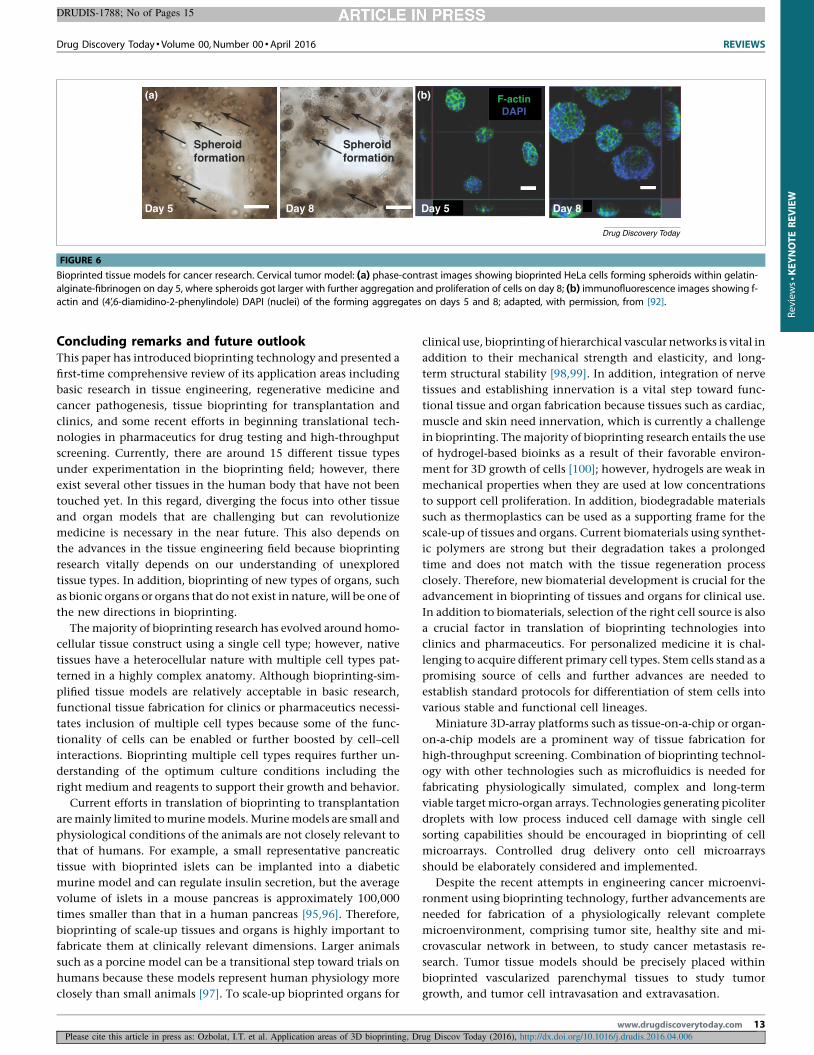
Drug Discovery Today � Volume 00, Number 00 �April 2016 REVIEWS
DRUDIS-1788; No of Pages 15
(a)
Day 5 Day 8
Spheroidformation
Spheroidformation
Day 5
F-actinDAPI
Day 8
(b)
Drug Discovery Today
FIGURE 6
Bioprinted tissue models for cancer research. Cervical tumor model: (a) phase-contrast images showing bioprinted HeLa cells forming spheroids within gelatin-
alginate-fibrinogen on day 5, where spheroids got larger with further aggregation and proliferation of cells on day 8; (b) immunofluorescence images showing f-
actin and (4’,6-diamidino-2-phenylindole) DAPI (nuclei) of the forming aggregates on days 5 and 8; adapted, with permission, from [92].
Reviews�KEYNOTEREVIEW
Concluding remarks and future outlookThis paper has introduced bioprinting technology and presented a
first-time comprehensive review of its application areas including
basic research in tissue engineering, regenerative medicine and
cancer pathogenesis, tissue bioprinting for transplantation and
clinics, and some recent efforts in beginning translational tech-
nologies in pharmaceutics for drug testing and high-throughput
screening. Currently, there are around 15 different tissue types
under experimentation in the bioprinting field; however, there
exist several other tissues in the human body that have not been
touched yet. In this regard, diverging the focus into other tissue
and organ models that are challenging but can revolutionize
medicine is necessary in the near future. This also depends on
the advances in the tissue engineering field because bioprinting
research vitally depends on our understanding of unexplored
tissue types. In addition, bioprinting of new types of organs, such
as bionic organs or organs that do not exist in nature, will be one of
the new directions in bioprinting.
The majority of bioprinting research has evolved around homo-
cellular tissue construct using a single cell type; however, native
tissues have a heterocellular nature with multiple cell types pat-
terned in a highly complex anatomy. Although bioprinting-sim-
plified tissue models are relatively acceptable in basic research,
functional tissue fabrication for clinics or pharmaceutics necessi-
tates inclusion of multiple cell types because some of the func-
tionality of cells can be enabled or further boosted by cell–cell
interactions. Bioprinting multiple cell types requires further un-
derstanding of the optimum culture conditions including the
right medium and reagents to support their growth and behavior.
Current efforts in translation of bioprinting to transplantation
are mainly limited to murine models. Murine models are small and
physiological conditions of the animals are not closely relevant to
that of humans. For example, a small representative pancreatic
tissue with bioprinted islets can be implanted into a diabetic
murine model and can regulate insulin secretion, but the average
volume of islets in a mouse pancreas is approximately 100,000
times smaller than that in a human pancreas [95,96]. Therefore,
bioprinting of scale-up tissues and organs is highly important to
fabricate them at clinically relevant dimensions. Larger animals
such as a porcine model can be a transitional step toward trials on
humans because these models represent human physiology more
closely than small animals [97]. To scale-up bioprinted organs for
Please cite this article in press as: Ozbolat, I.T. et al. Application areas of 3D bioprinting, Dr
clinical use, bioprinting of hierarchical vascular networks is vital in
addition to their mechanical strength and elasticity, and long-
term structural stability [98,99]. In addition, integration of nerve
tissues and establishing innervation is a vital step toward func-
tional tissue and organ fabrication because tissues such as cardiac,
muscle and skin need innervation, which is currently a challenge
in bioprinting. The majority of bioprinting research entails the use
of hydrogel-based bioinks as a result of their favorable environ-
ment for 3D growth of cells [100]; however, hydrogels are weak in
mechanical properties when they are used at low concentrations
to support cell proliferation. In addition, biodegradable materials
such as thermoplastics can be used as a supporting frame for the
scale-up of tissues and organs. Current biomaterials using synthet-
ic polymers are strong but their degradation takes a prolonged
time and does not match with the tissue regeneration process
closely. Therefore, new biomaterial development is crucial for the
advancement in bioprinting of tissues and organs for clinical use.
In addition to biomaterials, selection of the right cell source is also
a crucial factor in translation of bioprinting technologies into
clinics and pharmaceutics. For personalized medicine it is chal-
lenging to acquire different primary cell types. Stem cells stand as a
promising source of cells and further advances are needed to
establish standard protocols for differentiation of stem cells into
various stable and functional cell lineages.
Miniature 3D-array platforms such as tissue-on-a-chip or organ-
on-a-chip models are a prominent way of tissue fabrication for
high-throughput screening. Combination of bioprinting technol-
ogy with other technologies such as microfluidics is needed for
fabricating physiologically simulated, complex and long-term
viable target micro-organ arrays. Technologies generating picoliter
droplets with low process induced cell damage with single cell
sorting capabilities should be encouraged in bioprinting of cell
microarrays. Controlled drug delivery onto cell microarrays
should be elaborately considered and implemented.
Despite the recent attempts in engineering cancer microenvi-
ronment using bioprinting technology, further advancements are
needed for fabrication of a physiologically relevant complete
microenvironment, comprising tumor site, healthy site and mi-
crovascular network in between, to study cancer metastasis re-
search. Tumor tissue models should be precisely placed within
bioprinted vascularized parenchymal tissues to study tumor
growth, and tumor cell intravasation and extravasation.
ug Discov Today (2016), http://dx.doi.org/10.1016/j.drudis.2016.04.006
www.drugdiscoverytoday.com 13
REVIEWS Drug Discovery Today � Volume 00, Number 00 �April 2016
DRUDIS-1788; No of Pages 15
Review
s�K
EYNOTEREVIEW
AcknowledgementsThis work has been supported by National Science Foundation
CMMI Awards 1349716 and 1462232, Diabetes in Action Research
Please cite this article in press as: Ozbolat, I.T. et al. Application areas of 3D bioprinting, D
14 www.drugdiscoverytoday.com
and Education Foundation grant #426 and the China Scholarship
Council 201308360128.
References
1 Ozbolat, I.T. (2015) Bioprinting scale-up tissue and organ constructs for
transplantation. Trends Biotechnol. 33, 395–400
2 Ozbolat, I.T. (2015) Scaffold-based or scaffold-free bioprinting: competing or
complementing approaches? J. Nanotechnol. Eng. Med. 6, 24701
3 Jakab, K. et al. (2010) Tissue engineering by self-assembly and bio-printing of living
cells. Biofabrication 2, 22001
4 Moroni, L. et al. (2006) 3D fiber-deposited scaffolds for tissue engineering:
influence of pores geometry and architecture on dynamic mechanical properties.
Biomaterials 27, 974–985
5 Snyder, J.E. et al. (2011) Bioprinting cell-laden matrigel for radioprotection study
of liver by pro-drug conversion in a dual-tissue microfluidic chip. Biofabrication 3,
034112
6 Perkins, J.D. (2007) Are we reporting the same thing? Liver Transpl. 13, 465–466
7 Knowlton, S. et al. (2015) Bioprinting for cancer research. Trends Biotechnol. 33,
504–513
8 Ozbolat, I.T. and Yu, Y. (2013) Bioprinting toward organ fabrication: challenges
and future trends. IEEE Trans. Biomed. Eng. 60, 691–699
9 Itoh, M. et al. (2015) Correction: scaffold-free tubular tissues created by a Bio-3D
printer undergo remodeling and endothelialization when implanted in rat aortae.
PLoS ONE 10, e0145971
10 Gao, G. et al. (2014) Bioactive nanoparticles stimulate bone tissue formation in
bioprinted three-dimensional scaffold and human mesenchymal stem cells.
Biotechnol. J. 9, 1304–1311
11 Keriquel, V. et al. (2010) In vivo bioprinting for computer- and robotic-assisted
medical intervention: preliminary study in mice. Biofabrication 2, 14101
12 Fedorovich, N.E. et al. (2008) Three-dimensional fiber deposition of
cell-laden, viable, patterned constructs for bone tissue printing. Tissue Eng. A
14, 127–133
13 Phillippi, J.A. et al. (2008) Microenvironments engineered by inkjet bioprinting
spatially direct adult stem cells toward muscle- and bone-like subpopulations. Stem
Cells 26, 127–134
14 Hirt, M.N. et al. (2014) Cardiac tissue engineering: state of the art. Circ. Res. 114,
354–367
15 Eulalio, A. et al. (2012) Functional screening identifies miRNAs inducing cardiac
regeneration. Nature 492, 376–381
16 Jakab, K. et al. (2008) Tissue engineering by self-assembly of cells printed into
topologically defined structures. Tissue Eng. A 14, 413–421
17 Xu, T. et al. (2009) Fabrication and characterization of bio-engineered cardiac
pseudo tissues. Biofabrication 1, 35001
18 Gaebel, R. et al. (2011) Patterning human stem cells and endothelial cells with laser
printing for cardiac regeneration. Biomaterials 32, 9218–9230
19 Gaetani, R. et al. (2012) Cardiac tissue engineering using tissue printing
technology and human cardiac progenitor cells. Biomaterials 33, 1782–1790
20 Makris, E.A. et al. (2015) Repair and tissue engineering techniques for articular
cartilage. Nat. Rev. Rheumatol. 11, 21–34
21 Gruene, M. et al. (2010) Laser printing of stem cells for biofabrication of scaffold-
free autologous grafts. Tissue Eng. C: Methods 17, 79–87
22 Cui, X. et al. (2012) Direct human cartilage repair using three-dimensional
bioprinting technology. Tissue Eng. A 18, 1304–1312
23 Cui, X. et al. (2012) Synergistic action of fibroblast growth factor-2 and
transforming growth factor-beta1 enhances bioprinted human neocartilage
formation. Biotechnol. Bioeng. 109, 2357–2368
24 Xu, T.X. et al. (2013) Hybrid printing of mechanically and biologically
improved constructs for cartilage tissue engineering applications. Biofabrication 5,
15001
25 Ozbolat, I.T. et al. (2014) Development of ‘Multi-arm Bioprinter’ for hybrid
biofabrication of tissue engineering constructs. Robot. Comput. Integr. Manuf. 30,
295–304
26 Mannoor, M.S. et al. (2013) 3D printed bionic ears. Nano Lett. 13, 2634–2639
27 Markstedt, K. et al. (2015) 3D bioprinting human chondrocytes with
nanocellulose–alginate bioink for cartilage tissue engineering applications.
Biomacromolecules 16, 1489–1496
28 Hockaday, L.A. et al. (2014) 3D-printed hydrogel technologies for tissue-
engineered heart valves. 3D Print. Addit. Manuf. 1, 122–136
29 Jana, S. and Lerman, A. (2015) Bioprinting a cardiac valve. Biotechnol. Adv. 33,
1503–1521
30 Hockaday, L.A. et al. (2012) Rapid 3D printing of anatomically accurate and
mechanically heterogeneous aortic valve hydrogel scaffolds. Biofabrication 4,
35005
31 Malone, E. and Lipson, H. (2007) Fab@Home: the personal desktop fabricator
kitnull. Rapid Prototyp. J. 13, 245–255
32 Duan, B. et al. (2013) 3D bioprinting of heterogeneous aortic valve conduits with
alginate/gelatin hydrogels. J. Biomed. Mater. Res. Part A 101A, 1255–1264
33 Duan, B. et al. (2014) Three-dimensional printed trileaflet valve conduits
using biological hydrogels and human valve interstitial cells. Acta Biomater. 10,
1836–1846
34 No, D.Y. et al. (2015) 3D liver models on a microplatform: well-defined culture,
engineering of liver tissue and liver-on-a-chip. Lab Chip 15, 3822–3837
35 Faulkner-Jones, A. et al. (2015) Bioprinting of human pluripotent stem cells and
their directed differentiation into hepatocyte-like cells for the generation of mini-
livers in 3D. Biofabrication 7, 44102
36 Bertassoni, L.E. et al. (2014) Direct-write bioprinting of cell-laden methacrylated
gelatin hydrogels. Biofabrication 6, 024105
37 Bertassoni, L.E. et al. (2014) Hydrogel bioprinted microchannel networks for
vascularization of tissue engineering constructs. Lab Chip 14, 2202–2211
38 Horvath, L. et al. (2015) Engineering an in vitro air–blood barrier by 3D bioprinting.
Sci. Rep. 5, 7974
39 Lee, Y-B. et al. (2010) Bio-printing of collagen and VEGF-releasing fibrin gel
scaffolds for neural stem cell culture. Exp. Neurol. 223, 645–652
40 Hsieh, F-Y. et al. (2015) 3D bioprinting of neural stem cell-laden thermoresponsive
biodegradable polyurethane hydrogel and potential in central nervous system
repair. Biomaterials 71, 48–57
41 Owens, C.M. et al. (2013) Biofabrication and testing of a fully cellular nerve graft.
Biofabrication 5, 45007
42 Pagliuca, F.W. et al. (2015) Generation of functional human pancreatic b cells in
vitro. Cell 159, 428–439
43 Marchioli, G. (2015) Fabrication of three-dimensional bioplotted hydrogel
scaffolds for islets of Langerhans transplantation. Biofabrication 7, 25009
44 Proks, P. and Ashcroft, F.M. (1995) Effects of divalent cations on exocytosis and
endocytosis from single mouse pancreatic beta-cells. J. Physiol. 487, 465–477
45 Akkouch, A. et al. (2015) Microfabrication of scaffold-free tissue strands for three-
dimensional tissue engineering. Biofabrication 7, 31002
46 Coruh, A. and Yontar, Y. (2012) Application of split-thickness dermal grafts in
deep partial- and full-thickness burns: a new source of auto-skin grafting. J. Burn
Care Res. 33, e94–e100
47 Leon-Villapalos, J. et al. (2010) The use of human deceased donor skin allograft in
burn care. Cell Tissue Bank. 11, 99–104
48 Metcalfe, A.D. and Ferguson, M.W.J. (2007) Tissue engineering of replacement
skin: the crossroads of biomaterials, wound healing, embryonic development,
stem cells and regeneration. J. R. Soc. Interface 4, 413–437
49 Lee, V. et al. (2013) Design and fabrication of human skin by three-dimensional
bioprinting. Tissue Eng. C: Methods 20, 473–484
50 Koch, L. et al. (2012) Skin tissue generation by laser cell printing. Biotechnol. Bioeng.
109, 1855–1863
51 Michael, S. et al. (2013) Tissue engineered skin substitutes created by laser-assisted
bioprinting form skin-like structures in the dorsal skin fold chamber in mice. PLoS
ONE 8, e57741
52 Yanez, M. et al. (2014) In vivo assessment of printed microvasculature in a bilayer
skin graft to treat full-thickness wounds. Tissue Eng. A 21, 224–233
53 Skardal, A. et al. (2012) Bioprinted amniotic fluid-derived stem cells accelerate
healing of large skin wounds. Stem Cells Transl. Med. 1, 792–802
54 Higgins, C.A. et al. (2013) Microenvironmental reprogramming by three-
dimensional culture enables dermal papilla cells to induce de novo human hair-
follicle growth. Proc. Natl. Acad. Sci. U. S. A. 110, 19679–19688
55 Zhang, Y. et al. (2013) Direct bioprinting of vessel-like tubular microfluidic
channels. J. Nanotechnol. Eng. Med. 4, 020902
56 Yu, Y. et al. (2013) Evaluation of cell viability and functionality in vessel-like
bioprintable cell-laden tubular channels. J. Biomech. Eng. 135, 91011
57 Zhang, Y. et al. (2013) Characterization of printable cellular micro-fluidic channels
for tissue engineering. Biofabrication 5, 025004
58 Norotte, C. et al. (2009) Scaffold-free vascular tissue engineering using bioprinting.
Biomaterials 30, 5910–5917
rug Discov Today (2016), http://dx.doi.org/10.1016/j.drudis.2016.04.006

Drug Discovery Today � Volume 00, Number 00 �April 2016 REVIEWS
DRUDIS-1788; No of Pages 15
Reviews�KEYNOTEREVIEW
59 Christensen, K. et al. (2015) Freeform inkjet printing of cellular structures with
bifurcations. Biotechnol. Bioeng. 112, 1047–1055
60 Blaeser, A. et al. (2013) Biofabrication under fluorocarbon: a novel freeform
fabrication technique to generate high aspect ratio tissue-engineered constructs.
Biores. Open Access 2, 374–384
61 Xiong, R. (2015) Freeform drop-on-demand laser printing of 3D alginate and
cellular constructs. Biofabrication 7, 45011
62 Nishiyama, Y. et al. (2008) Development of a three-dimensional bioprinter:
construction of cell supporting structures using hydrogel and state-of-the-art
inkjet technology. J. Biomech. Eng. 131, 35001
63 Lee, V.K. et al. (2014) Generation of multi-scale vascular network system within 3D
hydrogel using 3D bio-printing technology. Cell. Mol. Bioeng. 7, 460–472
64 Zhao, L. et al. (2012) The integration of 3-D cell printing and mesoscopic
fluorescence molecular tomography of vascular constructs within thick hydrogel
scaffolds. Biomaterials 33, 5325–5332
65 Miller, J.S. et al. (2012) Rapid casting of patterned vascular networks for perfusable
engineered three-dimensional tissues. Nat. Mater. 11, 768–774
66 Wu, W. et al. (2011) Omnidirectional printing of 3D microvascular networks. Adv.
Mater. 23, H178–H183
67 Merceron, T.K. et al. (2015) 3D bioprinted complex structure for engineering the
muscle–tendon unit. Biofabrication 7, 35003
68 Fedorovich, N.E. et al. (2011) Biofabrication of osteochondral tissue equivalents by
printing topologically defined, cell-laden hydrogel scaffolds. Tissue Eng. C: Methods
18, 33–44
69 Park, J.Y. et al. (2014) A comparative study on collagen type I and hyaluronic acid
dependent cell behavior for osteochondral tissue bioprinting. Biofabrication 6,
35004
70 Shim, J-H. et al. (2012) Bioprinting of a mechanically enhanced three-dimensional
dual cell-laden construct for osteochondral tissue engineering using a multi-head
tissue/organ building system. J. Micromech. Microeng. 22, 85014
71 Gurkan, U.A. et al. (2014) Engineering anisotropic biomimetic fibrocartilage
microenvironment by bioprinting mesenchymal stem cells in nanoliter gel
droplets. Mol. Pharm. 11, 2151–2159
72 Yu, Y. et al. (2014) A hybrid bioprinting approach for scale-up tissue fabrication. J.
Manuf. Sci. Eng. 136, 61013
73 Lorber, B. et al. (2014) Adult rat retinal ganglion cells and glia can be printed by
piezoelectric inkjet printing. Biofabrication 6, 15001
74 Lozano, R. et al. (2015) 3D printing of layered brain-like structures using peptide
modified gellan gum substrates. Biomaterials 67, 264–273
75 Ozbolat, I.T. and Hospodiuk, M. (2016) Current advances and future perspectives
in extrusion-based bioprinting. Biomaterials 76, 321–343
76 Bose, S. et al. (2013) Bone tissue engineering using 3D printing. Mater. Today 16,
496–504
77 Zopf, D.A. et al. (2013) Bioresorbable airway splint created with a three-
dimensional printer. N. Engl. J. Med. 368, 2043–2045
78 Quint, C. et al. (2011) Decellularized tissue-engineered blood vessel as an arterial
conduit. Proc. Natl. Acad. Sci. U. S. A. 108, 9214–9219
Please cite this article in press as: Ozbolat, I.T. et al. Application areas of 3D bioprinting, Dr
79 Yoshida, Y. and Yamanaka, S. (2010) Recent stem cell advances: induced pluripotent
stem cells for disease modeling and stem cell-based regeneration. Circ. 122, 80–87
80 Nam, K-H. et al. (2015) Biomimetic 3D tissue models for advanced high-
throughput drug screening. J. Lab. Autom. 20, 201–215
81 Xu, F. et al. (2011) Microengineering methods for cell-based microarrays and high-
throughput drug-screening applications. Biofabrication 3, 34101
82 Chang, R. et al. (2008) Direct cell writing of 3D microorgan for in vitro
pharmacokinetic model. Tissue Eng. C: Methods 14, 157–166
83 Chang, R. et al. (2010) Biofabrication of a three-dimensional liver micro-organ as
an in vitro drug metabolism model. Biofabrication 2, 45004
84 Tourlomousis, F. and Chang, R.C. (2016) Numerical investigation of dynamic
microorgan devices as drug screening platforms. Part I: Macroscale modeling
approach & validation. Biotechnol. Bioeng. 113, 612–622
85 Rodrıguez-Devora, J.I. et al. (2012) High throughput miniature drug-screening
platform using bioprinting technology. Biofabrication 4, 35001
86 Demirci, U. and Montesano, G. (2007) Single cell epitaxy by acoustic picolitre
droplets. Lab Chip 7, 1139–1145
87 Xu, F. et al. (2009) Cell bioprinting as a potential high-throughput method for
fabricating cell-based biosensors (CBBs). IEEE Sensors http://dx.doi.org/10.1109/
ICSENS.2009.5398245
88 Vaidya, M. (2015) Startups tout commercially 3D-printed tissue for drug screening.
Nat. Med. 21, 2
89 Nelson, B. (2015) 3-dimensional bioprinting makes its mark: new tissue and organ
printing methods are yielding critical new tools for the laboratory and clinic.
Cancer Cytopathol. 123, 203–204
90 Roskos, K. et al. (2015) Bioprinting: an industrial perspective. In Essentials of 3D
Biofabrication and Translation (Atala, A. and Yoo, J.J., eds), pp. 395–412, Elsevier
91 Xu, F. et al. (2011) A three-dimensional in vitro ovarian cancer coculture model
using a high-throughput cell patterning platform. Biotechnol. J. 6, 204–212
92 Zhao, Y. et al. (2014) Three-dimensional printing of HeLa cells for cervical tumor
model in vitro. Biofabrication 6, 35001
93 Huang, T.Q. et al. (2014) 3D printing of biomimetic microstructures for cancer cell
migration. Biomed. Microdevices 16, 127–132
94 King, S.M. et al. (2014) Development of 3D bioprinted human breast cancer for in
vitro drug screening. Cancer Res. 74, 2034
95 Bock, T. et al. (2003) Increased islet volume but unchanged islet number in ob/ob
mice. Diabetes 52, 1716–1722
96 Ionescu-Tirgoviste, C. et al. (2015) A 3D map of the islet routes throughout the
healthy human pancreas. Sci. Rep. 5, 14634
97 Meurens, F. et al. (2012) The pig: a model for human infectious diseases. Trends
Microbiol. 20, 50–57
98 Zhang, Y. et al. (2015) In vitro study of directly bioprinted perfusable vasculature
conduits. Biomater. Sci. 3, 134–143
99 Dolati, F. et al. (2014) In vitro evaluation of carbon-nanotube-reinforced
bioprintable vascular conduits. Nanotechnology 25, 145101
100 Dababneh, A. and Ozbolat, I.T. (2014) Bioprinting technology: a current state-of-
the-art review. J. Manuf. Sci. Eng. 136, 061016
ug Discov Today (2016), http://dx.doi.org/10.1016/j.drudis.2016.04.006
www.drugdiscoverytoday.com 15