Embed Size (px)
Citation preview
Applicable Neuroradiology
For the Clinical Neurology Clerkship
LSU Medical School
New Orleans
Amy W Voigt, MD
Clerkship Director
Applicable Neuroradiology
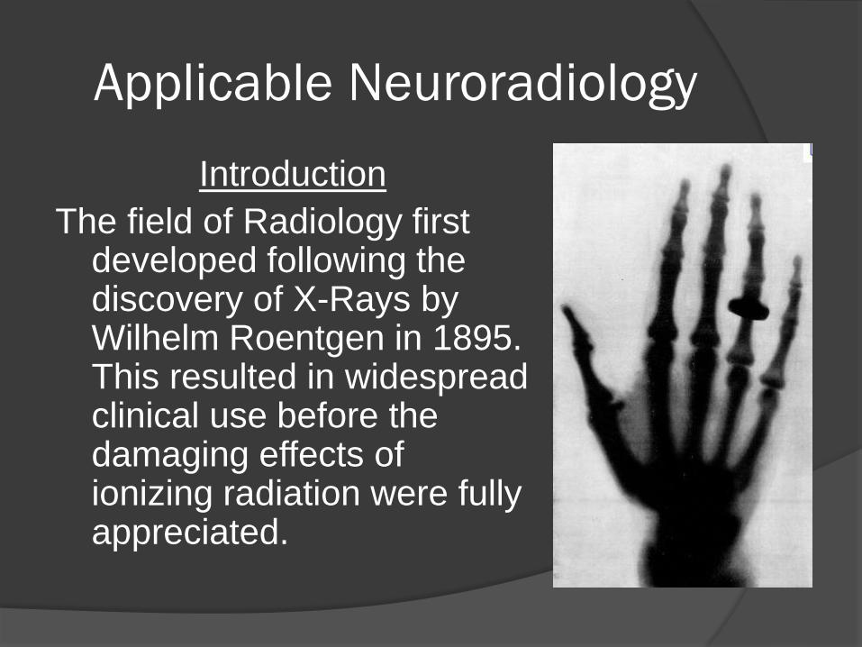
Introduction
The field of Radiology first developed following the discovery of X-Rays by Wilhelm Roentgen in 1895. This resulted in widespread clinical use before the damaging effects of ionizing radiation were fully appreciated.
Applicable Neuroradiology
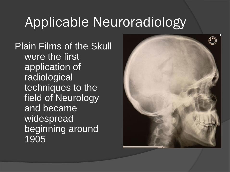
Plain Films of the Skull were the first application of radiological techniques to the field of Neurology and became widespread beginning around 1905
Applicable Neuroradiology
Plain Films of the Skull
Good for detecting Ca++
Good for Skull Fx’s
Good for Foreign Bodies
Quick way to look for pneumatization of cranial sinuses
Plain Spine Films
Good for vertebral fractures and dislocations
Used in evaluation of scoliosis
Does NOT image cord however
Applicable Neuroradiology
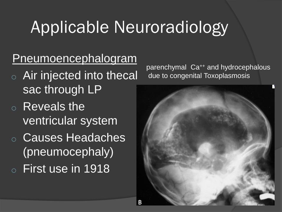
Pneumoencephalogram
o Air injected into thecal
sac through LP
o Reveals the
ventricular system
o Causes Headaches
(pneumocephaly)
o First use in 1918
parenchymal Ca++ and hydrocephalous
due to congenital Toxoplasmosis
Applicable Neuroradiology
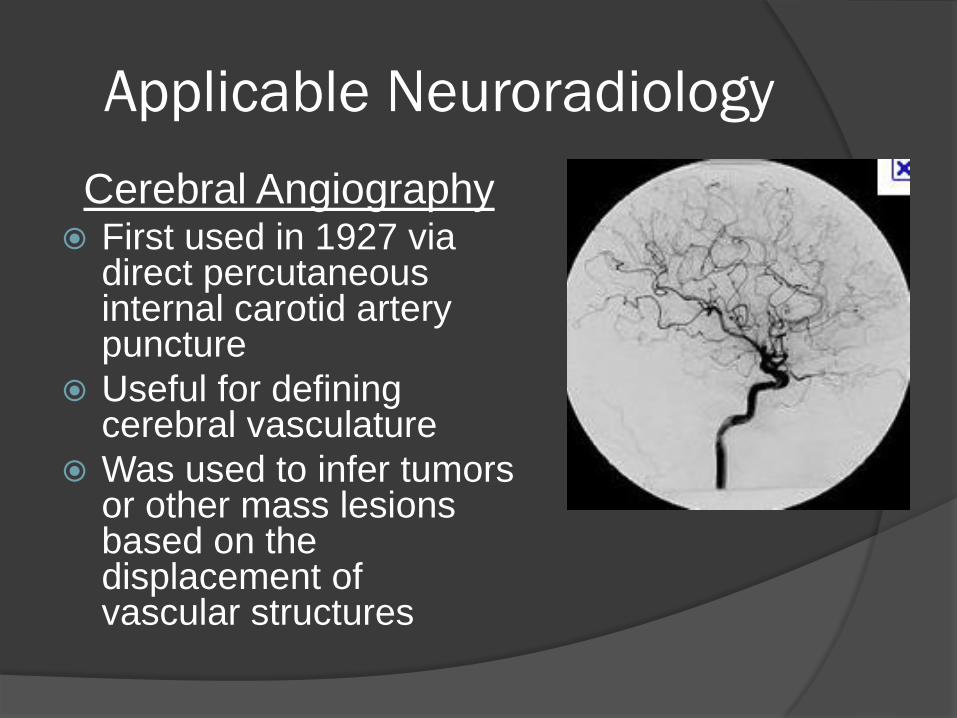
Cerebral Angiography First used in 1927 via
direct percutaneous internal carotid artery puncture
Useful for defining cerebral vasculature
Was used to infer tumors or other mass lesions based on the displacement of vascular structures
Applicable Neuroradiology
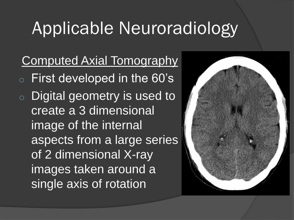
Computed Axial Tomography
o First developed in the 60’s
o Digital geometry is used to
create a 3 dimensional
image of the internal
aspects from a large series
of 2 dimensional X-ray
images taken around a
single axis of rotation
Applicable Neuroradiology
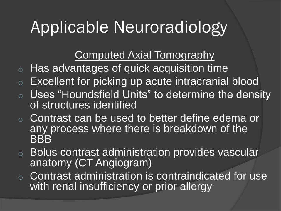
Computed Axial Tomography
o Has advantages of quick acquisition time
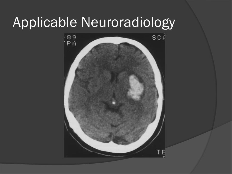
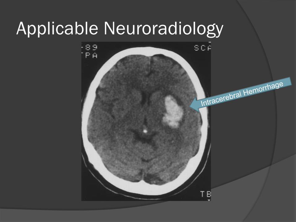
o Excellent for picking up acute intracranial blood
o Uses “Houndsfield Units” to determine the density of structures identified
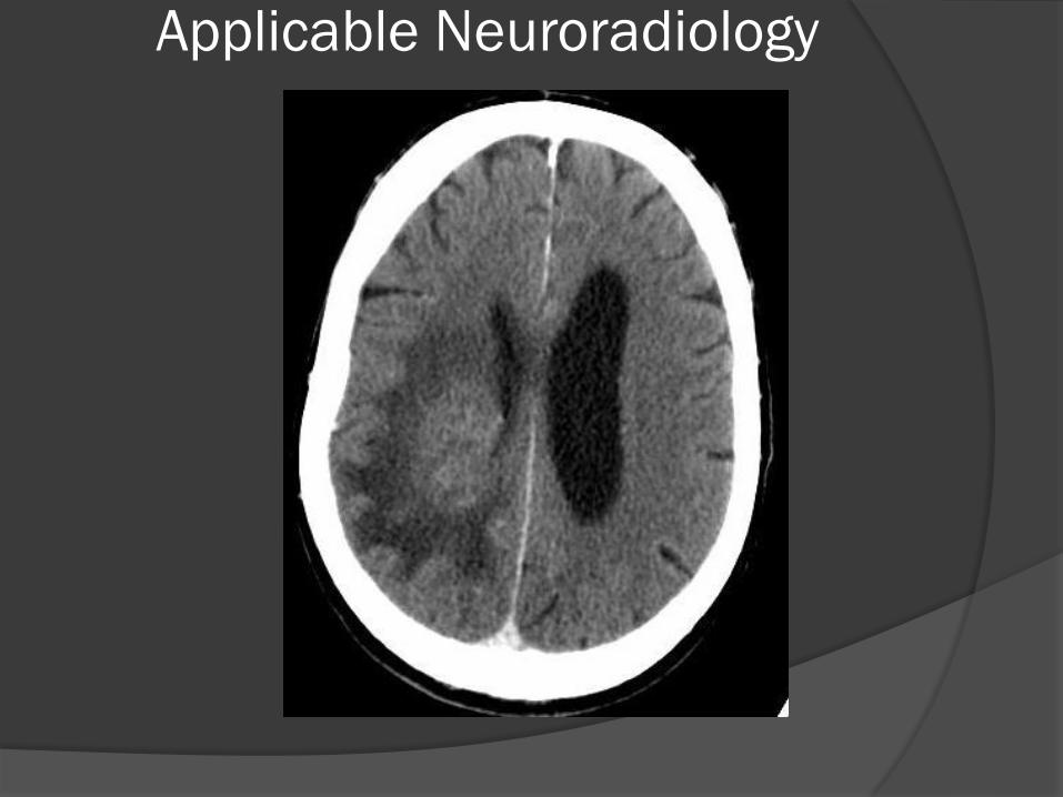
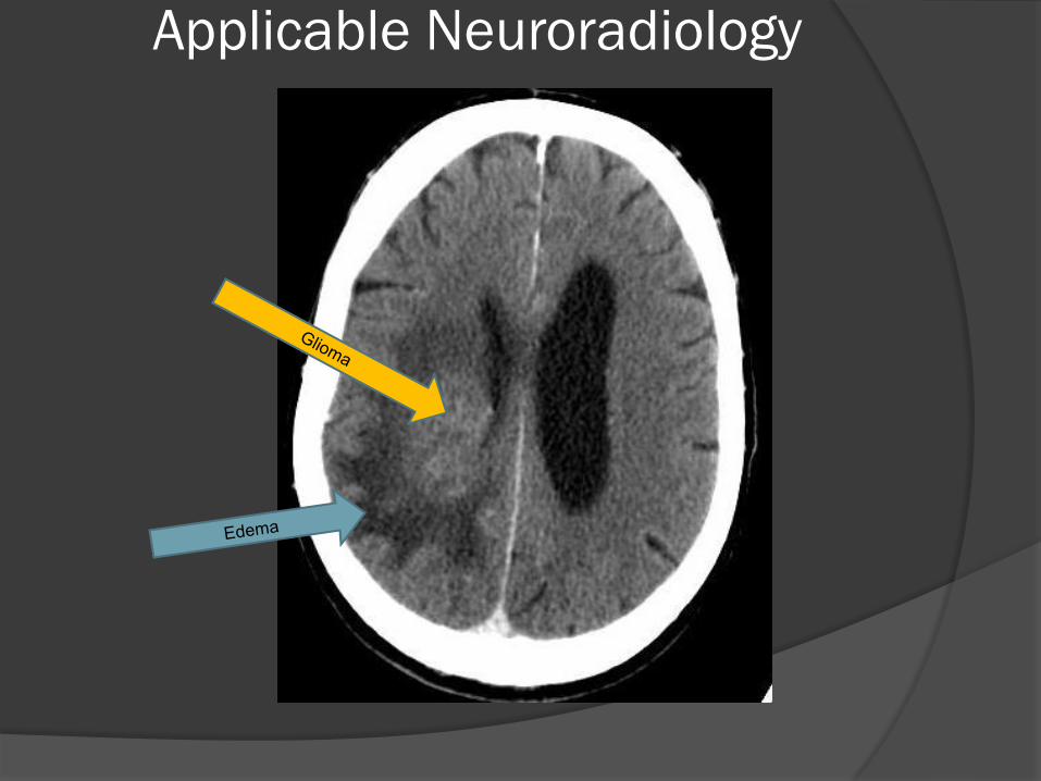
o Contrast can be used to better define edema or any process where there is breakdown of the BBB
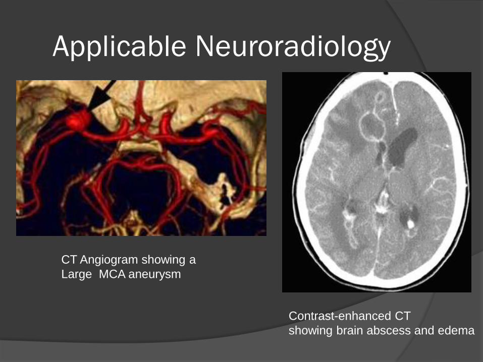
o Bolus contrast administration provides vascular anatomy (CT Angiogram)
o Contrast administration is contraindicated for use with renal insufficiency or prior allergy
Applicable Neuroradiology
CT Angiogram showing a
Large MCA aneurysm
Contrast-enhanced CT
showing brain abscess and edema
Applicable Neuroradiology
Computed Axial Tomography
o 5 “B” things that are bright (hyperdense) on CT
o Blood
o Bone (or Ca++)
o Brain
o Bullet (or foreign body)
o “Bontrast” for “Contrast”
Applicable Neuroradiology
Cranial Ultrasound
Cranial U/S developed in the 70’s
Used in infancy as a non-invasive way
to view ventricles and look for
intraventricular hemorrhage using the
anterior fontanelle as a portal
Used in adults for carotid
stenosis/dissection or for cerebral
vasospasm
Applicable Neuroradiology
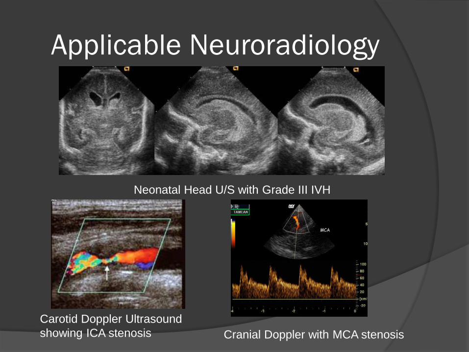
Neonatal Head U/S with Grade III IVH
Carotid Doppler Ultrasound
showing ICA stenosis Cranial Doppler with MCA stenosis
Applicable Neuroradiology
SPECT
Single Photon Emission Computed Tomography
Developed in 60’s (along with CT)
gamma ray-emitting long-acting isotope
(Technetium-99m) shows regional CBF
Can help localize seizure onset (Ictal-SPECT)
Can be superimposed on CT or MRI
More available than PET
Applicable Neuroradiology
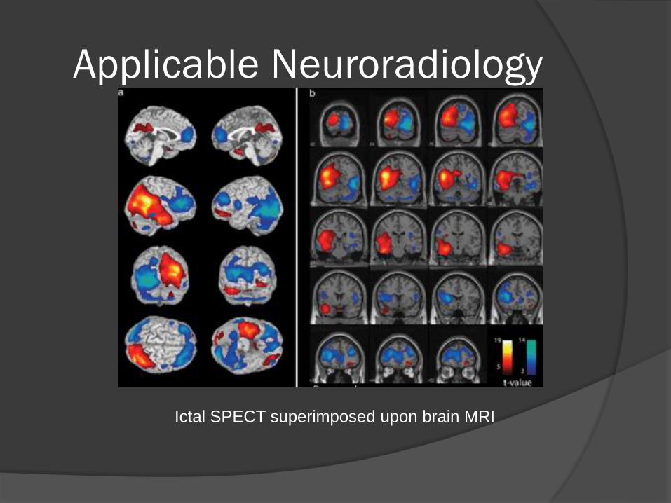
Ictal SPECT superimposed upon brain MRI
Applicable Neuroradiology
PET
Positron Emission Tomography
Developed in the 70’s
Detects gamma rays released by a radionuclide tracer linked to a marker
FDG (Fludeoxyglucose) most commonly used
Other markers include specific neurotransmitters or their receptors
Requires cyclotron to make short half-life tracers so not as available as PET
Applicable Neuroradiology
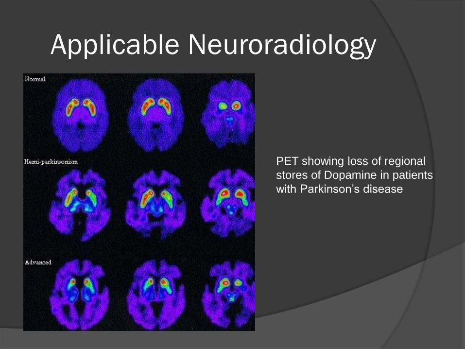
PET showing loss of regional
stores of Dopamine in patients
with Parkinson’s disease
Applicable Neuroradiology
Magnetic Resonance Imaging Developed in the 80’s
Powerful magnetic fields cause water molecules to align along their dipoles
Radiofrequency waves produce an electromagnetic field which transiently knocks the molecules out of alignment
When water molecules re-align within the magnetic field they release energy (photons) which are detected by scanners and following a lot of computer mumbo-jumbo an image is produced
Applicable Neuroradiology
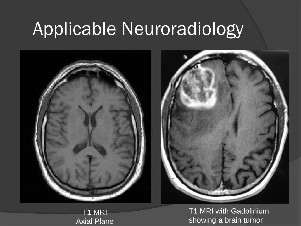
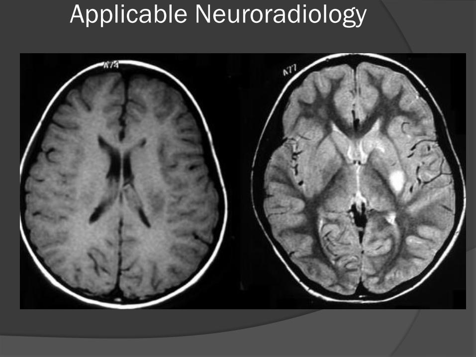
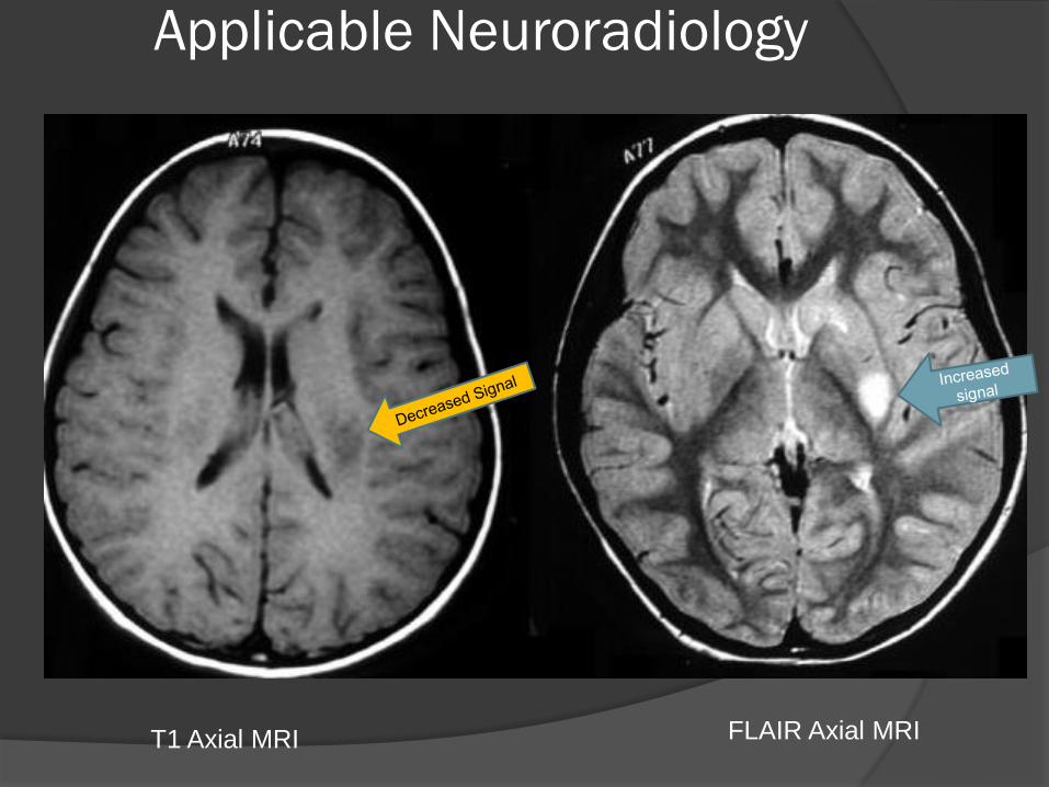
Magnetic Resonance Imaging T-1 Imaging
Water is dark. Fat (Myelin) is bright
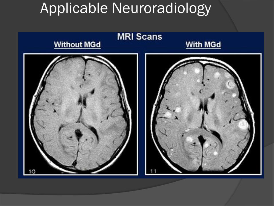
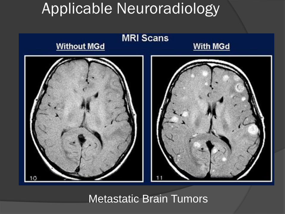
Gadolinium contrast used to show breakdown of BBB
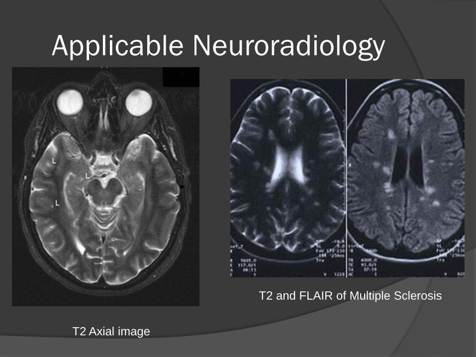
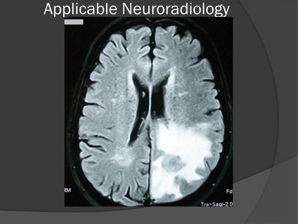
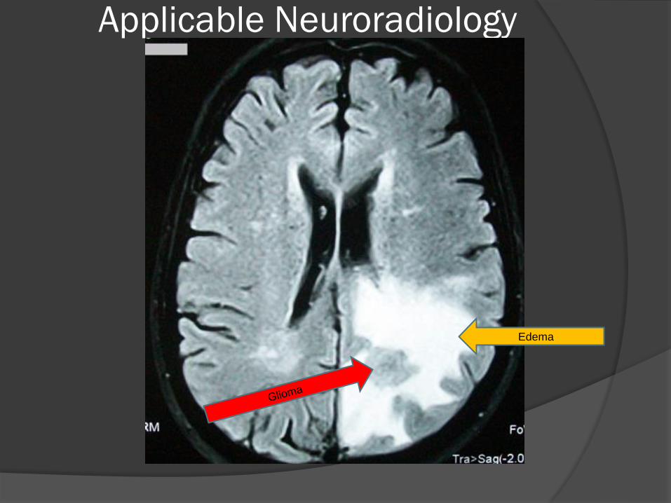
T-2 Imaging Water is bright. Fat is dark.
FLAIR (same as T2 except water is “blacked out”)
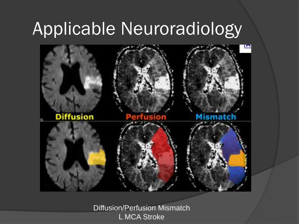
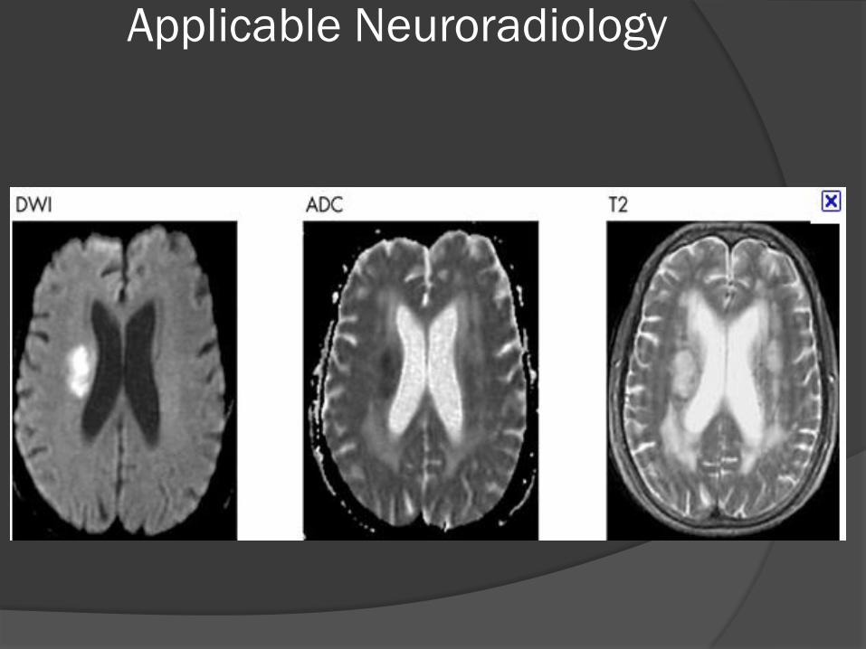
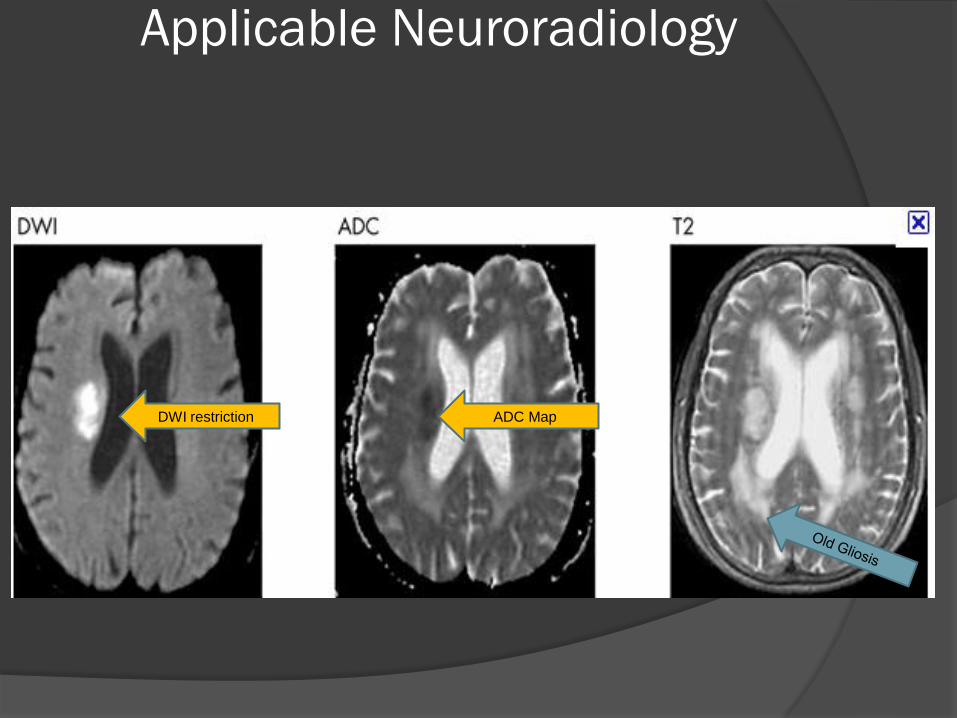
Diffusion Imaging Shows restricted Diffusion of water suggesting cell death
ADC Mapping takes into account brightness of background T2 signal
Applicable Neuroradiology
Blood on MRI is paradoxical and evolves
Note the intensity changes as the blood
cells break down and lose oxygen
For ACUTE Blood they are similar to FAT
T1 = White
T2 = Black
Applicable Neuroradiology
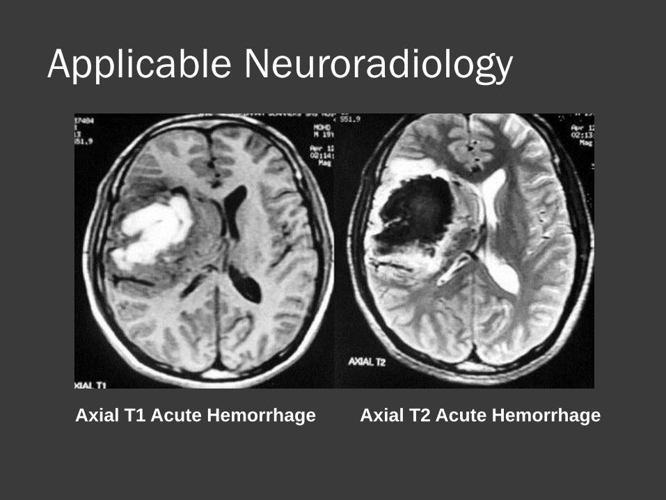
Axial T1 Acute Hemorrhage Axial T2 Acute Hemorrhage
Applicable Neuroradiology
T1 MRI
Axial Plane
T1 MRI with Gadolinium
showing a brain tumor
Applicable Neuroradiology
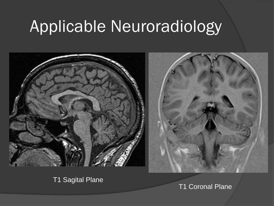
T1 Sagital Plane T1 Coronal Plane
Applicable Neuroradiology
T2 and FLAIR of Multiple Sclerosis
T2 Axial image
Applicable Neuroradiology
Diffusion/Perfusion Mismatch
L MCA Stroke
Applicable Neuroradiology
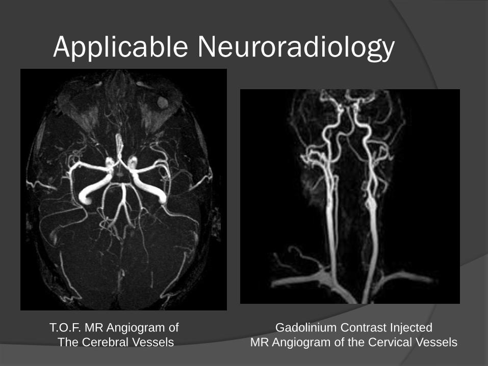
T.O.F. MR Angiogram of
The Cerebral Vessels
Gadolinium Contrast Injected
MR Angiogram of the Cervical Vessels

Applicable Neuroradiology
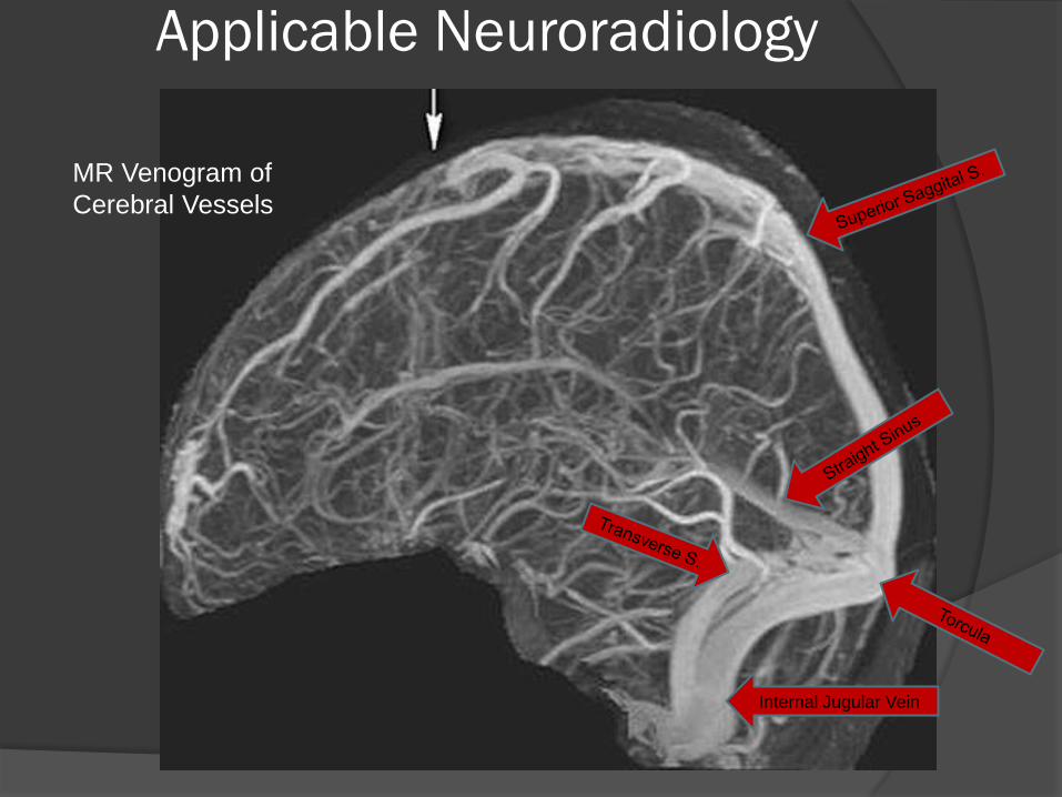
MR Venogram of the
Cerebral Sinuses and Draining Veins
Applicable Neuroradiology
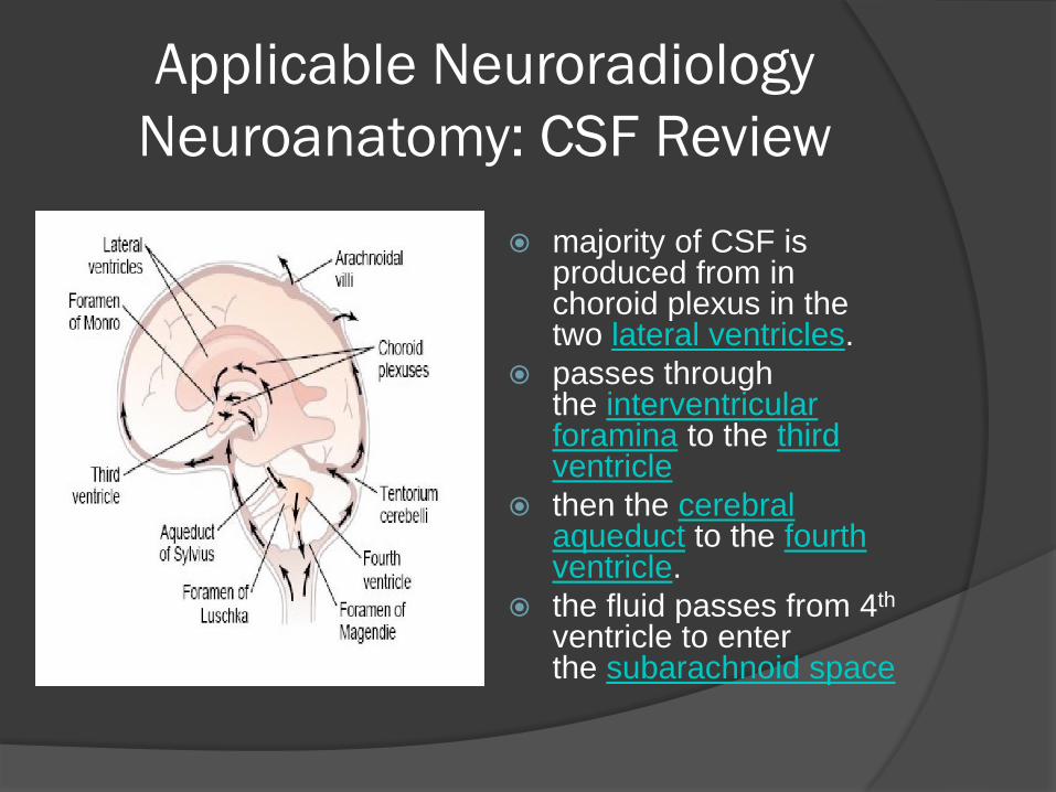
Neuroanatomy: CSF Review
majority of CSF is produced from in choroid plexus in the two lateral ventricles.
passes through the interventricular foramina to the third ventricle
then the cerebral aqueduct to the fourth ventricle.
the fluid passes from 4th ventricle to enter the subarachnoid space
Applicable Neuroradiology
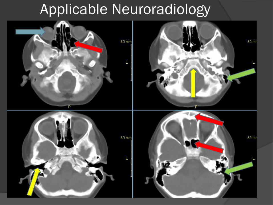
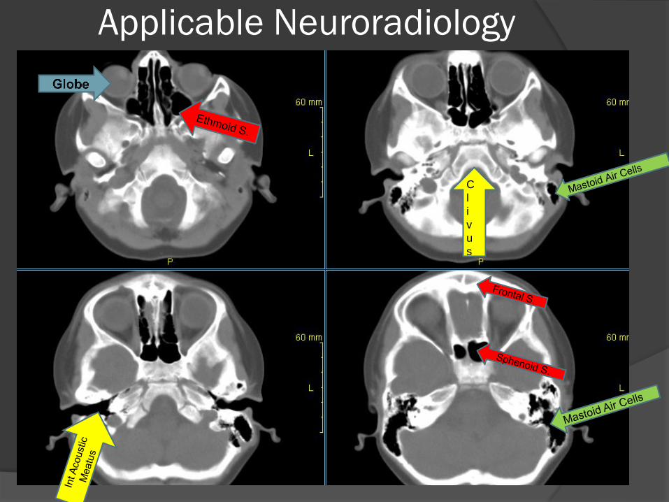
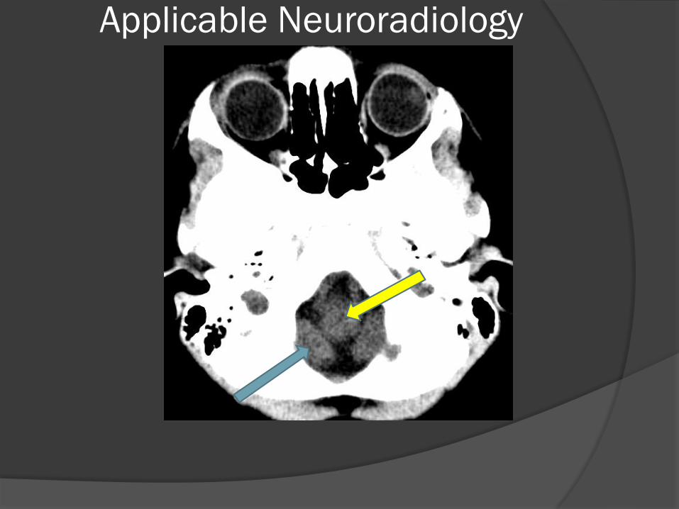
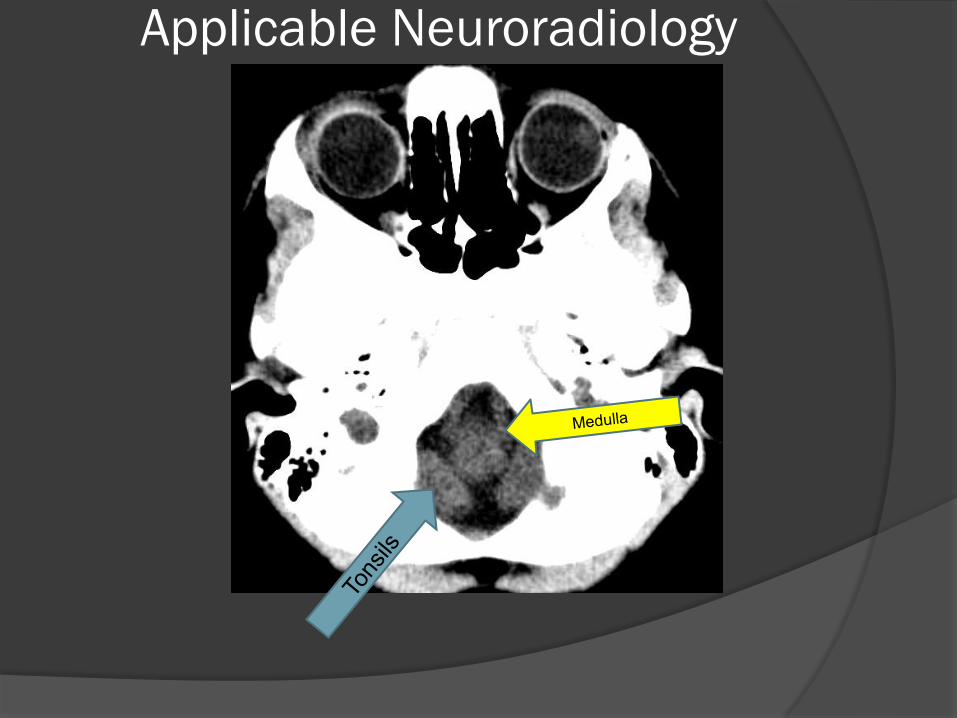
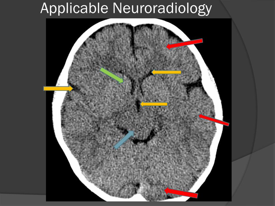
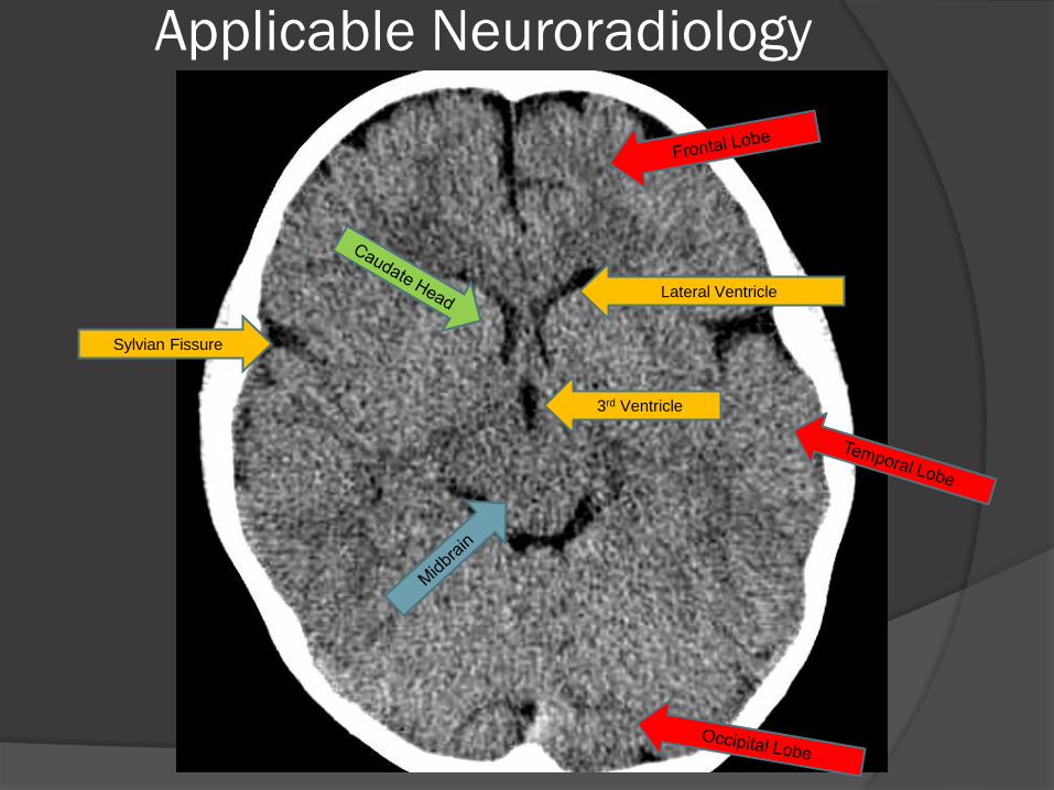
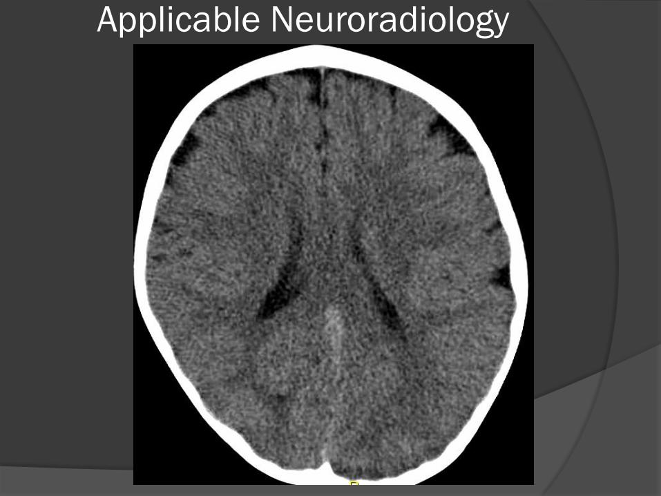
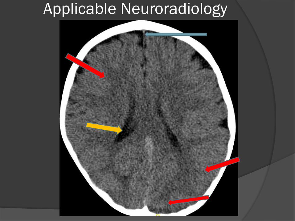
Neuroanatomy
Name The Structures
Applicable Neuroradiology
Applicable Neuroradiology
Applicable Neuroradiology
C
l
i
v
u
s
Applicable Neuroradiology
Applicable Neuroradiology
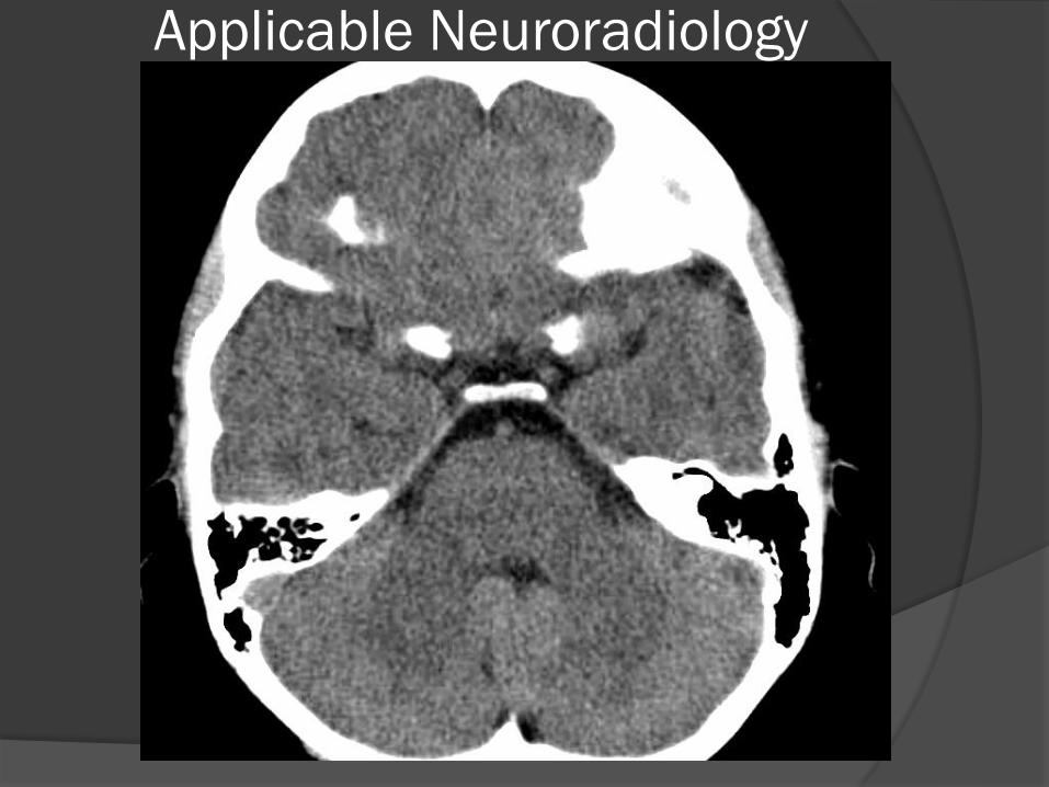
Applicable Neuroradiology
Applicable Neuroradiology
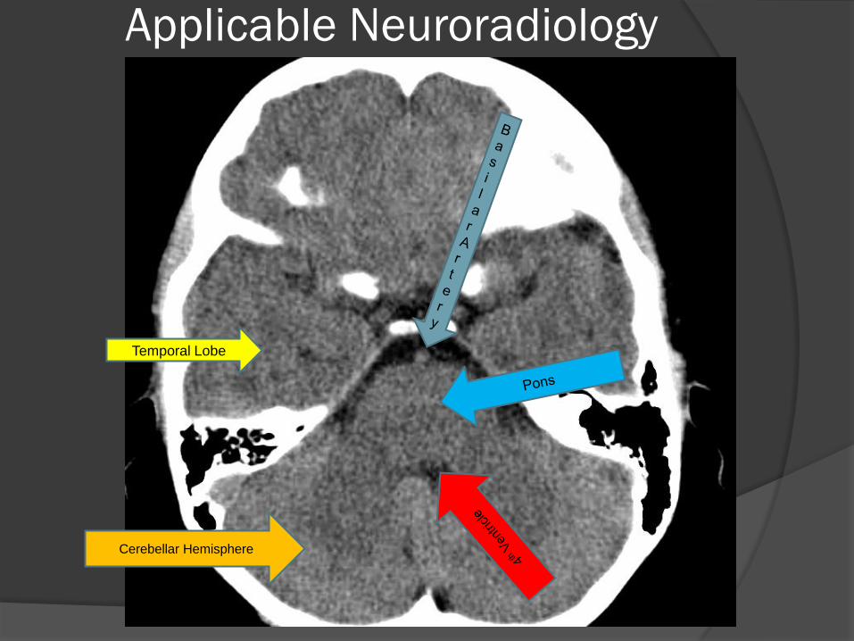
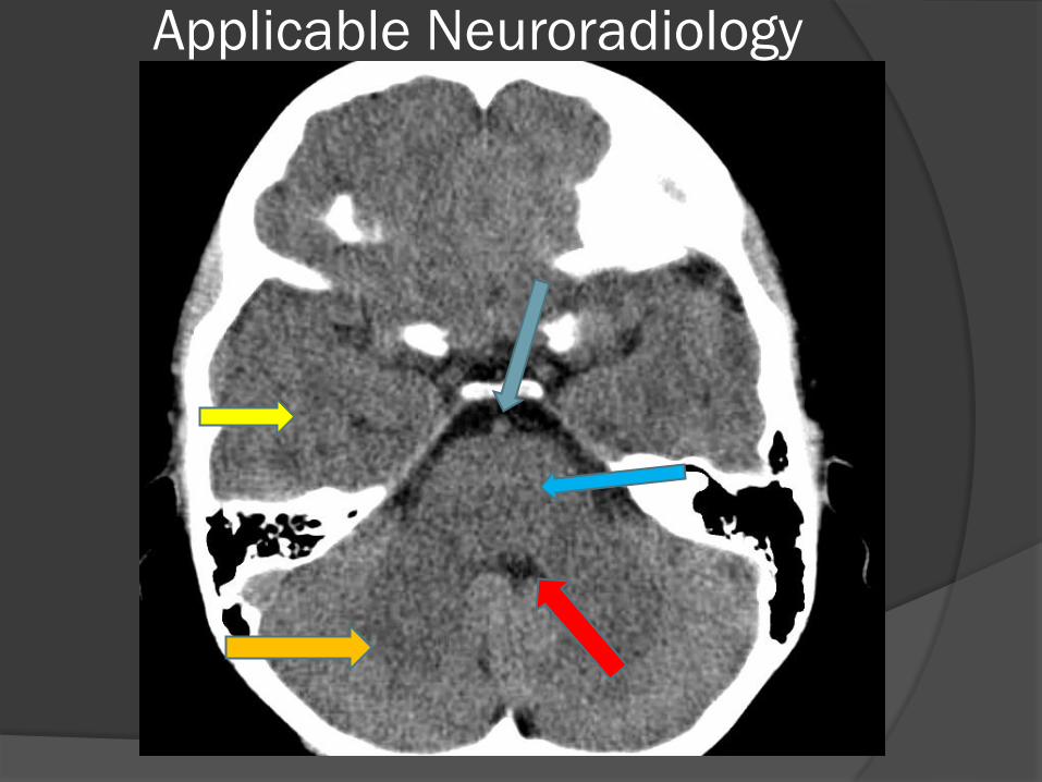
Applicable Neuroradiology
Temporal Lobe
Cerebellar Hemisphere
Applicable Neuroradiology
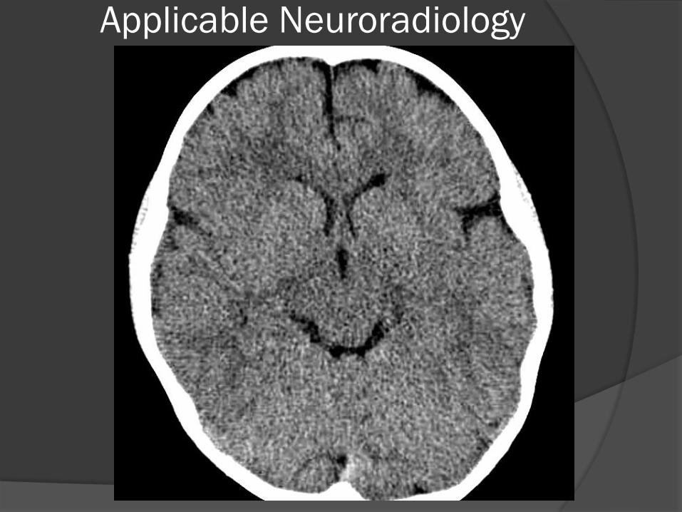
Applicable Neuroradiology
Applicable Neuroradiology
Sylvian Fissure
3rd Ventricle
Lateral Ventricle
Applicable Neuroradiology
Applicable Neuroradiology
Applicable Neuroradiology
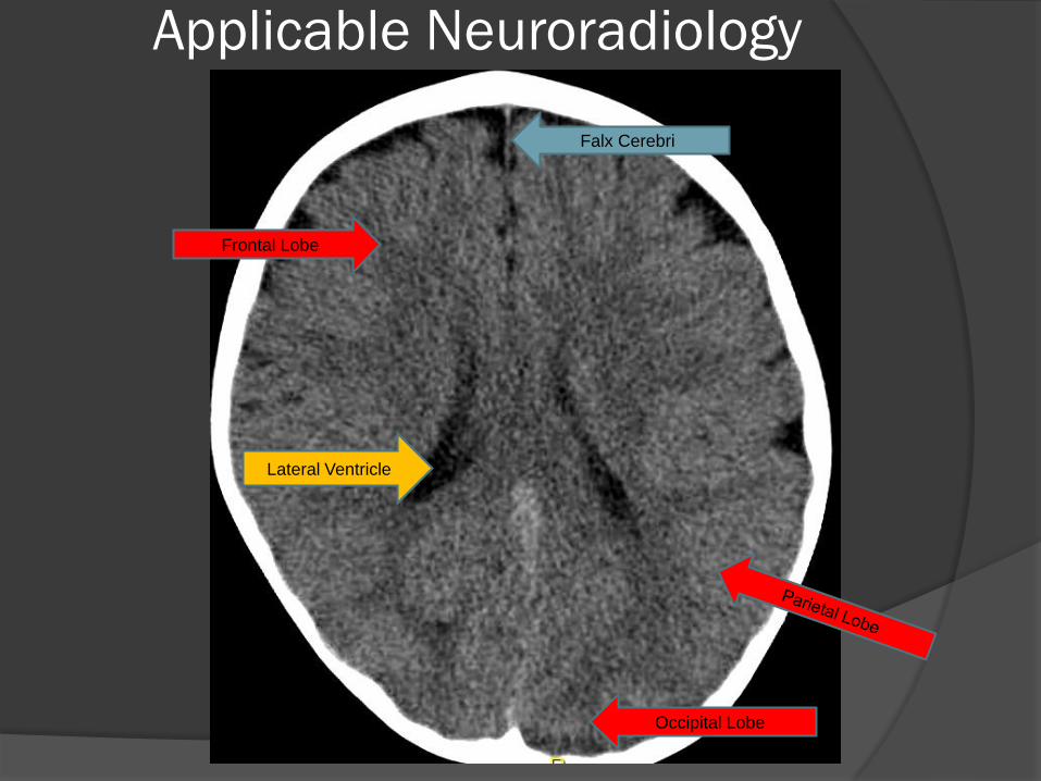
Lateral Ventricle
Falx Cerebri
Frontal Lobe
Occipital Lobe
Applicable Neuroradiology
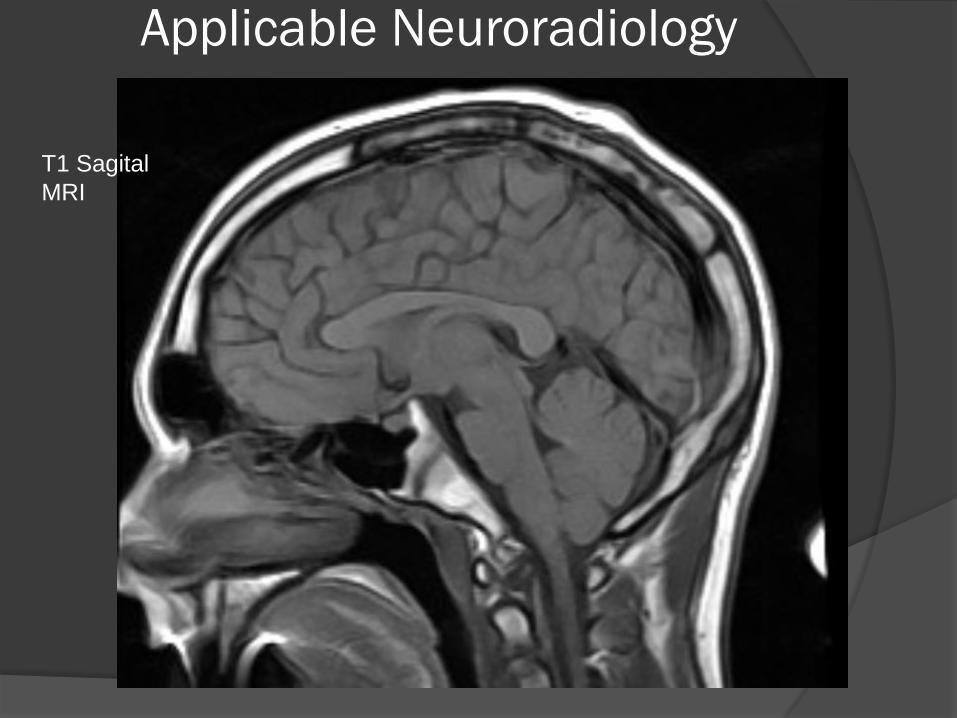
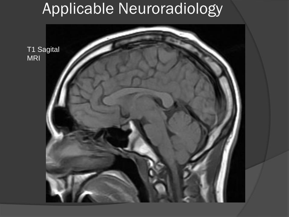
T1 Sagital
MRI
Applicable Neuroradiology
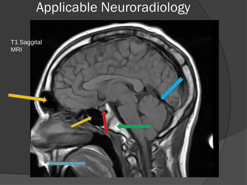
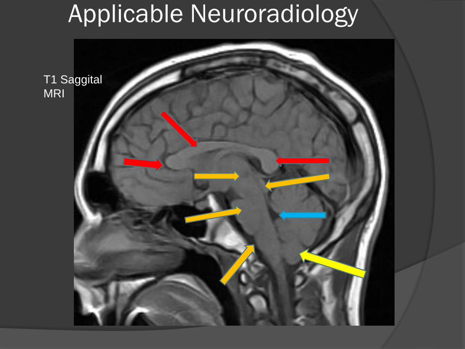
T1 Saggital
MRI
Applicable Neuroradiology
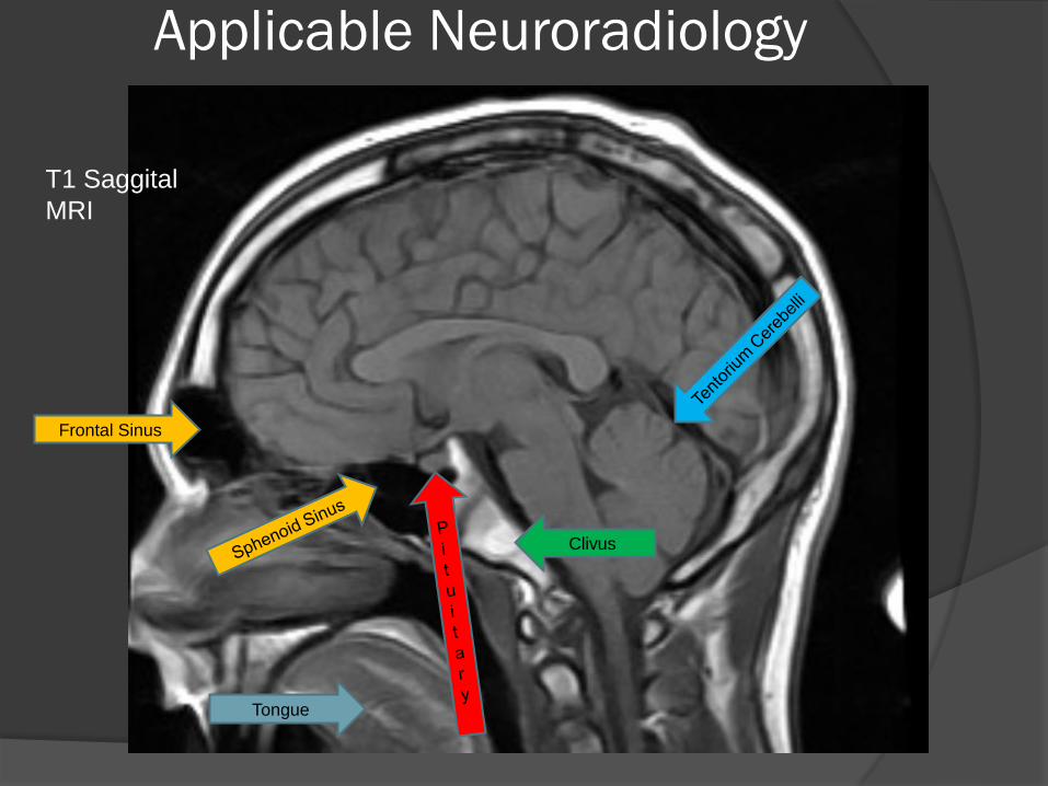
Frontal Sinus
Clivus
Tongue
T1 Saggital
MRI
Applicable Neuroradiology
T1 Sagital
MRI
Applicable Neuroradiology
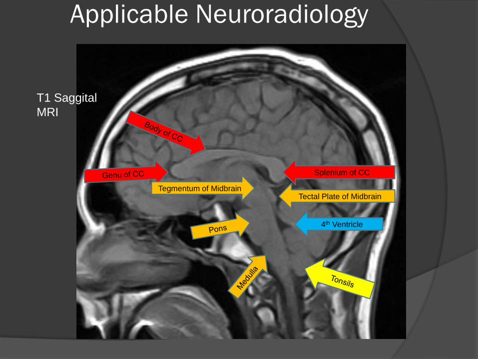
T1 Saggital
MRI
Applicable Neuroradiology
Splenium of CC
Tegmentum of Midbrain Tectal Plate of Midbrain
4th Ventricle
T1 Saggital
MRI

Applicable Neuroradiology
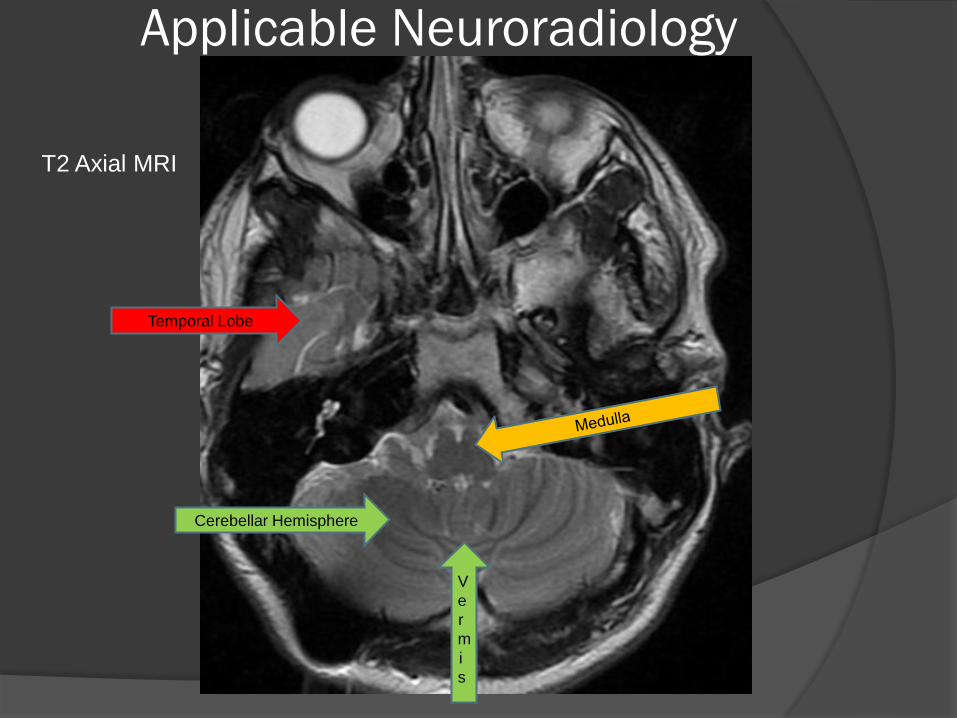
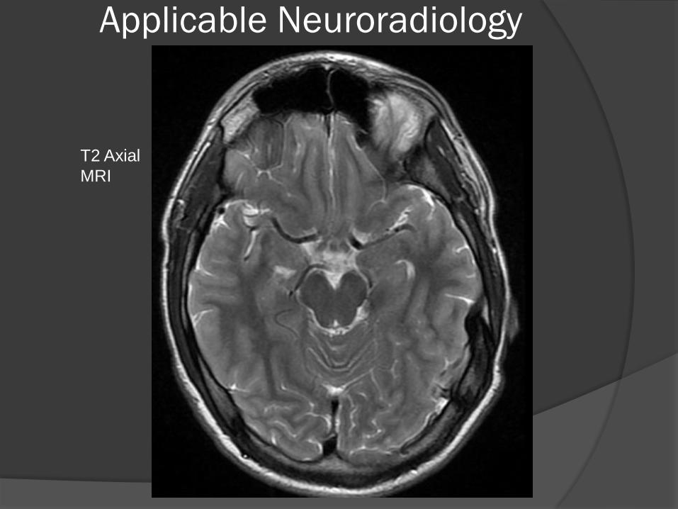
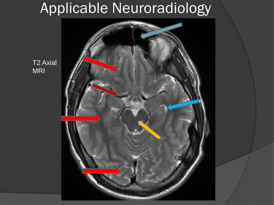
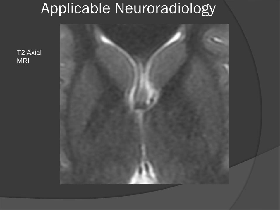
T2 Axial MRI
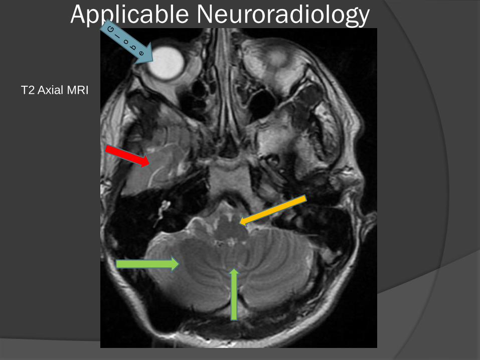
Applicable Neuroradiology
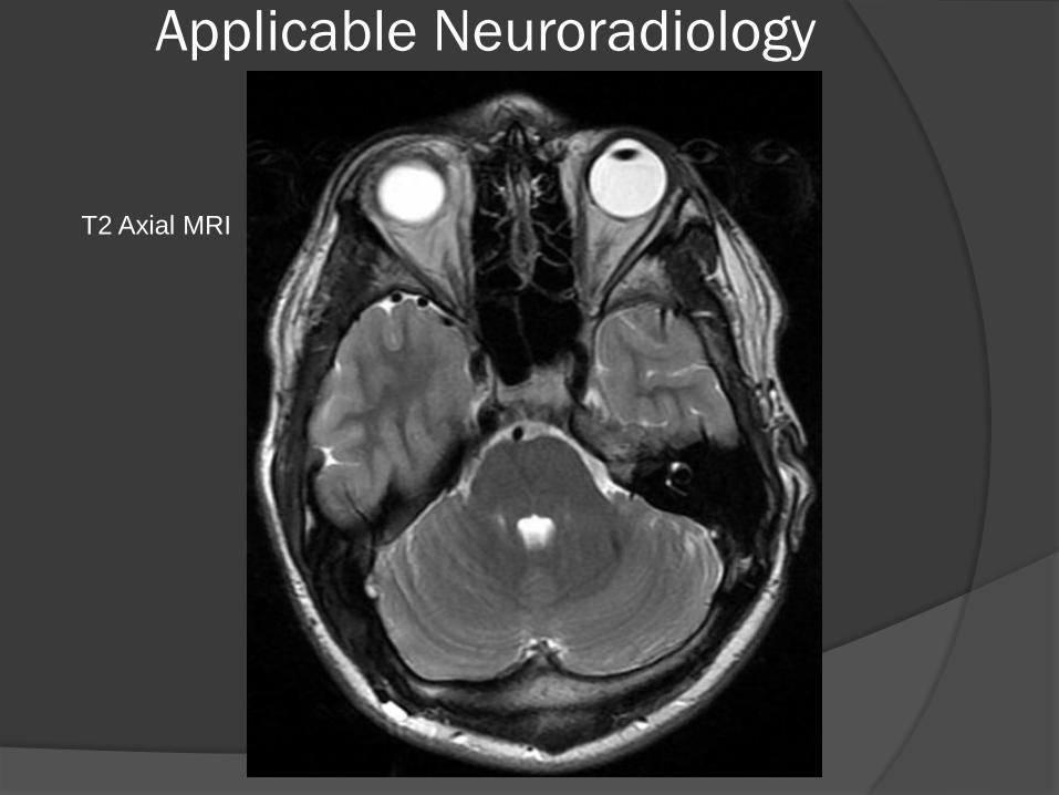
T2 Axial MRI
Applicable Neuroradiology
Temporal Lobe
Cerebellar Hemisphere
V
e
r
m
i
s
T2 Axial MRI
Applicable Neuroradiology
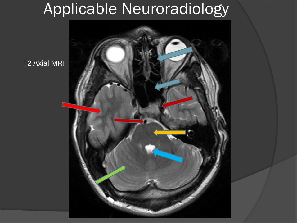
T2 Axial MRI
Applicable Neuroradiology
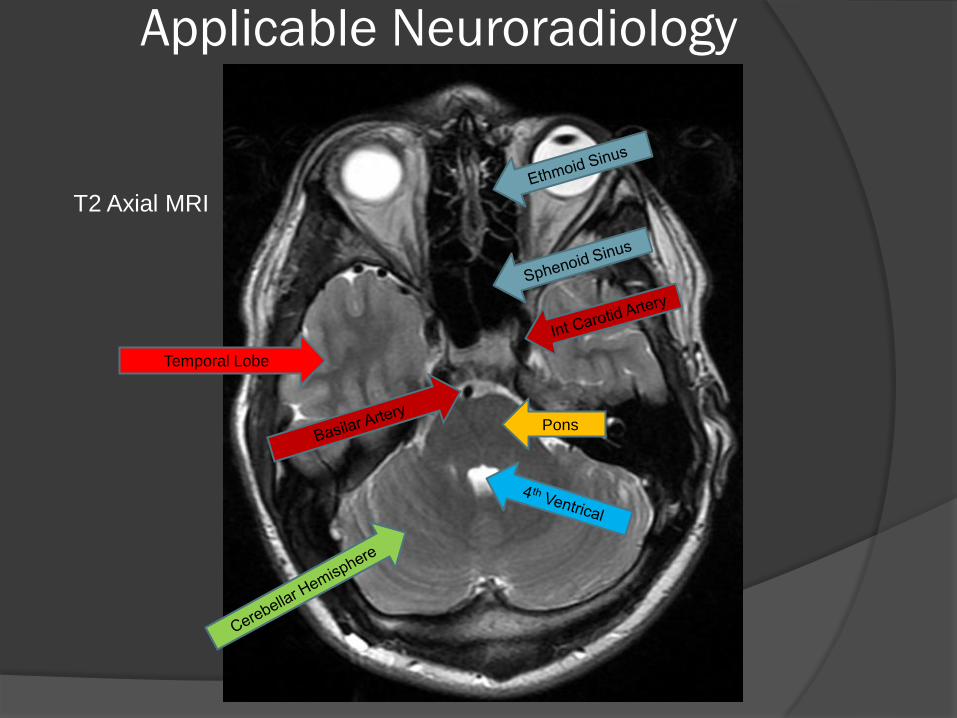
T2 Axial MRI
Applicable Neuroradiology
Temporal Lobe
Pons
T2 Axial MRI
Applicable Neuroradiology
T2 Axial
MRI
Applicable Neuroradiology
T2 Axial
MRI
Applicable Neuroradiology
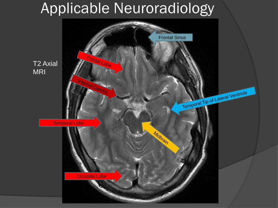
Frontal Sinus
Temporal Lobe
Occipital Lobe
T2 Axial
MRI
Applicable Neuroradiology
T2 Axial MRI
Applicable Neuroradiology
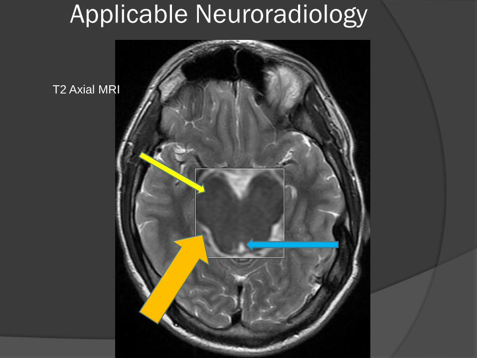
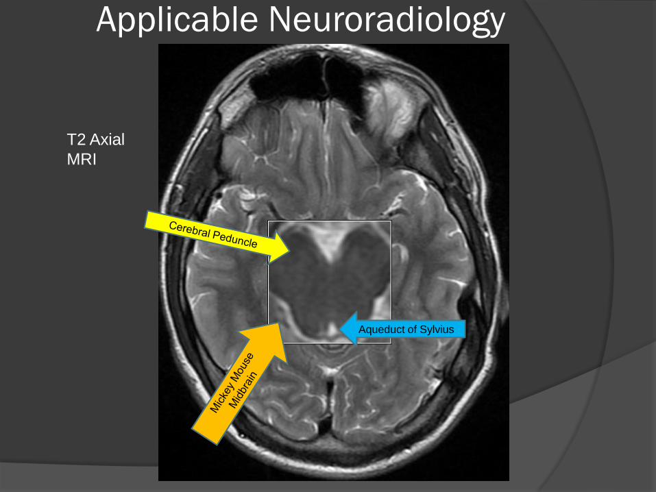
Aqueduct of Sylvius
T2 Axial
MRI
Applicable Neuroradiology
T2 Axial
MRI
Applicable Neuroradiology
T2 Axial
MRI
Applicable Neuroradiology
Splenium of CC
T2 Axial
MRI
Applicable Neuroradiology
T2 Axial
MRI

Applicable Neuroradiology
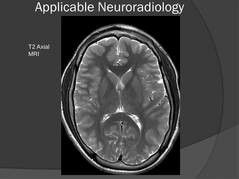
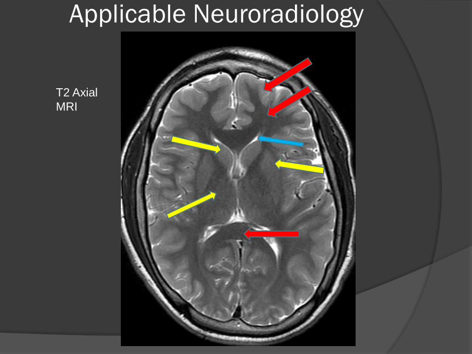
T2 Axial
MRI
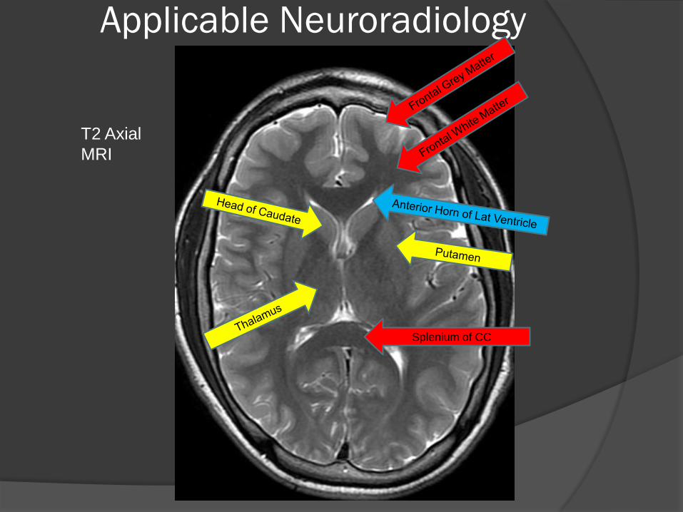
Applicable Neuroradiology
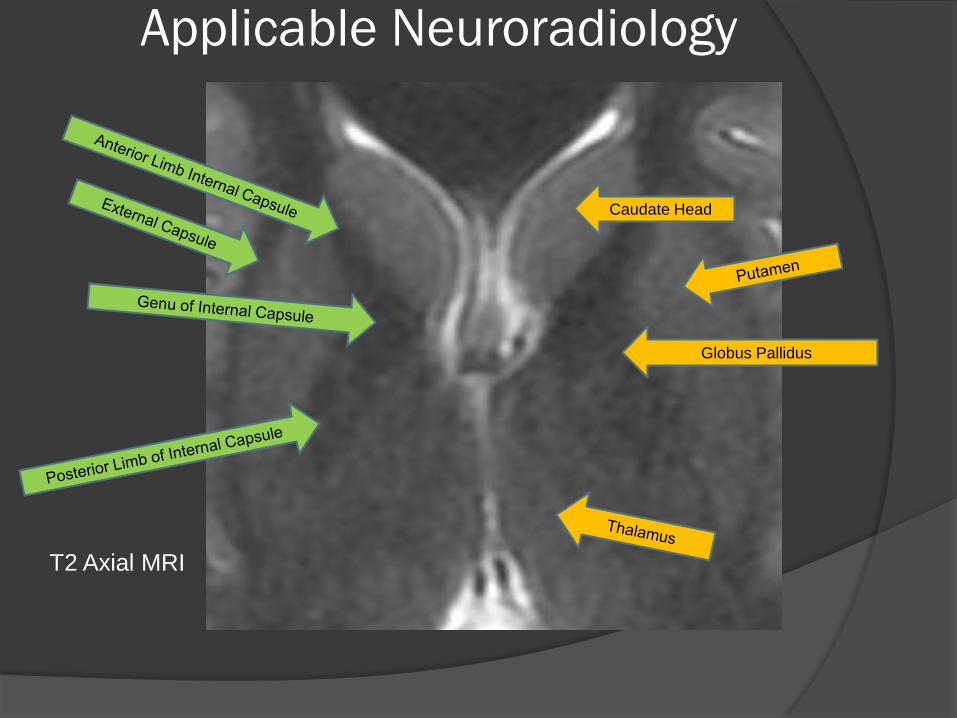
Caudate Head
Globus Pallidus
T2 Axial MRI
Applicable Neuroradiology
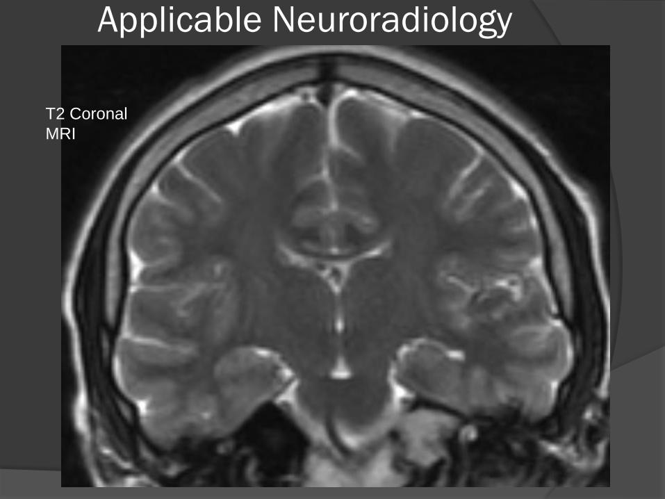
T2 Coronal
MRI

Applicable Neuroradiology
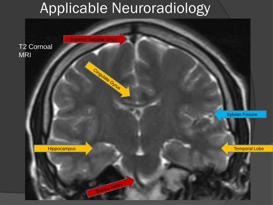
T2 Cornoal
MRI
Applicable Neuroradiology
T2 Cornoal
MRI
Hippocampus Temporal Lobe
Sylvian Fissure
Superior Saggital Sinus
Applicable Neuroradiology
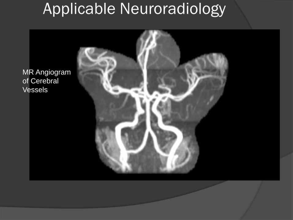
MR Angiogram
of Cerebral
Vessels
Applicable Neuroradiology
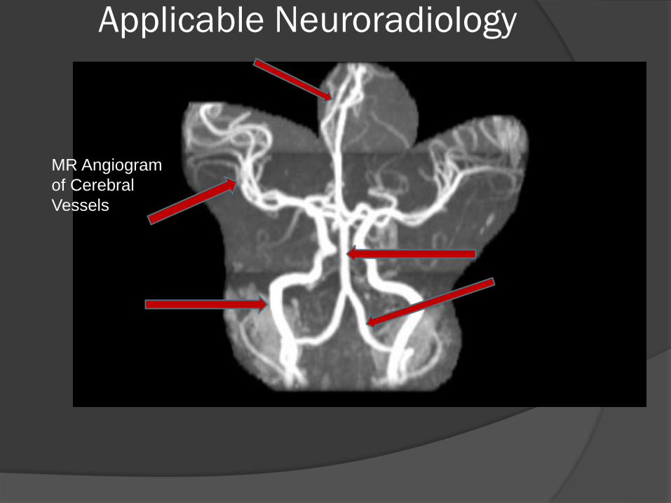
MR Angiogram
of Cerebral
Vessels
Applicable Neuroradiology
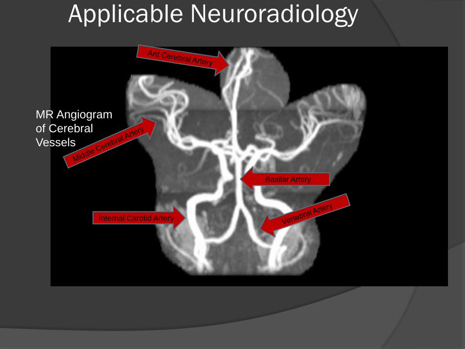
MR Angiogram
of Cerebral
Vessels
Internal Carotid Artery
Basilar Artery
Applicable Neuroradiology
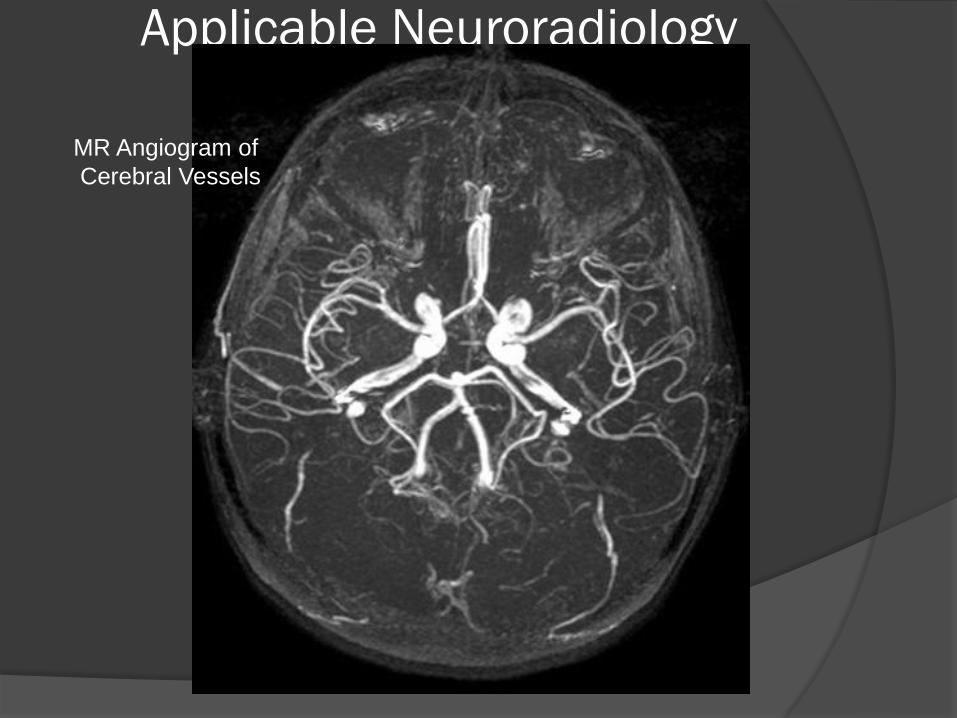
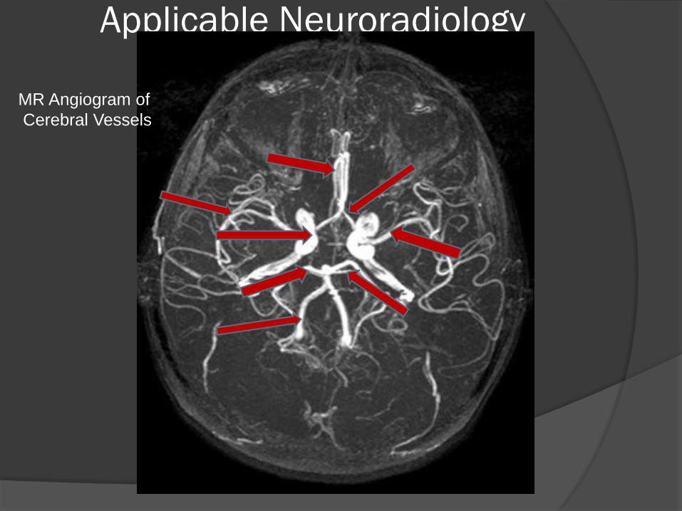
MR Angiogram of
Cerebral Vessels
Applicable Neuroradiology
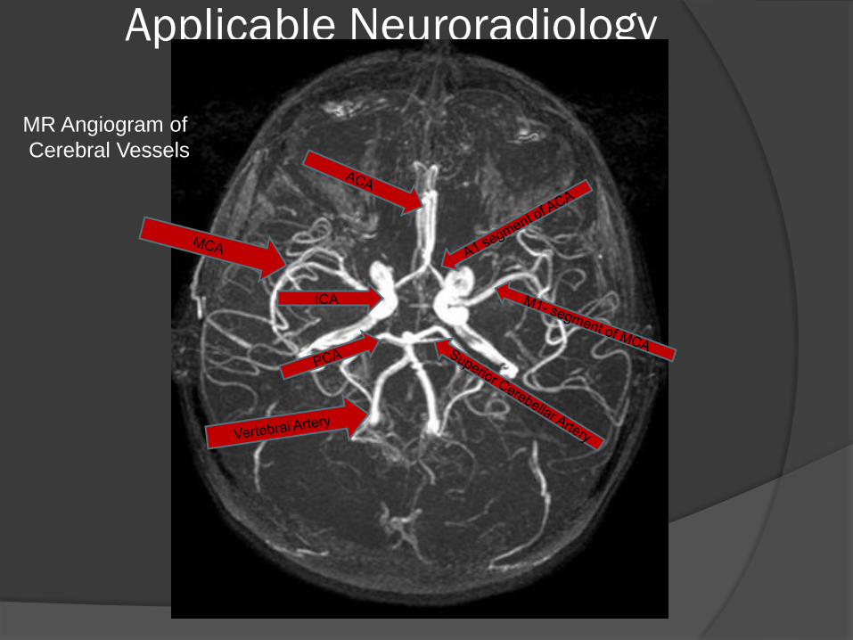
MR Angiogram of
Cerebral Vessels
Applicable Neuroradiology
MR Angiogram of
Cerebral Vessels
ICA
Applicable Neuroradiology
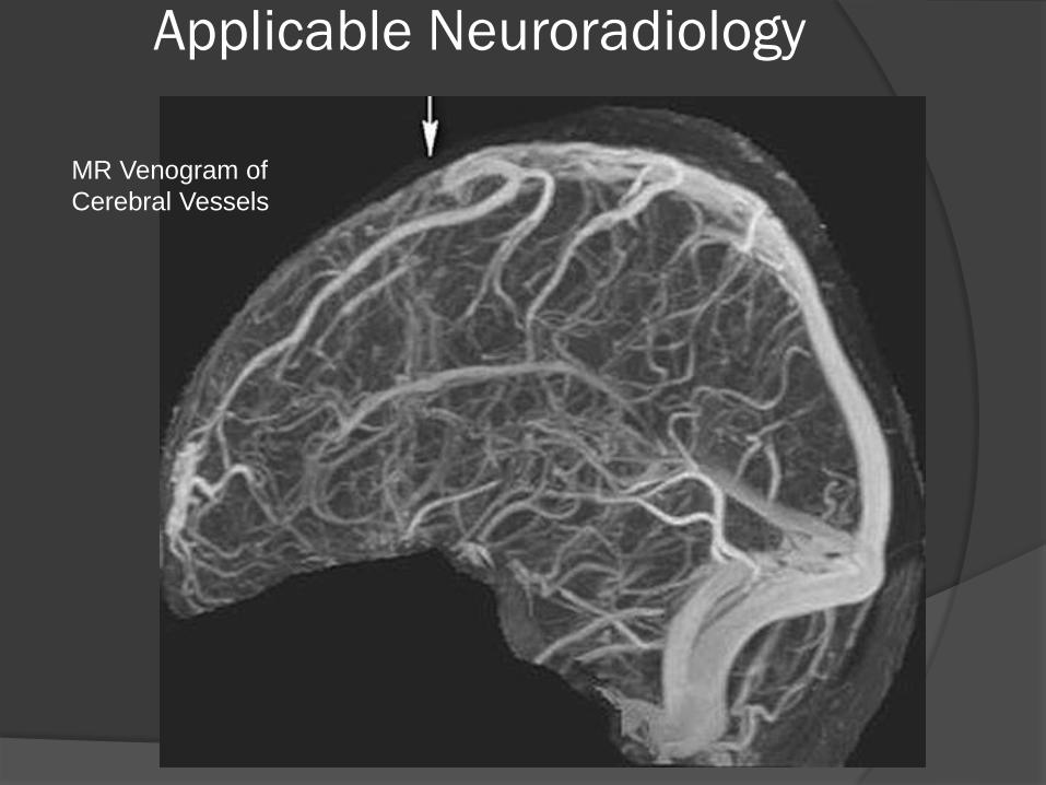
MR Venogram of
Cerebral Vessels
Applicable Neuroradiology
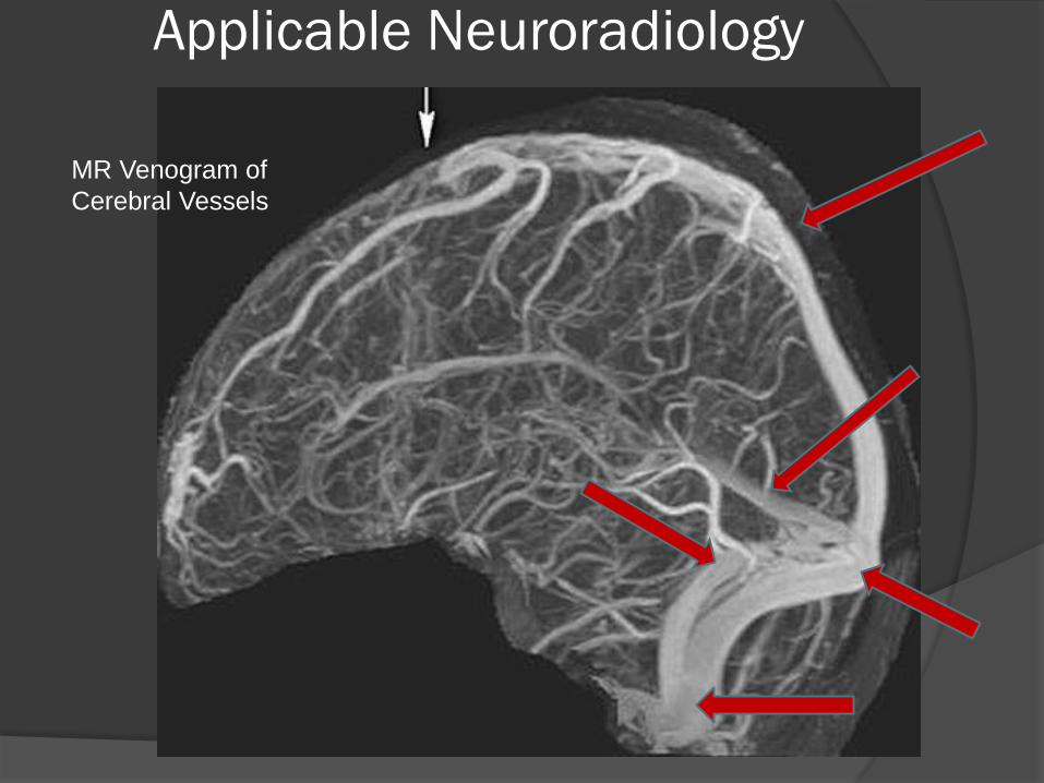
MR Venogram of
Cerebral Vessels
Applicable Neuroradiology
MR Venogram of
Cerebral Vessels
Internal Jugular Vein
Applicable Neuroradiology
What is the Abnormality?
Applicable Neuroradiology
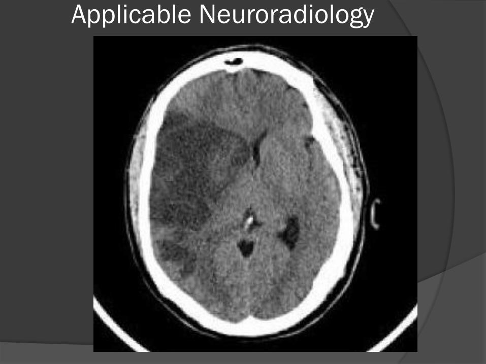
Applicable Neuroradiology
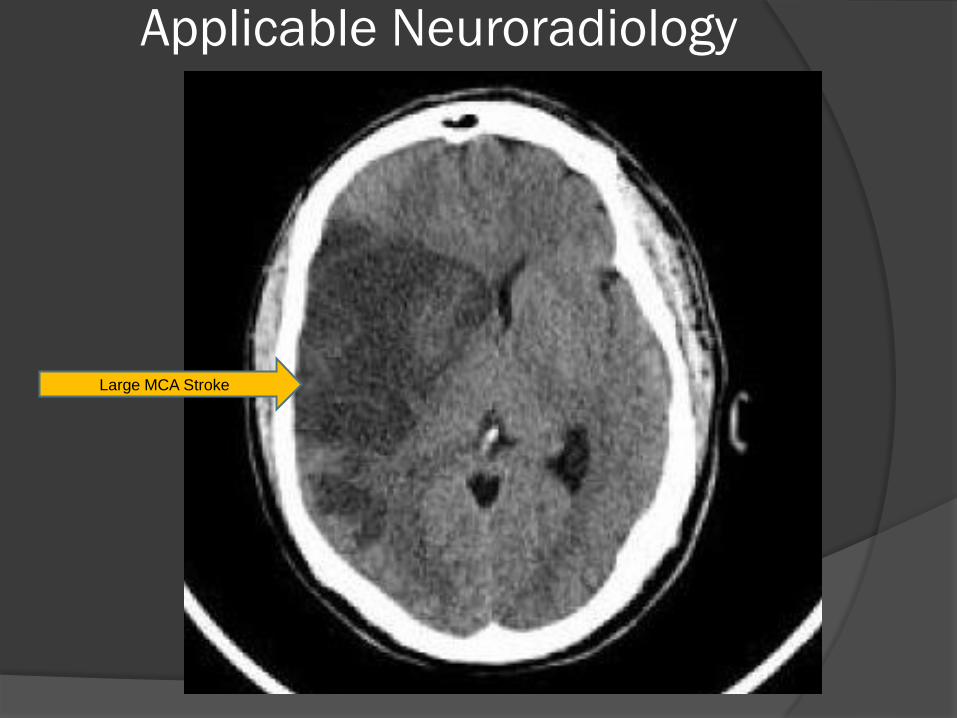
Large MCA Stroke
Applicable Neuroradiology
Applicable Neuroradiology
Applicable Neuroradiology
Applicable Neuroradiology
Applicable Neuroradiology
Applicable Neuroradiology
T1 Axial MRI FLAIR Axial MRI
Applicable Neuroradiology
Applicable Neuroradiology
DWI restriction ADC Map
Applicable Neuroradiology

Applicable Neuroradiology
Applicable Neuroradiology

Applicable Neuroradiology
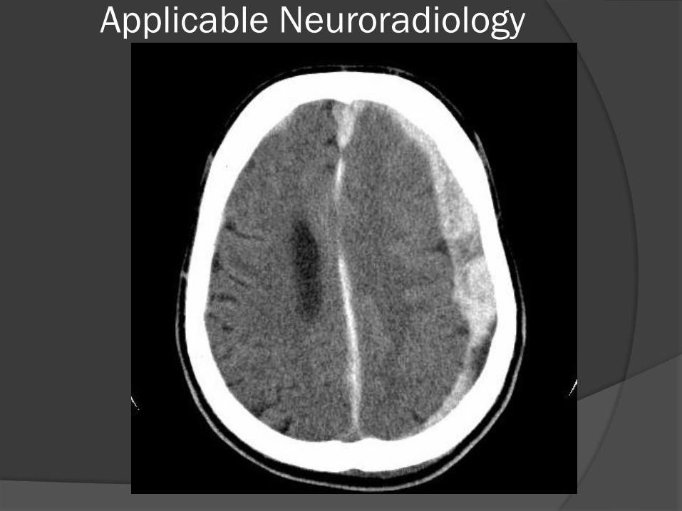
Mass Effect
Applicable Neuroradiology
Applicable Neuroradiology
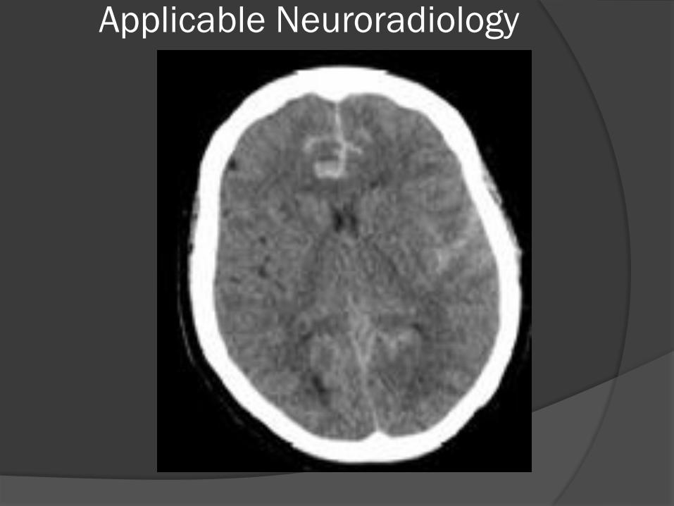
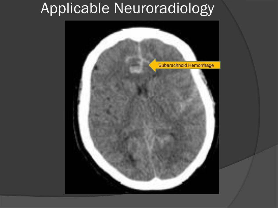
Subarachnoid Hemorrhage
Applicable Neuroradiology
Applicable Neuroradiology
Applicable Neuroradiology
Applicable Neuroradiology
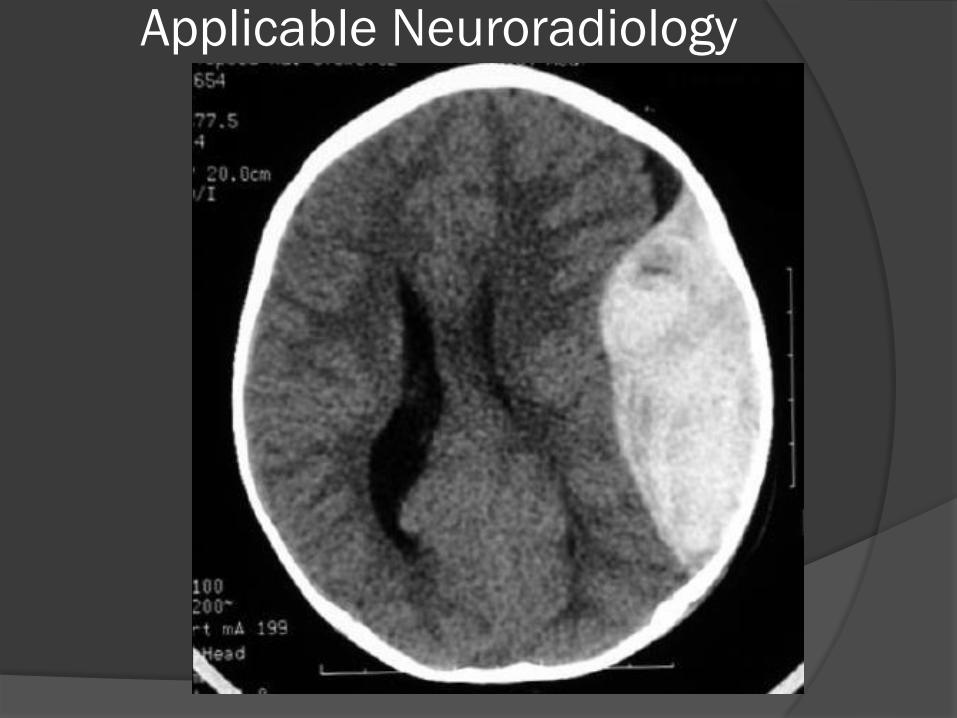
Edema
Applicable Neuroradiology
Applicable Neuroradiology
Metastatic Brain Tumors
Applicable Neuroradiology
Applicable Neuroradiology
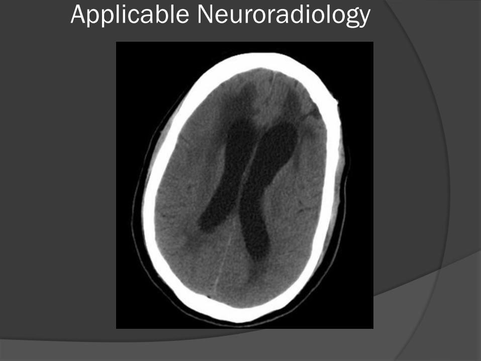
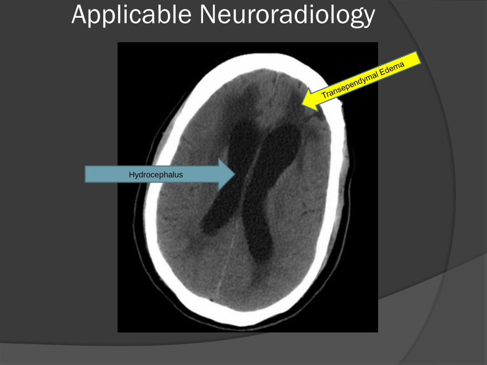
Hydrocephalus
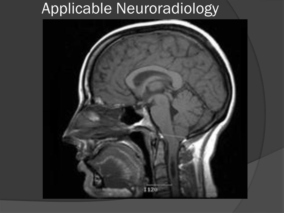
Applicable Neuroradiology
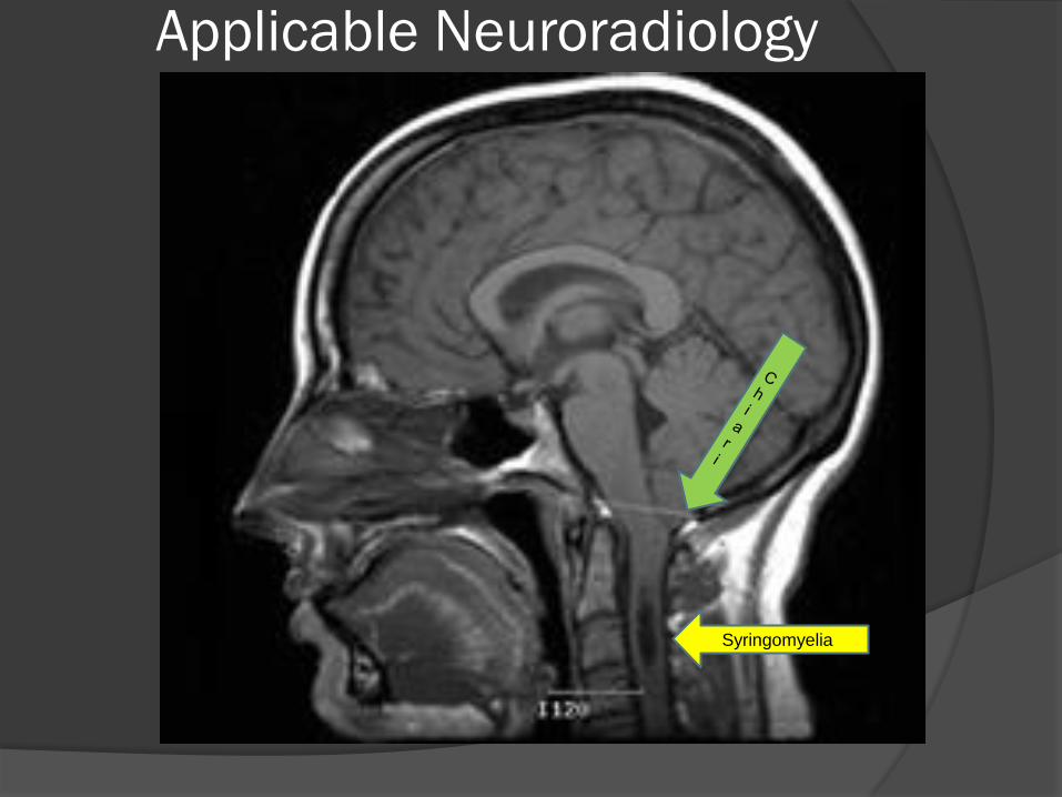
Applicable Neuroradiology
Syringomyelia
Applicable Neuroradiology
Bonus Round

Applicable Neuroradiology
T2 Axial MRI 4 y.o. with
Gelastic Seizures
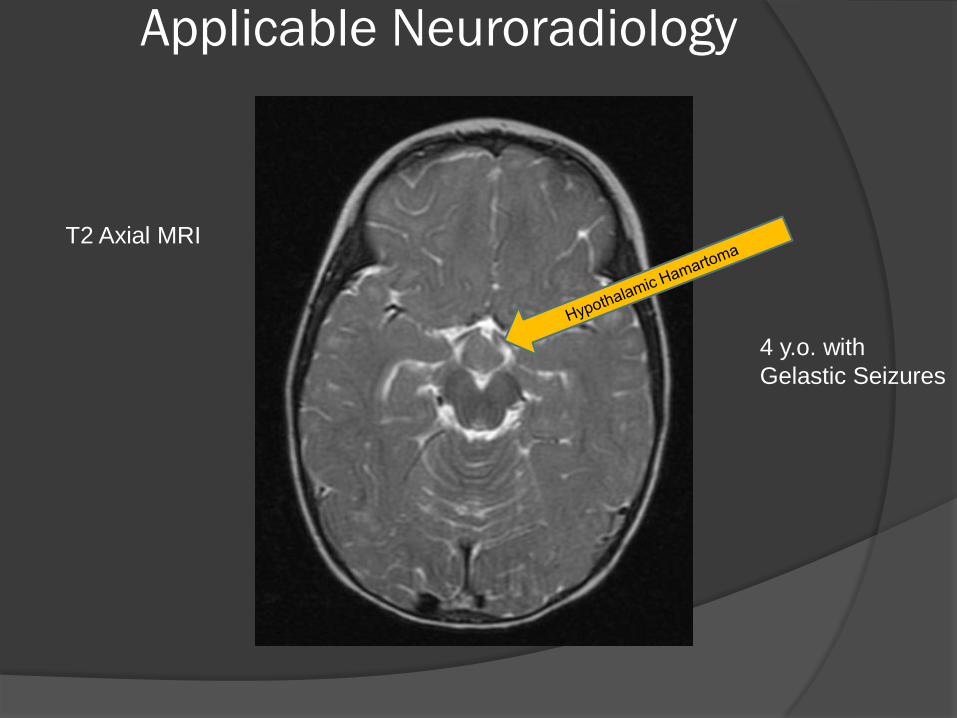
Applicable Neuroradiology
T2 Axial MRI
4 y.o. with
Gelastic Seizures
Applicable Neuroradiology
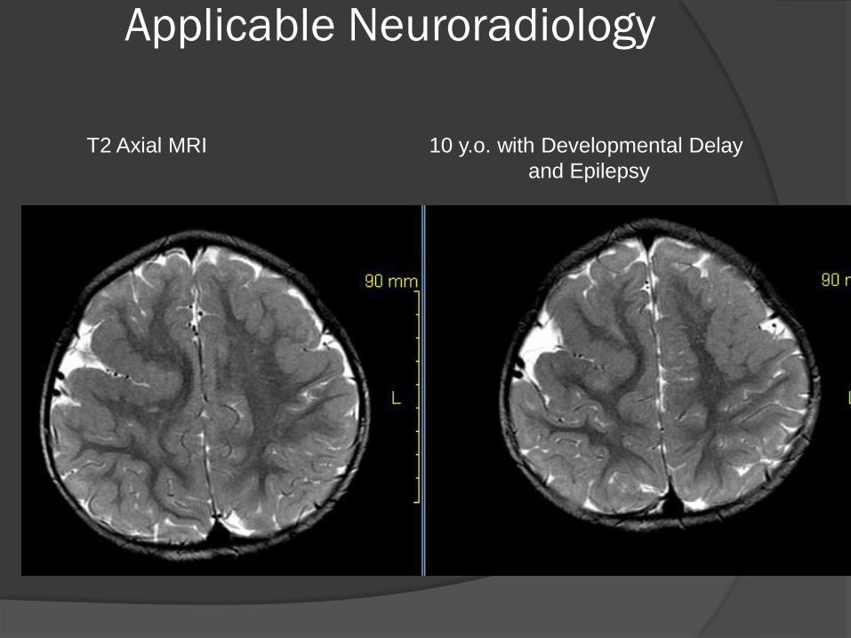
T2 Axial MRI 10 y.o. with Developmental Delay
and Epilepsy
Applicable Neuroradiology
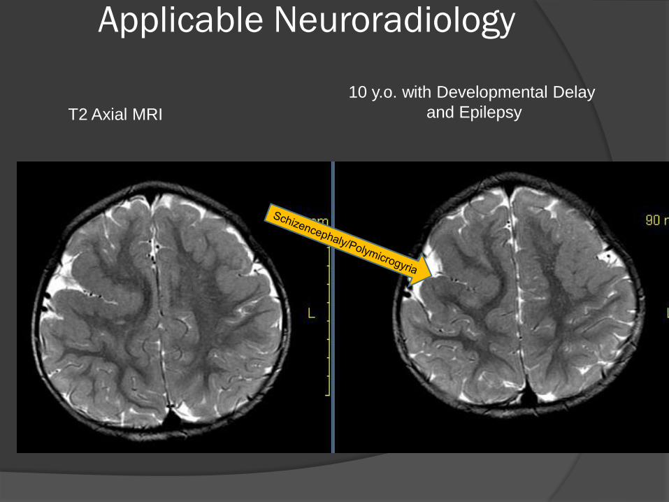
T2 Axial MRI
10 y.o. with Developmental Delay
and Epilepsy
Applicable Neuroradiology
The End