Embed Size (px)
Citation preview

403
Appendix C—Calculating Excited States using Gaussian This appendix contains methods for using Gaussian 0378 and Gaussian 09121 to
calculate excited states of molecules. Such methods are useful for estimating the
electronic transition frequencies of molecules, particularly for the open shell peroxy
radicals studied in our lab. In particular, these methods have been used to study
hydroxymethylperoxy (HMP), described in Part 3 of this thesis. Testing and verification
of these methods was performed on alkyl peroxy radicals, since extensive experimental
and computational work has been performed on these species.44
Methods described in this appendix:
1) Configuration Interaction Singles (CIS)
2) Time-Dependent HF and DFT (TD-HF and TD-DFT)
3) Exploiting Orbital Symmetry
4) Freezing the Initial Orbital Guess Using Guess=(Alter,Always)
5) Scaling of Transition Frequencies
6) Composite Quantum Chemistry Methods: G2, CBS-QB3, and W1
7) Generating Potential Energy Surfaces
8) A Caution on Using EOM-IP
1) Configuration Interaction Singles (CIS)
The Configuration Interaction Singles method (CIS) is a “zeroth order method” to
approximate excited state energies.196 Excited states are determined by combinations of
single excitations from the Hartree Fock wavefunction. CIS calculations tend to give

404
very rough estimates of transition frequencies (±4000 cm−1). In general, one should select
a more accurate method to estimate electronic transition frequencies. On the other hand,
CIS calculations are very cheap, and can be used on larger molecules where ab initio or
DFT methods would be unfeasible.
A CIS calculation can be requested using the following route line in Gaussian:
#p opt CIS/6-31+g(d,p) geom=connectivity Density=Current
There are a few things to point out in this route line. First, CIS calculations cannot
be paired with a higher level of theory (such as CCSD). Specifying both CIS and a level
of theory will default to a CIS calculation. Second, a CIS calculation should always be
run with the Opt keyword to ensure that the excited state analysis is performed on an
optimized geometry. Finally, note that the Density=Current command requests that
all analysis be done on the CIS wavefunction, rather than the HF wavefunction. As a
general rule, Density=Current should be included in all calculations where the
dipole moment integrals are needed.
The relevant CIS data are found near the end of the output file. These data include
transition dipole moments, transition frequencies, oscillator strengths, and information on
which orbitals are involved in the excitation. Shown below is the output for a CIS
calculation on the HMP radical (discussed in Part 3 of this thesis).
*********************************************************************** Excited states from <AA,BB:AA,BB> singles matrix: *********************************************************************** 1PDM for each excited state written to RWF 633 Ground to excited state Transition electric dipole moments (Au):

405 state X Y Z Osc. 1 0.0043 0.0068 0.0048 0.0000 2 -0.0526 -0.1119 -0.1460 0.0058 3 -0.0183 0.0033 -0.0028 0.0001 Ground to excited state transition velocity dipole Moments (Au): state X Y Z Osc. 1 0.0005 -0.0027 -0.0022 0.0003 2 0.0069 0.0121 0.0247 0.0023 3 -0.0109 -0.0028 0.0108 0.0006 Ground to excited state transition magnetic dipole Moments (Au): state X Y Z 1 -0.5531 0.7356 -0.3320 2 -0.7098 -0.2665 0.6301 3 0.0279 0.0486 0.0881 <0|del|b> * <b|rxdel|0> (Au), Rotatory Strengths (R) in cgs (10**-40 erg-esu-cm/Gauss) state X Y Z R(velocity) 1 -0.0003 -0.0020 0.0007 -12.7346 2 -0.0049 -0.0032 0.0156 7.4106 3 -0.0003 -0.0001 0.0010 0.4657 <0|r|b> * <b|rxdel|0> (Au), Rotatory Strengths (R) in cgs (10**-40 erg-esu-cm/Gauss) state X Y Z R(length) 1 -0.0024 0.0050 -0.0016 -0.2556 2 0.0374 0.0298 -0.0920 5.8450 3 -0.0005 0.0002 -0.0002 0.1418 <0|del|b> * <b|r|0> (Au) state X Y Z Osc.(frdel) 1 0.0000 0.0000 0.0000 0.0000 2 -0.0004 -0.0014 -0.0036 0.0036 3 0.0002 0.0000 0.0000 -0.0001 Ground to excited state transition densities written to RWF 633 Excitation energies and oscillator strengths: Excited State 1: ?Spin -A 0.7752 eV 1599.45 nm f=0.0000 12B -> 17B 0.13373 14B -> 17B -0.27905 14B -> 18B 0.19594 14B -> 19B 0.16934 14B -> 20B -0.14236 14B -> 21B 0.11446 14B -> 26B -0.11191 15B -> 17B 0.23868 15B -> 18B -0.16647 15B -> 19B -0.14434 15B -> 20B 0.12165 16B -> 17B -0.44498 16B -> 18B 0.31356 16B -> 19B 0.26832 16B -> 20B -0.22451 16B -> 21B 0.17976 16B -> 26B -0.16624 16B -> 29B -0.12826 16B -> 30B 0.11778 This state for optimization and/or second-order correction. Total Energy, E(CIS) = -264.047852507

406
In this case, the HMP A-X frequency is predicted to be 0.7752 eV (1599.45 nm,
or 6252 cm−1). This is 1100 cm−1 less than the experimental frequency, determined by
CRDS (7391 cm−1). It is predicted to have an oscillator strength of 0.0000, but Gaussian
predicts this for many peroxy radicals (HO2, CH3O2•, etc.). This is because the minimum
oscillator strength that Gaussian will report is 0.0001, corresponding to an integrated
cross section of 8.85 × 10⁻¹⁷ cm molec−1. Electronic transitions with integrated cross
sections less than this will appear to have an oscillator strength of 0, a reasonable result
for the weak A-X transition in peroxy radicals. 44
Table C.1 shows a comparison of CIS A-X transition frequency to experiment for
HO2, CH3OO•, and HMP. The quantitative and qualitative agreement between theory and
experiment is absolutely terrible. CIS calculations underestimate the A-X frequency by
1000–1500 cm−1. Additionally, the CIS calculation places the A-X transition frequency
of CH3OO• lower than HO2, while experiment shows the A-X transition frequency of
CH3OO• is higher than HO2.
Table C.1. Comparison of CIS/6-31+G(d,p) to experiment for A-X electronic transitions of peroxy radicals
Molecule A-X, CIS (cm−1) A-X, Experiment (cm−1) HO2 6331 7029 54, 55
CH3OO• 5857 7382 44 HOCH2OO• 6252 7391
2) Time-Dependent HF and DFT (TD-HF and TD-DFT)
The time-dependent versions of Hartree-Fock and Density Functional Theory
(TD-HF and TD-DFT) can also predict excited state energies. Both methods are
extensions of the respective time-independent theories (HF and DFT). While the

407
time-independent methods do not report excited state frequencies, the time dependent
formulations do.
To request a time-dependent calculation in Gaussian, simply append the TD
keyword to the rest of the route section. The program will automatically select TD-HF or
TD-DFT based on the level of theory requested. Shown below is the route line for a TD-
DFT calculation.
#p Opt B3LYP/6-31+G(d,p) TD geom=connectivity
The relevant section of the output looks similar to the CIS output. Shown below is the
result of a TD-B3LYP calculation on HO2.
***********************************************************************
Excited states from <AA,BB:AA,BB> singles matrix: *********************************************************************** 1PDM for each excited state written to RWF 633 Ground to excited state Transition electric dipole moments (Au): state X Y Z Osc. 1 0.0000 0.0000 -0.0198 0.0000 2 -0.0400 -0.5619 0.0000 0.0418 3 0.0159 0.1112 0.0000 0.0017 Ground to excited state transition velocity dipole Moments (Au): state X Y Z Osc. 1 0.0000 0.0000 0.0043 0.0003 2 0.0078 0.1050 0.0000 0.0374 3 -0.0041 -0.0206 0.0000 0.0015 Ground to excited state transition magnetic dipole Moments (Au): state X Y Z 1 -0.0795 -0.8866 0.0000 2 0.0000 0.0000 0.0309 3 0.0000 0.0000 -0.0447 <0|del|b> * <b|rxdel|0> (Au), Rotatory Strengths (R) in cgs (10**-40 erg-esu-cm/Gauss) state X Y Z R(velocity) 1 0.0000 0.0000 0.0000 0.0000 2 0.0000 0.0000 0.0000 0.0000 3 0.0000 0.0000 0.0000 0.0000 <0|r|b> * <b|rxdel|0> (Au), Rotatory Strengths (R) in cgs (10**-40 erg-esu-cm/Gauss) state X Y Z R(length)

408 1 0.0000 0.0000 0.0000 0.0000 2 0.0000 0.0000 0.0000 0.0000 3 0.0000 0.0000 0.0000 0.0000 <0|del|b> * <b|r|0> (Au) state X Y Z Osc.(frdel) 1 0.0000 0.0000 -0.0001 0.0001 2 -0.0003 -0.0590 0.0000 0.0395 3 -0.0001 -0.0023 0.0000 0.0016 Ground to excited state transition densities written to RWF 633 Excitation energies and oscillator strengths: Excited State 1: ?Spin -A" 0.9919 eV 1249.97 nm f=0.0000 8B -> 9B 1.08132 This state for optimization and/or second-order correction. Total Energy, E(RPA) = -150.876870963 Copying the excited state density for this state as the 1-particle RhoCI density. Excited State 2: ?Spin -A' 5.3799 eV 230.46 nm f=0.0418 7A -> 10A -0.17402 8A -> 13A 0.11876 9A -> 10A -0.19873 7B -> 9B 0.95400 Excited State 3: ?Spin -A' 5.4896 eV 225.85 nm f=0.0017 9A -> 10A -0.63390 9A -> 11A 0.25689 9A -> 14A -0.15605 7B -> 9B -0.17966 8B -> 10B 0.65845 8B -> 11B -0.35617 8B -> 14B 0.20378 Leave Link 914 at Tue Apr 06 09:17:52 2010, MaxMem= 167772160 cpu: 4.0 (Enter C:\G03W\l114.exe)
There are a couple of things to note about the TD-DFT calculations. Similar to the
CIS calculation, dipole moment integrals, excited state frequencies, oscillator strengths,
and the molecular orbitals involved in excitation are reported. The above output reports
the A-X frequency of HO2 to be 0.9919 eV (1249.97 nm, or 8000 cm−1). This
overestimates the A-X frequency by 1000 cm−1 (experimental value of 7029 cm−1).54, 55
Additionally, the TD-DFT calculations are very computationally expensive when paired
with the Opt keyword. One workaround is to optimize the geometry using

409
time-independent DFT, and then run a single point TD-DFT calculation. While this
method will reduce the computational expense, it is also less accurate than a full TD-DFT
optimization.
Table C.2 shows a comparison of TD-DFT A-X transition frequency to
experiment for HO2, CH3OO•, and HMP. TD-DFT values were calculated both with and
without the Opt keyword, as described above. When the Opt keyword was used on the
calculations of CH3OO• and HMP, Gaussian would crash after a few optimization steps,
reporting that the Hessian structure was not correct. We do notice that the single point
TD-DFT calculations are able to correctly predict the trend in A-X frequency amongst
the three radicals, suggesting that the single point TD-DFT method can be used to predict
relative positions in peroxy radicals.
Table C.2. Comparison of TD-DFT to experiment for A-X electronic transitions of peroxy radicals (TD-B3LYP/6-31+G(d,p))
Molecule A-X, TDDFT SP (cm−1)
A-X, TDDFT Opt (cm−1)
A-X, Experiment (cm−1)
HO2 8932 8000 7029 54, 55 CH3OO• 9287 not computed 7382 44
HOCH2OO• 9303 not computed 7391
The authors of NWChem,197 a computational chemistry program published by
Pacific Northwest National Laboratories, offer a comment on the accuracy of TD-DFT:
“The accuracy of TDDFT may vary depending on the exchange-correlation functional. In general, the exchange-correlation functionals that are widely used today and are implemented in NWChem work well for low-lying valence excited states. However, for high-lying diffuse excited states and Rydberg excited states in particular, TDDFT employing these conventional functionals breaks down and the excitation energies are substantially underestimated.”

410
TD-HF is roughly comparable in accuracy to CIS. My experience with TD-DFT is
absolute accuracy of ±1000 cm−1 (based on the above calculations on HO2, CH3OO•, and
HMP). In general, I would not ever run a TD-HF calculation, since the CIS calculation
will be of comparable accuracy and less expensive. TD-DFT can be thought of as one
step more accurate than CIS. However, the computational expense of TD-DFT
calculations makes them generally unsuitable for anything with more than a few carbons.
3) Exploiting Orbital Symmetry
The optimization algorithm in Gaussian will do its best to keep the same
molecular symmetry as the input geometry.79 Thus, if the input file contains a molecule in
Cs symmetry, the optimized structure will also be Cs. The same idea holds true for the
symmetry of the electronic wavefunction. If the input file contains an electronic
wavefunction with A’’ symmetry, then the optimization will attempt to keep the
electronic wavefunction in A’’ symmetry at every step.
The idea of restricting symmetries can be exploited to obtain excited state
geometries. Consider, for example, the first two states of HO2. The ground state has A’’
symmetry, while the first excited state has A’ symmetry. Suppose that the input file
requests a geometry optimization, with an initial electronic state guess of A’ symmetry.
During the optimization, Gaussian will always keep the electronic state symmetry as A’.
The result of the optimization will be the lowest energy structure with A’ electronic
symmetry, i.e., the first excited state.
The initial orbital population can be changed using Guess=Alter in the route
line. This command allows users to switch pairs of orbitals that are to be populated. (For

411
example, switching orbitals 4 and 5 will cause Gaussian to populate the orbitals in this
order: 1, 2, 3, 5, 4, 6, 7, …) Switching two core orbitals will have no effect. (However,
such an operation is not completely useless; it may be necessary to switch core orbital
filling in a CASSCF job.) Switching a core or valence orbital with a virtual orbital will
allow the user to change the electronic symmetry. These pairs of orbitals are listed at the
end of the input file.
If the calculation uses a restricted method (RHF, RCCSD, etc.), then only one line
is necessary to change orbitals. If the calculation uses an unrestricted method (UHF,
UCCSD, etc.), then separate lines are necessary for and electrons. Both lines must be
present in the input file. If no electrons of one type are to be changed, then the line should
be blank.
The following example file calculates the first excited state of HO2. Since HO2
has 17 electrons (9 , 8 ), we will change the order of orbitals 8 and 9. Since no
electrons are being altered, we put a blank line before the orbital line (this is in
addition to a blank line needed to separate the connectivity section from the orbital
alteration section).

412 %chk=HO2 B3LYP A Opt.chk %nproc=1 #p opt B3LYP/6-31+g(d,p) geom=connectivity Guess=Alter HO2 A State MKS, 4/6/10 0 2 O H 1 B1 O 1 B2 2 A1 B1=0.98052408 B2=1.33330037 A1=105.52948981 1 2 1.0 3 1.0 2 3 8 9
The output file will look exactly like a normal optimization file. However, the
user should ensure that the final symmetry is the excited state electronic symmetry,
and not the ground state. Sometimes, the optimization will “relax” down to the ground
state symmetry. I am not sure what the reason for this is, but it does happen from time to
time. The symmetry can be found in the final output (highlighted below).
(Enter C:\G03W\l9999.exe) Final structure in terms of initial Z-matrix: O H,1,B1 O,1,B2,2,A1 Variables: B1=0.97637273 B2=1.39529609 A1=103.55791628 1|1|UNPC-UNK|FOpt|UB3LYP|6-31+G(d,p)|H1O2(2)|PCUSER|06-Apr-2010|4||#P OPT B3LYP/6-31+G(D,P) GEOM=CONNECTIVITY GUESS=ALTER|Title Ca rd Required||0,2|O,-0.6348300196,0.,0.1172381389|H,-0.6030213526,0.,1. 0930925935|O,0.7102076887,0.,-0.253874713|||8,9||Version=IA32W-G03RevC .01|State=2-A'|HF=-150.8824122|S2=0.753042|S2-1=0.|S2A=0.750007|RMSD=3 .483e-009|RMSF=8.428e-005|Dipole=-0.1481518,0.,0.8045714|PG=CS [SG(H1O 2)]||@

413
In this case, the electronic state is listed as 2-A’, which is the electronic
symmetry of the first excited state. The final energy can be compared to the ground state
(calculated separately), and the A-X frequency can be computed. For HO2 at
B3LYP/6-31+G(d,p), the A-X frequency is computed as 7251 cm−1, 222 cm−1 higher than
the experimental value.
4) Freezing the Initial Orbital Guess Using Guess=(Alter,Always)
Note: The paper on alkyl peroxy spectra by Sharp et al. describe populating the
electronic orbitals in a low level calculation, then running a high level calculation using
the orbitals obtained from the previous calculation.44 It is unknown to me whether Sharp
uses a separate script or the method described below. As will be shown, our method can
reproduce Sharp’s results, and is, at a minimum, equivalent to their method. The
discussion below should help future students perform these calculations.
Most conformations of the molecules that we are interested in do not have any
point group symmetry (i.e., they belong to the C1 point group). As such, there is no
electronic wavefunction symmetry to take advantage of, and simply using the keyword
Guess=Alter will result in the optimization relaxing back to the ground state.
However, by changing this command to Guess=(Alter,Always), Gaussian 03W
will repopulate the electron orbitals at each step of the SCF calculation. The result of this
is “freezing” the electrons in a non-ground-state configuration. Such a calculation will
allow for the excited state energy of any molecule to be computed. Unlike a CIS
calculation, the geometry and orbitals are optimized in the excited state as opposed to the

414
ground state, and thus the energies obtained should be vastly more accurate than a CIS
calculation.
The syntax and output are the same as described in the previous section, with the
exception of changing Guess=Alter to Guess=(Alter,Always). The A-X
transition frequencies obtained on C1 conformers of a molecule are consistent with results
obtained from Cs conformers (i.e., exploiting orbital symmetry as described in the
previous section). This can be illustrated by mapping out the full potential energy
surfaces, and showing that the Cs transition frequencies agree with the surrounding C1
transition frequencies, as has been done for HMP (see the surfaces presented in Part 3 of
this thesis). This consistency strongly implies that this method of obtaining excited states
is valid for molecules with no symmetry.
Table C.3 shows a comparison of Guess=(Alter,Always) A-X transition
frequency to experiment for HO2, CH3OO•, and HMP. The absolute transition
frequencies are too high by 100–200 cm−1, though this represents a vast improvement
over CIS or TD-DFT. For the most part, the transition frequencies qualitatively follow
the experimental trends. Guess=(Alter,Always) is able to predict the transition
frequencies of both CH3OO• and HOCH2OO• to be higher than the transition frequency
of HO2. Unfortunately, it also predicts HOCH2OO• to have a lower frequency than
CH3OO•. Sharp et al. make similar observations: the Guess=(Alter,Always)
calculations become less accurate as the peroxy radical of interest becomes more
complicated (either additional functional groups, or a longer carbon chain).

415
Table C.3. Comparison of the Guess=(Alter,Always) method to experiment for A-X electronic transitions of peroxy radicals (B3LYP/6-31+G(d,p))
Molecule A-X, Alter,Always (cm−1)
A-X, Experiment (cm−1)
HO2 7251 7029 54, 55 CH3OO• 7622 7382 44
HOCH2OO• 7501 7391
5) Scaling of Transition Frequencies
The calculation methods presented above can often get the correct qualitative
behavior for the transition frequencies. For example, the Guess=(Alter,Always)
method at B3LYP/6-31+G(d,p) is able to predict that CH3OO• and HOCH2OO• both
have a higher A-X transition frequency than HO2. However, the quantitative agreement
between calculated and experimental transition frequencies is not very good, ranging in
accuracy from ±300 cm−1 (Guess=Alter,Always) to ±1500 cm−1 (single point
TD-DFT). As presented so far, the computed transition frequencies are of limited aid for
predicting electronic transitions that have not yet been detected.
A careful look at Tables C.1–C.3 reveals that each particular method tends to
overestimate or underestimate the transition frequency by a similar amount, regardless of
the specific peroxy radical being studied. This observation suggests that if we scale the
calculated frequencies to a well-studied peroxy radical, such as HO2, we will be able to
obtain quantitatively accurate transition frequencies. Scaling should eliminate systematic
errors from the level of theory and basis being used. Additionally, if the reference
molecule is chemically similar to the molecules of interest, we expect that the electronic
surfaces will have similar curvature and harmonic frequencies. Thus, we will also
account for any changes in the zero point vibrational energy (ZPVE).

416
Table C.4 contains the scaled A-X transition frequencies for CH3OO• and
HOCH2OO•, using each of the methods to obtain this frequency described above (CIS,
TD-DFT, Guess=Alter,Always). The frequencies are scaled to the experimentally
determined A-X transition of HO2 of 7029 cm−1.54, 55 The scaled frequencies are in
general closer to the experimental values than the unscaled frequencies. While CIS still
gives a poor prediction for the transition frequency, TD-DFT and
Guess=(Alter,Always) are able to predict the A-X frequency within 100 cm−1.
Although this is still considered considerable error, both TD-DFT and
Guess=(Alter,Always) can be used to aid in experimental detection of A-X
transitions in peroxy radicals. In order to obtain better quantitative agreement, we must
move on to more sophisticated (and thus more expensive) methods, described in the next
section.
Table C.4. Comparison of scaled frequencies to experiment for A-X electronic transitions of peroxy radicals. Frequencies scaled to A-X (HO2) = 7029 cm−1. (B3LYP/6-31+G(d,p))
Molecule CIS (cm−1)
TD-DFT, no opt(cm−1)
Guess=Alter,Always (cm−1)
Expt. (cm−1)
HO2 — — — 7029 54, 55 CH3O2• 6503 7309 7390 7382 44
HOCH2O2• 6942 7321 7272 7391
6) Composite Quantum Chemistry Methods: G2, CBS-QB3, and W1
Composite quantum chemistry methods are designed for high-accuracy
thermochemical calculations. The purpose of these calculations is to approximate the
energy at a high level of theory and large basis by performing many smaller calculations

417
(high level and small basis, or low level and large basis). Three methods used in the
current chemical literature are G2,123, 198-201 CBS-QB3,202-205 and W1.206-211 Of these, G2
has been used frequently in calculations of alkyl peroxy radicals. It has been shown to
predict the A-X transition frequencies within ±50 cm−1 without the need for scaling.44
Sharpe et al. show that as the alkyl chain gets larger, the frequencies predicted by the G2
method become less accurate. The accuracy may or may not get worse when additional
functional groups are present.
The purpose of the G2 method is to estimate the QCISD(T)/6-311+G(3df,2p)
energy by computing less expensive energies at lower levels of theory, or smaller basis
sets. These energies are used to estimate the contributions to the energy due to increasing
the basis set. A G2 calculation consists of the following steps:
1) Geometry optimization at HF/6-31G(d)
2) Frequency calculation at the geometry from step 1, to obtain the zero point
energy
3) Geometry optimization at MP2(Full)/6-31G(d)
4) Using the geometry from step 3, single point calculations at
a) QCISD(T,E4T)/6-311G(d,p) (see below for explanation of “E4T”)
b) MP4(SDTQ)/6-311G(2df,p)
c) MP4(SDTQ)/6-311+G(d,p)
d) MP2/6-311+G(3df,2p)
Note that the MP4(SDTQ) and QCISD(T) calculations will also provide MP2 and
MP4 energies, at no additional cost. The user must request for triples to be calculated
during the QCISD(T) calculation by using the method QCISD(T,E4T).

418
The G2 energy can then be computed from Equation C.1, as defined in Pople’s
G1 and G2 papers:123, 198, 201
E G2 = QCISD(T)/6-311G d,p
+ MP4/6-311G 2df,p - MP4/6-311G d,p
+ MP4/6-311+G d,p - MP4/6-311G d,p
+ MP2/6-311+G 3df,2p - MP2/6-311G 2df,p
+ MP2/
6-311G d,p - MP2/6-311+G d,p
+ 0.8929×ZPVE
+ HLC,
(C.1)
where the terms labeled {xx/yy} denote the single point energy at
xx/yy//MP2(Full)/6-31G(d), ZPVE is the zero point vibrational energy, 0.8929 is the
HF/6-31(d) scaling factor for the harmonic frequencies, and HLC is a “high level
correction.” The HLC is defined as
V UHLC 4.81 mhartree × N 0.19 mhartree × N , (C.2)
where NV is the total number of valence electrons, and NU is the number of unpaired
valence electrons. For example, in HMP, there are 33 total electrons. 8 of these 33 are
core (1s) electrons, leaving 25 valence electrons (NV), 1 of which is unpaired (NU). The
HLC is -60.19 mhartree.
For ground state molecules, the G2 method can be directly requested in G03W,
via the following route line:
#p G2 geom=connectivity
Gaussian will automatically carry out the calculations described above. The end
of the output file contains information on the G2 energies. Shown below is the output for
a G2 calculation on HMP:

419 Temperature= 298.150000 Pressure= 1.000000 E(ZPE)= 0.047510 E(Thermal)= 0.051972 E(QCISD(T))= -264.903868 E(Empiric)= -0.073870 DE(Plus)= -0.015881 DE(2DF)= -0.141516 G1(0 K)= -265.087624 G1 Energy= -265.083162 G1 Enthalpy= -265.082218 G1 Free Energy= -265.115071 E(Delta-G2)= -0.017592 E(G2-Empiric)= 0.013680 G2(0 K)= -265.091536 G2 Energy= -265.087074 G2 Enthalpy= -265.086130 G2 Free Energy= -265.118983 DE(MP2)= -0.166853 G2MP2(0 K)= -265.083401 G2MP2 Energy= -265.078938 G2MP2 Enthalpy= -265.077994 G2MP2 Free Energy= -265.110847
The G2 energy is listed as G2 (0 K). In this case, the G2 energy is −265.091536 au.
It is not possible to use the built in G2 method for calculating excited states,
because Gaussian will not pass the Guess=(Alter,Always) command correctly to
each step. Instead, the user must manually request the calculations described above (steps
1-4), and extract the correct energies from each file. I wrote a G2 calculator in Excel to
automatically calculate the G2 energy: the user types in the energies from each part, and
the program determines the G1 and G2 energies (Figure C.1 shows a screenshot of the
calculator). Typically, the G2 energy obtained through the calculator only differs from
the G2 energy directly reported by G03W by a few microhartree (<1 cm−1). Once the X
and A states have been calculated, the energy difference should be used directly as the
transition frequency. No further energy scaling is required (such as the scaling described
in the previous section), because differences in the ZPVE for the X and A states have
already been accounted for.

420
Figure C.1. Screenshot of the G2 energy calculator spreadsheet. The calculated G1 and G2 energies are circled.
Table C.5 shows a comparison of G2 A-X transition frequency to experiment for
HO2, CH3OO•, and HOCH2OO•. The quantitative agreement between calculation and
experiment is quite good. HO2 and HOCH2OO• are calculated to within 40 cm−1 of their
experimental values, while CH3OO• is calculated to better than 10 cm−1. It should be

421
noted that G2 is generally a much more expensive method than using Guess=(Alter,
Always), although this depends on the level of theory and basis used. It may be
unfeasible to run G2 calculations on larger alkyl peroxies without access to a
supercomputer.
Table C.5. Comparison of G2 calculated frequencies to experiment for A-X electronic transitions of peroxy radicals
Molecule A-X (G2) (cm−1) A-X, Experiment (cm−1)
HO2 7060 7029 54, 55 CH3OO• 7375 7382 44
HOCH2OO• 7424 7391
As shown in Part 3 of this thesis, the G2 method cannot locate all three
conformers (local minima) of the HMP radical. The MP2(Full)/6-31+G(d,p) potential
energy surface of HMP reveals that one of the local minima from CCSD and B3LYP
calculations becomes a shelf at MP2(Full). Because the CCSD calculation is likely more
accurate than the MP2(Full) calculation, my thought is that the G2 method is inadequate
for conformer searches of substituted peroxy radicals.
Two other composite chemistry methods are also available in Gaussian 03: the
complete basis set methods by Petersson (denoted as CBS methods),202-205 and the
Weizmann-1 method (W1).206-211 In particular, two methods of interest are CBS-QB3
(based on a B3LYP geometry optimization) and W1U (based on a UB3LYP geometry
optimization and UCCSD energy corrections). The work described in Part 3 of this thesis
shows that the B3LYP and CCSD potential energy surfaces of HMP are in qualitative
agreement. Furthermore, both methods are able to predict the A-X transition of HMP to
within ±80 cm−1 when scaled to HO2 (as seen in the previous section). Thus, composite
422
chemistry methods based on these levels of theory could lead to improvements in A-X
transition predictions.
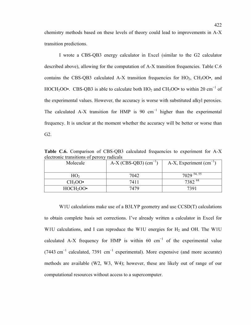
I wrote a CBS-QB3 energy calculator in Excel (similar to the G2 calculator
described above), allowing for the computation of A-X transition frequencies. Table C.6
contains the CBS-QB3 calculated A-X transition frequencies for HO2, CH3OO•, and
HOCH2OO•. CBS-QB3 is able to calculate both HO2 and CH3OO• to within 20 cm−1 of
the experimental values. However, the accuracy is worse with substituted alkyl peroxies.
The calculated A-X transition for HMP is 90 cm−1 higher than the experimental
frequency. It is unclear at the moment whether the accuracy will be better or worse than
G2.
Table C.6. Comparison of CBS-QB3 calculated frequencies to experiment for A-X electronic transitions of peroxy radicals
Molecule A-X (CBS-QB3) (cm−1) A-X, Experiment (cm−1)
HO2 7042 7029 54, 55 CH3OO• 7411 7382 44
HOCH2OO• 7479 7391
W1U calculations make use of a B3LYP geometry and use CCSD(T) calculations
to obtain complete basis set corrections. I’ve already written a calculator in Excel for
W1U calculations, and I can reproduce the W1U energies for H2 and OH. The W1U
calculated A-X frequency for HMP is within 60 cm−1 of the experimental value
(7443 cm−1 calculated, 7391 cm−1 experimental). More expensive (and more accurate)
methods are available (W2, W3, W4); however, these are likely out of range of our
computational resources without access to a supercomputer.

423
It should be noted that the 32-bit version of Gaussian cannot perform a W1
calculation on HO2 (or anything larger), due to the CCSD and CCSD(T) calculations
exceeding the 16 GB scratch space limit for 32-bit programs. There are no problems
running the W1 calculations on the 64-bit version of Gaussian in Linux. Furthermore,
other 64-bit programs are also be capable of these calculations (CFour, Molpro, Q-Chem,
etc.). Significant amounts of memory are required for these calculations: the W1
calculation on HMP exceeded the 10 GB of RAM that I initially had allocated to
Gaussian.
7) Generating Potential Energy Surfaces
It is generally useful to determine the potential energy surface (PES) of a
molecule as a function of molecular coordinates (bond distances, bond angles, dihedral
angles). These potential energy functions can be used to assess energy barriers to
molecular motion or reaction, coupling of vibrational and torsional normal modes due to
intramolecular interactions such as hydrogen bonding, and explicit calculation of
wavefunctions and quantum energy levels. As has been shown repeatedly in this thesis
(HOONO, HMP, 2-HIPP), normal mode coupling causes additional complexity in the IR
spectra, and must be explicitly modeled in order to correctly simulate the spectra.
Two types of potential energy scans can be requested in Gaussian: a rigid scan in
which all other molecular coordinates remain fixed, or a relaxed scan in which all other
molecular coordinates are optimized at each step. Rigid scans are useful in the adiabatic
approximations: fast vibrational modes can be completely separated from slow modes.
This is true when separating OH stretch motions from torsional motions in many

424
molecules, including HOONO and HMP. Relaxed scans are useful when an adiabatic
approximation cannot be made. This is true when scanning over dihedral angles in a
molecule: the torsional normal modes associated with these angles are typically of low
frequency, and many other low frequency modes are generally present.
Scans are typically performed across internal coordinates rather than a Cartesian
coordinate; therefore, it is easiest to request the potential energy scan directly from the
Z-matrix input. The following example requests a relaxed potential energy scan across
the two torsional angles (HOCO, OOCO) on the A state of the 2-HIPP radical, with the
essential parts of requesting the scan highlighted.

425 %chk=C:\G09W\MKS\2-HIPP PES\2-HIPP A B3LYP PES.chk #p Opt=Z-matrix B3LYP/6-31+g(d,p) Guess=(Alter,Always) NoSymm Title Card Required 0 2 C C 1 B1 H 2 B2 1 A1 H 2 B3 1 A2 3 D1 0 H 2 B4 1 A3 4 D2 0 C 1 B5 2 A4 5 D3 0 H 6 B6 1 A5 2 D4 0 H 6 B7 1 A6 2 D5 0 H 6 B8 1 A7 2 D6 0 O 1 B9 2 A8 6 D7 0 H 10 B10 1 A9 2 D8 0 O 1 B11 10 A10 11 HOCO 0 O 12 B12 1 A11 10 OOCO 0 B1=1.52722762 B2=1.0933087 B3=1.09422741 B4=1.09247629 B5=1.51904907 B6=1.09171903 B7=1.09207247 B8=1.09289496 B9=1.39714561 B10=0.96792656 B11=1.48479112 B12=1.37990918 A1=109.09594428 A2=111.00707754 A3=110.52283361 A4=114.39312339 A5=110.61869101 A6=109.68521782 A7=108.76323525 A8=112.59779458 A9=109.77029369 A10=109.73656718 A11=112.28122574 D1=119.77962987 D2=120.04129575 D3=-60.2624729 D4=55.87981057 D5=176.84253762 D6=-64.27682433 D7=-123.2116855 D8=-120.0 HOCO=0.0 S 18 10.0 OOCO=0.0 S 35 10.0 24 25

426
There are four essential features to this input file. First, the keyword
Opt=Z-matrix must be entered in the route line. This ensures that all optimization
proceeds using Z-matrix coordinates. If only Opt is entered, the scan will fail. Second,
the command NoSymm must be entered in the route line. If the scan breaks point group
symmetry and NoSymm has not been requested, the scan will fail and Gaussian will exit
with an error message. Third, all of the coordinates within the Z-matrix have been entered
as variables (R1, A1, D1, etc.). The variables of interest have been specially labeled
(HOCO and OOCO for the dihedral angles). Although this is not strictly necessary, it is
good practice to highlight which variables are being scanned. Fourth, the entries for the
variables being scanned over are not a single number; rather, they specify the initial
parameter, number of steps, and step size. For example, the line HOCO=0.0 S 18
10.0 requests that the variable HOCO start at a value 0.0, and then be scanned (S) with
18 steps of size 10.0 (final value of 180.0).
Gaussian will run all of the geometries consecutively and print the energies at the
end of the file. It is more useful to compile all of the energies into one text file for further
use in Excel (for data manipulation) and SigmaPlot (for surface plotting). GaussView can
compile all of the energies, although versions 3 and 5 do this differently. Version 3
compiles the energies in the same order in which Gaussian ran them, while version 5
reorders them in ascending order by coordinate. Additionally, GaussView 5 will display a
3-dimensional surface for multidimensional scans. Figure C.2 shows the graphical output
of GaussView 3.09 and GaussView 5.09 for scans of the X state of 2-HIPP.

427
Figure C.2. Visualization of energies generated from a 2-D potential energy scan of 2-HIPP in GaussView 3.09 (left) and GaussView 5.09 (right). GaussView 3.09 simply displays the energies in the order in which they were calculated, while GaussView 5.09 generates a contour plot.
The 2-dimensional potential energy surfaces presented in this thesis were
generated in SigmaPlot. SigmaPlot requires a periodic arrangement of data. In other
words, to generate a plot across 2 dihedral angles of 360° apiece, the input data must be
arranged in the form (−180°, −180°), (−180°, −170°), …, (−180°, 180°), (−170°, −180°),
(−170°, −170°), etc. For many scans, only half of these points are explicitly calculated
(the other half are equal in energy by symmetry). Before inputting energies into
SigmaPlot, the user must generate all of the necessary energies in Excel by exploiting
symmetry. Afterwards, the energies should be sorted into a periodic form, typically
achieved by sorting one variable followed by a second variable. Finally, the sorted values
can be copied into SigmaPlot to generate a contour plot similar to the ones presented in
Parts 2 and 3 of this thesis.

428
8) A Caution on Using Equation of Motion (EOM) Methods
Note that a full discussion of Equation of Motion (EOM) methods is not included
here because Gaussian 03 does not contain these methods. Gaussian 09 as well as many
other programs (such as CFour and Q-Chem) do support EOM methods. These methods
may prove useful for future calculations; however, future students are advised to use
caution with these methods. Terry Miller’s group found that EOM-IP overestimated the
A-X transition frequency for alkyl peroxies by ~1000 cm−1.44 Even if these values are
scaled to experimental results on HO2 or CH3OO•, the transition frequencies do not
follow the experimental trends any better than the previously described methods. The
Miller group attributes the poor agreement to EOM methods not properly accounting for
electron correlation.
Acknowledgements
We thank J. Sigrid Barklund and Leah G. Dodson for suggestions on how to
improve the readability and utility of this appendix.