Embed Size (px)
Citation preview
Apoptotic Bax Gene Expression in Glioblastoma
Cells Exposed to Estradiol valerate
Farahmandlou N, Noroozi S*
Nooshin Farahmandlou (PhD) is with the Department of Basic Sciences, Faculty of Pharmacy, Pharmaceutical
Sciences Branch, Islamic Azad University, Tehran - Iran (IAUPS ) (e-mail: [email protected])
(e-mail: [email protected]).
Sanaz Noroozi (MSc) (*corresponding author) is with Department of Biology, Faculty of Basic Sciences,
GhiamDasht (East Tehran Branch), Islamic Azad University, Tehran-Iran ([email protected])
Abstract: Cancers may be influenced by sex steroids in vivo and in vitro. This research aims to determine the
effects of estradiol valerate on Bax gene expression level in glioblastoma (A172) cells in cell culture. A172 cells
were exposed to cytotoxic dose (0.1 mg/ml ) of estradiol valerate solution. Real time PCR was used to measure
Bax gene expression level. Our results indicated that exposure to 0.1 mg/ml of estradiol valerate led to
significant increase in viability compared to control cells (P<0.05). Our findings indicated that cytotoxic dose of
estradiol have apoptotic effects on brain tumor (A172) cells in cell culture.
Keywords: Estradiol , A172, Bax.
1. Introduction
Glioblastoma is the most common and aggressive primary brain tumor in adults. Defining histopathologic
features are necrosis and endothelial proliferation, resulting in the assignment of grade IV, the highest grade in
the World Health Organization (WHO) classification of brain tumors. The classic clinical term "secondary
glioblastoma" refers to a minority of glioblastomas that evolve from previously diagnosed WHO grade II or
grade III gliomas [1].
Glioblastomas have an annual incidence of 5.26 per 100,000 population or 17,000 new diagnoses per year.
These tumors are typically associated with a dismal prognosis and poor quality of life. Glioblastoma also
remains a difficult cancer to treat, although therapeutic options have been improving. Optimal management
requires a multidisciplinary approach and knowledge of potential complications from both the disease and its
treatment [2].
Estrogen exhibits a broad spectrum of physiological functions ranging from regulation of the menstrual cycle
and reproduction to modulation of bone density, brain function, and cholesterol mobilization. Despite the
beneficial actions of endogenous estrogen, sustained exposure to exogenous estrogen is a well-established risk
factor for various cancers [3]-[6].
This research was exerted to determine the apoptotic effects of cytotoxic dose of estradiol Bax gene
expression level in glioblastoma (A172) cells in cell culture.
2. Material and methods
A172 cells (glioblastoma cell line) were purchased from National Cell Bank of Iran (Pasteur Institute, Tehran,
Iran). Cells were grown and incubated in standard situation. Then, cells were sub-cultured into 75cm2 flasks, 96-
well plates or 6-well plates. Cytotoxicity of different doses of the estradiol was assayed using MTT method.
ISBN 978-93-86878-07-6
8th International Conference on Agricultural, Environment, Biology and Medical Sciences
(AEBMS-2017)
Dec. 21-22, 2017 Dubai (UAE)
https://doi.org/10.15242/HEAIG.C1217232 90
Real time PCR was used to measure Bax gene expression level. Analyses were conducted using the SPSS20 and
ANOVA.
3. Results
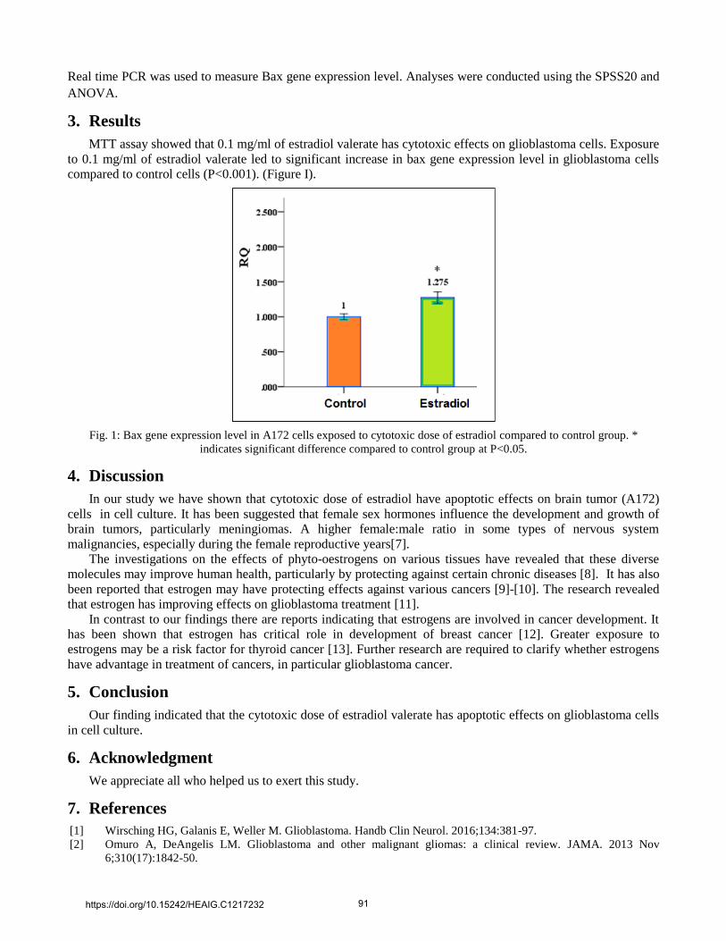
MTT assay showed that 0.1 mg/ml of estradiol valerate has cytotoxic effects on glioblastoma cells. Exposure
to 0.1 mg/ml of estradiol valerate led to significant increase in bax gene expression level in glioblastoma cells
compared to control cells (P<0.001). (Figure I).
Fig. 1: Bax gene expression level in A172 cells exposed to cytotoxic dose of estradiol compared to control group. *
indicates significant difference compared to control group at P<0.05.
4. Discussion
In our study we have shown that cytotoxic dose of estradiol have apoptotic effects on brain tumor (A172)
cells in cell culture. It has been suggested that female sex hormones influence the development and growth of
brain tumors, particularly meningiomas. A higher female:male ratio in some types of nervous system
malignancies, especially during the female reproductive years[7].
The investigations on the effects of phyto-oestrogens on various tissues have revealed that these diverse
molecules may improve human health, particularly by protecting against certain chronic diseases [8]. It has also
been reported that estrogen may have protecting effects against various cancers [9]-[10]. The research revealed
that estrogen has improving effects on glioblastoma treatment [11].
In contrast to our findings there are reports indicating that estrogens are involved in cancer development. It
has been shown that estrogen has critical role in development of breast cancer [12]. Greater exposure to
estrogens may be a risk factor for thyroid cancer [13]. Further research are required to clarify whether estrogens
have advantage in treatment of cancers, in particular glioblastoma cancer.
5. Conclusion
Our finding indicated that the cytotoxic dose of estradiol valerate has apoptotic effects on glioblastoma cells
in cell culture.
6. Acknowledgment
We appreciate all who helped us to exert this study.
7. References
[1] Wirsching HG, Galanis E, Weller M. Glioblastoma. Handb Clin Neurol. 2016;134:381-97.
[2] Omuro A, DeAngelis LM. Glioblastoma and other malignant gliomas: a clinical review. JAMA. 2013 Nov
6;310(17):1842-50.
https://doi.org/10.15242/HEAIG.C1217232 91
[3] Liang J, Shang Y. Estrogen and cancer. Annu Rev Physiol. 2013;75:225-40.
[4] Barakat R, Oakley O, Kim H, Jin J, Ko CJ. Extra-gonadal sites of estrogen biosynthesis and function. BMB Rep.
2016 Sep;49(9):488-96.
[5] Mauvais-Jarvis F, Clegg DJ, Hevener AL. The role of estrogens in control of energy balance and glucose
homeostasis. Endocr Rev. 2013 Jun;34(3):309-38
[6] Zhao Z, Wang H, Jessup JA, Lindsey SH, Chappell MC, Groban L. Role of estrogen in diastolic dysfunction. Am J
Physiol Heart Circ Physiol. 2014 Mar 1;306(5):H628-40.
[7] Helseth A. Incidence and survival of intracranial meningioma patients in Norway 1963–1992. Neuroepidemiology
1997;16:53–9.
[8] Anderson JJ, Anthony M, Messina M, Garne SC. Effects of phyto-oestrogens on tissues. Nutr Res Rev. 1999
Jun;12(1):75-116
[9] Suba Z. Circulatory estrogen level protects against breast cancer in obese women. Recent Pat Anticancer Drug
Discov. 2013 May;8(2):154-67.
[10] Honma N, Hosoi T, Arai T, Takubo K. Estrogen and cancers of the colorectum, breast, and lung in postmenopausal
women. Pathol Int. 2015 Sep;65(9):451-9.
[11] Plunkett RJ, Lis A, Barone TA, Fronckowiak MD, Greenberg SJ. Hormonal effects on glioblastoma multiforme in
the nude rat model. J Neurosurg. 1999 Jun;90(6):1072-7.
[12] Samavat H, Kurzer MS. Estrogen metabolism and breast cancer.Cancer Lett. 2015 Jan 28;356(2 Pt A):231-43.
[13] Zahid M, Goldner W, Beseler CL, Rogan EG, Cavalieri EL.Unbalanced estrogen metabolism in thyroid cancer.
Int J Cancer. 2013 Dec 1;133(11):2642-9.
https://doi.org/10.15242/HEAIG.C1217232 92

![Phytophthora sojaeAvirulence Effector Avr3b is a Secreted ... · in plants by BAX (a mammalian pro-apoptotic factor), by the PAMP infestin1 (INF1) and by several effectors [30,31]](https://img.pdfslide.us/doc/110x75/5f7a1bbd88775a60713d767e/phytophthora-sojaeavirulence-effector-avr3b-is-a-secreted-in-plants-by-bax-a.jpg)