Embed Size (px)
Citation preview
Apoptosis Detection Using the BD Accuri™ C6 Flow Cytometer
Stacey Roys, Marketing Applications Specialist, BD Biosciences
Cyndy Lane, Senior Product Manager, BD Biosciences
23-14519-00
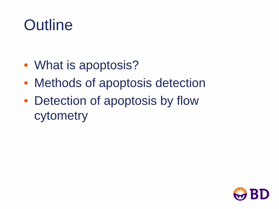
Outline
• What is apoptosis?• Methods of apoptosis detection• Detection of apoptosis by flow
cytometry

Apoptosis Definition
The process leading to controlled self destruction of a cell. Cells undergo death neatly without damaging their neighbors. Apoptosis is a “programmed event.”
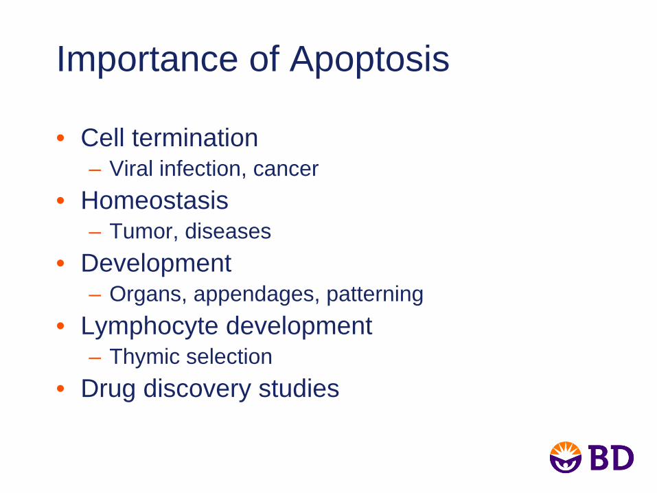
Importance of Apoptosis
• Cell termination– Viral infection, cancer
• Homeostasis– Tumor, diseases
• Development– Organs, appendages, patterning
• Lymphocyte development– Thymic selection
• Drug discovery studies
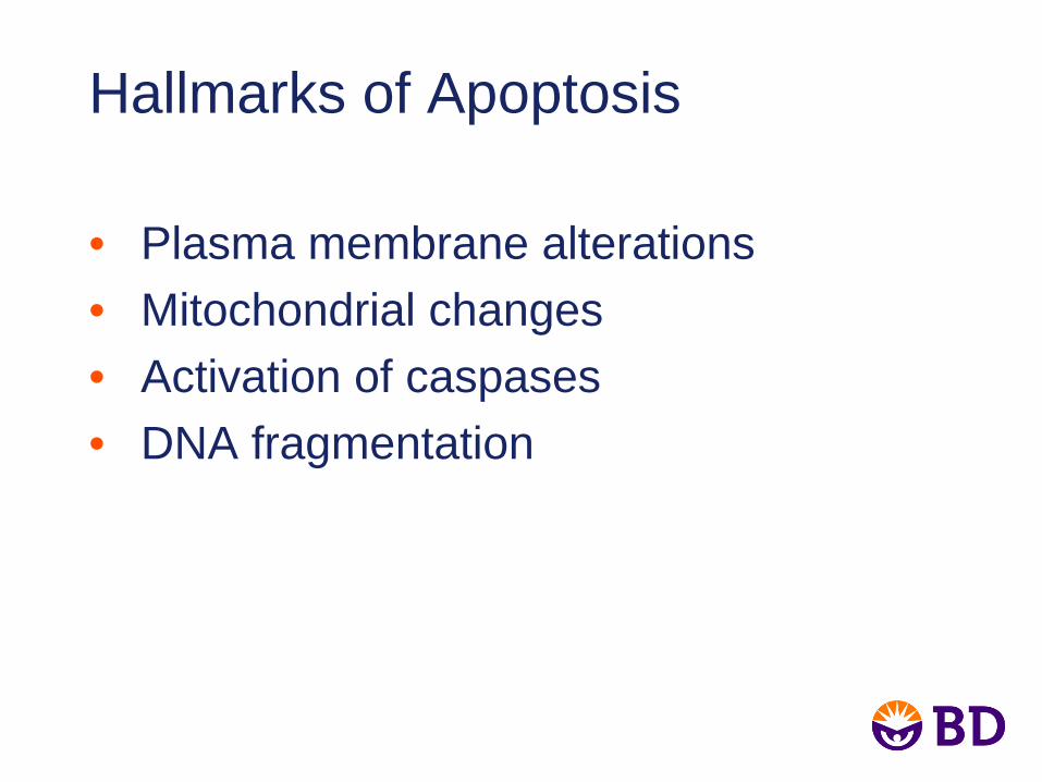
Hallmarks of Apoptosis
• Plasma membrane alterations• Mitochondrial changes• Activation of caspases• DNA fragmentation
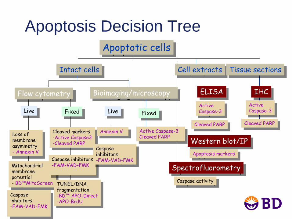
Apoptosis Decision TreeApoptotic cellsApoptotic cells
IHCIHC
ActiveCaspase-3
ActiveCaspase-3
Cleaved PARPCleaved PARP
ELISAELISA
ActiveCaspase-3
ActiveCaspase-3
Cleaved PARPCleaved PARP
Bioimaging/microscopyBioimaging/microscopyFlow cytometryFlow cytometry
SpectrofluorometrySpectrofluorometry
Caspase
activityCaspase
activity
LiveLive
Loss of membrane asymmetry
-
Annexin
V
Loss of membrane asymmetry-
Annexin
V
Mitochondrial membrane potential
-
BD™MitoScreen
Mitochondrial membrane potential-
BD™MitoScreen
FixedFixed
Cleaved markers-Active Caspase3-Cleaved PARP
Cleaved markers-Active Caspase3-Cleaved PARP
Caspase
inhibitors-FAM-VAD-FMK
Caspase
inhibitors-FAM-VAD-FMK
TUNEL/DNAfragmentation-BD™
APO-Direct-APO-BrdU
TUNEL/DNAfragmentation-BD™
APO-Direct-APO-BrdU
Western blot/IPWestern blot/IP
Apoptosis markersApoptosis markers
LiveLive
Annexin
VAnnexin
V
FixedFixed
Active Caspase-3Cleaved PARP
Active Caspase-3Cleaved PARP
Caspase
inhibitors
-FAM-VAD-FMK
Caspase
inhibitors-FAM-VAD-FMK
Intact cellsIntact cells Cell extractsCell extracts Tissue sectionsTissue sections
Caspase
inhibitors
-FAM-VAD-FMK
Caspase
inhibitors-FAM-VAD-FMK
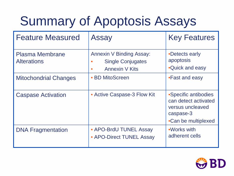
Summary of Apoptosis AssaysFeature Measured Assay Key Features
Plasma Membrane Alterations
Annexin V Binding Assay:• Single Conjugates• Annexin V Kits
•Detects early apoptosis•Quick and easy
Mitochondrial Changes • BD MitoScreen •Fast and easy
Caspase Activation • Active Caspase-3 Flow Kit •Specific antibodies can detect activated versus uncleaved caspase-3•Can be multiplexed
DNA Fragmentation • APO-BrdU TUNEL Assay• APO-Direct TUNEL Assay
•Works with adherent cells
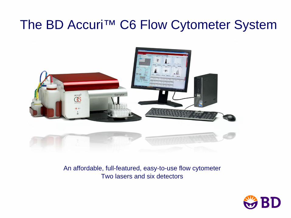
The BD Accuri™ C6 Flow Cytometer System
An affordable, full-featured, easy-to-use flow cytometerTwo lasers and six detectors
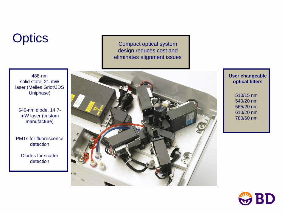
Optics Compact optical system design reduces cost and
eliminates alignment issues
488-nm solid state, 21-mW
laser (Melles Griot/JDS Uniphase)
640-nm diode, 14.7- mW laser (custom
manufacture)
PMTs for fluorescencedetection
Diodes for scatter detection
510/15 nm540/20 nm565/20 nm610/20 nm780/60 nm
User changeable optical filters
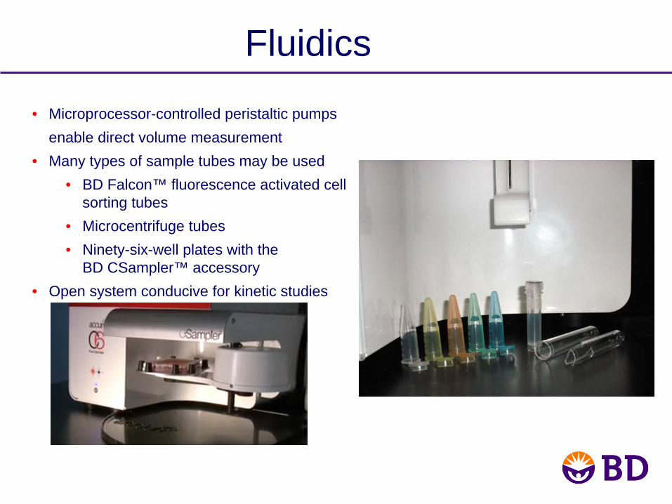
Fluidics
• Microprocessor-controlled peristaltic pumps enable direct volume measurement
• Many types of sample tubes may be used• BD Falcon™ fluorescence activated cell
sorting tubes• Microcentrifuge tubes• Ninety-six-well plates with the
BD CSampler™ accessory• Open system conducive for kinetic studies

Detection: Wide Dynamic Range
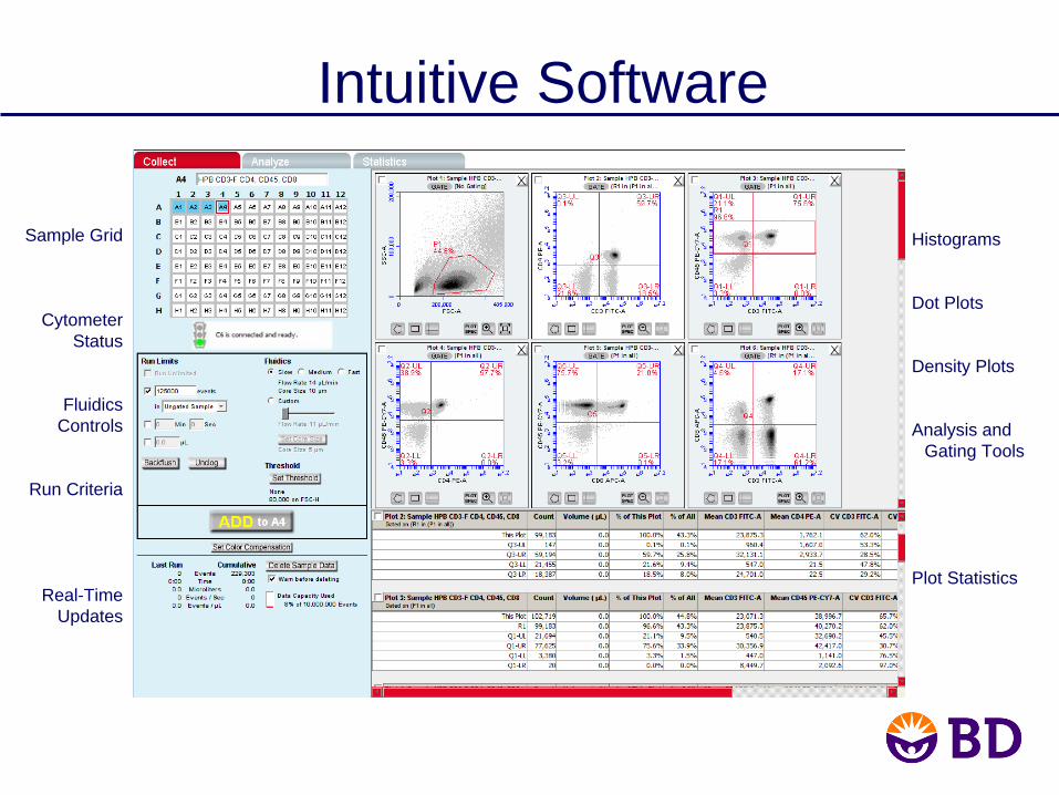
Intuitive Software
Sample Grid
CytometerStatus
Fluidics Controls
Run Criteria
Real-Time Updates
Histograms
Dot Plots
Density Plots
Analysis and Gating Tools
Plot Statistics
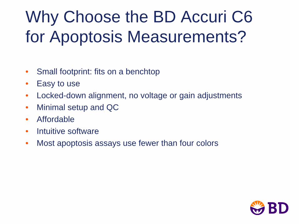
Why Choose the BD Accuri C6 for Apoptosis Measurements?
• Small footprint: fits on a benchtop• Easy to use• Locked-down alignment, no voltage or gain adjustments • Minimal setup and QC• Affordable• Intuitive software• Most apoptosis assays use fewer than four colors
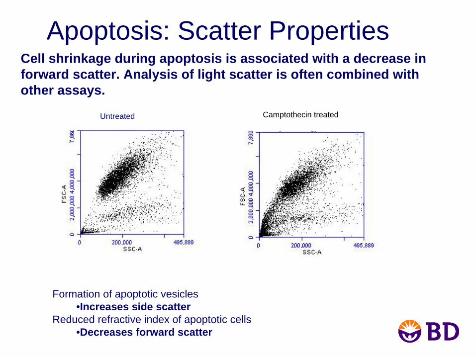
Apoptosis: Scatter Properties
Formation of apoptotic vesicles •Increases side scatter
Reduced refractive index of apoptotic cells •Decreases forward scatter
Cell shrinkage during apoptosis is associated with a decrease in forward scatter. Analysis of light scatter is often combined with other assays.
Untreated Camptothecin treated
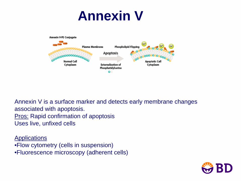
Annexin V
Annexin V is a surface marker and detects early membrane changes associated with apoptosis.Pros: Rapid confirmation of apoptosisUses live, unfixed cells
Applications•Flow cytometry (cells in suspension)•Fluorescence microscopy (adherent cells)
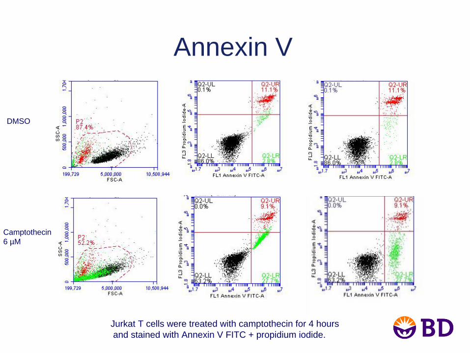
Annexin V
DMSO
Camptothecin6 µM
Jurkat T cells were treated with camptothecin for 4 hoursand stained with Annexin V FITC + propidium iodide.
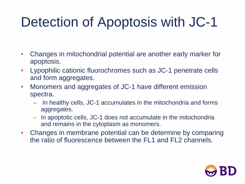
Detection of Apoptosis with JC-1
• Changes in mitochondrial potential are another early marker for apoptosis.
• Lypophilic cationic fluorochromes such as JC-1 penetrate cells and form aggregates.
• Monomers and aggregates of JC-1 have different emission spectra.– In healthy cells, JC-1 accumulates in the mitochondria and forms
aggregates.– In apoptotic cells, JC-1 does not accumulate in the mitochondria
and remains in the cytoplasm as monomers.• Changes in membrane potential can be determine by comparing
the ratio of fluorescence between the FL1 and FL2 channels.
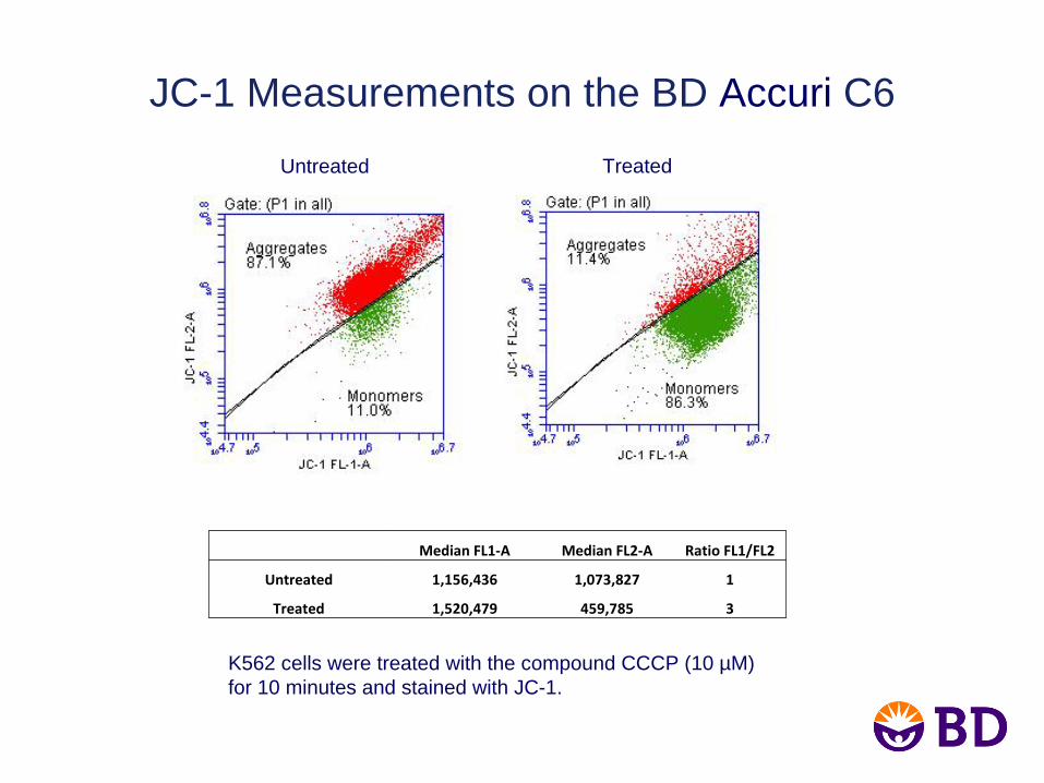
JC-1 Measurements on the BD Accuri C6
K562 cells were treated with the compound CCCP (10 µM)for 10 minutes and stained with JC-1.
Median FL1‐A Median FL2‐A Ratio FL1/FL2
Untreated 1,156,436 1,073,827 1
Treated 1,520,479 459,785 3
Untreated Treated
Caspase-3
• Caspases are proteases that are activated upon cleavage at aspartate residues at the earliest stages of apoptosis.
• Several caspases are important for apoptosis, including caspases-3, 8, and 9.
• Methods to measure caspase cleavage include fluorogenic substrates and detection with antibodies specific to the cleaved (activated) forms of caspases.
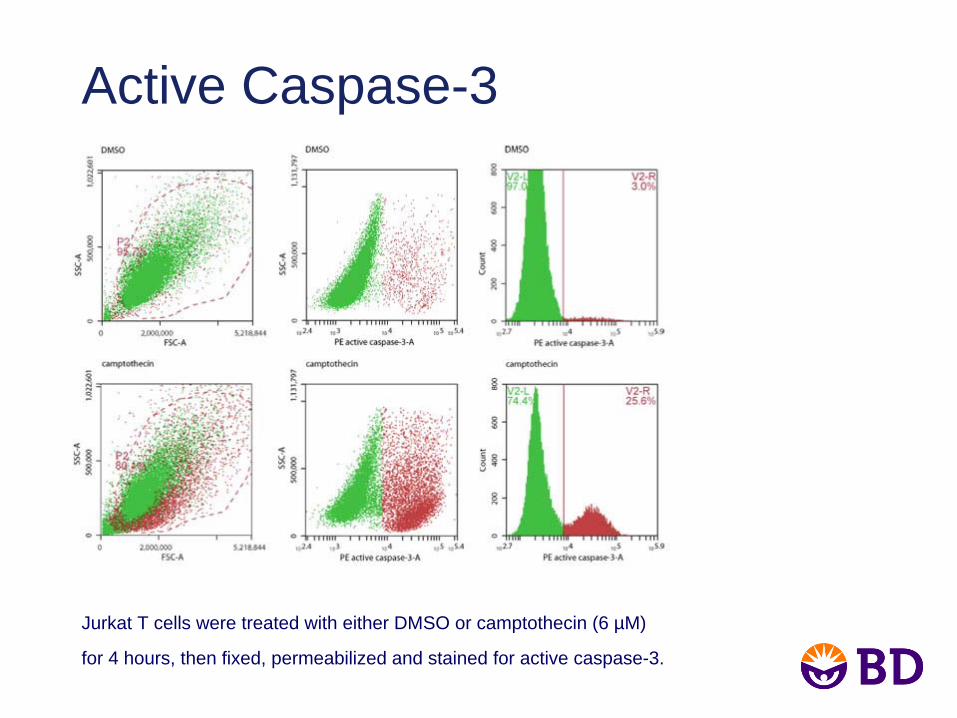
Active Caspase-3
Jurkat T cells were treated with either DMSO or camptothecin (6 µM)
for 4 hours, then fixed, permeabilized and stained for active caspase-3.
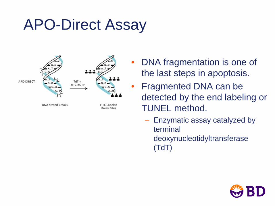
APO-Direct Assay
• DNA fragmentation is one of the last steps in apoptosis.
• Fragmented DNA can be detected by the end labeling or TUNEL method.– Enzymatic assay catalyzed by
terminal deoxynucleotidyltransferase (TdT)
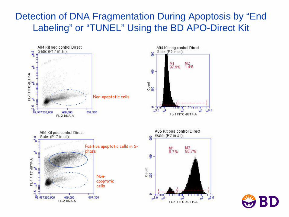
Detection of DNA Fragmentation During Apoptosis by “End Labeling” or “TUNEL” Using the BD APO-Direct Kit
NonNon--apoptotic cellsapoptotic cells
NonNon--
apoptotic apoptotic cellscells
Positive apoptotic cells in SPositive apoptotic cells in S--
phasephase
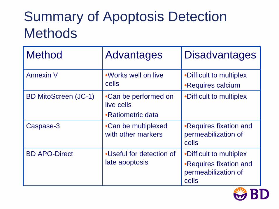
Summary of Apoptosis Detection MethodsMethod Advantages Disadvantages
Annexin V •Works well on live cells
•Difficult to multiplex•Requires calcium
BD MitoScreen (JC-1) •Can be performed on live cells•Ratiometric data
•Difficult to multiplex
Caspase-3 •Can be multiplexed with other markers
•Requires fixation and permeabilization of cells
BD APO-Direct •Useful for detection of late apoptosis
•Difficult to multiplex•Requires fixation and permeabilization of cells
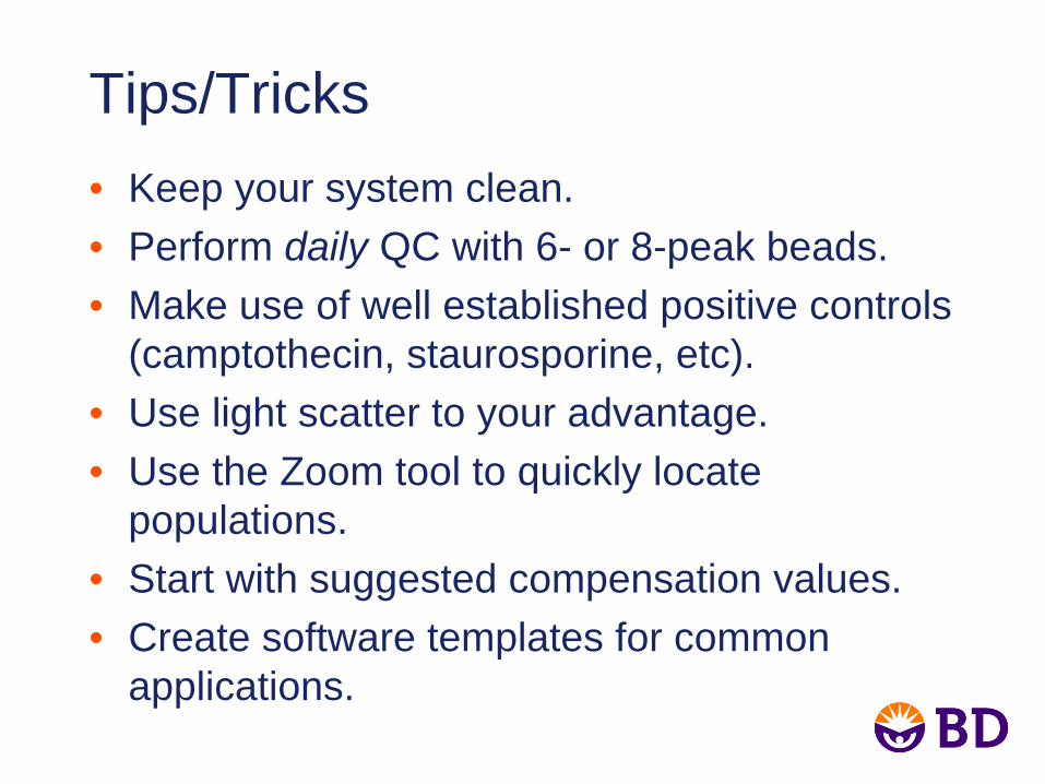
Tips/Tricks• Keep your system clean.• Perform daily QC with 6- or 8-peak beads.• Make use of well established positive controls
(camptothecin, staurosporine, etc).• Use light scatter to your advantage.• Use the Zoom tool to quickly locate
populations.• Start with suggested compensation values.• Create software templates for common
applications.
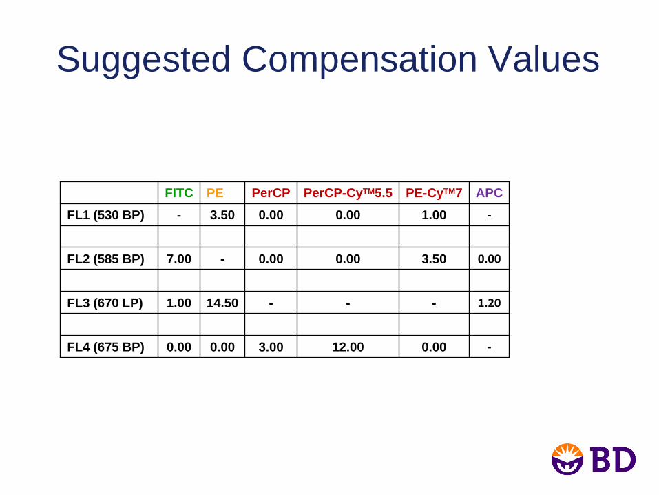
Suggested Compensation Values
FITC PE PerCP PerCP-CyTM5.5 PE-CyTM7 APCFL1 (530 BP) - 3.50 0.00 0.00 1.00 ‐
FL2 (585 BP) 7.00 - 0.00 0.00 3.50 0.00
FL3 (670 LP) 1.00 14.50 - - - 1.20
FL4 (675 BP) 0.00 0.00 3.00 12.00 0.00 ‐
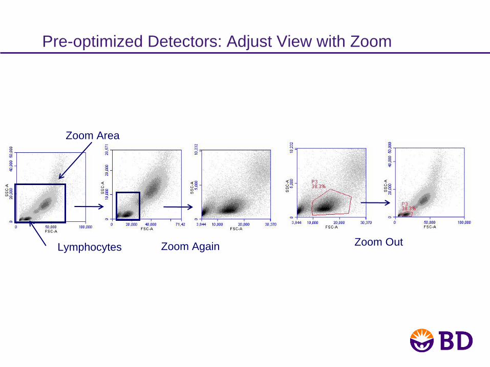
Lymphocytes
Zoom Area
Zoom OutZoom Again
Pre-optimized Detectors: Adjust View with Zoom
Conclusions• There are multiple methods currently
available to measure apoptosis.• Apoptosis detection by flow cytometry
can be done easily, using two colors.• The BD Accuri C6 is a perfect analysis
tool for new and experienced users.

Acknowledgments:
Maria DinkelmannMette EjrnaesMonisha SundarrajanKoen Verbrugghe
For Research Use Only. Not for use in diagnostic or therapeutic procedures.Class 1 Laser Product.Cy™ is a trademark of Amersham Biosciences Corp. Cy™ dyes are subject to proprietary rights of Amersham Biosciences Corp and Carnegie Mellon University and are made and sold under license from Amersham Biosciences Corp only for research and in vitro diagnostic use. Any other use requires a commercial sublicense from Amersham Biosciences Corp, 800 Centennial Avenue, Piscataway, NJ 08855-1327, USA.BD, BD Logo and all other trademarks are property of Becton, Dickinson and Company. © 2012 BD


![storage.googleapis.com · Mack the Knife [C6] [Dm] [G7] [C6] [Am] [Dm] [G7] [C6] (stop) Well the [C6] shark has pretty [Dm] teeth dear And he [G7] keeps them pearly [C6] white Just](https://img.pdfslide.us/doc/110x75/5b5bff6a7f8b9ac6028b54cf/-mack-the-knife-c6-dm-g7-c6-am-dm-g7-c6-stop-well-the-c6.jpg)