Embed Size (px)
Citation preview
Research ArticleApoptosis and Mobilization of Lymphocytes to Cardiac Tissue IsAssociated with Myocardial Infarction in a Reperfused PorcineModel and Infarct Size in Post-PCI Patients
Maria J. Forteza ,1,2,3 Isabel Trapero,2,3 Arantxa Hervas,3,4 Elena de Dios,3,4
Amparo Ruiz-Sauri,3,5 Gema Minana,2,3,4 Clara Bonanad,3,4 Cristina Gómez,4
Ricardo Oltra,6 Cesar Rios-Navarro,3,4 Daniel F. J. Ketelhuth,1 Julio Nunez,2,3,4
Francisco J. Chorro,2,3,4,7 and Vicente Bodi 2,3,4,7
1Department of Medicine, Cardiovascular Medicine Unit, Center for Molecular Medicine, Karolinska Institute,Karolinska University Hospital, Stockholm, Sweden2Department of Medicine, University of Valencia, Valencia, Spain3Institute of Health Research INCLIVA, Valencia, Spain4Department of Cardiology, Hospital Clinico Universitario de Valencia, Valencia, Spain5Department of Pathology, University of Valencia, Valencia, Spain6Intensive Care Unit, Hospital Clinico Universitario de Valencia, Valencia, Spain7Centro de Investigación Biomédica en Red-Cardiovascular (CIBERCV), Madrid, Spain
Correspondence should be addressed to Maria J. Forteza; [email protected] Vicente Bodi; [email protected]
Received 1 March 2017; Accepted 13 November 2017; Published 18 March 2018
Academic Editor: Nageswara Madamanchi
Copyright © 2018Maria J. Forteza et al. This is an open access article distributed under the Creative Commons Attribution License,which permits unrestricted use, distribution, and reproduction in any medium, provided the original work is properly cited.
ST-segment elevation myocardial infarction (STEMI) is the most severe outcome of coronary artery disease. Despite rapidreperfusion of the artery, acute irrigation of the cardiac tissue is associated with increased inflammation. While innate immuneresponse in STEMI is well described, an in-depth characterization of adaptive immune cell dynamics and their potential roleremains elusive. We performed a translational study using a controlled porcine reperfusion model of STEMI and the analysis oflymphocyte subsets in 116 STEMI patients undergoing percutaneous coronary intervention (PCI). In the animal model, a sharpdrop in circulating T lymphocytes occurred within the first hours after reperfusion. Notably, increased apoptosis of circulatinglymphocytes and infiltration of proinflammatory Th1 lymphocytes in the heart were observed 48 h after reperfusion. Similarly,in STEMI patients, a sharp drop in circulating T lymphocyte subsets occurred within the first 24 h post-PCI. A cardiac magneticresonance (CMR) evaluation of these patients revealed an inverse association between 24 h circulating T lymphocyte numbersand infarction size at 1-week and 6-month post-PCI. Our translational approach revealed striking changes in the circulating andtissue-infiltrating T lymphocyte repertoire in response to ischemia-reperfusion. These findings may help in developing newdiagnostic and therapeutic approaches for coronary diseases.
1. Introduction
Coronary artery disease is the single most frequent cause ofdeathworldwide. ST-segment elevationmyocardial infarction(STEMI) is the most severe outcome of coronary disease,
accounting for 12.8% of all deaths worldwide [1]. STEMIcommonlyoccurswhen thrombus formation leads to the com-plete occlusion of a major epicardial coronary vessel. Thus,myocardial infarction is associatedwithan inflammatory reac-tion,which is a prerequisite for healing and scar formation [2].
HindawiOxidative Medicine and Cellular LongevityVolume 2018, Article ID 1975167, 9 pageshttps://doi.org/10.1155/2018/1975167

STEMI must be diagnosed and treated promptly, usuallyby means of percutaneous coronary intervention (PCI).Despite rapid reperfusion of the coronary artery, the acute irri-gation of tissue has been associated with acceleration andincrease in local inflammation [3]. Several mechanisms havebeen proposed to explain ischemia-reperfusion-induced localinflammation, including activation of complement and reac-tive oxygen species [4]. Hence, post–ischemic-reperfusioninflammation is characterized by the recruitment and activa-tion of immune cells from the innate and adaptive immunesystems [5, 6].
Upon activation, adaptive immune CD4+ T lymphocytescan develop into T-helper (Th) 1, Th2, Th17, or regulatory Tcells (Tregs), depending on the set of costimulatory mole-cules and cytokines expressed by antigen-presenting cells.In general terms, Th1 and Th17 cells are considered proin-flammatory, while Tregs have been described to maintainimmune tolerance and homeostasis [7].
Previous studies from our group and others haveshown an acute decrease in the blood lymphocyte countin patients with STEMI [8], which has been associatedwith more severe prognosis, represented by an increasein infarct size and microvascular obstruction, measuredby cardiac magnetic resonance (CMR) [9]. While in general,the dynamics of innate immune cells in post–ischemic-reperfusion inflammation is well described, especiallymonocyte-derived cells [10]; the role of adaptive immunecells remains poorly characterized. In the present study,using a well-standardized porcine ischemia-reperfusionmodel and investigating STEMI patients, we show thatapoptosis and tissue mobilization of lymphocytes toinfarcted myocardium are associated with ischemic injuryand infarct size.
2. Methods
2.1. Porcine STEMI Model and Experimental Design. Twelvejuvenile domestic female pigs weighing 25–30 kg were usedin the study. In short, animals were sedated using IM 8mg/kg ketamine and 0.1mg/kg medetodimine and anaesthetizedusing a 10mg/kg/h continuous IV infusion of 2% propofol.Infarction was induced inflating a 2.5× 10mm angioplastyballoon in the mid left anterior descending coronary arteryin anaesthetized pigs. After 90min, the balloon was deflated,and the restoration of normal coronary flow was documentedby angiography. No coronary dissection or closure wasdetected at reperfusion or at the 48 h angiogram. After 48h,the pigs were anaesthetized again.
Blood samples were obtained using a multipurposecatheter placed in the coronary sinus of swine before bal-loon inflation, after 90min (immediately before balloondeflation) and 2h and 48 h after reperfusion. PBMCs wereisolated and frozen following the same protocol as inpatients. The hearts were then arrested with potassiumchloride and removed. The left ventricle was sectionedinto 5mm thick short-axis slices and incubated with a2% 2,3,5-triphenyltetrazolium chloride (TTZC) solutionfor 20min at 37°C. Finally, sections were photographed,
and the infarct size was defined as the myocardial areathat failed to stain with TTZC.
The Animal Care and Use Committee of the University ofValencia approved the study, which conforms to “The Guidefor the Care andUse of Laboratory Animals” published by theUS National Institutes of Health (NIH Publication number85-23, revised 1996). Further details of the experimentalstudy are described in the supplementary material.
2.2. RNA Isolation and Real-Time Quantitative PCR(RT-qPCR). Frozen myocardial tissue from the infarcted,adjacent, and remote areas of the pigs was homogenized inTRIzol isolation reagent (Life Technologies, Madrid, Spain)for RNA isolation. RT-qPCR was performed using an ABIPrism 7900 sequence detection system (Life Technologies,Madrid, Spain) with TaqMan Gene Expression Assays (LifeTechnologies, Madrid, Spain). The fold change in geneexpression from the control group was calculated using the2−ΔΔCt method [10].
2.3. Immunohistochemical Characterization of LymphocyteInfiltrates in Porcine Hearts. Tissue samples were obtainedafter the extraction and slicing of the heart. The samples werefixed in 10% formalin and embedded in paraffin. Afterward,4μm thick myocardial samples from paraffin-embeddedsamples were histologically characterized in the infarcted,adjacent, and remote areas. The following primary antibodieswere used: rabbit anti-humanCD3 for T cells andmouse anti-humanCD79a for B cells (both fromDako, Barcelona, Spain).Sections were then incubated with a HRP-conjugated second-ary antibody and developed with 3,3′-diaminobenzidinetetrahydrochloride (Dako, Barcelona, Spain).
2.4. Patient Study Design and Groups. One hundred andthirty-five consecutive STEMI patients that referred to PCIduring December 2011 to June 2013 were prospectivelyincluded in this study. STEMI was defined according to thelatest universal definition of myocardial infarction [11].Patients with a history of previous myocardial infarctionwere not considered for participation.
Thefinal study group comprised 116 patients who fulfilledthe inclusion criteria. From all patients, ninety-eight wereassigned for blood sampling 24 h post-PCI and CMR withinthe first week after STEMI. Seventy-two of the ninety-eightpatients repeated CMR after 6 months—twenty-six patientswere excluded due to death (n = 3), contraindications toCMR (n = 7), or the cardiologist’s decision (n = 16). EighteenSTEMI patients were assigned to serial blood sampling,including pre-PCI, and 24h, 96 h, and 30-day post-PCI(flowchart of the overall study design is shown in Figure 1).
Individuals were managed both in-hospital and afterdischarge by a specific STEMI unit, and current recommen-dations were strictly followed. ECG and angiographic charac-teristics were prospectively recorded in all cases upon patientadmission. Written informed consent was obtained from allpatients. The study was approved by the ethical committeeof clinical investigations of the Hospital Clinico Universitariode Valencia (approved in April 2008) and was conducted in
2 Oxidative Medicine and Cellular Longevity

agreement with the ethical principles for medical researchinvolving humans from the Declaration of Helsinki.
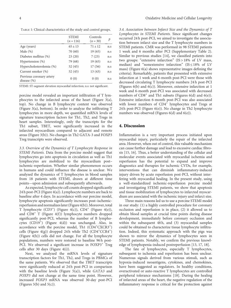
The clinical characteristics of both groups are shown inTable 1. The electrocardiographic, laboratory, and angio-graphic characteristics of STEMI patients are shown inSupplementary Table 1. STEMI patients were examined witha 1.5-Tesla system (Sonata Magnetom, Siemens, Erlangen,Germany) in accordance with our clinic’s protocol [12](see Supplementary Materials for detailed CMR protocol).
2.5. Blood Collection and PBMC Isolation. Peripheral venousblood (20ml) was drawn from all patients and controls. Totalleukocyte cell count was determined using an automatedblood cell counter. The peripheral blood mononuclear cells(PBMCs) were obtained using a density gradient centrifuga-tion with Lymphoprep© (Axis-Shield, Norway). Followingisolation, the PBMCs were frozen in freezing medium (10%DMSO and 90% fetal bovine serum) and stored at −80°C.
2.6. Flow Cytometry Analysis. Flow cytometric analysis wasused to characterize lymphocyte subsets in isolated PBMCs.In brief, frozen PBMC aliquots were quickly thawed andcounted with a Neubauer chamber. Discrimination betweenlive and dead cells was carried out prior to analysis with 7-aminoactinomycin D (7-AAD; Beckman Coulter). Thefollowing conjugated human antibodies were used: PerCP-anti-CD3 andPCy5-anti-CD3as panT-cellmarker (BeckmanCoulter, CA, USA), FITC-anti-CD4 for T-helper cells, FITC-anti-CD8 for cytotoxic T cells, APC-anti-CXCR3 and PE-anti-CCR4 for Th1 and Th2 cells, PE-anti-FOXP3 for Tregs,and APC-anti-CD19 for B cells. For porcine samples, thefollowing antibodies were used: FITC-anti-CD1 for B cellsand APC-anti-FOPX3 for Tregs (Beckton Dickinson, NJ,USA). Lymphocyte apoptosis was analyzed in fresh bloodsamples by dual selectivity with Annexin V and 7-AAD. Inbrief, the lymphocyte population was gated, and apoptosiswas determined as the percentage of cells positive for AnnexinV but negative for 7-ADD.
Samples were analyzed using a BD FACSVerse flowcytometer (standard 2-laser configuration, BD, USA), and aminimum of 10,000 events was acquired. FlowJo 8.7 software(TriStar, Oregon, USA) was used for the analysis of all theacquired data.
2.7. Statistical Analysis. The Shapiro-Wilk normality test wasapplied to test for a normal distribution. Continuous variableswere expressed as themean± SD, and comparisonsweremadeusing the repeated measures or ordinary one-way ANOVAwith a Bonferroni post hoc test when applicable. Percentageswere compared using a chi-square test and Fisher’s exact testwhen appropriate. Statistical significance was considered fora two-tailed p value < 0.05. All statistical tests were performedusing SPSS 19.0 (SPSS Inc., Chicago, IL, USA).
3. Results
3.1. Dynamics of Adaptive Immune Cells in Pigs Subjected toSTEMI. We analyzed the dynamics of the adaptive immunecells in blood of porcine experimental model of reperfusedSTEMI. Corroborating with previous clinical studies fromour group [13], we observed that the postischemic condition,induced by coronary occlusion and followed by reperfusion,is associated with a significant decrease circulating totallymphocyte counts (Figure 2(a)). A substantial increasedapoptosis among lymphocytes was seen immediately afterreperfusion and persisted over 48 h (Figure 2(b)). Amongthe lymphocyte subsets, we observed that T- but not B lym-phocytes accounted to the drop in lymphocytes’ count(Figures 2(c) and 2(d)). Interestingly, CD8+ T lymphocyteand Treg numbers were reduced within 2 h post-MI andstayed low till 48 h (Figures 2(e) and 2(g)). Only a modestand nonsignificant decreased CD4+ lymphocyte numberswere observed (Figure 2(f)).
3.2. T Lymphocytes Are Mobilized to Infarcted Myocardium.Immunohistochemical analysis of myocardium of our
In-hospital death (n = 6)In-hospital re-infraction (n = 4)In-hospital severe clinical instability (n = 9)Claustrophobia (n = 3)
Neoplasic disease (n = 3)Autoimmune disease (n = 1)Chronic infectious disease (n = 2)
Pacemakers or internal cardiac defibrillators (n = 9)
135 STEMIpatients
116 STEMIpatients
18 STEMIpatients
98 STEMIpatients
98 STEMIpatients
CMR 1st week
72 STEMIpatients
CMR 6th month
Death (n = 3)Contraindications to CMR (n = 7)Patient/cardiologist decision (n = 16)
24 h post-PCI
24 h post-PCI
Pre-PCI
PCI
PCI
96 h post-PCI 30 d post-PCI
(I)(II)
(III)
(I)(II)
(III)
(IV)(V)
(VI)(VII)
(VIII)
Figure 1: Flowchart of the study group. This flowchart illustrates the STEMI patients and controls and the different blood samples that wereobtained. In total, 116 STEMI patients were selected after exclusion criteria: 18 STEMI patients were selected for serial blood extractions atdifferent time points and 98 STEMI patients were selected for 24 h post-PCI blood extraction and CMR during 1st week and after 6months. STEMI: ST-segment elevation myocardial infarction; CMR: cardiac magnetic resonance.
3Oxidative Medicine and Cellular Longevity
porcine model revealed an important infiltration of T lym-phocytes to the infarcted areas of the heart (Figure 3(a),top). No change in B lymphocyte content was observed(Figure 3(a), bottom). In order to analyze the infiltrating Tlymphocytes in more depth, we quantified mRNA levels ofsignature transcription factors for Th1, Th2, and Tregs inheart samples. Interestingly, only the transcripts for theTh1 subset, TBET, were significantly increased in theinfarcted myocardium compared to adjacent and remoteareas (Figure 3(b)). No changes in Th2 GATA-3 and FOXP3Treg transcripts were observed.
3.3. Overview of the Dynamics of T Lymphocyte Response inSTEMI Patients. Data from the porcine model suggest thatlymphocytes go into apoptosis in circulation as well as Th1lymphocytes are mobilized to the myocardium post–ischemic-reperfusion. Whether similar phenomenon occursin humans and could influence the disease is unclear. Weanalyzed the dynamics of T lymphocytes in blood samplesfrom 18 patients with STEMI, drawn at different timepoints—upon admission and subsequently afterwards.
As expected, lymphocyte cell counts dropped significantly24 h post-PCI (Figure 4(a)). Lymphocyte numbers are back tobaseline after 4 days. In accordance with our porcine model,lymphocyte apoptosis significantly increases post–ischemic-reperfusionandnormalizes later (Figure4(b)).Moreover, totalT lymphocyte (CD3+) (Figure 4(c)), CD4+ (Figure 4(e)),and CD8+ T (Figure 4(f)) lymphocyte numbers droppedsignificantly post-PCI, whereas the number of B lympho-cytes (CD19+) (Figure 4(d)) was unchanged. Also, inaccordance with the porcine model, Th1 (CD4+CXCR3+)cells (Figure 4(g)) dropped 24h while Th2 (CD4+CCR4+)(Figure 4(h)) cells did not change. For all the lymphocytepopulations, numbers were restored to baseline 96h post-PCI. We observed a significant increase in FOXP3+ Tregcells after 30 days (Figure 4(i)).
Next, we analyzed the mRNA levels of signaturetranscription factors for Th1, Th2, and Tregs in PBMCs ofthe same patients. We observed that the TBET transcriptswere significantly reduced at 24 h post-PCI in comparisonwith the baseline levels (Figure 5(a)), while GATA3 andFOXP3 did not change at the same time point. However,increased FOXP3 mRNA was observed 30-day post-PCI(Figures 5(b) and 5(c)).
3.4. Association between Infarct Size and the Dynamics of TLymphocytes in STEMI Patients. Since significant changesoccurred 24h post-PCI, we aimed to investigate the associa-tion between infarct size and the T lymphocyte numbers inSTEMI patients. CMR was performed in 98 STEMI patients1 week and 6 months after PCI (Supplementary Table 2).Similar to previous studies [14], we classified patients intotwo groups: “extensive infarction” (IS> 18% of LV mass,median) and “nonextensive infarction” (IS≤ 18% of LVmass) (Figure 6(a) shows representative images defining thecriteria). Remarkably, patients that presented with extensiveinfarction at 1 week and 6-month post-PCI were those withdecreased circulating T lymphocyte numbers 24 h post-PCI(Figures 6(b) and 6(c)). Moreover, extensive infarction at 1week and 6-month post-PCI was associated with decreasednumbers of CD8+ and Th1 subsets (Figures 6(d) and 6(e)).Extensive infarction 6-month post-PCI was also associatedwith lower numbers of CD4+ lymphocytes and Tregs at24 h post-PCI (Figure 6(e)). No change in Th2 lymphocytenumbers was observed (Figures 6(d) and 6(e)).
4. Discussion
Inflammation is a very important process initiated uponmyocardial injury, particularly the repair of the infarctedarea. However, when out of control, this valuable mechanismcan cause further damage and lead to excessive cardiac fibro-sis [15, 16]. Thus, a better understanding of the cellular andmolecular events associated with myocardial ischemia andreperfusion has the potential to expand and improvediagnostics and therapies for ischemic CVDs, for example,interventions that can diminish inflammatory-inducedinjury driven by acute reperfusion post-PCI, without inter-fering with myocardial healing. In the present study, usinga well-standardized ischemic-reperfusion porcine modeland investigating STEMI patients, we show that apoptosisand tissue mobilization of lymphocytes to infarcted myocar-dium are associated with the ischemic injury and infarct size.
Three main reasons led us to use a porcine STEMI modelin our study: (1) a highly controlled procedure for coronaryocclusion and reperfusion is in place, (2) it allowed us toobtain blood samples at crucial time points during diseasedevelopment, immediately before coronary occlusion andwithin the subsequent 48 h, and (3) myocardial samplescould be obtained to characterize tissue lymphocyte infiltra-tion. Indeed, this systematic approach with the pigs wasshown to mirror the dynamics of lymphocytes seen inSTEMI patients. Notably, we confirm the previous knowl-edge of lymphopenia-induced postreperfusion [13, 17, 18].
The fate of lymphocytes, especially T lymphocytes,subsequent to ischemia and reperfusion has been unclear.Numerous signals derived from various stimuli, such ashypoxia-induced neoantigens, cytokines, and chemokines,have been suggested as regulators. In healthy conditions,overactivated or auto-reactive T lymphocytes are controlledperipheral tolerance mechanisms [18]. During the healingof infarcted areas of the heart, the negative regulation of theinflammatory response is critical for the protection against
Table 1: Clinical characteristics of the study and control groups.
STEMI(n = 116)
Controls(n = 30) p
Age (years) 65± 13 71± 12 n.s
Male (%) 70 (60) 19 (65) n.s
Diabetes mellitus (%) 23 (20) 7 (23) n.s
Hypertension (%) 79 (68) 19 (65) n.s
Hypercholesterolemia (%) 52 (45) 17 (56) n.s
Current smoker (%) 52 (45) 13 (43) n.s
Previous coronary arterydisease (%)
0 (0) 0 (0) n.s
STEMI: ST-segment elevation myocardial infarction; n.s: not significant.
4 Oxidative Medicine and Cellular Longevity

adverse effects that could lead to excessive remodeling andfibrosis [19].
It is well established that transient T lymphocytedepletion, largely through apoptosis, is a very importantmechanism of immunoregulation [17]. This knowledge is
well exemplified in the case of administration of specificT lymphocyte depleting anti-CD3 antibodies, which throughthe induction of apoptosis of these cells can induce a short-term immunosuppression followed by long-term tolerance[20]. In our study, ischemia-reperfusion led to a substantial
p < 0.01
0
4000
8000
12000
Lym
phoc
ytes
(cel
ls/𝜇
l)
90 min 2 h 48 h0 h
(a)
p < 0.01p < 0.05
0
100
200
300
400
500
Ann
exin
V+
(cel
ls/𝜇
l)90 min 2 h 48 h0 h
(b)
p < 0.05
90 min 2 h 48 h0 h0
500
1000
1500
2000
2500
CD3+ (c
ells/𝜇
l)
(c)
0
1000
2000
3000
4000
CD1+ (c
ells/𝜇
l)
90 min 2 h 48 h0 h
(d)
p < 0.05
0
500
1000
1500
CD8+ (c
ells/𝜇
l)
48 h0 h 90 min 2 h
(e)
0
500
1000
1500
2000
CD4+ (c
ells/𝜇
l)
90 min 2 h 48 h0 h
(f)
p < 0.05p < 0.01
0
5
10
15
20
CD4+ CD
25+ FO
XP3+
(cel
ls/𝜇
l)
90 min0 h 2 h 48 h
(g)
Figure 2: Lymphocyte and T cell dynamics in swine with induced infarction. Dynamics of lymphocyte and T cell subsets in blood from swinewith induced infarction at different time points: preinfarction (0 h), postinfarction and prereperfusion (90min), and postreperfusion (2 h and48 h). (a) Lymphocyte cell count. (b) Lymphocyte apoptosis. Number of Annexin V-positive cells selected in the lymphocyte gate. (c) T cells(CD3). (d) B cells (CD1). (e) CD8 cells. (f) CD4 cells. (g) Treg cells (CD4+CD25+FOXP3+). Data are expressed as mean± SD. versuspreinfarction (0 h). SD: standard deviation; Treg: T regulatory cells.
CD3
CD79a
Remote Adjacent Infarcted
(a)
FOXP3 GATA3 TBETX
p < 0.001
RemoteAdjacentInfarcted
0
2
4
6
8
10
mRN
A fo
ld ch
ange
(b)
Figure 3: T cell infiltration and lymphocyte apoptosis in swine myocardial samples. (a) Microscopic captions of porcine myocardium; top: Tcell (CD3) infiltration; bottom: B cell (CD79a) in infarcted, adjacent, and remote areas. (b) T cell subset gene expression: FOXP3, GATA 3,and TBETX in infarcted, adjacent, and remote areas. Data are expressed as mean± SD. ∗p < 0 05 versus remote. SD: standard deviation.
5Oxidative Medicine and Cellular Longevity

p < 0.05
0
1000
2000
3000
4000
Lym
phoc
yte (
cells
/𝜇l)
Pre-
PCI
96 h
pos
t-PCI
30 d
pos
t-PCI
24 h
pos
t-PCI
(a)
p < 0.05
p < 0.01
0
200
400
600
Ann
exin
V (c
ells/𝜇
l)
Pre-
PCI
96 h
pos
t-PCI
30 d
pos
t-PCI
24 h
pos
t-PCI
(b)
p < 0.05
0
500
1000
1500
2000
2500
CD3+ ce
lls (c
ells/𝜇
l)
Pre-
PCI
96 h
pos
t-PCI
30 d
pos
t-PCI
24 h
pos
t-PCI
(c)
0
100
200
300
400
CD19
+ cells
(cel
ls/𝜇
l)
Pre-
PCI
30 d
pos
t-PCI
96 h
pos
t-PCI
24 h
pos
t-PCI
(d)
p < 0.01
0
500
1000
1500
2000
CD4+ (c
ells/𝜇
l)
Pre-
PCI
30 d
pos
t-PCI
96 h
pos
t-PCI
24 h
pos
t-PCI
(e)
p < 0.01
0
500
1000
1500
CD8+ (c
ells/𝜇
l)
Pre-
PCI
30 d
pos
t-PCI
96 h
pos
t-PCI
24 h
pos
t-PCI
(f)
p < 0.05
0
500
1000
1500
CD4+ CX
CR3+ (c
ells/𝜇
l)
Pre-
PCI
30 d
pos
t-PCI
96 h
pos
t-PCI
24 h
pos
t-PCI
(g)
0
200
400
600
800
CD4+ CC
R4+ (c
ells/𝜇
l)
24 h
pos
t-PCI
96 h
pos
t-PCI
30 d
pos
t-PCI
Pre-
PCI
(h)
p < 0.01
0
20
40
60
CD4+ CD
25+ FO
XP3+ (c
ells/𝜇
l)
Pre-
PCI
30 d
pos
t-PCI
96 h
pos
t-PCI
24 h
pos
t-PCI
(i)
Figure 4: Lymphocyte and T cell dynamics STEMI patients. Total lymphocyte, lymphocyte apoptosis (Annexin V+ cells), and T cell subsetcount (cells/μl) from blood of STEMI patients at different time points: after MI and before reperfusion (pre-PCI) and after MI and afterreperfusion (24 h, 96 h, and 30 d post-PCI). A significant drop of lymphocytes, T cell (CD3+), CD4, CD8, and Th1 (CD4+CXCR3+) 24 hpost-PCI was observed in STEMI patients. While B cells (CD19+) and Th2 (CD4+CCR4+) did not change. Treg (CD4+CD25+FOXP3+)increased after one month. Values are expressed as mean± SD. p < 0 05 or p < 0 01 versus pre-PCI. PCI: percutaneous coronaryintervention; SD: standard deviation; STEMI: ST-segment myocardial infarction; Treg: T regulatory cells.
6 Oxidative Medicine and Cellular Longevity

increase in apoptosis of circulating lymphocytes on ourpigs and patients. Although, we have not deeply investigatedthe molecular mechanisms that could drive this phenome-non, our data suggest that lymphopenia post–ischemic-reperfusion could be an attempt protective mechanism.
The clearance of dead cells by phagocytes has been shownto activate inhibitory programs, serving as a key mechanismfor the termination of the proinflammatory cascade [20].Along with this mechanism, increased Treg numbers mayalso represent an inhibitory process to stop further andunnecessary damage [21]. In line with this, increasedFOXP3+ Treg numbers were found one month after reperfu-sion in STEMI patients.
Considering lymphocyte populations and subsets, themost significant changes were observed at 24 h post-PCIin patients and 48 h post coronary occlusion in the por-cine model. A significant decrease in CD4+, Th1, andCD8+ lymphocytes was observed, while other subsets suchas Th2 and B cells were unchanged in blood. Interestingly,analysis of pig hearts revealed an increased T lymphocyteinfiltration and the expression of the signature Th1 tran-scription factor, TBET, in infarcted areas. Altogether, thesedata suggest another potential mechanism involved in thepostreperfusion lymphopenia, the mobilization of cells tothe myocardium.
While Th1 lymphocytes were increased in theinfarcted myocardium, neither Th2 nor Treg infiltrationseems to be influenced by post–ischemic-reperfusion.This pattern of cells could have direct consequences tothe inflammatory process triggered in the heart. It hasbeen shown that recombination-activating gene knockoutmice (Rag1−/−), which lack T and B lymphocytes, pres-ent significantly smaller infarct size when subjected to
left coronary artery ligation, compared to immunocom-petent controls. Interestingly, reconstitution of Rag1−/−
mice with CD4+ T lymphocytes from only wild typebut not IFNγ−/− mice reversed the protective phenotype[22]. In clinical studies, it has been shown that patientswith ACS present increased T lymphocyte-derived-IFNγresponse [23, 24]. Thus, it has been recently proposedthat IFNγ can influence TGFβ-induced healing processesin the heart [25]. Altogether, these and our data suggestthat Th1 lymphocytes and their major produced cyto-kine, IFNγ, can play a deleterious role in MI promotingcardiac damage.
The translational approach used in the present studyreveals that changes in the T lymphocyte repertoire occurin a clinical scenario of STEMI patients treated with pri-mary PCI and in a controlled experimental porcine modelof induced anterior infarction. We show that the acutedecrease in the proinflammatory circulating T lymphocytesin blood is due to increased apoptosis and the mobiliza-tion of these cells to the infarcted areas of the heart.Patients with extensive infarctions presented less Th1 cellsin blood soon after PCI, suggesting that this early infiltra-tion of cells could have a direct impact on myocardialinflammation and healing. These findings are very impor-tant and can help guide the development of novel diagnos-tic approaches and therapies for coronary diseases. Ofnote, in experimental models, boosting of Tregs andconsequent modulation of macrophage responses haveshown promising results, improving myocardium healingand increasing survival [26, 27]. The continued researchin this field as well as the refinement of immunomodula-tory therapies will hopefully allow us to see such strategiesmoving into clinical trials in the near future.
p < 0.01
Tbetx
0
5
10
15
mRN
A fo
ld ch
ange
24h
post-
PCI
96 h
pos
t-PCI
300
h po
st-PC
I
Pre-
PCI
(a)
Gata3
0
5
10
mRN
A fo
ld ch
ange
24h
post-
PCI
Pre-
PCI
300
h po
st-PC
I
96 h
pos
t-PCI
(b)
p < 0.05
Foxp3
0
2
4
6
8
mRN
A fo
ld ch
ange
300
h po
st-PC
I
24h
post-
PCI
96 h
pos
t-PCI
Pre-
PCI
(c)
Figure 5: CD4 T cell subsets dynamics in STEMI patients. mRNA gene expression analysis of the dynamics of different CD4 subsets inSTEMI patients. Tbetx gene expression indicates Th1 cells, Gata3 gene expression indicates Th2 cells, and FOXP3 gene expressionindicates Treg cells. A significant decrease of proinflammatory Th1 cells was observed 24 h post-PCI while Treg cells increased within thenext month. Values are expressed as mean± SD. p < 0 05 or p < 0 01 versus pre-PCI. PCI: percutaneous coronary intervention; SD:standard deviation; STEMI: ST-segment myocardial infarction.
7Oxidative Medicine and Cellular Longevity

1 week post-PCI 6-months post-PCI
(a)
Nonextensive infarction (n = 51)
1 week post-PCI
0
500
1000
1500
2000
24 h
pos
t-PCI
(T el
ls/𝜇
l)
Nonextensive infarctionExtensive infarction
Extensive infarction (n = 47)
(b)
Nonextensive infarction (n = 35)
6-months post-PCI
0
500
1000
1500
2000
2500
24 h
pos
t-PCI
(T el
ls/𝜇
l)
Nonextensive infarctionExtensive infarction
Extensive infarction (n = 37)
(c)
Nonextensive infarction (n = 51)
1 week post-PCI
⁎
⁎
0
50
500
1000
1500
2000
24 h
pos
t-PCI
(T el
ls/𝜇
l)
CD8 CD4+CD25+
FOXP3+CD4+
CCR4+CD4+
CXCR3+CD4
Extensive infarction (n = 47)
(d)
Nonextensive infarction (n = 35)
6-months post-PCI
⁎
⁎
⁎
⁎
0
50
500
1000
1500
2000
24 h
pos
t-PCI
(T el
ls/𝜇
l)
CD4 CD4+ CD25+
FOXP3+CD8 CD4+
CCR4+CD4+
CXCR3+
Extensive infarction (n = 37)
(e)
Figure 6: Association of CMR-derived infarct size with T cell count in STEMI patients 24 h post-PCI. (a) Example of CMR images from apatient with an anterior STEMI. (b) Association of 1-week CMR-derived infarct size with T cell count (cells/μl) in blood of STEMIpatients at 24 h post-PCI. (c) Association of 6-month CMR-derived infarct size with T cell count (cells/μl) in blood of STEMI patients at24 h post-PCI. (d) Association of 1-week CMR-derived infarct size with T cell subsets (cells/μl) in blood of STEMI patients at 24 h post-PCI. (e) Association of 6-month CMR-derived infarct size with T cell subsets (cells/μl) in blood of STEMI patients 24 h post-PCI. Data areexpressed as mean± SD. ∗p < 0 05. PCI: percutaneous coronary intervention; SD: standard deviation; STEMI: ST-segment myocardialinfarction.
8 Oxidative Medicine and Cellular Longevity

Conflicts of Interest
The authors declare that they have no conflicts of interest.
Acknowledgments
The present study was supported by the “Instituto de SaludCarlos III” and cofunded by “FEDER” (PI14/00271, PIE15/00013, and CB16/11/00486 grants) and “Conselleria deEducacio, Cultura i Esport de la Generalitat Valenciana”(PROMETEO/2013/007 grant). Maria J. Forteza and DanielF. J. Ketelhuth are supported by grants from the SwedishHeart-Lung Foundation.
Supplementary Materials
Supplementary Table 1: electrocardiographic, laboratory,and angiographic characteristics of STEMI patients. Supple-mentary Table 2: cardiovascular magnetic resonance imagingdata of STEMI patients. (Supplementary Materials)
References
[1] F. A. Masoudi, A. Ponirakis, R. W. Yeh et al., “Cardiovascularcare facts: a report from the national cardiovascular dataregistry: 2011,” Journal of the American College of Cardiology,vol. 62, no. 21, pp. 1931–1947, 2013.
[2] N. G. Frangogiannis, C. W. Smith, and M. L. Entman, “Theinflammatory response in myocardial infarction,” Cardiovas-cular Research, vol. 53, no. 1, pp. 31–47, 2002.
[3] D. J. Hausenloy and D. M. Yellon, “Myocardial ischemia-reperfusion injury: a neglected therapeutic target,” The Journalof Clinical Investigation, vol. 123, no. 1, pp. 92–100, 2013.
[4] D. M. Yellon and D. J. Hausenloy, “Myocardial reperfusioninjury,” The New England Journal of Medicine, vol. 357,no. 11, pp. 1121–1135, 2007.
[5] A. Abbate, E. Bonanno, A. Mauriello et al., “Widespreadmyocardial inflammation and infarct-related artery patency,”Circulation, vol. 110, no. 1, pp. 46–50, 2004.
[6] G. M. Fröhlich, P. Meier, S. K. White, D. M. Yellon, and D. J.Hausenloy, “Myocardial reperfusion injury: looking beyondprimary PCI,” European Heart Journal, vol. 34, no. 23,pp. 1714–1722, 2013.
[7] M. DuPage and J. A. Bluestone, “Harnessing the plasticity ofCD4+ T cells to treat immune-mediated disease,” NatureReviews Immunology, vol. 16, no. 3, pp. 149–163, 2016.
[8] M. Madjid, I. Awan, J. T. Willerson, and S. W. Casscells, “Leu-kocyte count and coronary heart disease: implications for riskassessment,” Journal of the American College of Cardiology,vol. 44, no. 10, pp. 1945–1956, 2004.
[9] S. E. Boag, R. Das, E. V. Shmeleva et al., “T lymphocytes andfractalkine contribute to myocardial ischemia/reperfusioninjury in patients,” The Journal of Clinical Investigation,vol. 125, no. 8, pp. 3063–3076, 2015.
[10] T. Heidt, G. Courties, P. Dutta et al., “Differential contributionof monocytes to heart macrophages in steady-state and aftermyocardial infarction,” Circulation Research, vol. 115, no. 2,pp. 284–295, 2014.
[11] K. Thygesen, J. S. Alpert, A. S. Jaffe et al., “Third universal def-inition of myocardial infarction,” Circulation, vol. 126, no. 16,pp. 2020–2035, 2012.
[12] V. Bodí, J. Sanchis, M. P. López-Lereu et al., “Usefulness of acomprehensive cardiovascular magnetic resonance imagingassessment for predicting recovery of left ventricular wall motionin the setting of myocardial stunning,” Journal of the AmericanCollege of Cardiology, vol. 46, no. 9, pp. 1747–1752, 2005.
[13] V. Bodí, J. Sanchis, J. Núñez et al., “Post-reperfusion lympho-penia and microvascular obstruction in ST-segment elevationacute myocardial infarction,” Revista Española de Cardiología(English Edition), vol. 62, no. 10, pp. 1109–1117, 2009.
[14] O. Husser, J. V. Monmeneu, C. Bonanad et al., “Head-to-headcomparison of 1 week versus 6 months CMR-derived infarctsize for prediction of late events after STEMI,” The Interna-tional Journal of Cardiovascular Imaging, vol. 29, no. 7,pp. 1499–1509, 2013.
[15] F. K. Swirski and M. Nahrendorf, “Leukocyte behavior inatherosclerosis, myocardial infarction, and heart failure,”Science, vol. 339, no. 6116, pp. 161–166, 2013.
[16] N. G. Frangogiannis, “Inflammation in cardiac injury, repairand regeneration,” Current Opinion in Cardiology, vol. 30,no. 3, pp. 240–245, 2015.
[17] A. Liston and D. H. D. Gray, “Homeostatic control of regula-tory T cell diversity,” Nature Reviews Immunology, vol. 14,no. 3, pp. 154–165, 2014.
[18] P. H. Krammer, R. Arnold, and I. N. Lavrik, “Life and death inperipheral T cells,” Nature Reviews Immunology, vol. 7, no. 7,pp. 532–542, 2007.
[19] U. Hofmann, N. Beyersdorf, J. Weirather et al., “Activation ofCD4+ T lymphocytes improves wound healing and survivalafter experimental myocardial infarction in mice,” Circulation,vol. 125, no. 13, pp. 1652–1663, 2012.
[20] C. Penaranda, Q. Tang, and J. A. Bluestone, “Anti-CD3 ther-apy promotes tolerance by selectively depleting pathogeniccells while preserving regulatory T cells,” The Journal of Immu-nology, vol. 187, no. 4, pp. 2015–2022, 2011.
[21] V. Bodi, J. Sanchis, J. Nunez et al., “Uncontrolled immuneresponse in acutemyocardial infarction: unraveling the thread,”American Heart Journal, vol. 156, no. 6, pp. 1065–1073, 2008.
[22] Z. Yang, Y.-J. Day, M.-C. Toufektsian et al., “Myocardialinfarct–sparing effect of adenosine A2A receptor activation isdue to its action on CD4+ T lymphocytes,” Circulation,vol. 114, no. 19, pp. 2056–2064, 2006.
[23] X. Cheng, Y.-H. Liao, H. Ge et al., “TH1/TH2 functionalimbalance after acute myocardial infarction: coronary arterialinflammation or myocardial inflammation,” Journal of Clini-cal Immunology, vol. 25, no. 3, pp. 246–253, 2005.
[24] A. L. Pasqui, M. Di Renzo, A. Auteri, and L. Puccetti, “Cyto-kines in acute coronary syndromes,” International Journal ofCardiology, vol. 105, no. 3, pp. 355-356, 2005.
[25] X. Yan, H. Zhang, Q. Fan et al., “Dectin-2 deficiency modulatesTh1 differentiation and improves wound healing after myocar-dial infarction,” Circulation Research, vol. 120, no. 7, pp. 1116–1129, 2017.
[26] J. Weirather, U. D. W. Hofmann, N. Beyersdorf et al., “Foxp3+
CD4+ T cells improve healing after myocardial infarction bymodulating monocyte/macrophage differentiation,” Circula-tion Research, vol. 115, no. 1, pp. 55–67, 2014.
[27] E. H. Choo, J.-H. Lee, E.-H. Park et al., “Infarctedmyocardium-primed dendritic cells improve remodeling andcardiac function after myocardial infarction by modulatingthe regulatory T cell and macrophage polarization,” Circula-tion, vol. 135, no. 15, pp. 1444–1457, 2017.
9Oxidative Medicine and Cellular Longevity

Stem Cells International
Hindawiwww.hindawi.com Volume 2018
Hindawiwww.hindawi.com Volume 2018
MEDIATORSINFLAMMATION
of
EndocrinologyInternational Journal of
Hindawiwww.hindawi.com Volume 2018
Hindawiwww.hindawi.com Volume 2018
Disease Markers
Hindawiwww.hindawi.com Volume 2018
BioMed Research International
OncologyJournal of
Hindawiwww.hindawi.com Volume 2013
Hindawiwww.hindawi.com Volume 2018
Oxidative Medicine and Cellular Longevity
Hindawiwww.hindawi.com Volume 2018
PPAR Research
Hindawi Publishing Corporation http://www.hindawi.com Volume 2013Hindawiwww.hindawi.com
The Scientific World Journal
Volume 2018
Immunology ResearchHindawiwww.hindawi.com Volume 2018
Journal of
ObesityJournal of
Hindawiwww.hindawi.com Volume 2018
Hindawiwww.hindawi.com Volume 2018
Computational and Mathematical Methods in Medicine
Hindawiwww.hindawi.com Volume 2018
Behavioural Neurology
OphthalmologyJournal of
Hindawiwww.hindawi.com Volume 2018
Diabetes ResearchJournal of
Hindawiwww.hindawi.com Volume 2018
Hindawiwww.hindawi.com Volume 2018
Research and TreatmentAIDS
Hindawiwww.hindawi.com Volume 2018
Gastroenterology Research and Practice
Hindawiwww.hindawi.com Volume 2018
Parkinson’s Disease
Evidence-Based Complementary andAlternative Medicine
Volume 2018Hindawiwww.hindawi.com
Submit your manuscripts atwww.hindawi.com