Embed Size (px)
Citation preview
Rev
ised
20
10
The diagnosis of a blood cancer can be a devastating event for patients,
families and friends. It is therefore vital for everyone to have access to
reputable and understandable information to help cope with the illness.
Whenever possible our booklets are written in line with national guidelines
for the treatment of patients with a blood cancer. The information in our
booklets is more detailed than in many others but is written in a clear style
with all scientific terms explained for the general reader.
We recognise that the amount and level of information needed is a personal
decision and can change over time. Particularly at the time of diagnosis,
patients may prefer less detailed information. A number of alternative sources
of information are available which complement our publications.
The booklets in this series are intended to provide general information about
the topics they describe. In many cases the treatment of individual patients will
differ from that described in the booklets.
At all times patients should rely on the advice of their specialist
who is the only person with full information about their diagnosis
and medical history.
For further advice contact the clinical information team on 020 7269 9060.
Leukaemia & Lymphoma Research,43 Great Ormond Street, London WC1N 3JJT: 020 7405 0101 • F: 020 7405 3139E: [email protected]
Series compiled by Ken Campbell MSc, revised 2010.
The design of this booklet has been produced with kind assistance from Euro RSCG Life.
© All rights reserved. No part of this publication may be reproduced or transmitted without permission in writing from Leukaemia & Lymphoma Research.
Registered charity 216032 (England & Wales) SC037529 (Scotland)
What is aplastic anaemia?
1
Aplastic anaemia is a rare acquired disorder in which there is a failure
of the bone marrow to produce sufficient blood cells for the circulation.
Acquired means that the condition is neither present at birth nor inherited
but has developed during the patient’s life. Blood cells come from special
cells in the bone marrow, called stem cells. Less than 1 in 5,000 of the
marrow cells is a stem cell. The stem cells give rise to a progressively
maturing series of different cell types which eventually lead to all the
functional blood cells found in the circulation. In aplastic anaemia blood
production by stem cells fails resulting in a lack of red cells (anaemia),
white cells (leading to an increased risk of infection) and platelets (which
are needed to prevent bleeding and bruising). Aplastic anaemia is not
a form of cancer. There is a lack of cells within the blood and the blood
forming stem cells within the marrow are replased by fat cells. Any
remaining cells look more or less normal in contrast to leukaemia and
other blood cancers.
The failure of the stem cells to produce mature blood cells can vary from
partial to almost complete thus producing a disease of varying severity
in different people. The degree of marrow failure may change with time
in a given patient. Symptoms are slow to emerge because the loss of stem cell
function is gradual. Often patients only realise that they have been less than
fully fit for some time after their symptoms have been investigated with
a blood test.
The diagnosis is suggested by the blood count and by examination of the
blood film but it can only be confirmed by a bone marrow examination. There
are two parts to a bone marrow examination, the ‘aspirate’ and the ‘trephine’.
The aspirate is obtained by sucking a small amount of cells from the marrow
through a hollow needle, spreading it on a glass slide and then stained and
examined in the laboratory. This reveals the appearance of individual cells and
2
helps to exclude other diagnoses such as leukaemia. The trephine gives
an overall picture of the marrow and is important in determining the severity
of the marrow failure. In normal bone marrow about half of the space is taken
up by fat cells with the remainder being blood producing (haemopoietic)
cells. In aplastic anaemia almost the entire space is taken up by fat cells.
Occasionally there may be patchy loss of haemopoietic cells within the bone
marrow. It may be necessary to repeat the examination at a different site
to be quite sure of the diagnosis.
Though aplastic anaemia can occur at any age, it appears to be more
common in two age groups, those aged between 10 and 20 years and
in people aged 40 years or over. The condition appears to be slightly more
common in men. People of all ethnic groups may be affected. There is a higher
frequency in tropical countries and the Far East. This is probably related
to some factor in the environment rather than any particular race. People
who move from these regions to Europe or the USA seem to acquire the same
chance of developing aplastic anaemia as the local population.
The lack of blood cells produces a potentially very serious or fatal disease
unless properly managed. Until about 1980 the majority of patients with
severe disease did not survive more than a year but fortunately new methods
of support and treatment have completely changed this gloomy outlook.
Successful treatment requires a long time. Patience and care are required
by all involved including the family, friends and the medical team.
What are the types of aplastic anaemia?
The term aplastic anaemia is usually understood to refer to the acquired
disorder. The deficiency of the cells in the blood and the appearances
of the bone marrow, however, are not seen just in acquired aplastic
anaemia but may develop in a number of other conditions. These other
conditions have to be ruled out when reaching the final diagnosis of
acquired aplastic anaemia. Some of these are congenital disorders (i.e.
present at birth). Others which develop during life have some similarity
to acquired aplastic anaemia.
Treatment with high doses of chemotherapy or radiation will produce an
aplastic appearance in the bone marrow as well as loss of cells in the blood.
The timing and duration of the bone marrow depression are predictable
from the type of treatment given and the dose received. Rarely aplastic
anaemia may follow accidental exposure to large doses of radiation such
as the Chernobyl nuclear power station accident in the Ukraine. Such
cases are excluded from the definition of aplastic anaemia. Failure of blood
production for these reasons is often termed ‘inevitable aplastic anaemia’.
In this situation the fall in blood cells occurs rapidly over one or two days and
recovery takes place in the majority of patients if they are supported through
the period of marrow depression with blood transfusions. In rare cases
damage to the bone marrow may be more severe and a stem cell transplant
may be needed to restore blood cell production.
Inherited aplastic anaemias. There are a number of rare inherited diseases
which develop into bone marrow failure with a fatty marrow and loss of
circulating blood cells. The most common is called Fanconi anaemia.1 There
is wide variation in the presentation of this type of aplastic anaemia but it is
3
1 There is a separate publication on Fanconi anaemia available fromLeukaemia & Lymphoma Research.
4
usually fairly easy to differentiate from acquired aplastic anaemia. Fanconi
anaemia affects brothers and sisters within a family whereas the parents
have a normal blood system. Typically the children are rather short and have
abnormalities of the forearms or hands and a gradual but progressive failure
of the bone marrow. The aplastic phase usually develops in childhood but may
not present until adolescence or even early adult life.
Fanconi anaemia can be diagnosed and clearly distinguished from acquired
disease by a special test on the blood.
Malignant conditions. Acute leukaemias usually present with an increase
in white blood cells, although red blood cell and platelet numbers may
be reduced. Typically, regardless of the blood count, the bone marrow will be
packed with very large numbers of very immature white cells called blasts.
In rare cases, particularly but not exclusively in children, acute leukaemia
may present with blood and marrow appearances indistinguishable from
aplastic anaemia. In these instances the aplastic anaemia seems to recover
spontaneously after about three to six weeks and the blood count returns
to normal. The acute leukaemia then reveals itself some six weeks to six
months later (or even longer).
This presentation of acute leukaemia usually suggests a good response to
treatment so any delay in arriving at a diagnosis does not affect the outcome.
A change in diagnosis from aplastic anaemia to acute leukaemia may cause
distress and confusion to families unless they have been forewarned of the
possibility. In fact the outlook for both diseases in childhood is broadly similar
even though the treatment is entirely different.
The most difficult disorder to distinguish from aplastic anaemia is called
hypoplastic myelodysplastic syndrome (hypoplastic MDS). MDS is
a collection of disorders which affect mainly older patients in which failure
of bone marrow function is associated with the appearance of slightly
5
abnormal looking (dysplastic) cells in the blood and bone marrow.2 In most
cases the marrow is overactive and this, together with the dysplastic cells
makes the diagnosis straightforward. Sometimes the MDS marrow has few
cells and increased fat (hypoplastic marrow) and abnormal cells are hard
to find. It is fortunate that the distinction is not critical since both aplastic
anaemia and hypoplastic MDS respond to similar treatment. Indeed there
is close similarity between the two conditions and they are sometimes called
‘overlap syndromes’.
2 There is a separate publication on the myelodysplastic syndromes available fromLeukaemia & Lymphoma Research.
The apparent cause of the disease does not influence the response to
treatment. All patients have the same chance of responding to treatment
when the severity of the bone marrow failure is taken into account. It
is thought that in most cases of acquired aplastic anaemia the damage
to bone marrow stem cells is caused by an auto-immune reaction. This
happens when the body’s immune cells become confused and start
to attack body tissues. In about three quarters of all cases this auto-
immune reaction has no clear underlying cause. This is called idiopathic
acquired aplastic anaemia (idiopathic means ‘of unknown cause’). In the
remaining cases there seems to be an identifiable factor triggering the
auto-immune response.
The basis for suspecting that certain drugs, chemicals or virus can cause
aplastic anaemia is the occurrence of the disease following exposure.
There are no specific tests that can be used to identify with certainty the
cause of the aplastic anaemia in any particular case. Despite all the difficulties
in deciding whether a given factor may have caused the aplastic anaemia, it
is very important to obtain a careful account of all drug or chemical exposure
from each patient or their family. It is known that the delay between exposure
and the development of a fall in blood count is typically about eight weeks.
Drugs which have been taken for the first time less than eight weeks before
symptoms develop are less likely to have triggered the disease, unless the drug
had been taken intermittently or regularly before. If a drug was stopped more
than a year before the symptoms develop it becomes less probable that the
drug caused the illness.
Drugs which are most strongly suspected of causing aplastic anaemia include
several which are used to treat arthritis, (particularly anti-inflammatory which
6
What causes acquired aplastic anaemia?
7
are used for the treatment of rheumatoid arthritis), drugs used to control
overactive thyroid, some drugs used in psychiatry and a few antibiotics. The
risk of developing aplastic anaemia as a side effect of drug treatment is very
small; for the antibiotic chloramphenicol, one of the more widely studied
triggers for the disease, about one person in 25,000 taking the drug may
develop aplastic anaemia.3 For most drugs, monitoring the blood count
is not of use in preventing the disease because of the delay between taking
the drug and the risk of developing aplastic anaemia. Once the disease has
been triggered, withdrawal of the drug will not reverse the process. One
exception is gold salts used to treat rheumatoid arthritis where monitoring
is essential as it may allow the drugs to be withdrawn before there has been
irreversible damage.
It is more difficult to assess a possible link between a workplace chemical
exposure and the development of aplastic anaemia. Many factors have
to be weighed and it may be impossible to make a definite judgement. The list
of chemicals which have been suspected of causing aplastic anaemia is very
long but the evidence against any individual compound is often rather weak.
There is a need to monitor the patient carefully if they return to their previous
place of work following treatment. Non-workplace exposures to chemicals are
even more difficult to assess; examples of chemicals to be suspected are wood
preservatives, adhesives and other organic compounds.
Some patients, perhaps one in ten, have had a virus infection shortly before
developing aplastic anaemia. The most common indication of this is a patient
with abnormalities of the liver which suggest hepatitis. The patient may first
notice jaundice or a general fatigue and loss of appetite. The bone marrow
failure follows a few weeks later. Although several viruses have been identified
as causing hepatitis no particular one has been found to cause aplastic
anaemia as well.
3 Chloramphenicol eye-drops are widely used, often for children, but very little drug is absorbed into the body and careful studies have shown no risk of aplastic anaemia from chloramphenicol eye-drops.
8
What are the signs and symptoms of aplastic anaemia?
The signs and symptoms derive entirely from the deficiency of the blood
cells. These are not specific to aplastic anaemia but occur in any condition
where there is a significant deficiency of all blood cells. This differs from
blood cancers (leukaemia and related conditions) where there may also
be symptoms caused by the large numbers of abnormal cells and these are
the main diseases from which aplastic anaemia has to be separated.
This can only be achieved by blood and marrow tests.
Deficiency of red blood cells produces anaemia and the patient may notice:
Fatigue and tiredness
Shortness of breath on moderate exercise, for example going up stairs
Paleness
Pulsating noise in the ears and/or headache
Deficiency of white blood cells, in particular neutrophils, causes an increased
risk of infection. Common infections include:
Recurrent or persistent sore throat
Skin infections
Chest infection
Deficiency of platelets causes a tendency to bleed. Common sites include:
Gum bleeding, particularly after brushing the teeth
Nose bleeds
Heavy or prolonged periods
Blood blisters in the mouth
Skin, shown by easy bruising even in the absence of a knock, or a rash
composed of small red spots most prominent on the legs (called
a ‘petechial’ rash)
These signs and symptoms are not specific to aplastic anaemia but occur in
any condition where there is a significant deficiency of all blood cells. They are
also typical of acute leukaemias and these are the main diseases from which
aplastic anaemia has to be separated. This can only be achieved by blood and
marrow tests.
9
10
Aplastic anaemia cannot be diagnosed on clinical findings alone. The
results of laboratory tests are necessary to exclude other causes of low
numbers of cells in the blood and to confirm the diagnosis. The main
problem in diagnosing aplastic anaemia is the exclusion of other diseases
with similar signs and symptoms.
The main tests used in diagnosis of aplastic anaemia are a full blood count
and a bone marrow biopsy. The bone marrow sample(s) indicate whether the
marrow contains normal amounts of active blood forming tissue. This is called
the cellularity of the marrow. This must be interpreted according to the age of
the patient because the proportion of the marrow which is actively producing
blood cells decreases with increasing age. Terms used to describe the amount
of active marrow include ‘hypocellular’ which means low numbers of cells and
‘hypoplastic’ which means reduced blood forming tissue. The opposite terms
‘hypercellular’ and ‘hyperplastic’ indicate increased activity. Most patients
with aplastic anaemia will have reduced numbers of white cells, red cells and
platelets in their blood. It is a part of the definition of aplastic anaemia that
marrow is hypoplastic.
Most other conditions in which low blood counts are seen involve either
a malignancy of the blood forming cells (leukaemia, myeloma etc.) or an
increase in blood cell production but with many immature blood cells being
destroyed before they leave the marrow (e.g. myelodysplastic syndromes).
The marrow in these conditions is typically hyperplastic. A hypoplastic
(empty) marrow is not always an indication of aplastic anaemia but
a hyperplastic (overactive) marrow rules out this diagnosis.
How is aplastic anaemia diagnosed?
11
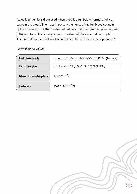
Aplastic anaemia is diagnosed when there is a fall below normal of all cell
types in the blood. The most important elements of the full blood count in
aplastic anaemia are the numbers of red cells and their haemoglobin cont ent
(Hb), numbers of reticulocytes, and numbers of platelets and neutrophils.
The normal number and function of these cells are described in Appendix A.
Normal blood values
Reticulocytes
Red blood cells
Absolute neutrophils
50-150 x 109/l (0.5-2.5% of total RBC)
4.5-6.5 x 10¹²/l (male) 4.0-5.5 x 10¹²/l (female)
1.5-8 x 10⁹/l
Platelets 150-400 x 109/l
It is important to have a measure of the amount of damage to the bone
marrow because this influences the treatment strategy. There is a simple
but effective classification based mainly on the findings in the blood
count. The disease can be divided into three groups, non-severe aplastic
anaemia (NSAA), severe aplastic anaemia (SAA) and very severe aplastic
anaemia (VSAA). The names were devised when there was less effective
treatment than there is now and the differences are not as alarming
as the names might suggest. Nevertheless the classification is still a guide
as to which type of treatment is most likely to produce the best result for
a particular patient.
Non-severe aplastic anaemia (NSAA)The blood counts are low with a hypoplastic marrow but the condition is not
severe enough to meet the criteria for severe aplastic anaemia. These patients
may require red blood cell transfusions but not usually platelet transfusions.
They are not particularly at risk from spontaneous infection.
Severe aplastic anaemia (SAA)This is present when the marrow is hypoplastic for the age of the patient and
any two of the following features are present:
A low platelet count (less than 20 x109/l)
A reticulocyte count less than 25 x 109/l (corrected reticulocyte count
of less than 1%)
An absolute neutrophil count less than 0.5x 10⁹/l.
12
How is the severity of aplastic anaemia determined?
13
Very severe aplastic anaemia (VSAA)The definition is the same as for severe aplastic anaemia but with a very low
absolute neutrophil count, less than 0.2 x 10⁹/l.
Principles of treatmentTreatment of aplastic anaemia can be divided into supportive therapy, which
is directed at treating consequences of low red cell, white cell and platelet
counts, and definitive therapy which is aimed at restoring the function of the
marrow so that the patient becomes independent of blood transfusion, is no
longer at risk from bleeding or abnormal infections, and can lead a normal
life. Definitive treatment takes weeks to months to achieve its greatest effect.
Supportive therapy is essential during this time when patients will be at risk
from anaemia, bleeding/bruising problems and infections.
Supportive therapySupportive therapy will be needed for patients with anything other than very
mild aplastic anaemia.
Red blood cell transfusionsVirtually all patients with aplastic anaemia who need treatment will require
transfusions of red blood cells. They may also need transfusions of platelets.
Since patients may require many transfusions they should always receive
blood products from which as many as possible of the white cells have been
removed. This is done because white cells are likely to provoke the patient’s
immune system to produce antibodies. These antibodies can cause problems
in future transfusions and may cause complications if a stem cell transplant
is performed. Removal of white blood cells from all blood transfusion products
is called ‘leucodepletion’. This is now routinely applied to all transfusion units
collected in the UK before delivery to hospitals.
14
How is aplastic anaemia treated?
15
If a patient is being considered for a stem cell transplant it is particularly
important that they do not receive transfusions of blood or blood products
from any family member. This is because such transfusions may seriously
compromise the likelihood of success of a later transplant from
a related donor.
Red cell transfusions may (rarely) produce problems in patients with aplastic
anaemia. Some patients produce antibodies to so-called ‘minor’ blood groups.
This sometimes means that they need carefully selected blood which may
need to be specially ordered from the National Blood Service. Without these
precautions the presence of antibodies would mean the transfused blood
would not last very long.
Rarely a red cell transfusion may cause fever and even rigors (shaking).
These can be controlled with injections of hydrocortisone and Piriton™.
Piriton™ is a strong antihistamine which often makes people sleepy.
One unit of transfused red blood cells typically raises the haemoglobin level
by about 1.0g/dl. It is usual to need three to four units each month. Repeated
red cell transfusions given over many years may lead to accumulation of iron
in the body. This can produce problems with the heart and hormone producing
(endocrine) glands unless precautions are taken. Few patients with aplastic
anaemia need transfusion programmes which are be prolonged enough
to cause such problems.
Platelets are shorter-lived cells; a single unit transfused should raise the
platelet count significantly for about two to three days. Response to platelet
transfusions is much more variable from one patient to another. Sometimes
patients develop antibodies to platelets and special donors have to be found
by the transfusion service.
16
All patients who are being treated with only supportive therapy need to be
monitored regularly to detect any worsening of their condition. In milder
cases this involves visits to the out-patients or day care unit to check the full
blood count and general health and to determine the time of any transfusions
needed. In more severe cases, where patients are very vulnerable to infection,
they may need to spend some time in the hospital in a special protected
environment. If patients with a low neutrophil count develop a fever they must
be treated promptly with antibiotics without waiting for proof of infection.
If symptoms persist despite first line antibiotics then additional drugs,
which counteract fungal infections, are given. Any patient with aplastic
anaemia who thinks they are developing an infection should consult with
their doctor so that any necessary treatment can be commenced as soon
as possible.
Definitive therapyExcept for a minority of patients with mild aplastic anaemia, all patients will
require some form of definitive therapy. The long-term outcome is very poor
for patients with severe or very severe aplastic anaemia who receive only
supportive treatment.
The underlying disease process in acquired aplastic anaemia is destruction
of blood-forming stem cells by the immune system. Definitive therapy
is aimed at either blocking this process by suppressing the immune system
or, alternatively, at curing the disease by replacing the diseased marrow and
abnormal immune system using a stem cell transplant from a donor.
Once a decision has been made that definitive treatment is necessary, this
should be initiated as soon as possible. This is because patients remain
at risk of life-threatening infection and/or bleeding until their blood counts
can be restored to normal. In the case of stem cell transplants the fewer blood
transfusions a patient has received prior to the transplant the lower the risk
of graft rejection. This is another reason why if a transplant is planned
it should be done as soon as possible.
Stem cell transplant4
The only truly curative treatment for a patient with severe or very severe
aplastic anaemia is a stem cell transplant using normal stem cells from
a donor. These replace the damaged marrow with healthy marrow.
More importantly, they also replace the cells in the patient’s immune system
which are responsible for destruction of the bone marrow stem cells in the
first place. Stem cell transplantation is most successful in children and young
adults who have a brother or sister (a sibling) as a suitable donor. This is
known as an allogeneic sibling transplant. Patients who are aged below 40
years and who have SAA or VSAA and are eligible for a transplant are usually
offered this treatment as the first option. The risk of transplantation increases
with age and for older patients a transplant is usually suggested only for
patients with VSAA or whose disease has not responded to a first course of
immunosuppressive treatment. Transplants from volunteer matched donors
may be successful, particularly in children, but the risk is much higher than
with sibling transplants.
Although a sibling-donor transplant is the preferred treatment for SAA
or VSAA only a minority of patients are of the appropriate age group and
have a donor available. For this reason transplantation is not widely used
as a treatment for adults. Any patient who is being considered for a transplant
will have detailed discussions of the risks and benefits with their specialist.
ImmunosuppressionFor patients who are not eligible for a transplant, treatment is aimed
at suppressing the immune activity which causes the damage or prevents the
recovery of the marrow stem cells. This is called immunosuppressive treatment
and offers a good chance of long-term survival and a normal or near normal
lifestyle. The usual therapy used to suppress the immune system in aplastic
anaemia is the use of antilymphocyte or antithymocyte globulins (ALG
or ATG) and cyclosporin.
17
4 There is a separate publication on stem cell transplantation available from Leukaemia & Lymphoma Research.
18
ALG (or ATG) is a preparation of antibodies which can remove certain cells
of the immune system. They are typically obtained from the serum of either
rabbits or horses which have been immunised with human lymphocytes.
The differences between ALG and ATG are determined by the type of human
lymphocytes which are used to immunise the donor animal in order to produce
the antibodies. Because they are biological products produced by different
techniques they are not standardised and dose schedules vary from one type
to another.
All preparations of ALG and ATG contain proteins from species other than
man and so they can provoke an immune reaction. There is a delayed reaction
which comes on seven to fourteen days after the treatment. This is called
serum sickness and is the result of the patient producing antibodies against
the foreign protein. The patient may experience fever, joint pains and skin rash
which can be managed with steroids and analgesics.
There may also be an acute reaction which produces high fever and sometimes
rigors on the first two days of administration. This affects nearly all patients
and is related to the destruction of the lymphocytes. There may also be a
skin rash.
Giving ALG or ATG to patients with aplastic anaemia briefly increases the risk
of infection through suppression of the immune system. This treatment should
always be given in a hospital where strict isolation from infection is available.
The normal course of treatment is given over five days through a drip line
(called a central line) which is fed into a large vein to avoid problems with local
inflammation. The patient usually stays in hospital for about three weeks
to allow the immune system to recover and to ensure there is no further risk
of serum sickness.
There is no clear evidence to prove that either ALG or ATG is better for
treatment of severe aplastic anaemia. ATG or ALG is most commonly used
in combination with ciclosporin, another immunosuppressive drug.
Ciclosporin is usually started after completion of the ALG treatment and is
continued for at least six months. Side effects of ciclosporin are fortunately not
very troublesome for most patients though care has to be taken in adjusting
the dose for each patient so that maximum effect is achieved without causing
kidney or liver damage
Other treatmentsA number of other approaches have been used in the treatment of severe
aplastic anaemia. Anabolic (androgenic) steroids were the first specific
therapy for aplastic anaemia before immunosuppressive treatment became
available. This treatment is not effective in severe or very severe aplastic
anaemia but may have a place in less severe cases that fail to respond
to immunosuppression. The side effects are often unpleasant, particularly
for female patients, and so this treatment should not be offered without
a considerable amount of explanation and discussion. Anabolic steroids are
useful for patients with Fanconi anaemia, the inherited type of
aplastic anaemia.
Growth factors are naturally occurring substances which are essential for the
normal control of blood production. Granulocyte colony stimulating factor
(G-CSF) is the natural protein which stimulates neutrophil production and
function. Therapy with additional G-CSF may increase neutrophil numbers to
a level which reduces the risk of spontaneous infections. An exception to this is
in patients with very severe aplastic anaemia in whom G-CSF is not effective.
There are two G-CSF preparations available, Filgrastim™ and Lenograstim™.
The best way to use this additional treatment is still under investigation. Most
growth factors such as erythropoietin (which increases red cell production)
and thrombopoietin (which controls platelet production), are produced at high
level in patients with aplastic anaemia and additional therapy is not helpful.
19
Treatment planningThe treatment plan for each patient will be determined by the severity of the
disease, their age, the availability of a suitable potential stem cell donor and
their general health at the time of diagnosis. A very important part of the early
discussion of treatment options includes the recognition that recovery may
take a long time, that different treatments are not mutually exclusive, and
that it may be necessary to change between different treatments.
There is always a delay in confirming the diagnosis of aplastic anaemia, in part
so that other causes of the symptoms and low blood count can be excluded.
During this time patients often require transfusions of red cells and platelets.
Such small numbers of transfusions do not compromise the outcome of later
treatment and are important in keeping the patient as well as possible. During
this time patients may need antibiotics to control infection.
Once the initial investigations and discussions have been completed the
treatment options will be discussed and explained and a treatment plan
will be agreed upon. For most patients the decision will centre on whether
immunosuppressive therapy or a stem cell transplant should be the first
option. For children and younger adults with SAA who have a brother or sister
as a potential donor, a stem cell transplant will usually be the first choice.
For others immunosuppression may be preferred as initial treatment, with
transplantation as a later option if immunosuppression is unsuccessful.
ImmunosuppressionFor most patients immunosuppression will be the treatment of choice for the
disease. The treatment and the response fall into a number of stages.
To receive treatment with ALG (ATG) the patient needs to be in hospital and
preferably in an isolation room. The main side effects and problems with ALG
(ATG) have already been described. These are fever and rigors on the first two
days of receiving the treatment and the delayed serum sickness. The patient
20
21
usually spends two to three weeks in hospital. In many schedules the patient
continues to take ciclosporin after leaving hospital.
Initially nothing will seem to have happened and the patient will still require
transfusions. The first signs of recovery usually do not appear before six
to eight weeks and may be delayed much longer. A rise in the neutrophil
count or a lessening of the need for platelet transfusions is often the first
sign of recovery. A full reassessment of marrow activity is usually made at
about three to four months post treatment. When patients show signs of
recovery cyclosporin is continued and the blood counts carefully checked. As
the recovery becomes more clearly established the intervals between tests
get longer so that only a test every three to four weeks may be needed. It is
important to realise that this treatment is aimed at achieving freedom from
transfusion and protection from excessive infections rather than cure. This
is called remission and occurs in about two out of every three patients so
treated. The blood count may remain mildly abnormal for many years and
even in patients who achieve a truly normal blood count it is possible to show
residual damage in the bone marrow stem cells.
If the first course of treatment fails there are a number of options. For most
patients a second course of treatment can be given using the same or different
type of ALG (ATG). Indeed, several courses of immunosuppression can
be given for repeated failure or for relapse. For patients with a family donor
this may be the time to consider a stem cell transplant.
It is important to realise that recovery from aplastic anaemia can occur
at any time, even after several years of transfusion dependence. It is therefore
necessary to continue to keep the patient in the best possible health with
transfusions and antibiotics even though this may prove a great burden to the
patient. None the less patients and families should never give up the hope and
expectation that remission may come.
22
5 Paroxysmal nocturnal haemoglobinuria (PNH) is a rare condition affecting mainly the red blood cells but also other blood cells. In PNH the red blood cells are destroyed more rapidly than normal. Occasionally this causes the urine to turn very dark (haemoglobinuria). PNH is so called because the colour may occur intermittently (paroxysmal) when the urine is generally darkest first thing in the morning (nocturnal haemoglobinuria).
Long term follow up after immunosuppressionThe marrow shows some residual damage even after complete return
to normal blood counts. For just over half the patients who have a good
response the counts remain constant for many years and they may eventually
be considered cured. For other patients some change or deterioration in the
blood count may occur sometime in the ten years following completion of
treatment and this often leads to a return to the need for blood transfusions.
True relapse, that is a return of the original condition of aplastic anaemia,
affects perhaps one in ten patients. Relapse is especially likely to occur
in pregnancy or following a heavy immune challenge such as multiple
immunisations at the same time. Only very rarely can relapse be attributed
to re-exposure to the original cause of the disease.
In twenty to thirty percent of cases a new, abnormal type of blood cell will
emerge. This is perhaps a result of abnormal cells escaping the damage
which caused the aplastic anaemia in the first place. These cells lack certain
proteins on their surface and the condition is called after its most dramatic
manifestations, paroxysmal nocturnal haemoglobinuria5 (PNH). The most
dangerous type of relapse is the change from aplastic to preleukaemic blood
production (myelodysplastic syndrome) or even to acute leukaemia (acute
myeloid leukaemia). Such changes are rare but if they do occur then
the risks associated with unrelated donor transplant may become much
more acceptable.
The possibility of relapse means that patients need to be followed up for many
years after treatment. As time goes by the frequency of checks can be reduced
so that eventually only annual blood counts are needed or even just occasional
contact with the specialist unit.
23
Such contacts can provide reassurance and a chance to discuss special issues,
for example the risks and management of pregnancy or of foreign travel.
Stem cell transplantationStem cell transplantation for aplastic anaemia from a matched brother
or sister has a very good outcome in the majority of younger patients (under
30 to 40 years). Overall, about seven out of ten patients so treated will have
a successful transplant without long-term side effects and many may
be considered cured.
For children and young adults, particularly if the disease is severe,
transplantation from a suitably matched volunteer donor may be appropriate.
Although the success rates for such transplants are not quite as good as
from a brother or sister, they still offer a good chance for a cure. The risks and
benefits of unrelated transplants will be discussed in detail before a patient
or parent is asked to make a decision. The results of unrelated transplants
are beginning to improve but such procedures should only be carried out
in specialist units when all treatment options have been thoroughly discussed.
The main risks are those of the transplant and include graft failure,
the reaction of the graft against the patient (graft versus host disease,
GvHD) and development of severe infections. The most disabling long-term
complication is chronic GvHD which may result in an extensive, scarring
skin rash and liver disease and/or diarrhoea. The risk of these complications
increases with age. This is why this treatment is not recommended as first line
for all patients with a suitable donor.
Unlike a transplant to treat leukaemia or a related blood cancer, the pre-
transplant conditioning needed to prevent graft rejection is relatively mild
for patients with aplastic anaemia. Radiation treatment is not required and
patients who have a successful transplant are fertile and have a normal
24
chance of becoming a parent. The conditioning does not even abolish the
possibility of later recovery of the patient’s own marrow.
The schedule for transplantation starts with admission to a specialist
transplant unit. The patient is given an isolation room. This is a room with a
clean filtered air supply, from which as many potentially dangerous organisms
as possible have been removed. A diet of specially prepared food is started
which again aims to remove potentially harmful organisms. Discussion with
the dietician on particular needs, likes and dislikes is helpful at this time, as is
an explanation to friends and relatives of what is and is not permitted in the
way of supplementary food. Rules concerning visitors may vary from unit
to unit but in general visitors are restricted to close family or friends who are
instructed in the protocols of hand washing and so on needed to preserve the
clean environment. In most cases a detailed discussion of what is going
to happen will take place before admission.
During the transplant process many blood transfusions, antibiotics and other
fluids will need to be given directly into the circulation by a vein. To achieve this
with minimum discomfort and inconvenience to the patient a central line is
put in place which will remain there for the whole of the transplant procedure.
Occasionally these lines may become infected and have to be changed but
in general they have proved to be of enormous benefit in coping with the
requirements of treatment.
Conditioning is the word used to describe the drugs given to prepare the
patient to receive the graft. The combinations of drugs that are used vary
from one centre to another so explanations of side effects and duration
of conditioning will be different. A common schedule includes specific
immunosuppressive drugs like ALG followed by cyclophosphamide, a drug
used in chemotherapy. The immediate side effects are usually not very
troublesome though some patients need treatment to stop them being sick.
Conditioning also further increases the risk of infection, hence the need
25
for isolation. A major side effect of conditioning is subsequent loss of hair.
Fortunately it will grow again in about six months.
The stem cells, which may be collected from the donor either from the marrow
or from the blood after mobilisation, are given through the central line.
Reactions are rare and usually consist of mild fever as the stem cells are given.
The patient is given ciclosporin to reduce the risk of graft rejection and GvHD.
This may commence either a few days before the transplant or at the time the
stem cells are given to the patient. The dose is adjusted for individual patients.
Patients transplanted for aplastic anaemia need to continue on this drug for
up to a year post transplant. Despite taking this drug the patient can usually
return to a normal life style after about six months.
It takes about three weeks to a month for the new graft to become established
and the patient remains in isolation during this time. Quite commonly in this
period the patient develops fevers which need treatment with antibiotics.
Some degree of GvHD is also not unusual, perhaps with a skin rash or mild
diarrhoea. This may resolve without treatment or require a course of cortisone.
More severe GvHD needs more aggressive management.
Once the graft is established the patient normally leaves hospital but
still needs to take care to avoid infections as far as possible and careful
explanations of what is allowable are given before discharge. Some infections,
particularly certain virus infections, may be reactivated in the first few months
after discharge and so careful checking of the patient and the blood counts
is required. Although the patient with a successful graft feels well after about
six months and may go back to work or school, complete recovery of the
immune system takes about two years. Re-vaccination is needed once the
ciclosporin is stopped.
Summary
Acquired aplastic anaemia is a rare, potentially life threatening disorder.
In most cases the cause is unknown; in a few cases drug or chemical
exposure is suspected. The mainstays of management are support with
blood transfusions and protection from infection. A few patients may
receive only this but most patients will require definitive treatment with
immunosuppression or a stem cell transplant. The condition is chronic
but recovery may occur even after several failed attempts at achieving
remission. The disease used to have a fearsome reputation but now,
thanks to the greatly improved means of support and treatment, seventy
to eighty percent of patients should recover.
Useful contactsThe Aplastic Anaemia Trust
Tel: 0870 487 0099
Fax: 020 7840 7841
Website: www.theaat.org.uk
Email: [email protected]
26
27
Appendix A
Red Blood Cells (RBC): The normal red blood cell count is 4-5.5x10¹²/l in
women and 4.5-6.5x10¹²/l in men. For assessing the degree of anaemia, the
usual measurement is the haemoglobin (Hb), which is normally 11.5-15.5 g/dl
for women and 13.5-17.5 g/dl for men. The degree of anaemia is not used to
assess severity because the value is often altered by blood transfusion.
The reticulocyte count estimates the numbers of newly produced red cells
present in the blood. The count is often given as a percentage of all red blood
cells. With modern cell counting machines in the laboratory the actual number
of reticulocytes can be measured, which is more useful than an estimate of
the percentage of reticulocytes. The normal range is 50-150 x 109/l (0.5-2.5%
of the normal RBC count). A low reticulocyte count is an indication of the
marrow’s failure to produce red cells and is used in the assessment of severity.
The absolute neutrophil count (ANC) indicates whether white blood cells are
being produced normally. Neutrophils have a short survival once they leave
the marrow and so they give a useful estimate of current marrow activity.
The neutrophil count is often reported as a percentage of the total white
count. The absolute neutrophil count is the percentage count multiplied by
the total number of white cells. This gives a more accurate indication of
numbers of white cells than the percentage count. The normal range is about
1.5 to 8.0 x10⁹/l, (a slightly lower value may be seen in healthy people
of Afro-Caribbean descent).
The platelet count is an automated count of the number of platelets in the
blood. The normal range is between 150 and 400x109/l.

28
Notes
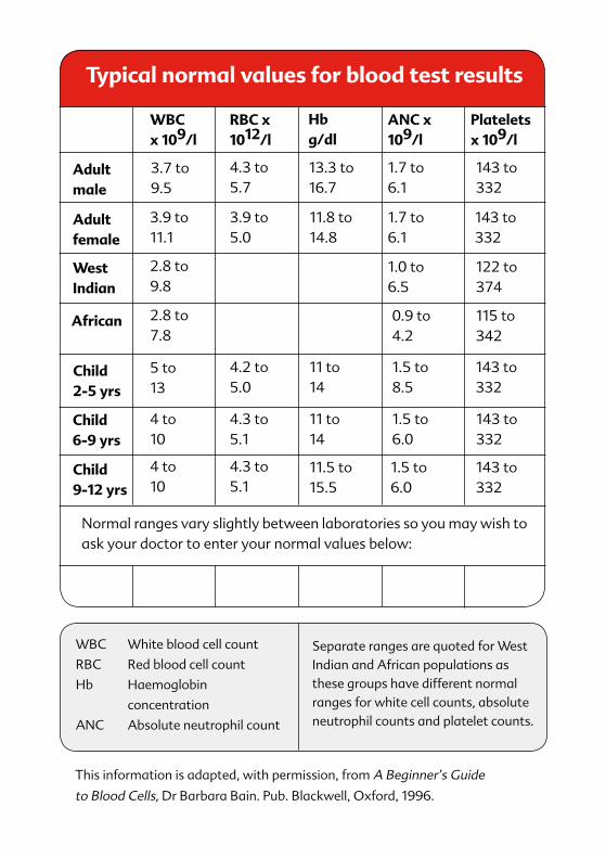
Adult female
WBCx 109/l
RBC x 1012/l
ANC x 109/l
Platelets x 109/l
Hb g/dl
Adult male
3.7 to 9.5
4.3 to 5.7
13.3 to 16.7
1.7 to 6.1
143 to 332
143 to 332
122 to 374
115 to 342
143 to 332
143 to 332
143 to 332
1.7 to 6.1
1.0 to 6.5
0.9 to 4.2
1.5 to 8.5
1.5 to 6.0
1.5 to 6.0
11.8 to 14.8
11 to 14
11 to 14
11.5 to 15.5
3.9 to 5.0
4.2 to 5.0
4.3 to 5.1
4.3 to 5.1
3.9 to 11.1
2.8 to 9.8
2.8 to 7.8
5 to 13
4 to 10
4 to 10
West Indian
African
Normal ranges vary slightly between laboratories so you may wish to ask your doctor to enter your normal values below:
Child 2-5 yrs
Child 6-9 yrs
Child 9-12 yrs
WBC White blood cell count
RBC Red blood cell count
Hb Haemoglobin
concentration
ANC Absolute neutrophil count
Separate ranges are quoted for West Indian and African populations as these groups have different normal ranges for white cell counts, absolute neutrophil counts and platelet counts.
This information is adapted, with permission, from A Beginner’s Guide
to Blood Cells, Dr Barbara Bain. Pub. Blackwell, Oxford, 1996.
Typical normal values for blood test results
02/10
Leukaemia and Related Diseases
Acute Promyelocytic Leukaemia (APL)
Adult Acute Lymphoblastic Leukaemia (ALL)
Adult Acute Myeloid Leukaemia (AML)
Childhood Acute Lymphoblastic Leukaemia (ALL)
Childhood Acute Myeloid Leukaemia (AML)
Chronic Lymphocytic Leukaemia (CLL)
Chronic Myeloid Leukaemia (CML)
Aplastic Anaemia (AA)
The Myelodysplastic Syndromes (MDS)
The Myeloproliferative Disorders (MPD)
Multiple Myeloma (MM)
Hodgkin’s Lymphoma (HL)
Non-Hodgkin’s Lymphoma (NHL)
Bone Marrow and Stem Cell Transplantation (BMT) – for children and adults
Donating stem cells – what's involved?
Donor Lymphocyte Infusion (DLI) – what’s involved?
The Seven Steps – Blood & bone marrow transplantation
Undergoing high dose therapy and autologous stem cell transplant
Chemotherapy – what do I need to know?
Clinical Trials
Complementary and Alternative Medicine (CAM)
Dietary advice for patients with neutropenia
Supportive care
Treatment decisions
Watch and wait
Young adults with a blood cancer – what do I need to know?
Jack's Diary: an illustrated children's book to help young patients understand and deal with blood cancers, treatment and life changes
Wiggly's World: a colourful A-Z illustrated booklet, designed to take the anxiety out of treatment for children and their parents
Leukaemia & Lymphoma Research, 43 Great Ormond Street, London WC1N 3JJT: 020 7405 0101 • F: 020 7405 3139
E: [email protected] • www.llresearch.org.uk
Registered charity 216032 (England & Wales) SC037529 (Scotland)
The following patient information booklets are available free of charge from Leukaemia & Lymphoma Research. You can download them from our website or request copies by phone or post (see form inside):
Leaflets on a range of associated blood disorders are also available from Leukaemia & Lymphoma Research