Embed Size (px)
Citation preview

APITHERAPY IN IMMUNE
MEDIATED DISORDERSMEDIATED DISORDERS

BY
EHAB AHMED KAMAL
LECTURER OF COMPLEMENTRY MEDICINE
NATIONAL RESEARCH CENTRE
GIZA - EGYPTGIZA - EGYPT
CONSULTANT OF GASTROENTROLOGY AND
HEPATOLOGY

WHAT IS APITHERAPY
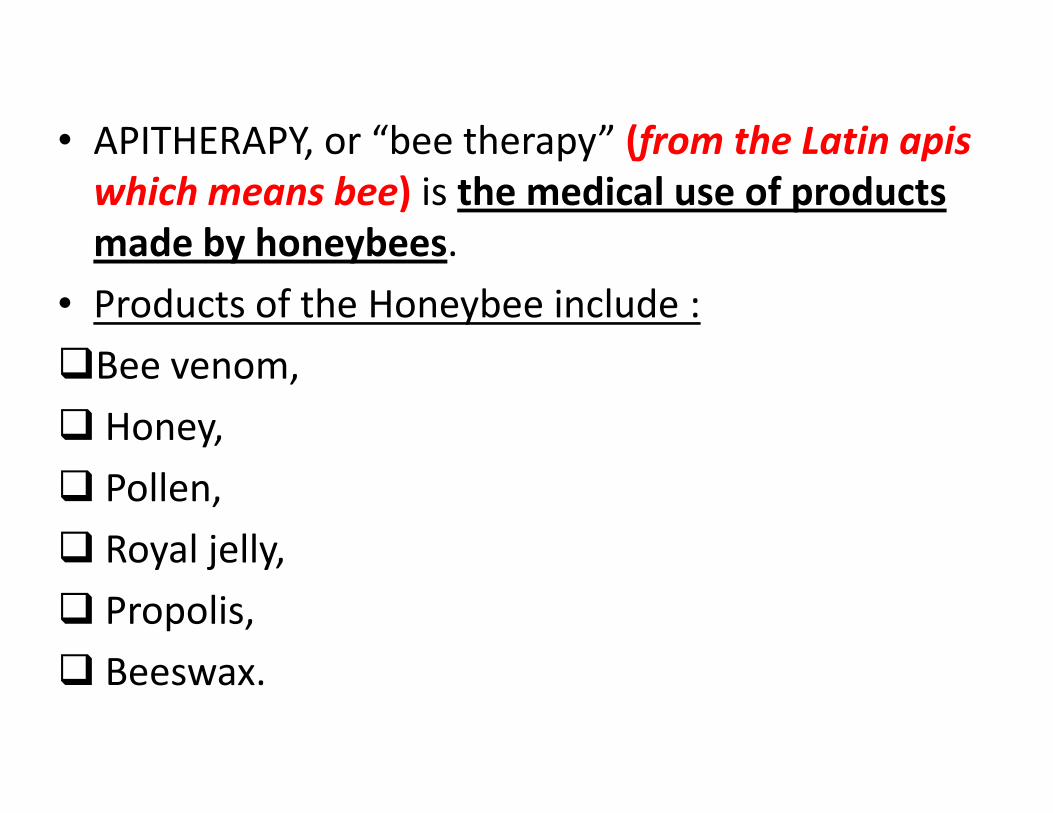
• APITHERAPY, or “bee therapy” (from the Latin apis
which means bee) is the medical use of products
made by honeybees.
• Products of the Honeybee include :
�Bee venom,
� Honey,� Honey,
� Pollen,
� Royal jelly,
� Propolis,
� Beeswax.

• It is important to note that Apitherapy is not only
the use of the venom for healing, often called BEE
STING THERAPY, but the use of all the hive
products, and usually a combination of them.
• These products are also sometimes mixed with• These products are also sometimes mixed with
other ingredients, specifically different essential
oils, dependent on the condition being treated.

HISTORY OF APITHERAPY

• The exact place and pattern of origin of apitherapy
is not clear.
• History of apitherapy can be traced back to ancient
Egypt, Greece, and China (Hegazi, 1998)
• Even Hippocrates, the great Greek physician
renowned as the "father of medicine," used bee
venom to treat joint pain and arthritis. Ancient
Greeks athletes used honey to boost an energy.
(Broffman, 1999).

• The modern systematic study of apitherapy was
initiated through the efforts of the Austrian
physician PHILLIP TERC.
• He published the results of intentional bee sting
and bee in his article "Report about a Peculiarand bee in his article "Report about a Peculiar
Connection Between the Beestings and
Rheumatism" in 1888.

• The holly Quran 1400 years ago mentioned that the
bee products contain cure to people.
Wherein is healing for people
Al Nahl :69

Active components of apitherapy
1-Peptide constituents1-Peptide constituents

PEPTIDE 401
• Mast cell degranulating (MCD) peptide—MCD
peptide, also known as peptide 401, a bee venom
polypeptide with 22 amino acids and constituting
2–3% of dry bee venom.
• It was originally named due to its biological action• It was originally named due to its biological action
of causing release of histamine from mast cells
(Banks et al., 1990).

APAMIN
• Another important bee venom neurotoxic
polypeptide of 18 amino acids comprising 2–3% of
dry bee venom.
• It possesses a selective inhibitory action on calcium-
dependent potassium channels that are involved independent potassium channels that are involved in
regulation of the after-hyperpolarization period and
frequency of action potential generation in the
central nervous system (CNS) (Hugues et al., 1982).

• Afterhyperpolarization, describes the phase of a
neuron's action potential where the
cell's membrane potential falls below the
normal resting potential.
• This is also commonly referred to as an action• This is also commonly referred to as an action
potential's undershoot phase (M. Shah, and D. G.
Haylett, 2000).

MELLITIN

• A strongly basic 26 amino-acid polypeptide which
constitutes 40–60% of the whole dry honeybee
venom.
• It has various biological, pharmacological and
toxicological actions including strong surfacetoxicological actions including strong surface
activity on cell lipid membranes, hemolyzing
activity, antibacterial and antifungal activities
(Lariviere and Melzack, 1996).

• The cytotoxic effect through the activation of PLA2
by melittin is believed to be an important
mechanism of anti-cancer activity of BV.
• Several cancer cells, including renal, lung, liver,
prostate, bladder, and mammary cancer cells asprostate, bladder, and mammary cancer cells as
well as leukemia cells, can be targets of melittin
(Moon et al., 2006).

• The induction of apoptotic cell death through
several cancer cell death mechanisms, including the
activation of caspase and matrix metalloproteinases
(MMP), is important for the melittin-induced anti-
cancer effects (Holle et al., 2003).
• The binding of the cell lytic peptide (melittin) to the
hormone receptors as well as gene therapy carrying
melittin can be useful as a novel targeted treatment
for some types of cancer, such as prostate and
breast cancers (Li et al., 2004).

• Recently, Melittin has also been demonstrated to
cause neural plastic changes along pain-signaling
pathways by activation and sensitization of
nociceptor cells via phosphorylation of mitogen-
activated protein kinases (MAPK) (Hao et al.,
2008;Yu et al., 2009).2008;Yu et al., 2009).
• The effect of mellitin was studied in animal models
with amyotrophic lateral sclerosis (ALS) it was
found that administering melittin decreased
microglial activity and the expression of the pro-
inflammatory factor TNF-α (Yang EJ., et al 2010).

ADOLAPIN

• ADOLAPIN, a basic polypeptide with 103 amino
acids residues and comprising 1% of dry bee
venom, it has been shown to have anti-nociceptive
“decreasing pain sensation” anti-inflammatory and
antipyretic effects (Koburova et al., 1984,1985).
• Adolapin can inhibit prostaglandin synthesis via
inhibition of cyclooxygenase activity (Shkenderov
and Koburova, 1982).

ENZYMES

PHOSPHOLIPASE A2
• PLA2, which constitutes 10–12% of dry bee venom,
has inflammatory and nociceptive effects (Landucci
et al., 2000).
• PLA2 is a membrane-associated phospholipid
converting enzyme that is important in theconverting enzyme that is important in the
production of arachidonic acid, which is further
metabolized to protaglandins by cyclooxygenase
and to leukotrienes by lipoxygenase (Landucci et
al., 2000).

• PLA2 exhibits complex interactions with melittin
that can result in potentiation of secretory PLA2
effects or in inhibition depending on the
peptide/phospholipid ratio (Koumanov et al.,
2003).
• PLA2 has effects in a range of cells related to
nociception including astrocytes and neurons and
possibly microglial cells , it is also involved in nerve
regeneration (Sun et al., 2004a).

HYALURONIDASE

• HYALURONIDASE constitutes 1.5–2% of dry bee
venom (Lariviere and Melzack, 1996).
• Hyaluronidases break down hyaluronic acid in
tissues such as in synovial bursa of rheumatoid
arthritis patients (Barker et al., 1964).arthritis patients (Barker et al., 1964).
• Hyaluronidase in bee venom shares this property
with endogenous hyaluronidase (Barker et al.,
1963).

IMMUNE EFFECTS OF APITHERPAY

• In most of the diseases which are considered to
benefit from propolis, cellular immune reaction is
activated, neopterin levels in body fluids are
increased and enhanced tryptophan degradation is
observed.
• Increased amounts of neopterin are produced by
human monocytes/macrophages upon stimulation
with the cytokine interferon-y (Murr C., et al 2002).

• Caffeic acid phenethyl ester (CAPE) is a biologically
active component of propolis, a resinous material
obtained from bee hives (Girgin et al., 2009).
• CAPE has several positive effects, including anti-• CAPE has several positive effects, including anti-
inflammatory, anti-oxidation, anti-cancer, anti-
bacterial, anti-viral, anti-fungal, and
immunomodulatory effects (Jung et al., 2008).

• Song et al., (2008) evaluated the anti-inflammatory
effect of CAPE on cultured human middle ear
epithelial cells (HMEECs).
• They suggested that the anti-inflammatory effect of• They suggested that the anti-inflammatory effect of
caffeic acid phenethyl ester ( CAPE ) is due to its
inhibition of tumor necrosis factor (TNF)-alpha
expression and interleukin (IL)-8 production (Song
et al., 2008) .

• Márquez et al., (2004) evaluated the
immunosuppressive activity of CAPE in human T-
cells, discovering that this phenolic compound is a
potent inhibitor of early and late events in T-cell
receptor-mediated T-cell activation.
• They found that CAPE specifically inhibited both
interleukin (IL)-2 gene transcription and IL-2
synthesis in stimulated T-cells.

• Kohno et al., (2004) examined the anti-
inflammatory actions of Royal Jelly (RJ) at a
cytokine level. When supernatants of RJ
suspensions were added to a culture of mouse
peritoneal macrophages stimulated with
lipopolysaccharide and IFN-gamma.lipopolysaccharide and IFN-gamma.
• the production of proinflammatory cytokines, such
as TNF-alpha, IL-6, and IL-1, was efficiently inhibited
in a dose-dependent manner without having
cytotoxic effects on macrophages.

POLLEN

• At the mucosal surfaces, pollen grains do not only
release allergens but also proinflammatory and
immunomodulatory lipids, termed pollen-
associated lipid mediators.
• Among these, the E1-phytoprostanes (PPE1) were• Among these, the E1-phytoprostanes (PPE1) were
identified to modulate dendritic cell (DC) function:
PPE1 inhibit the DC's capacity to produce IL-12 and
enhance DC mediated TH2 polarization of naive T
cells (Gilles et al., 2009).

ULCERATIVE COLITISULCERATIVE COLITIS

WHAT IS ULCERATIVE COLITIS?
• Ulcerative colitis (UC) is one of the 2 major types of
inflammatory bowel disease (IBD), along with Crohn
disease.
• Unlike Crohn disease (CD), which can affect any• Unlike Crohn disease (CD), which can affect any
part of the gastrointestinal (GI) tract, UC
characteristically involves only the large bowel.
Signs and symptoms
Patients with UC predominantly complain of the
following:
• Rectal bleeding.
• Frequent stools.
• Mucous discharge from the rectum.
• Tenesmus (occasionally).
• Lower abdominal pain .

In some cases, UC has a fulminate course marked by
the following:
• Severe diarrhea and cramps
• Fever
• Leukocytosis
• Abdominal distention

UC is associated with various extracolonic
manifestations, as follows:
• Uveitis
• Pyoderma gangrenosum
• Pleuritis
• Erythema nodosum
• Ankylosing spondylitis
• Spondyloarthropathies

EPIDEMIOLOGY

• In North America, incidence rates range from 2.2 to
19.2 cases per 100,000 person-years for ulcerative
colitis and 3.1 to 20.2 cases per 100,000 person-
years for Crohn disease (Molodecky NA et al,.
2012).
• The incidence and prevalence of Crohn disease and
ulcerative colitis appear to be lower in Asia and the
Middle East (Ng SC,. Gastroenterology 2013).

DIAGNOSIS

Laboratory studies are useful principally inexcluding other diagnoses and assessing thepatient’s nutritional status. They may include thefollowing:
• Complete blood count (CBC).
• Comprehensive metabolic panel.
• Inflammation markers (eg, erythrocytesedimentation rate [ESR], C-reactive protein [CRP]).
• Stool assays.
• Serologic markers (eg, antineutrophil cytoplasmicantibodies [ANCA], anti–Saccharomyces
cerevisiae antibodies [ASCA]).

Diagnosis is best made with endoscopy and biopsy,
on which the following are characteristic:
• Abnormal erythematous mucosa, with or without
ulceration, extending from the rectum to a part or
all of the colon
• Uniform inflammation, without intervening areas of • Uniform inflammation, without intervening areas of
normal mucosa (skip lesions tend to characterize
Crohn disease)
• Contact bleeding may also be observed, with mucus
identified in the lumen of the bowel


HISTOLOGY

• In untreated disease, UC usually exhibits a
histological pattern of CHRONIC ACTIVE COLITIS,
which refers to the presence of active inflammation
accompanied by features of chronic mucosal injury.
• Activity is defined as the presence of neutrophil-• Activity is defined as the presence of neutrophil-
mediated epithelial injury, which may take the form
of neutrophils infiltrating crypt epithelium
(cryptitis), collections of neutrophils within crypt
lumens (crypt abscesses), or by infiltration of
surface epithelium with or without mucosal
ulceration ( Gupta RB., et al 2007).

• Chronicity is defined by crypt architectural
distortion, basal lymphoplasmacytosis, or cell
metaplasia.
• Architectural distortion is represented by• Architectural distortion is represented by
shortening of the crypts ( Gupta RB., et al 2007).

ETIOLOGY

• The exact etiology of ulcerative colitis is unknown,
but certain factors have been found to be
associated with the disease, and some hypotheses
have been presented.
• Genetic factors , immune conditions, environmental• Genetic factors , immune conditions, environmental
factors and NSAIDs use may be associated with the
development and affect the course of ulcerative
coloitis (Jantchou P., et al 2010).

Genetics
• The current hypothesis is that genetically
susceptible individuals have abnormalities of
humoral and cell-mediated immunity and/or
generalized enhanced reactivity againstgeneralized enhanced reactivity against
commensal intestinal bacteria and that this
deregulated mucosal immune response predisposes
to colonic inflammation (Xavier RJ,et al 2007).

Immune reactions
• Immune reactions that compromise the integrity ofthe intestinal epithelial barrier may contribute toulcerative colitis.
• Serum and mucosal autoantibodies againstintestinal epithelial cells may be involved. Thepresence of antineutrophil cytoplasmic antibodies(ANCA) and anti– Saccharomycescerevisiae antibodies (ASCA) is a well-knownfeature of inflammatory bowel disease (DubinskyMC, et al 2001).

Environmental factors
• Environmental factors also play a role. For example,
sulfate-reducing bacteria, which produce sulfides,
are found in large numbers in patients with
ulcerative colitis, and sulfide production is higher inulcerative colitis, and sulfide production is higher in
patients with ulcerative colitis than in other people
(Almeida MG, et al 2008).

NSAID use
• Nonsteroidal anti-inflammatory drug (NSAID) use is
higher in patients with ulcerative colitis than in
control subjects, (Felder Jb et al 2000) .

PATH PHYSIOLOGYPATH PHYSIOLOGY

• Subsets of T cells accumulate in the lamina propria
of the diseased colonic segment.
• These T cells are cytotoxic to colonic epithelium,
with increased production of immunoglobulin G
(IgG) and immunoglobulin E (IgE) (Himmel ME, et(IgG) and immunoglobulin E (IgE) (Himmel ME, et
al 2008).

• Also this is linked to excessive immune responses to
intestinal microbiota which are triggered by
increased activity of effector T cells and/or
decreased activity of regulatory T cells, changes in
the composition of intestinal microflora, and/or
damaged epithelial barrier (N. A. Molodecky anddamaged epithelial barrier (N. A. Molodecky and
G. G. Kaplan, 2010).
• Elevated expression of TNF was detected in
IBD patients more than 20 years ago (D.
Owczarek., et al 2012).
• A recent report showed that elevated
concentration of TNF was present in bloodconcentration of TNF was present in blood
serum of IBD patients while other groups found
increased levels of TNF protein both in serum
and in the intestinal lamina propria of UC
patients (R. Matsuda., et al 2009)

MANAGEMENTMANAGEMENT

Medical treatment of mild UC includes thefollowing:
• Mild disease confined to the rectum: Topicalmesalazine via suppository or budesonide rectalfoam.
• Left-side colonic disease: Mesalazine suppositoryand oral aminosalicylate (oral mesalazine isand oral aminosalicylate (oral mesalazine ispreferred to oral sulfasalazine).
• Systemic steroids, when disease does not quickly
respond to aminosalicylates.
• Oral budesonide.
• After remission, long-term maintenance therapy(eg, once-daily mesalazine).

Medical treatment of acute, severe UC may include
the following:
• Hospitalization.
• Intravenous high-dose corticosteroids.
• Alternative induction medications: Cyclosporine,
tacrolimus, infliximab, adalimumab, golimumab.

• INFLIXIMAB: REMICADE an antibody administered
intravenously , it works by blocking the effects
of tumor necrosis factor alpha (TNF alpha).
“Dosing in UC: 5 mg/kg IV at 0, 2, and 6 weeks, then every 8“Dosing in UC: 5 mg/kg IV at 0, 2, and 6 weeks, then every 8
weeks”.

• ADALIMUMAB: HUMIRA other form of injectable
anti TNF used in autoimmune disorders.
“ Dosing in UC
Induction: 160 mg SC either as 4 injections of 40 mg on day
1 or as 2 injections of 40 mg daily on 2 consecutive days,
then 80 mg SC 2 weeks later (day 15).
Maintenance :(beginning Week 4 Day 29): 40 mg SC q2wk.”

• Adverse effects of biological therapy:
�Antinuclear antibodies (50%).
�Infection (36%).
�Nausea (21%).�Nausea (21%).
�Infusion reaction and Headache (18%).
�Antibodies to double-stranded DNA (17%).
�Elevated alanine transaminase (ALT; rarely >3 times
upper limit of normal)

• Increased risk for:
�Active tuberculosis.
� Invasive fungal infections.
�Infections caused by other opportunistic pathogens,�Infections caused by other opportunistic pathogens,
including bacteria (eg, Legionella, Listeria).
�Malignancy: Lymphoma and other malignancies.

Indications for urgent surgery include the following:
• Toxic megacolon refractory to medical management.
• Fulminant attack refractory to medical management.
• Uncontrolled colonic bleeding.
Indications for elective surgery include the following:Indications for elective surgery include the following:
• Long-term steroid dependence.
• Dysplasia or adenocarcinoma found on screening
biopsy.
• Disease present 7-10 years.
Surgical options include the following :
• Total colectomy (panproctocolectomy) and
ileostomy.
• Ileoanal pouch reconstruction or ileorectal
anastomosis.anastomosis.
• In an emergency, subtotal colectomy with end-
ileostomy (Shen B. 2009 ).

DISEASE COURSEDISEASE COURSE

• Approximately 67 % of patients have at least one
relapse 10 years following the diagnosis (Scand J.
2009).
• The risk of relapse depends on the age at initial
diagnosis (Ha CY., et al 2010).diagnosis (Ha CY., et al 2010).
• A disease flare within two years of the diagnosis,
the presence of fever or weight loss at diagnosis,
and active disease in the preceding year increase
the risk of subsequent relapse (Scand J. 2009).

• Extension of colonic disease is seen in up to 20 % of
patients within five years (Allison J Clin. 2008).
• Approximately 20 - 30 % of patients with ulcerative
colitis will require colectomy for acute
complications or for medically intractable diseasecomplications or for medically intractable disease
(Scand J. 2009).

• Patients with ulcerative colitis are at increased riskfor colorectal cancer (CRC) (Lutgens MW. 2013).
• EXTENSION: The risk of CRC appears to be highestin patients with pancolitis, while those withproctitis and proctosigmoiditis are probably not anincreased risk of CRC, regardless of the duration ofincreased risk of CRC, regardless of the duration ofdisease.
• TIMING: The CRC risk begins to increase 8 to 10years following the onset of symptoms in patientswith pancolitis (Gyde SN. 1988).

CLINICAL TRIALCLINICAL TRIAL

• A clinical trial was performed 2009 by inducing UC
by administering trinitrobenzene sulfonic acid in
expermintal rats.
• The rats were then treated, in groups of six, with a
single enema of manuka honey or sulfasalazinesingle enema of manuka honey or sulfasalazine
medication (as a positive control) or a combination
of manuka honey and sulfasalazine, or not treated
(as a negative control).

• Visual examination of the colon showed thatmanuka honey on its own significantly decreasedulcerative colitis compared with no treatment andtreatment with sulfasalazine (to about one sixth ofthat with the no-treatment control, being twice aseffective as sulfasalazine).
• Histopathology showed that there was severeinflammation (evidenced by infiltration ofinflammatory cells) with no treatment, but onlymild inflammation with the honey treatment, thisbeing less than with the sulfasalazine treatment.(Medhi B,.et al 2009)

CONCLUSIONCONCLUSION

• The immune disorders associated with
development of UC , the disease course and
progression , the need of various methods of
treatment with multiple relapses and major side
effects encourage for more safe and effective
methods of treatment.methods of treatment.
• The role of bee products as natural defense against
the precipitating factors of the disease as tumor
necrosing factor and T cells represents a safe and
effective way in the management of UC patients.

• The role of CAPE as inhibitor of stimulation of T
lymphocytes and its action as natural inhibitor of
the tumor necrosing factor (Song et al., 2008) .
• Also the role of royal jelly as inhibitor of tumor
necrosing factor without any cytotoxic effectsnecrosing factor without any cytotoxic effects
Kohno et al., (2004) rendering the usage of
apitherapy as a safe, effective and promising
modality in the treatment of UC .

RECOMMENDATIONSRECOMMENDATIONS

• Large multicenter study should be done to
investigate the long term effect and ability of the
apitherapy products to induce and maintain
remission in UC patients.
• Comparing the effects and results and adverse• Comparing the effects and results and adverse
effects with the traditional medical therapy
• Different routes of application should be studied
including topical application in the form of enemas.
• Bee sting therapy also should be studied in cases of
UC to detect the possible benefit to theses patientsUC to detect the possible benefit to theses patients
• Clinical , laboratory and histological studies should
be performed before and after the course of
apitherapy .
• Any side effects or abnormal results should be
considered to detect about the safety andconsidered to detect about the safety and
effectiveness of apitherapy in UC patients.

THANK YOUTHANK YOUTHANK YOUTHANK YOUTHANK YOUTHANK YOUTHANK YOUTHANK YOU