Embed Size (px)
Citation preview

1
A&P I Exam 3 Review Slides
Lectures 9-12
Ch. 4 and Ch. 5
2
Types/Functions of Epithelial Tissue
• Functions of Epithelial Tissue
– Physical protection
– Control of permeability
• Secretion, Absorption, Filtration
– Provide sensation
– Provide specialized secretions (glands)
• Types of epithelium
1. Covering and Lining Epithelium
– External Surfaces, e.g., skin, Internal surfaces
2. Glandular Epithelium
3
Characteristics of Epithelial Tissue
• Specialized contacts with other cells
• Polarity (different ends of cell do different
things)
• Avascularity (no blood supply)
• Regeneration (can divide to make new
cells)
• Cellularity (lots of cells in close contact)
Remember: Epithelial tissues always have a
free surface and a basement membrane
2
4
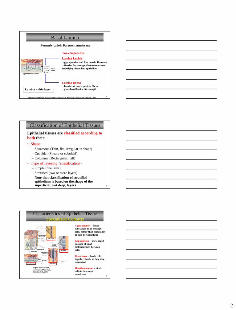
Basal Lamina
Two components:
Lamina Lucida
- glycoproteins and fine protein filaments
- Barrier for passage of substances from
underlying tissue into epithelium
Lamina Densa- bundles of coarse protein fibers
- gives basal lamina its strength
Formerly called: Basement membrane
Figure from: Martini, Fundamentals of Anatomy & Physiology, Benjamin Cummings, 2004
Lamina = thin layer
5
Classification of Epithelial Tissues
• Shape
– Squamous (Thin, flat, irregular in shape)
– Cuboidal (Square or cuboidal)
– Columnar (Rectangular, tall)
• Type of layering (stratification)
– Simple (one layer)
– Stratified (two or more layers)
– Note that classification of stratified epithelium is based on the shape of the superficial, not deep, layers
Epithelial tissues are classified according to
both their:
6
Characteristics of Epithelial Tissue
Specialized Contacts
Figure from: Martini,
Anatomy & Physiology,
Prentice Hall, 2001
Tight junction – forces
substances to go through
cells, rather than being able
to pass between them
Gap junction – allow rapid
passage of small
molecules/ions between
cells
Desmosome – binds cells
together firmly so they stay
connected
Hemidesmosome – binds
cells to basement
membrane
3
7
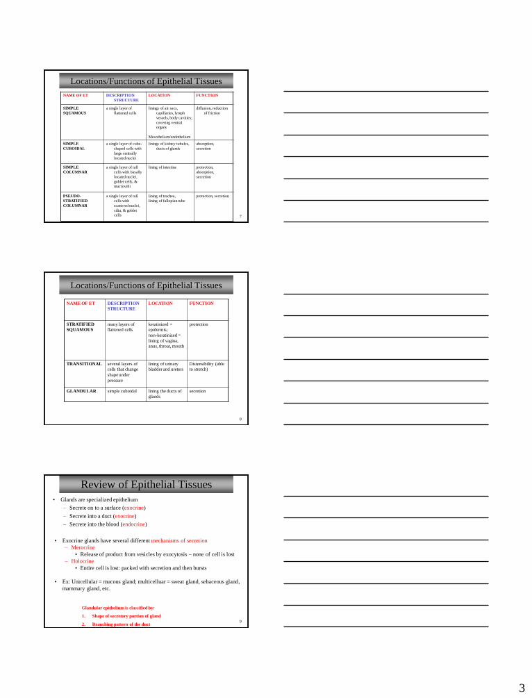
Locations/Functions of Epithelial Tissues
NAME OF ET DESCRIPTION
STRUCTURE
LOCATION FUNCTION
SIMPLE
SQUAMOUS
a single layer of
flattened cells
linings of air sacs,
capillaries, lymph
vessels, body cavities;
covering ventral organs
Mesothelium/endothelium
diffusion, reduction
of friction
SIMPLE
CUBOIDAL
a single layer of cube-
shaped cells with
large centrally
located nuclei
linings of kidney tubules,
ducts of glands
absorption,
secretion
SIMPLE
COLUMNAR
a single layer of tall
cells with basally
located nuclei,
goblet cells, & mucrovilli
lining of intestine protection,
absorption,
secretion
PSEUDO-
STRATIFIED
COLUMNAR
a single layer of tall
cells with
scattered nuclei,
cilia, & goblet cells
lining of trachea,
lining of fallopian tube
protection, secretion
8
Locations/Functions of Epithelial Tissues
NAME OF ET DESCRIPTION
STRUCTURE
LOCATION FUNCTION
STRATIFIED
SQUAMOUS
many layers of
flattened cells
keratinized =
epidermis;
non-keratinized =
lining of vagina,
anus, throat, mouth
protection
TRANSITIONAL several layers of
cells that change
shape under
pressure
lining of urinary
bladder and ureters
Distensibility (able
to stretch)
GLANDULAR simple cuboidal lining the ducts of
glands
secretion
9
Review of Epithelial Tissues
• Exocrine glands have several different mechanisms of secretion
– Merocrine
• Release of product from vesicles by exocytosis – none of cell is lost
– Holocrine
• Entire cell is lost: packed with secretion and then bursts
• Ex: Unicellular = mucous gland; multicelluar = sweat gland, sebaceous gland,
mammary gland, etc.
• Glands are specialized epithelium
– Secrete on to a surface (exocrine)
– Secrete into a duct (exocrine)
– Secrete into the blood (endocrine)
Glandular epithelium is classified by:
1. Shape of secretory portion of gland
2. Branching pattern of the duct
4
10
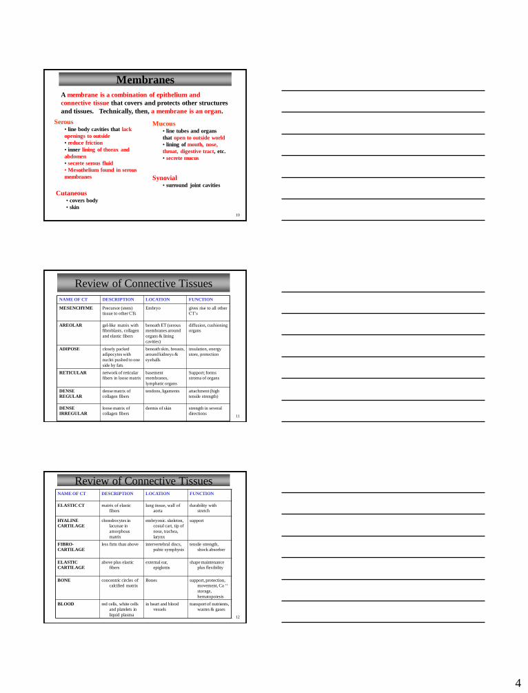
Membranes
Serous• line body cavities that lack
openings to outside
• reduce friction
• inner lining of thorax and
abdomen
• secrete serous fluid
• Mesothelium found in serous
membranes
Mucous• line tubes and organs
that open to outside world
• lining of mouth, nose,
throat, digestive tract, etc.
• secrete mucus
Cutaneous• covers body
• skin
A membrane is a combination of epithelium and
connective tissue that covers and protects other structures
and tissues. Technically, then, a membrane is an organ.
Synovial• surround joint cavities
11
Review of Connective Tissues
NAME OF CT DESCRIPTION LOCATION FUNCTION
MESENCHYME Precursor (stem)
tissue to other CTs
Embryo gives rise to all other
CT’s
AREOLAR gel-like matrix with
fibroblasts, collagen
and elastic fibers
beneath ET (serous
membranes around
organs & lining
cavities)
diffusion, cushioning
organs
ADIPOSE closely packed
adipocytes with
nuclei pushed to one
side by fats
beneath skin, breasts,
around kidneys &
eyeballs
insulation, energy
store, protection
RETICULAR network of reticular
fibers in loose matrix
basement
membranes,
lymphatic organs
Support; forms
stroma of organs
DENSE
REGULAR
dense matrix of
collagen fibers
tendons, ligaments attachment (high
tensile strength)
DENSE
IRREGULAR
loose matrix of
collagen fibers
dermis of skin strength in several
directions
12
Review of Connective TissuesNAME OF CT DESCRIPTION LOCATION FUNCTION
ELASTIC CT matrix of elastic
fibers
lung tissue, wall of
aorta
durability with
stretch
HYALINE
CARTILAGE
chondrocytes in
lacunae in
amorphous
matrix
embryonic. skeleton,
costal cart, tip of
nose, trachea,
larynx
support
FIBRO-
CARTILAGE
less firm than above intervertebral discs,
pubic symphysis
tensile strength,
shock absorber
ELASTIC
CARTILAGE
above plus elastic
fibers
external ear,
epiglottis
shape maintenance
plus flexibility
BONE concentric circles of
calcified matrix
Bones support, protection,
movement, Ca ++
storage,
hematopoiesis
BLOOD red cells, white cells
and platelets in
liquid plasma
in heart and blood
vessels
transport of nutrients,
wastes & gases

5
13
Name of
CT
Different types of
this CT
Main types of cells
present
Main types of
fibers present
Consistency of
matrix Examples of Locations
CT Proper
1) Areolar (Loose)
2) Dense regular
3) Dense irregular
4) Adipose
5) Reticular
6) Elastic
1) Fibroblasts
2) Fibroblasts
3) Fibroblasts
4) Adipocytes
5) Fibroblasts
6) Fibroblasts
1) Collagen, Elastic
2) Collagen
3) Collagen
4) Reticular
5) Reticular
6) Elastic
Semi-liquid
1) Skin, between muscles
2) Tendons, ligaments
3) Dermis
4) Body fat areas
5) Stroma of liver, spleen
6) Lungs, airways,
arteries/heart
Cartilage 1) Hyaline
2) Fibrocartilage
3) Elastic
(All) Chondrocytes
1) Collagen (sparse)
2) Collagen (dense)
3) Elastic
All types: Semi-
solid, gelatinous;
rubbery
1) Ribs, ends of bones
2) Intervertebral disks
3) Pinna of ear, epiglottis
Bone 1) Dense
2) Spongy
(All) Osteocytes Collagen Solid
(hydroxyapatite)
1) Outer portions of bone
2) Inner portions of bone
Blood
--
1) RBCs
2) WBCs
3) Platelets (cell
fragments)
Fibrinogen (soluble) Liquid Blood vessels, heart
Lymph -- Lymphocytes Reticular (in stroma
of lymphoid organs)
Liquid Lymph vessels
Connective Tissue (CT) Summary Table
Three main components of ALL types of CT: cell, fibers, ground substance
-cyte = fully differentiated; -blast = young, actively synthesizing cell
14
Connective Tissue - Major Cell Types
Fibroblasts
• fixed cell
• most common cell; always
in CT proper
• large, star-shaped
• produce fibers
• produce ground substance
Macrophages
• wandering cell
• phagocytic
• important in defense
• derived from circulating
monocytes
Mast cells are mediators of inflammation – see later…
15
Connective Tissue Fibers
Collagenous fibers
• thick
• composed of collagen
• great tensile strength
• hold structures together
• abundant in dense CT
• tendons, ligaments
Elastic fibers
• bundles of
microfibrils embedded
in elastin
• fibers branch
• elasticity
• vocal cords, air
passages
Reticular fibers
• very thin collagenous
fibers
• highly branched
• form supportive
networks

6
16
The “Ground Substance” of CT
Figures from: Alberts et al., Essential Cell Biology, Garland Press, 1998
VERY hydrophilic!
**Function: Very active in
controlling passage of
substances through this portion
of the matrix and keeping CT
hydratedGAGs = glycosaminoglycans (negatively charged
polysaccharides); a major molecule in ground
substance
glucosamine
17
Tendons and Ligaments
Tendons: Connect muscle to bone
Ligaments: Connect bone to bone
Aponeuroses: Broad, fibrous sheets; usually attach muscle to muscle (or bone)
18
CT Framework of the Body
Figure from: Martini, Anatomy & Physiology, Prentice Hall, 2001
Fascia: layers of fibrous
connective tissue covering
and separating muscle. It
connects the organs of the
dorsal and ventral cavities
with the rest of the body
Provide:
- Strength
- Stability
- Organ position
- Conduits

7
19
Muscle – Overview
• General characteristics
– Elongated cells with special properties
– Muscle cells (myocytes) = muscle fibers
– Contractile (major property of all muscle)
– Use actin (thin) and myosin (thick) for contraction
• Three types of muscle tissue
– Cardiac (involuntary)
– Skeletal (voluntary)
– Smooth (involuntary)
20
Review of Muscle TypesNAME OF
MUSCLE
TISSUE
DESCRIPTION OF
STRUCTURE
TYPE OF
CONTROL
LOCATION FUNCTION
SKELETAL
MUSCLE
long, thin fibers with
many nuclei and
striations
Voluntary attached to bones to move bones
SMOOTH
MUSCLE
spindle shaped cells with
one centrally
located nucleus,
lacking striations
Involuntary walls of visceral
hollow organs,
irises of eyes,
walls of blood vessels
to move substances
through
passageways (i.e.
food, urine, semen),
constrict blood
vessels, etc
CARDIAC
MUSCLE
a network of striated
cells with one
centrally located
nucleus attached by intercalated discs
- Intercalated disks
consist of : 1)gap
junctions and 2)
desmosomes
Involuntary heart pump blood to lungs
and body
21
Nervous Tissue
• found in brain, spinal cord, and peripheral nerves
• conduction of nerve impulses - neurons
• sensory reception
• neuroglial cells are supporting cells
8
22
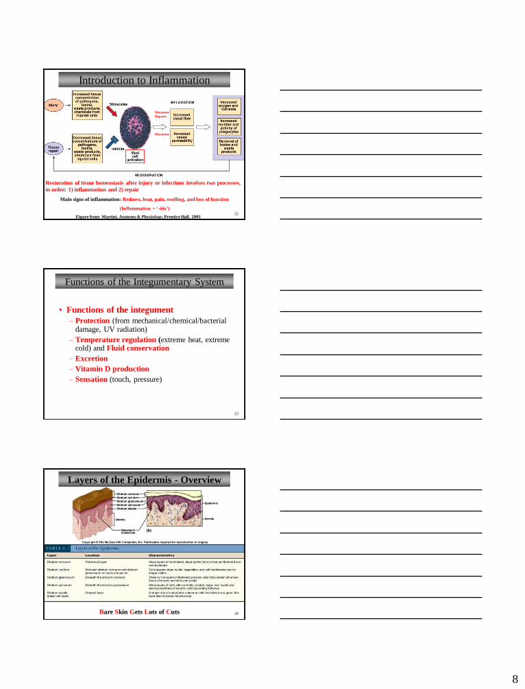
Introduction to Inflammation
Figure from: Martini, Anatomy & Physiology, Prentice Hall, 2001
Restoration of tissue homeostasis after injury or infections involves two processes,
in order: 1) inflammation and 2) repair
Main signs of inflammation: Redness, heat, pain, swelling, and loss of function
(Inflammation = ‘-itis’)
Histamine
Histamine
Heparin
23
Functions of the Integumentary System
• Functions of the integument
– Protection (from mechanical/chemical/bacterial damage, UV radiation)
– Temperature regulation (extreme heat, extreme cold) and Fluid conservation
– Excretion
– Vitamin D production
– Sensation (touch, pressure)
24
Layers of the Epidermis - Overview
Bare Skin Gets Lots of Cuts

9
25
Thick and Thin Skin
Thin (0.07-0.12 mm)(epidermal thickness) Thick (0.8-1.4 mm)
(epidermal thickness)
Thick skin - palms of hands, soles of feet; five epidermal layers
Thin skin - everywhere else; four epidermal layers (no s. lucidum)
Figure from: Martini, Anatomy & Physiology, Prentice Hall, 2001
26
Cells of the Epidermis
• Epidermis of the skin is classified as a keratinized stratified squamous epithelium
• Cells of the epidermis include
– Keratinocytes (90%)
• Keratin – a tough, fibrous intracellular protein (protection)
• Lamellar granules (waterproofing, extracellular)
– Melanocytes (8%)• Produce melanin (protection from UV radiation)
– Langerhans cells (1-2%)• Migrate to skin from bone marrow
• Participate in skin’s immune response (dendritic cells)
– Merkel cells (< 1%)
• Least numerous; specialized epithelial cells
• Function in sensation of touch
27
Skin Color
1. Genetic Factors
• varying amounts and
type of melanin
• varying size/number of
melanin granules
• albinos lack melanin
(but not melanocytes!)
2. Environmental Factors
• sunlight
• UV light from sunlamps
• X rays
3. Physiological Factors
• dilation of dermal blood vessels
(erythema)
• constriction of dermal blood
vessels (less pink, pale = pallor)
• level of oxygenation of blood
* normal = pink (fair-skinned)
* low = bluish (cyanosis)
• carotene -> Vit A (yellow)
• jaundice (yellow)

10
28
Skin Color and Melanin
Figure from: Martini, Fundamentals of Anatomy & Physiology, Pearson Education, 2004
Melanocytes produce melanin
- tyrosine melanin
- UV radiation up-regulates production of
melanin
- Dark-skinned individuals have
* same number, higher activity of melanocytes
* more pigmented layers of epidermis
Dark-skinned Fair-skinned
29
Keratin and Vitamin D
• Keratin (tough, fibrous intracellular protein)
– Protection
– Water resistance
• Vitamin D3 (“sunshine vitamin”)
– After UV irradiation epidermal cells in s. spinosum and
s. basale convert a cholesterol-related steroid to Vit D3
(cholecalciferol)
– Vit D3 – absorption of calcium and phosphorus by
small intestine
30
Two Layers of the Dermis
1. Papillary layer (near epiderm.)
- areolar connective tissue (CT)
- capillaries and sensory neurons
- dermal papillae
- fingerprints (with epi. ridges)
2. Reticular layer
- dense, irregular CT
- collagen fiber bundles extend
upward and downward
- also contains elastic fibers and
cells of CT proper
- accessory organs of
integumentary system (from epi.)
Figure adapted from: Martini, Anatomy & Physiology, Prentice Hall, 2001
11
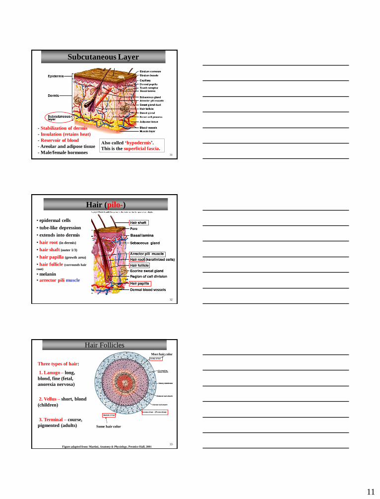
31
Subcutaneous Layer
- Stabilization of dermis
- Areolar and adipose tissue
- Male/female hormones
Also called ‘hypodermis’.
This is the superficial fascia.
- Insulation (retains heat)
Basal lamina
- Reservoir of blood
32
Hair (pilo-)
• epidermal cells
• tube-like depression
• extends into dermis
• hair root (in dermis)
• hair shaft (outer 1/3)
• hair papilla (growth area)
• hair follicle (surrounds hair
root)
• melanin
• arrector pili muscle
(from epidermis)
Basal lamina
33
Hair Follicles
Figure adapted from: Martini, Anatomy & Physiology, Prentice Hall, 2001
Some hair color
Three types of hair:
1. Lanugo – long,
blond, fine (fetal,
anorexia nervosa)
2. Vellus – short, blond
(children)
3. Terminal – course,
pigmented (adults)
Most hair color
(Protection)

12
34
Sebaceous (Oil) Glands
• multicellular, holocrine glands
• absent on palms and soles
• usually associated with hair
follicles
• secrete sebum, a waxy, oily
material
• inhibits growth of bacteria
• lubricates and protects keratin
of hair shaft, and conditions skin Sebaceous follicles – not
associated with hair.
Discharge directly on to
skin. On face, back, chest,
nipples and male sex
organs.
35
Sweat Glands (Multicelluar)
• apocrine (merocrine secr.)
glands
- associated with hair follicles
- thick, odorous secretion
• ceruminous glands
• mammary glands
• also called sudoriferous glands
• eccrine (merocrine secr.) glands
- most numerous
- palms, soles, forehead, neck,
back
- directly on to surface
- watery secretion
- for thermoregulation
Specialized (apocrine secretion)
Sweating with visible wetness = diaphoresis
36
Nails
Figure from: Saladin,
Anatomy & Physiology,
McGraw Hill, 2007
Be able to identify these structures by labeling this diagramHyponychium
(Perionychium)
13
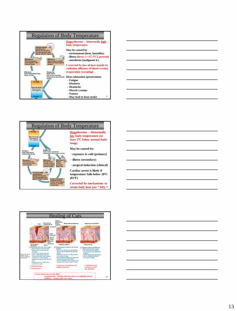
37
Regulation of Body TemperatureHyperthermia – Abnormally high
body temperature
May be caused by
- environment (heat, humidity)
- illness (fever [>=37.20C], pyrexia)
- anesthesia (malignant h.)
Corrected by loss of heat mainly by
radiation (dilation of blood vessels),
evaporation (sweating)
Heat exhaustion (prostration)
- Fatigue
- Dizziness
- Headache
- Muscle cramps
- Nausea
- May lead to heat stroke
38
Regulation of Body Temperature
Hypothermia – Abnormally
low body temperature (at
least 20C below normal body
temp)
May be caused by:
- exposure to cold (primary)
- illness (secondary)
- surgical induction (clinical)
Cardiac arrest is likely if
temperature falls below 28oC
(82oF)
Corrected by mechanisms to
retain body heat (see * left)
**
Healing of Cuts
39
Tissue repair can occur by either:
1) regeneration – healing with tissue that was originally present
2) fibrosis – healing with ‘scar’ tissue
Figure From: Marieb
& Hoehn, Human
Anatomy & Physiology,
9th ed., Pearson
1. Bleeding/clotting
2. Scab formation
3. Epidermal cell migration and
collagen production
4. Shedding of scab;
covering of wound
with epithelium


![100[ch] 0.28[ps/ch] 200[ch] 0.54[ps/ch] TDC-calibration](https://img.pdfslide.us/doc/110x75/56649c7d5503460f94931818/100ch-028psch-200ch-054psch-tdc-calibration.jpg)