Embed Size (px)
DESCRIPTION
3 Chapter PowerPoint® Lecture Slides prepared by Jason LaPres Lone Star College - North Harris Copyright © 2010 Pearson Education, Inc. Cells are the building blocks of all plants and animals All cells come from the division of preexisting cells Cells are the smallest units that perform all vital physiological functions Each cell maintains homeostasis at the cellular level Developed from Robert Hookes research Cell Theory Copyright © 2010 Pearson Education, Inc.
Citation preview

Copyright © 2010 Pearson Education, Inc.
C h a p t e r
3
The Cellular Level of
Organization
PowerPoint® Lecture Slides prepared by Jason LaPres
Lone Star College - North Harris

Copyright © 2010 Pearson Education, Inc.
An Introduction to Cells
Cell Theory Developed from Robert Hooke’s research
Cells are the building blocks of all plants and animals
All cells come from the division of preexisting cells
Cells are the smallest units that perform all vital
physiological functions
Each cell maintains homeostasis at the cellular level

Copyright © 2010 Pearson Education, Inc.
An Introduction to Cells
Sex cells (germ cells)
Reproductive cells
Male sperm
Female oocyte (a cell that develops into an egg)
Somatic cells (soma = body)
All body cells except sex cells
Copyright © 2010 Pearson Education, Inc.
An Introduction to Cells
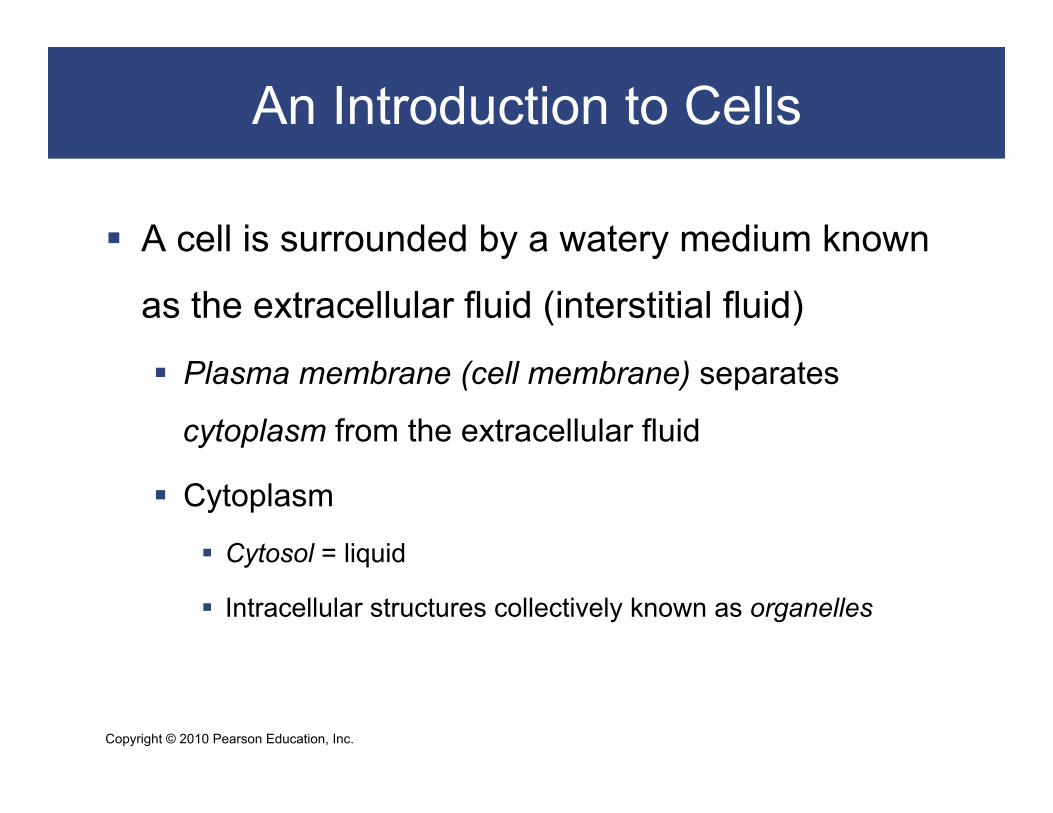
A cell is surrounded by a watery medium known
as the extracellular fluid (interstitial fluid)
Plasma membrane (cell membrane) separates
cytoplasm from the extracellular fluid
Cytoplasm
Cytosol = liquid
Intracellular structures collectively known as organelles

Copyright © 2010 Pearson Education, Inc.
An Introduction to Cells
Figure 3–1 The Anatomy of a Model Cell.

Copyright © 2010 Pearson Education, Inc.
Plasma Membrane
Functions of the Plasma Membrane Physical isolation
Barrier
Regulates exchange with environment Ions and nutrients enter Wastes eliminated and cellular products released
Monitors the environment Extracellular fluid composition Chemical signals
Structural support Anchors cells and tissues
Copyright © 2010 Pearson Education, Inc.
Plasma Membrane
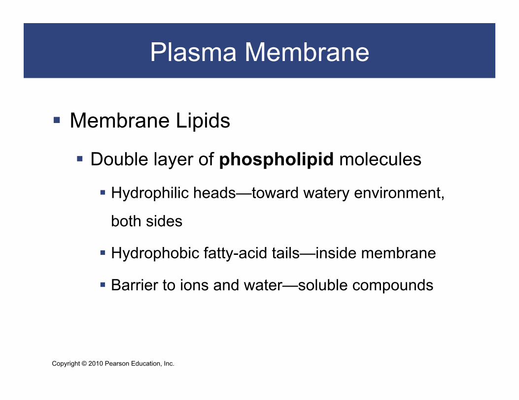
Membrane Lipids
Double layer of phospholipid molecules
Hydrophilic heads—toward watery environment,
both sides
Hydrophobic fatty-acid tails—inside membrane
Barrier to ions and water—soluble compounds

Copyright © 2010 Pearson Education, Inc.
Plasma Membrane
Membrane Proteins
Integral proteins
Within the membrane
Peripheral proteins
Bound to inner or outer surface of the membrane

Copyright © 2010 Pearson Education, Inc.
Plasma Membrane
Membrane Proteins Anchoring proteins (stabilizers)
Attach to inside or outside structures Recognition proteins (identifiers)
Label cells as normal or abnormal Enzymes
Catalyze reactions Receptor proteins
Bind and respond to ligands (ions, hormones) Carrier proteins
Transport specific solutes through membrane Channels
Regulate water flow and solutes through membrane

Copyright © 2010 Pearson Education, Inc.
Plasma Membrane
Membrane Carbohydrates Proteoglycans, glycoproteins, and glycolipids
Extend outside cell membrane
Form sticky “sugar coat” (glycocalyx)
Functions of the glycocalyx Lubrication and protection
Anchoring and locomotion
Specificity in binding (receptors)
Recognition (immune response)
Copyright © 2010 Pearson Education, Inc.
Plasma Membrane
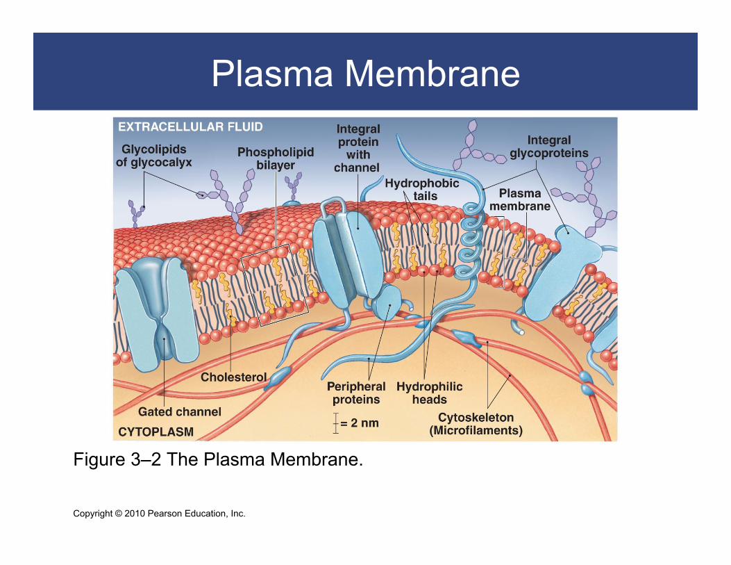
Figure 3–2 The Plasma Membrane.

Copyright © 2010 Pearson Education, Inc.
Organelles and the Cytoplasm
All materials inside the cell and outside the nucleus Cytosol (fluid)
Dissolved materials: – nutrients, ions, proteins, and waste products
High potassium/low sodium High protein High carbohydrate/low amino acid and fat
Organelles Structures with specific functions
Copyright © 2010 Pearson Education, Inc.
Organelles and the Cytoplasm
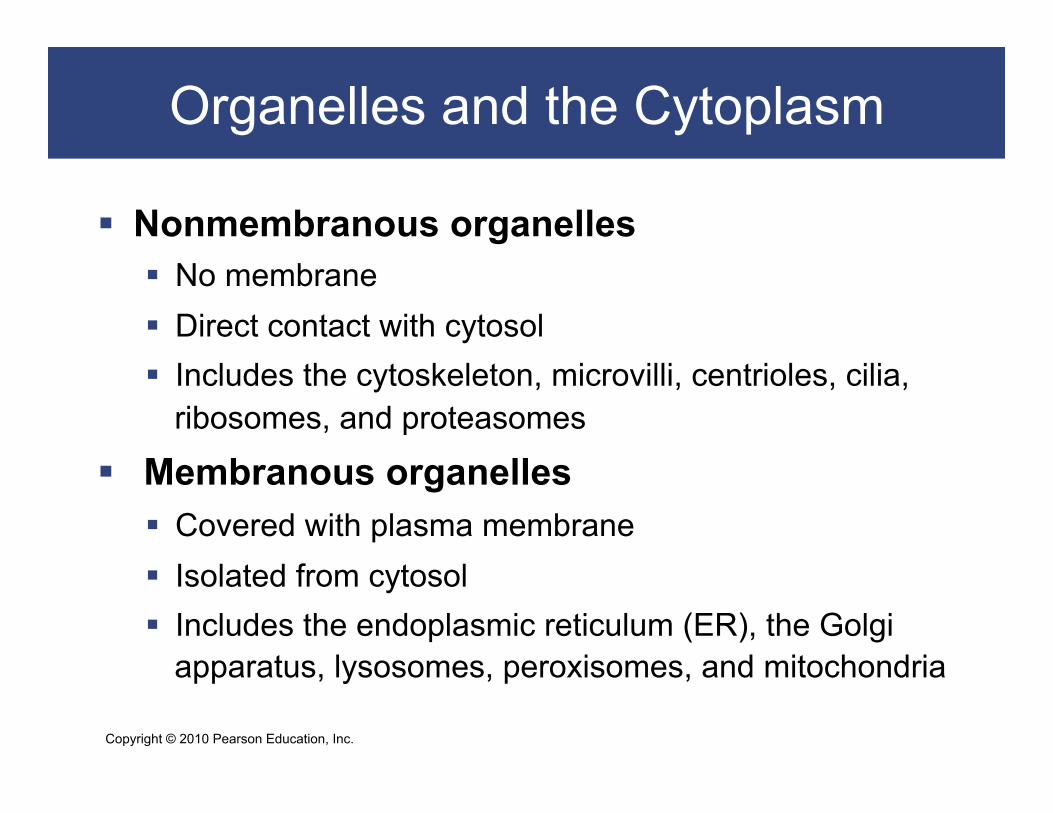
Nonmembranous organelles No membrane Direct contact with cytosol Includes the cytoskeleton, microvilli, centrioles, cilia,
ribosomes, and proteasomes
Membranous organelles Covered with plasma membrane Isolated from cytosol Includes the endoplasmic reticulum (ER), the Golgi
apparatus, lysosomes, peroxisomes, and mitochondria

Copyright © 2010 Pearson Education, Inc.
Organelles and the Cytoplasm
Nonmembranous Organelles
The Cytoskeleton — structural proteins for shape
and strength
Microfilaments
Intermediate filaments
Microtubules
Copyright © 2010 Pearson Education, Inc.
Organelles and the Cytoplasm
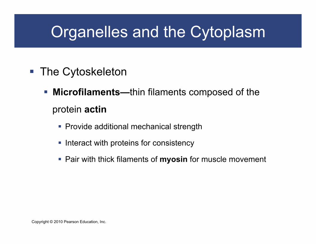
The Cytoskeleton
Microfilaments—thin filaments composed of the
protein actin
Provide additional mechanical strength
Interact with proteins for consistency
Pair with thick filaments of myosin for muscle movement

Copyright © 2010 Pearson Education, Inc.
Organelles and the Cytoplasm
The Cytoskeleton
Intermediate filaments—mid-sized between
microfilaments and thick filaments
Durable (collagen)
Strengthen cell and maintain shape
Stabilize organelles
Stabilize cell position
Copyright © 2010 Pearson Education, Inc.
Organelles and the Cytoplasm
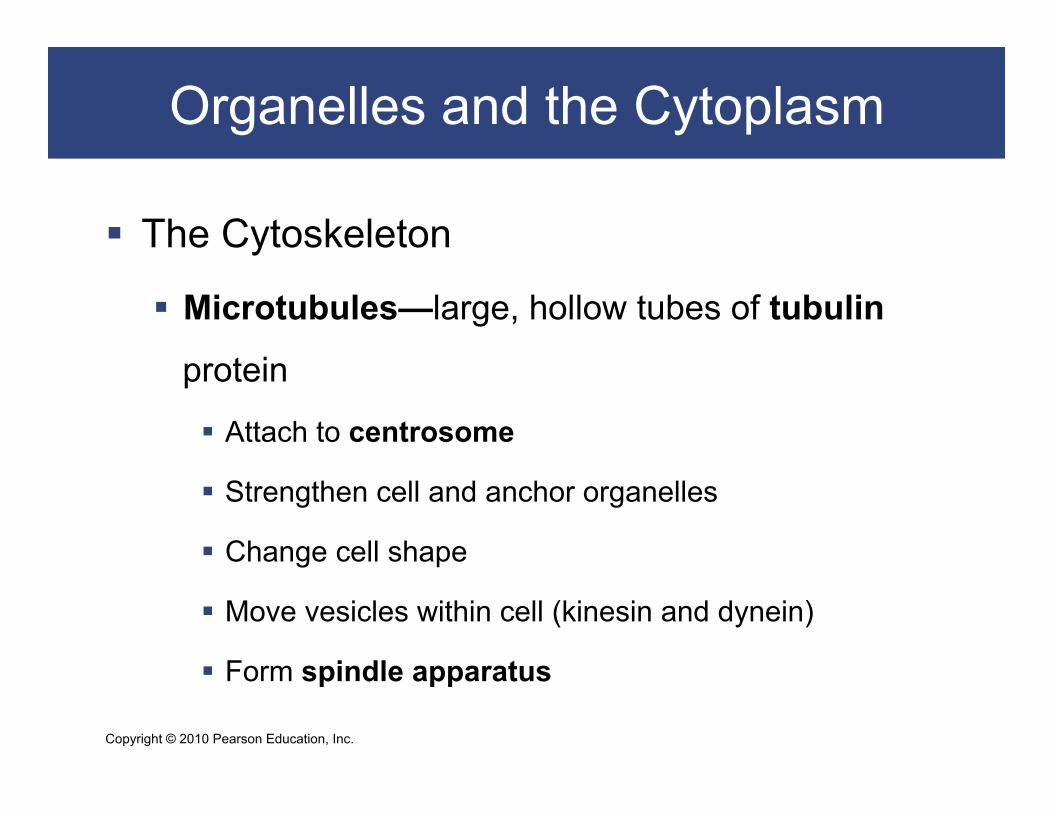
The Cytoskeleton
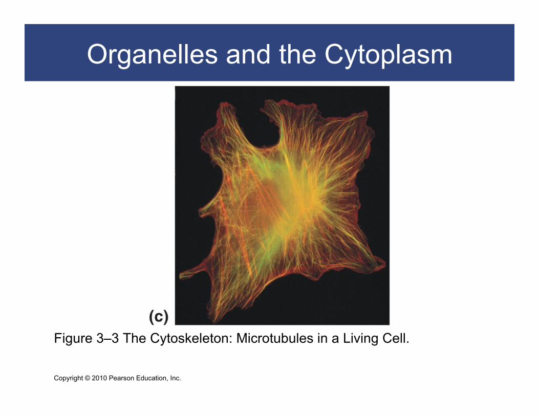
Microtubules—large, hollow tubes of tubulin
protein
Attach to centrosome
Strengthen cell and anchor organelles
Change cell shape
Move vesicles within cell (kinesin and dynein)
Form spindle apparatus

Copyright © 2010 Pearson Education, Inc.
Organelles and the Cytoplasm
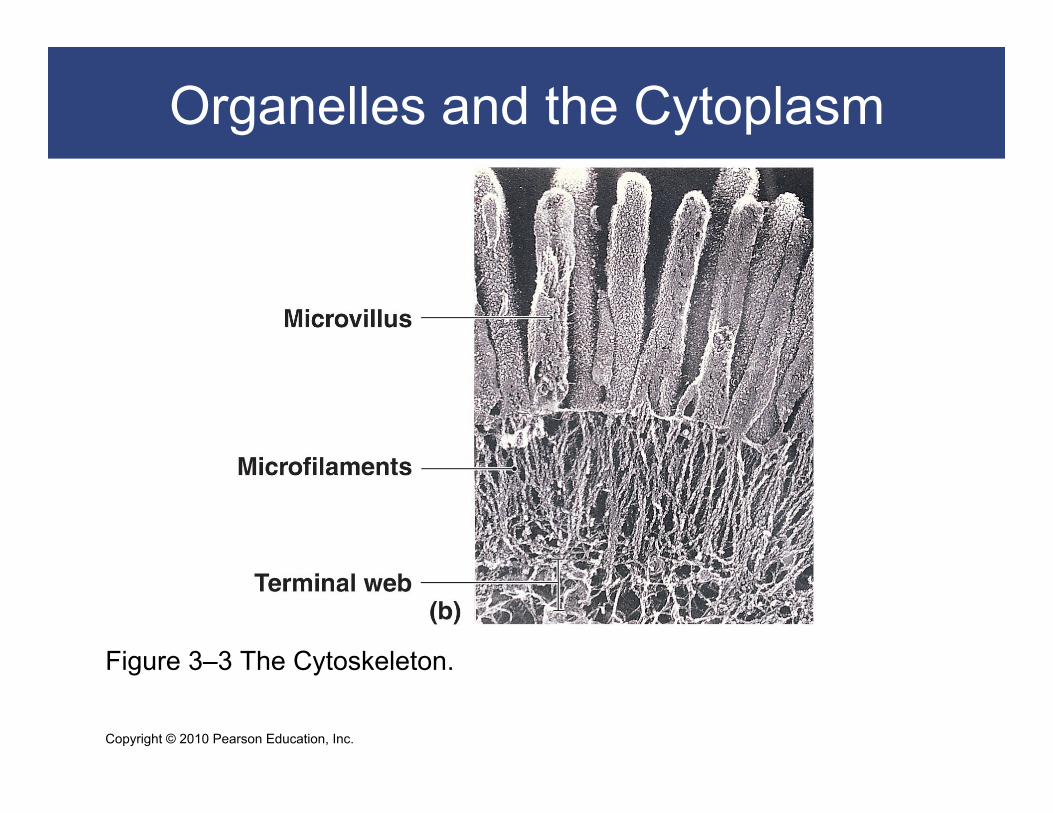
Figure 3–3 The Cytoskeleton.
Copyright © 2010 Pearson Education, Inc.
Organelles and the Cytoplasm
Figure 3–3 The Cytoskeleton.

Copyright © 2010 Pearson Education, Inc.
Organelles and the Cytoplasm
Microvilli Increase surface area for absorption Attach to cytoskeleton
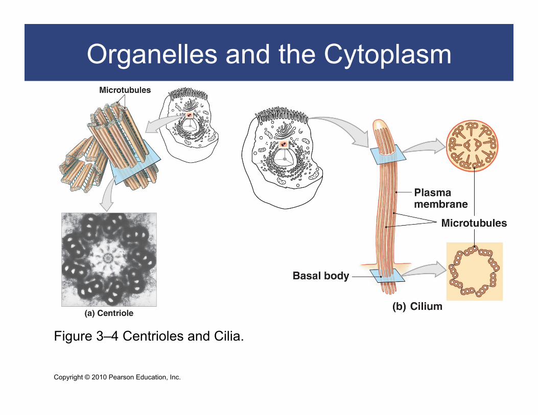
Centrioles in the Centrosome Centrioles form spindle apparatus during cell division Centrosome: cytoplasm surrounding centriole
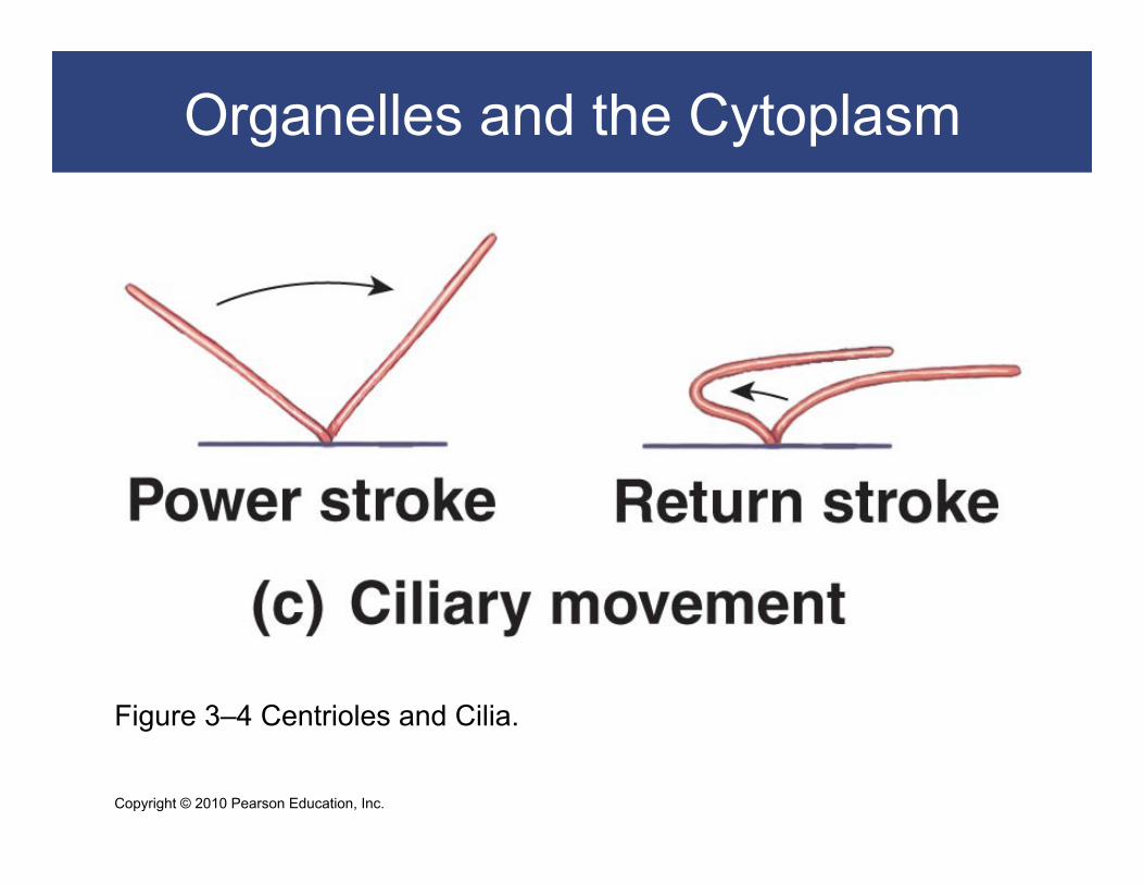
Cilia Small hair-like extensions Cilia move fluids across the cell surface
Copyright © 2010 Pearson Education, Inc.
Organelles and the Cytoplasm
Figure 3–3 The Cytoskeleton: Microtubules in a Living Cell.
Copyright © 2010 Pearson Education, Inc.
Organelles and the Cytoplasm
Figure 3–4 Centrioles and Cilia.
Copyright © 2010 Pearson Education, Inc.
Organelles and the Cytoplasm
Figure 3–4 Centrioles and Cilia.
Copyright © 2010 Pearson Education, Inc.
Organelles and the Cytoplasm
Ribosomes Build polypeptides in protein synthesis Two types
Free ribosomes in cytoplasm: – manufacture proteins for cell
Fixed ribosomes attached to ER: – manufacture proteins for secretion
Proteasomes Contain enzymes (proteases) Disassemble damaged proteins for recycling
Copyright © 2010 Pearson Education, Inc.
Organelles and the Cytoplasm
Membranous Organelles Five types of membranous organelles
Endoplasmic reticulum (ER)
Golgi apparatus
Lysosomes
Peroxisomes
Mitochondria
Copyright © 2010 Pearson Education, Inc.
Organelles and the Cytoplasm
Endoplasmic reticulum (ER)
Endo- = within, plasm = cytoplasm, reticulum =
network
Cisternae are storage chambers within membranes
Functions
Synthesis of proteins, carbohydrates, and lipids
Storage of synthesized molecules and materials
Transport of materials within the ER
Detoxification of drugs or toxins

Copyright © 2010 Pearson Education, Inc.
Organelles and the Cytoplasm
Endoplasmic reticulum (ER)
Smooth endoplasmic reticulum (SER) No ribosomes attached
Synthesizes lipids and carbohydrates:
– phospholipids and cholesterol (membranes)
– steroid hormones (reproductive system)
– glycerides (storage in liver and fat cells)
– glycogen (storage in muscles)
Copyright © 2010 Pearson Education, Inc.
Organelles and the Cytoplasm
Endoplasmic reticulum (ER)
Rough endoplasmic reticulum (RER)
Surface covered with ribosomes:
– active in protein and glycoprotein synthesis
– folds polypeptides protein structures
– encloses products in transport vesicles
Copyright © 2010 Pearson Education, Inc.
Organelles and the Cytoplasm
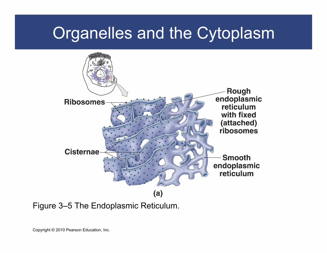
Figure 3–5 The Endoplasmic Reticulum.

Copyright © 2010 Pearson Education, Inc.
Organelles and the Cytoplasm
Figure 3–5 Rough Endoplasmic Reticulum.
Copyright © 2010 Pearson Education, Inc.
Organelles and the Cytoplasm
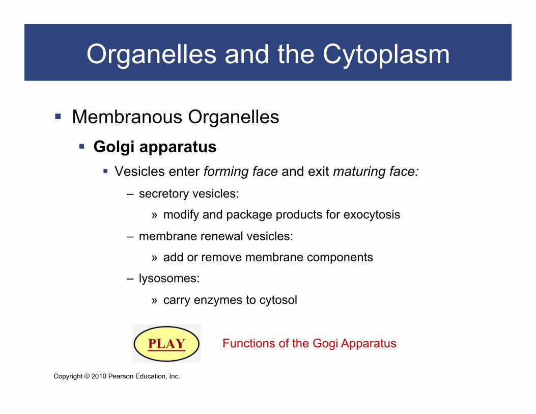
Membranous Organelles Golgi apparatus
Vesicles enter forming face and exit maturing face: – secretory vesicles:
» modify and package products for exocytosis
– membrane renewal vesicles:
» add or remove membrane components
– lysosomes:
» carry enzymes to cytosol Functions of the Gogi Apparatus

Copyright © 2010 Pearson Education, Inc.
Organelles and the Cytoplasm
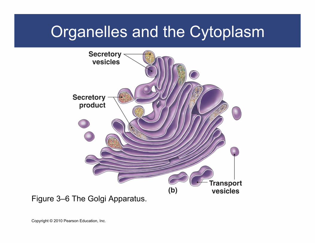
Figure 3–6 The Golgi Apparatus.
Copyright © 2010 Pearson Education, Inc.
Organelles and the Cytoplasm
Figure 3–6 The Golgi Apparatus.
Copyright © 2010 Pearson Education, Inc.
Organelles and the Cytoplasm
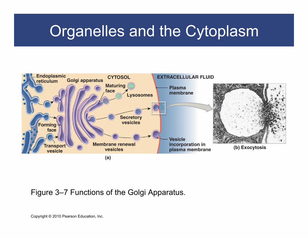
Figure 3–7 Functions of the Golgi Apparatus.

Copyright © 2010 Pearson Education, Inc.
Organelles and the Cytoplasm
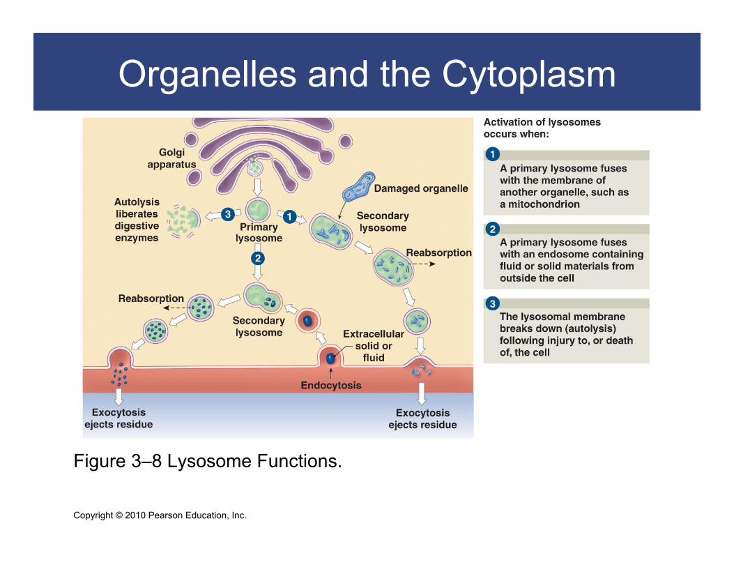
Membranous Organelles Lysosomes
Powerful enzyme-containing vesicles: – lyso- = dissolve, soma = body
Primary lysosome: – formed by Golgi apparatus and inactive enzymes
Secondary lysosome: – lysosome fused with damaged organelle – digestive enzymes activated – toxic chemicals isolated

Copyright © 2010 Pearson Education, Inc.
Organelles and the Cytoplasm
Functions of Lysosomes Clean up inside cells
Break down large molecules Attack bacteria Recycle damaged organelles Eject wastes by exocytosis
Autolysis Auto- = self, lysis = break Self-destruction of damaged cells:
– lysosome membranes break down – digestive enzymes released – cell decomposes – cellular materials recycle
Copyright © 2010 Pearson Education, Inc.
Organelles and the Cytoplasm
Figure 3–8 Lysosome Functions.
Copyright © 2010 Pearson Education, Inc.
Organelles and the Cytoplasm
Membranous Organelles
Peroxisomes
Are enzyme-containing vesicles:
– break down fatty acids, organic compounds
– produce hydrogen peroxide (H2O2)
– replicate by division

Copyright © 2010 Pearson Education, Inc.
Organelles and the Cytoplasm
Membranous Organelles
Membrane flow
A continuous exchange of membrane parts by
vesicles:
– all membranous organelles (except mitochondria)
– allow adaptation and change

Copyright © 2010 Pearson Education, Inc.
Organelles and the Cytoplasm
Membranous Organelles Mitochondria
Have smooth outer membrane and inner membrane with numerous folds (cristae)
Matrix: – fluid around cristae
Mitochondrion takes chemical energy from food (glucose): – produces energy molecule ATP
Copyright © 2010 Pearson Education, Inc.
Organelles and the Cytoplasm
Mitochondria Aerobic metabolism (cellular respiration)
Mitochondria use oxygen to break down food and produce ATP
glucose + oxygen + ADP → carbon dioxide + water + ATP
Glycolysis: – glucose to pyruvic acid (in cytosol)
Tricarboxylic acid cycle (TCA cycle): – pyruvic acid to CO2 (in matrix)
Electron transport chain – inner mitochondrial membrane

Copyright © 2010 Pearson Education, Inc.
Organelles and the Cytoplasm
Figure 3–9 Mitochondria.
Copyright © 2010 Pearson Education, Inc.
The Nucleus
Nucleus Largest organelle The cell’s control center
Nuclear envelope Double membrane around the nucleus
Perinuclear space Between the two layers of the nuclear envelope
Nuclear pores Communication passages

Copyright © 2010 Pearson Education, Inc.
The Nucleus
Figure 3–10 The Nucleus

Copyright © 2010 Pearson Education, Inc.
The Nucleus
Figure 3–10 The Nucleus

Copyright © 2010 Pearson Education, Inc.
The Nucleus
Contents of the Nucleus DNA
All information to build and run organisms
Nucleoplasm Fluid containing ions, enzymes, nucleotides, and some RNA
Nuclear matrix Support filaments
Nucleoli Are related to protein production Are made of RNA, enzymes, and histones Synthesize rRNA and ribosomal subunits

Copyright © 2010 Pearson Education, Inc.
The Nucleus
Contents of the Nucleus
Nucleosomes
DNA coiled around histones
Chromatin
Loosely coiled DNA (cells not dividing)
Chromosomes
Tightly coiled DNA (cells dividing)

Copyright © 2010 Pearson Education, Inc.
The Nucleus
Figure 3–11 The Organization of DNA within the Nucleus.

Copyright © 2010 Pearson Education, Inc.
The Nucleus
Information Storage in the Nucleus DNA
Instructions for every protein in the body
Gene DNA instructions for one protein
Genetic code The chemical language of DNA instructions:
– sequence of bases (A, T, C, G)
Triplet code: – 3 bases = 1 amino acid

Copyright © 2010 Pearson Education, Inc.
Organelles Review

Copyright © 2010 Pearson Education, Inc.
Organelles Review
Copyright © 2010 Pearson Education, Inc.
Protein Synthesis
The Role of Gene Activation in Protein Synthesis
The nucleus contains chromosomes
Chromosomes contain DNA
DNA stores genetic instructions for proteins
Proteins determine cell structure and function

Copyright © 2010 Pearson Education, Inc.
Protein Synthesis
Transcription
Copies instructions from DNA to mRNA (in nucleus)
Translation
Ribosome reads code from mRNA (in cytoplasm)
Assembles amino acids into polypeptide chain
Processing
By RER and Golgi apparatus produce protein

Copyright © 2010 Pearson Education, Inc.
Protein Synthesis
The Transcription of mRNA
A gene is transcribed to mRNA in three steps
Gene activation
DNA to mRNA
RNA processing
Transcription and Translation
Copyright © 2010 Pearson Education, Inc.
Protein Synthesis
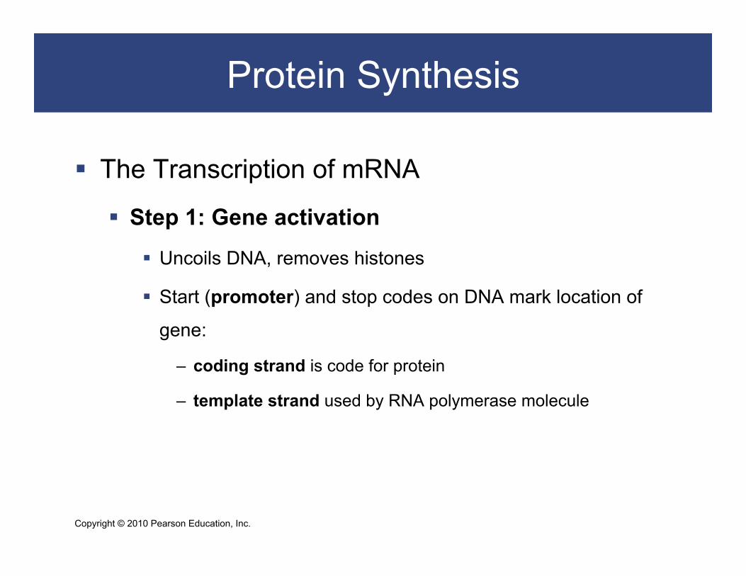
The Transcription of mRNA
Step 1: Gene activation
Uncoils DNA, removes histones
Start (promoter) and stop codes on DNA mark location of
gene:
– coding strand is code for protein
– template strand used by RNA polymerase molecule

Copyright © 2010 Pearson Education, Inc.
Protein Synthesis
The Transcription of mRNA
Step 2: DNA to mRNA
Enzyme RNA polymerase transcribes DNA:
– binds to promoter (start) sequence
– reads DNA code for gene
– binds nucleotides to form messenger RNA (mRNA)
– mRNA duplicates DNA coding strand, uracil replaces
thymine

Copyright © 2010 Pearson Education, Inc.
Protein Synthesis
The Transcription of mRNA Step 3: RNA processing
At stop signal, mRNA detaches from DNA molecule:
– code is edited (RNA processing)
– unnecessary codes (introns) removed
– good codes (exons) spliced together
– triplet of three nucleotides (codon) represents one amino acid

Copyright © 2010 Pearson Education, Inc.
Protein Synthesis
Figure 3–12 mRNA Transcription.

Copyright © 2010 Pearson Education, Inc.
Protein Synthesis
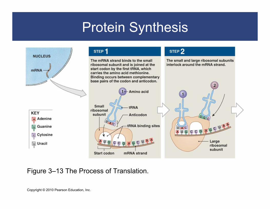
Translation mRNA moves
From the nucleus through a nuclear pore
mRNA moves To a ribosome in cytoplasm
Surrounded by amino acids
mRNA binds to ribosomal subunits tRNA delivers amino acids to mRNA
Copyright © 2010 Pearson Education, Inc.
Protein Synthesis
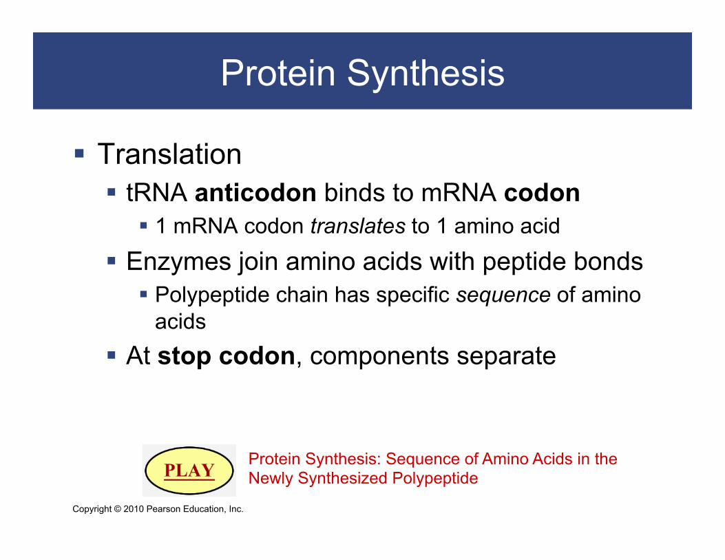
Translation tRNA anticodon binds to mRNA codon
1 mRNA codon translates to 1 amino acid
Enzymes join amino acids with peptide bonds Polypeptide chain has specific sequence of amino
acids At stop codon, components separate
Protein Synthesis: Sequence of Amino Acids in the Newly Synthesized Polypeptide
Copyright © 2010 Pearson Education, Inc.
Protein Synthesis
Figure 3–13 The Process of Translation.

Copyright © 2010 Pearson Education, Inc.
Protein Synthesis
Figure 3–13 The Process of Translation.
Copyright © 2010 Pearson Education, Inc.
Protein Synthesis
How the Nucleus Controls Cell Structure and Function Direct control through synthesis of
Structural proteins
Secretions (environmental response)
Indirect control over metabolism through enzymes

Copyright © 2010 Pearson Education, Inc.
Membrane Transport
The plasma (cell) membrane is a barrier, but
Nutrients must get in
Products and wastes must get out
Permeability determines what moves in and out of a
cell, and a membrane that
Lets nothing in or out is impermeable
Lets anything pass is freely permeable
Restricts movement is selectively permeable

Copyright © 2010 Pearson Education, Inc.
Membrane Transport
Plasma membrane is selectively permeable Allows some materials to move freely Restricts other materials
Selective permeability restricts materials based on Size Electrical charge Molecular shape Lipid solubility
Membrane Transport: Fat-and Water-Soluble Molecules
Copyright © 2010 Pearson Education, Inc.
Membrane Transport
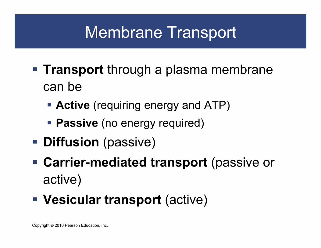
Transport through a plasma membrane can be Active (requiring energy and ATP) Passive (no energy required)
Diffusion (passive) Carrier-mediated transport (passive or
active) Vesicular transport (active)

Copyright © 2010 Pearson Education, Inc.
Membrane Transport
All molecules are constantly in motion Molecules in solution move randomly Random motion causes mixing Concentration is the amount of solute in
a solvent Concentration gradient
More solute in one part of a solvent than another
Copyright © 2010 Pearson Education, Inc.
Diffusion
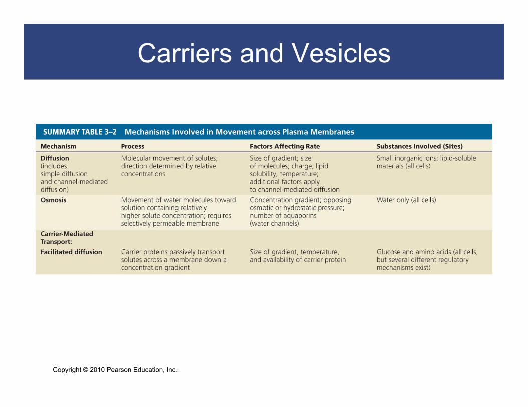
Diffusion is a Function of the Concentration Gradient Diffusion
Molecules mix randomly
Solute spreads through solvent
Eliminates concentration gradient
Solutes move down a concentration gradient
Membrane Transport: Diffusion

Copyright © 2010 Pearson Education, Inc.
Diffusion
Figure 3–14 Diffusion.
Copyright © 2010 Pearson Education, Inc.
Diffusion
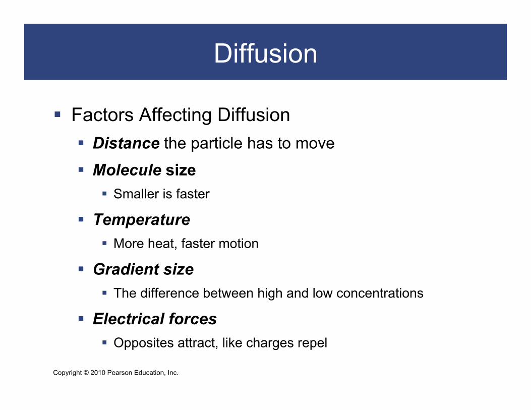
Factors Affecting Diffusion Distance the particle has to move
Molecule size Smaller is faster
Temperature More heat, faster motion
Gradient size The difference between high and low concentrations
Electrical forces Opposites attract, like charges repel

Copyright © 2010 Pearson Education, Inc.
Diffusion
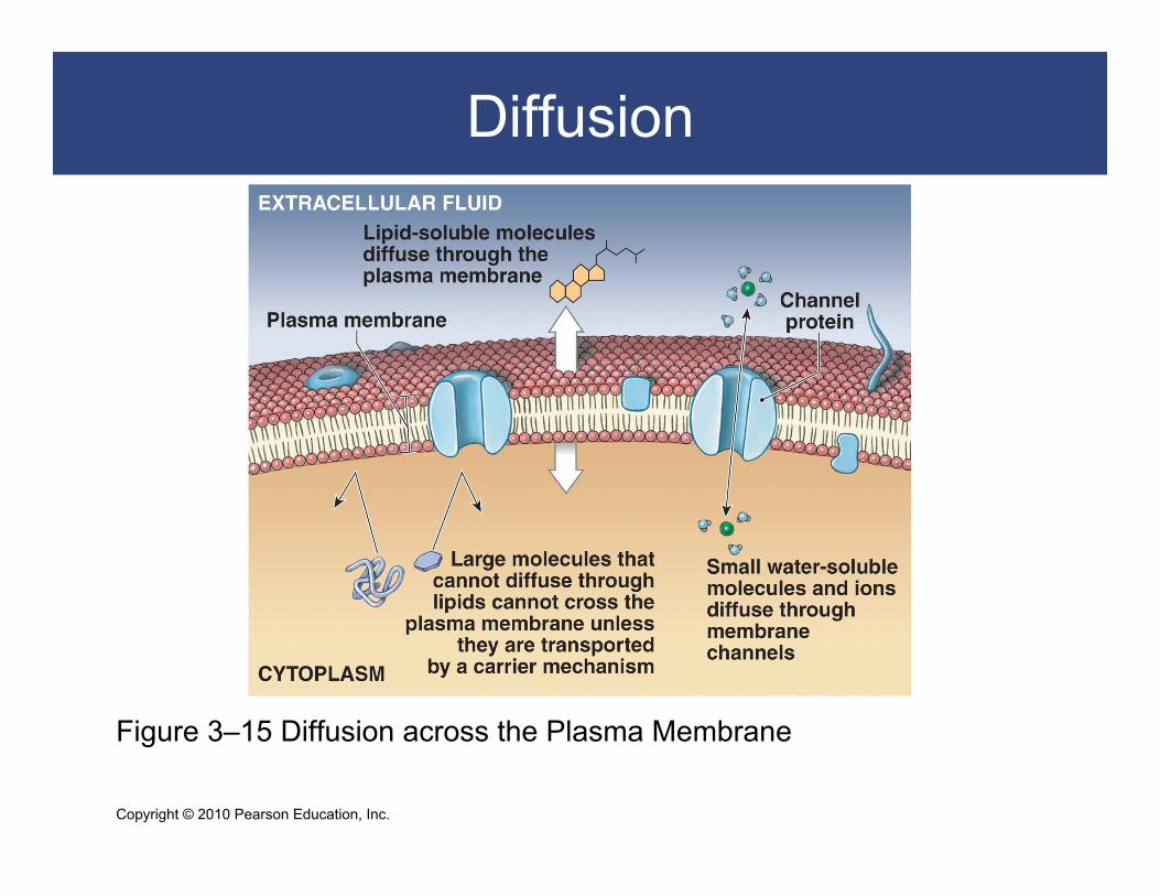
Diffusion Across Plasma Membranes Can be simple or channel mediated
Materials that diffuse through plasma membrane by simple diffusion:
– lipid-soluble compounds (alcohols, fatty acids, and steroids)
– dissolved gases (oxygen and carbon dioxide)
Materials that pass through transmembrane proteins (channels):
– are water–soluble compounds – are ions

Copyright © 2010 Pearson Education, Inc.
Diffusion
Diffusion Across Plasma Membranes
Factors in channel-mediated diffusion
Passage depends on:
– size
– charge
– interaction with the channel
Copyright © 2010 Pearson Education, Inc.
Diffusion
Figure 3–15 Diffusion across the Plasma Membrane

Copyright © 2010 Pearson Education, Inc.
Diffusion
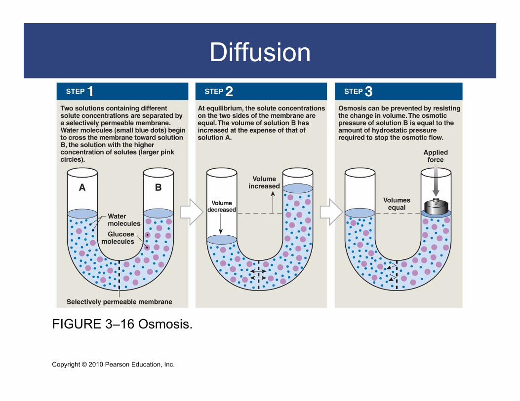
Osmosis: A Special Case of Diffusion Osmosis is the diffusion of water across the
cell membrane More solute molecules, lower concentration of
water molecules Membrane must be freely permeable to water,
selectively permeable to solutes Water molecules diffuse across membrane toward
solution with more solutes Volume increases on the side with more solutes
Copyright © 2010 Pearson Education, Inc.
Diffusion
FIGURE 3–16 Osmosis.

Copyright © 2010 Pearson Education, Inc.
Diffusion
Osmosis: A Special Case of Diffusion
Osmotic Pressure
Is the force of a concentration gradient of water
Equals the force (hydrostatic pressure) needed to
block osmosis
Copyright © 2010 Pearson Education, Inc.
Diffusion
Osmolarity and Tonicity The osmotic effect of a solute on a cell:
Two fluids may have equal osmolarity, but different tonicity
Isotonic (iso- = same, tonos = tension) A solution that does not cause osmotic flow of water in or out
of a cell
Hypotonic (hypo- = below) Has less solutes and loses water through osmosis
Hypertonic (hyper- = above) Has more solutes and gains water by osmosis
Copyright © 2010 Pearson Education, Inc.
Diffusion
Osmolarity and Tonicity
A cell in a hypotonic solution:
Gains water
Ruptures (hemolysis of red blood cells)
A cell in a hypertonic solution:
Loses water
Shrinks (crenation of red blood cells)

Copyright © 2010 Pearson Education, Inc.
Diffusion
Figure 3–17 Osmotic Flow across a Plasma Membrane.

Copyright © 2010 Pearson Education, Inc.
Carriers and Vesicles
Carrier-Mediated Transport Carrier-mediated transport of ions and organic
substrates Facilitated diffusion Active transport
Characteristics Specificity:
– one transport protein, one set of substrates Saturation limits:
– rate depends on transport proteins, not substrate Regulation:
– cofactors such as hormones

Copyright © 2010 Pearson Education, Inc.
Carriers and Vesicles
Carrier-Mediated Transport Cotransport
Two substances move in the same direction at the same time
Countertransport One substance moves in while another moves out
Copyright © 2010 Pearson Education, Inc.
Carriers and Vesicles
Carrier-Mediated Transport Facilitated diffusion
Passive
Carrier proteins transport molecules too large to fit through channel proteins (glucose, amino acids):
– molecule binds to receptor site on carrier protein
– protein changes shape, molecules pass through
– receptor site is specific to certain molecules
Membrane Transport: Facilitated Diffusion

Copyright © 2010 Pearson Education, Inc.
Carriers and Vesicles
FIGURE 3–18 Facilitated Diffusion.
Copyright © 2010 Pearson Education, Inc.
Carriers and Vesicles
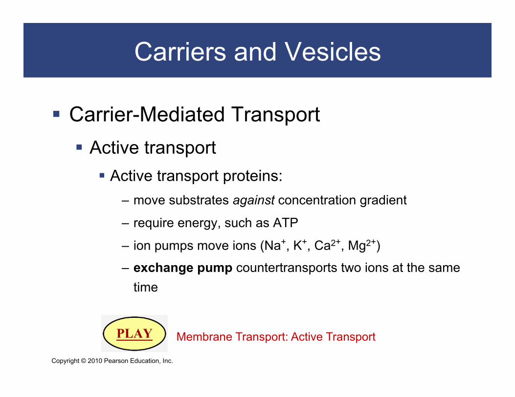
Carrier-Mediated Transport Active transport
Active transport proteins: – move substrates against concentration gradient
– require energy, such as ATP
– ion pumps move ions (Na+, K+, Ca2+, Mg2+)
– exchange pump countertransports two ions at the same time
Membrane Transport: Active Transport
Copyright © 2010 Pearson Education, Inc.
Carriers and Vesicles
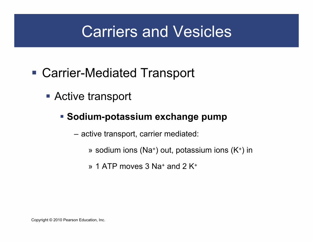
Carrier-Mediated Transport
Active transport
Sodium-potassium exchange pump
– active transport, carrier mediated:
» sodium ions (Na+) out, potassium ions (K+) in
» 1 ATP moves 3 Na+ and 2 K+

Copyright © 2010 Pearson Education, Inc.
Carriers and Vesicles
Figure 3–19 The Sodium–Potassium Exchange Pump

Copyright © 2010 Pearson Education, Inc.
Carriers and Vesicles
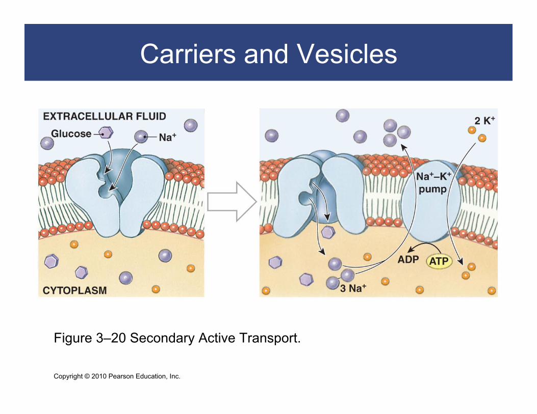
Carrier-Mediated Transport
Active transport
Secondary active transport
– Na+ concentration gradient drives glucose transport
– ATP energy pumps Na+ back out
Copyright © 2010 Pearson Education, Inc.
Carriers and Vesicles
Figure 3–20 Secondary Active Transport.

Copyright © 2010 Pearson Education, Inc.
Carriers and Vesicles
Vesicular Transport (or bulk transport)
Materials move into or out of cell in vesicles
Endocytosis (endo- = inside) is active transport using ATP:
– receptor mediated
– pinocytosis
– phagocytosis
Exocytosis (exo- = outside)
– Granules or droplets are released from the cell
Copyright © 2010 Pearson Education, Inc.
Carriers and Vesicles
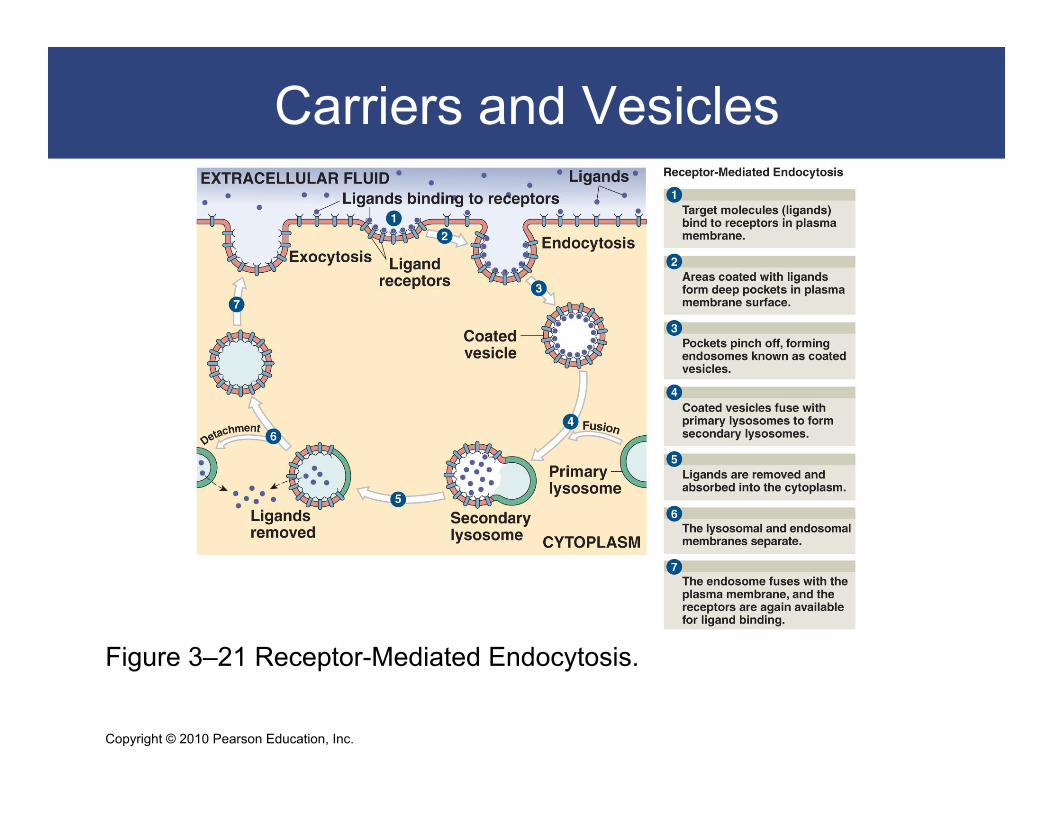
Endocytosis
Receptor-mediated endocytosis:
Receptors (glycoproteins) bind target molecules (ligands)
Coated vesicle (endosome) carries ligands and receptors
into the cell
Copyright © 2010 Pearson Education, Inc.
Carriers and Vesicles
Figure 3–21 Receptor-Mediated Endocytosis.

Copyright © 2010 Pearson Education, Inc.
Carriers and Vesicles
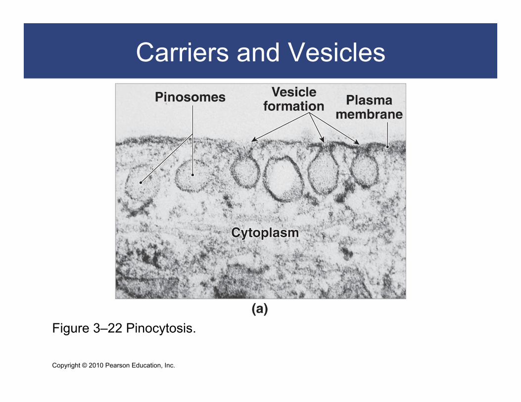
Endocytosis
Pinocytosis
Endosomes “drink” extracellular fluid
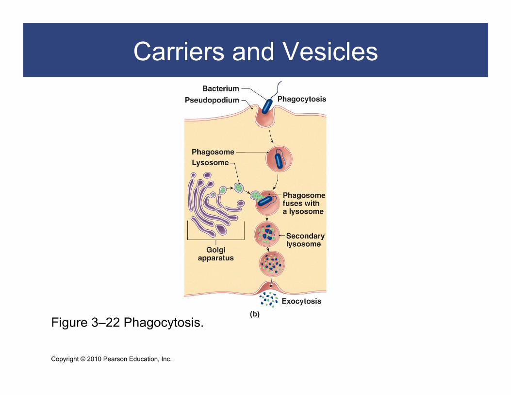
Phagocytosis
Pseudopodia (psuedo- = false, pod- = foot)
Engulf large objects in phagosomes
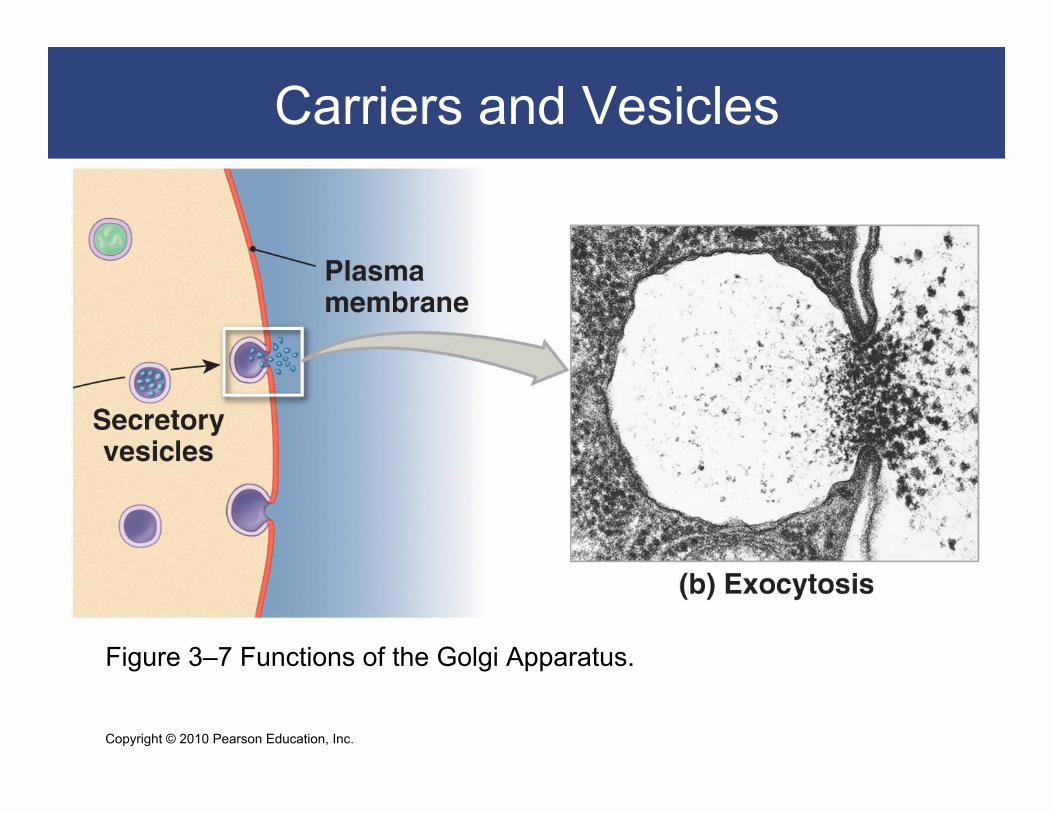
Exocytosis
Is the reverse of endocytosis
Copyright © 2010 Pearson Education, Inc.
Carriers and Vesicles
Figure 3–22 Pinocytosis.
Copyright © 2010 Pearson Education, Inc.
Carriers and Vesicles
Figure 3–22 Phagocytosis.
Copyright © 2010 Pearson Education, Inc.
Carriers and Vesicles
Figure 3–7 Functions of the Golgi Apparatus.
Copyright © 2010 Pearson Education, Inc.
Carriers and Vesicles

Copyright © 2010 Pearson Education, Inc.
Carriers and Vesicles

Copyright © 2010 Pearson Education, Inc.
Transmembrane Potential
Interior of plasma membrane is slightly negative,
outside is slightly positive
Unequal charge across the plasma membrane is
transmembrane potential
Resting potential ranges from –10 mV to
–100 mV, depending on cell type
Copyright © 2010 Pearson Education, Inc.
A Cell’s Life Cycle
Most of a cell’s life is spent in a nondividing state (interphase)
Body (somatic) cells divide in three stages DNA replication duplicates genetic material exactly
Mitosis divides genetic material equally
Cytokinesis divides cytoplasm and organelles into two daughter cells
Interphase, Mitosis, and Cytokinesis
Copyright © 2010 Pearson Education, Inc.
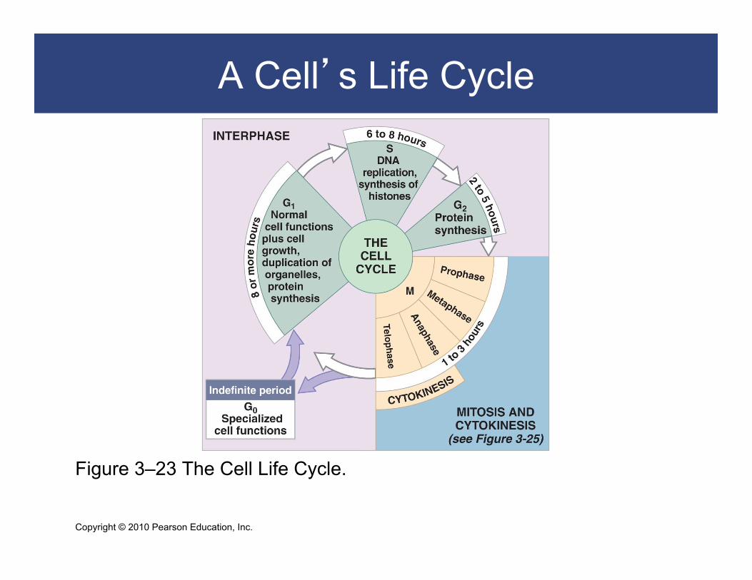
A Cell’s Life Cycle
Figure 3–23 The Cell Life Cycle.
Copyright © 2010 Pearson Education, Inc.
A Cell’s Life Cycle
Interphase The nondividing period
G-zero (G0) phase—specialized cell functions only
G1 phase—cell growth, organelle duplication, protein synthesis
S phase—DNA replication and histone synthesis
G2 phase—finishes protein synthesis and centriole replication

Copyright © 2010 Pearson Education, Inc.
A Cell’s Life Cycle
Interphase S phase
DNA replication: – DNA strands unwind
– DNA polymerase attaches complementary nucleotides

Copyright © 2010 Pearson Education, Inc.
A Cell’s Life Cycle
FIGURE 3–24 DNA Replication.

Copyright © 2010 Pearson Education, Inc.
A Cell’s Life Cycle
Mitosis Divides duplicated DNA into two sets of
chromosomes DNA coils tightly into chromatids
Chromatids connect at a centromere
Protein complex around centromere is kinetochore
Copyright © 2010 Pearson Education, Inc.
A Cell’s Life Cycle
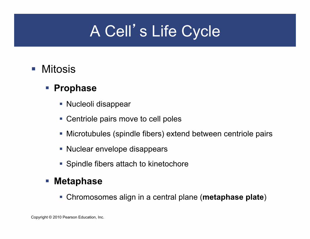
Mitosis
Prophase Nucleoli disappear
Centriole pairs move to cell poles
Microtubules (spindle fibers) extend between centriole pairs
Nuclear envelope disappears
Spindle fibers attach to kinetochore
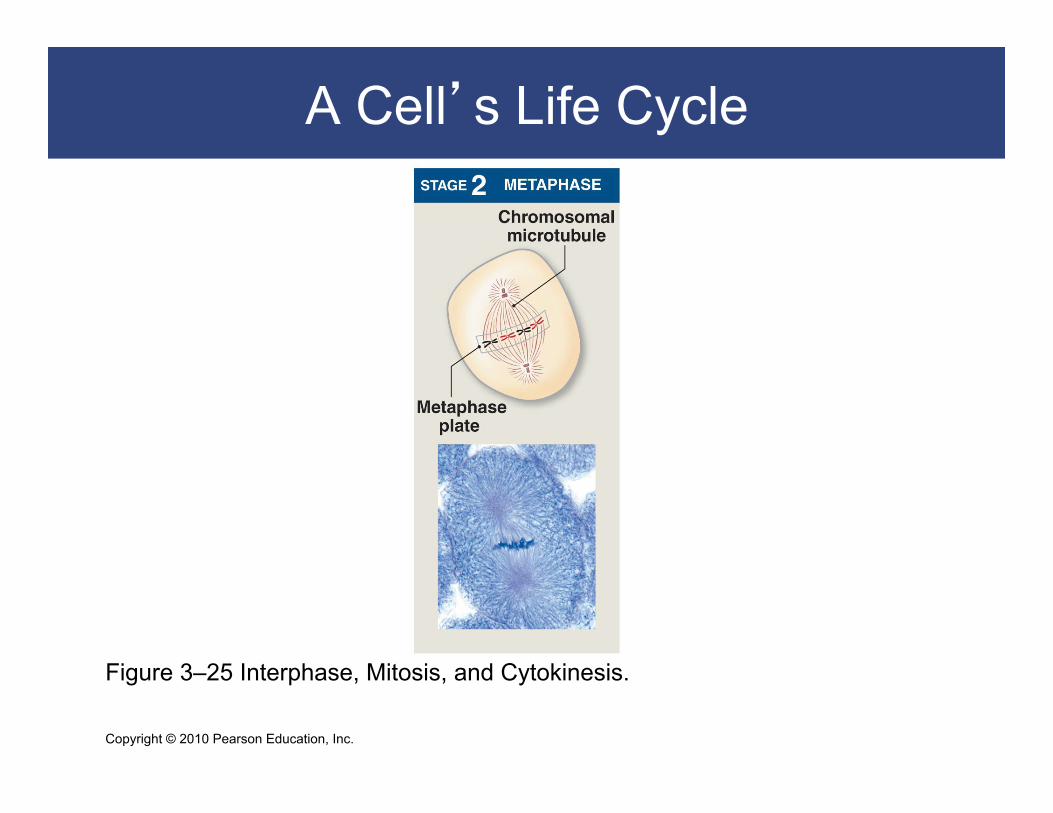
Metaphase Chromosomes align in a central plane (metaphase plate)
Copyright © 2010 Pearson Education, Inc.
A Cell’s Life Cycle
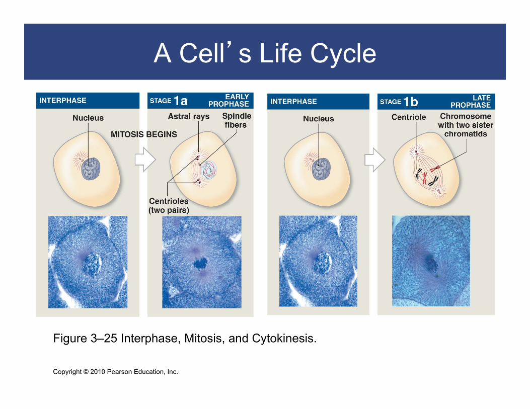
Figure 3–25 Interphase, Mitosis, and Cytokinesis.
Copyright © 2010 Pearson Education, Inc.
A Cell’s Life Cycle
Figure 3–25 Interphase, Mitosis, and Cytokinesis.

Copyright © 2010 Pearson Education, Inc.
A Cell’s Life Cycle
Mitosis Anaphase
Microtubules pull chromosomes apart
Daughter chromosomes group near centrioles
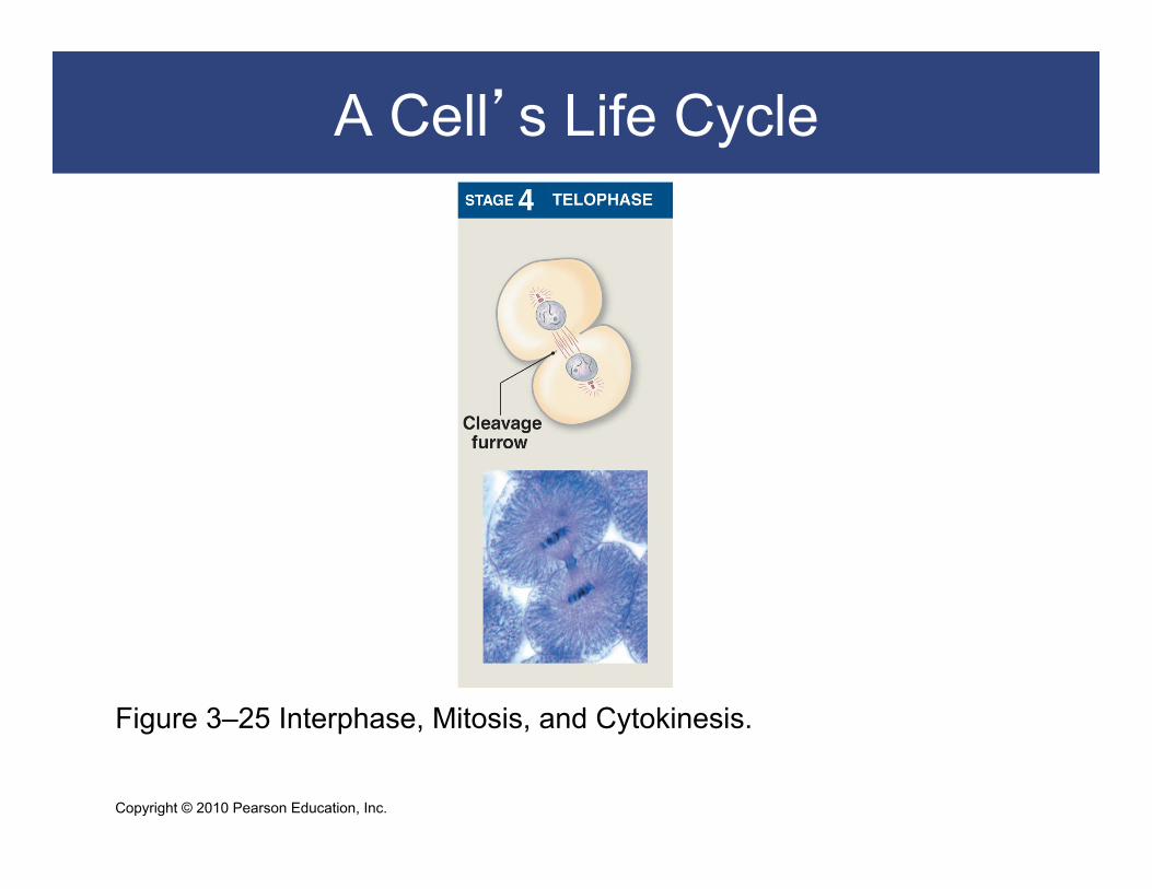
Telophase Nuclear membranes reform
Chromosomes uncoil
Nucleoli reappear
Cell has two complete nuclei

Copyright © 2010 Pearson Education, Inc.
A Cell’s Life Cycle
Figure 3–25 Interphase, Mitosis, and Cytokinesis.

Copyright © 2010 Pearson Education, Inc.
A Cell’s Life Cycle
Cytokinesis
Division of the cytoplasm
Cleavage furrow around metaphase plate
Membrane closes, producing daughter cells
Copyright © 2010 Pearson Education, Inc.
A Cell’s Life Cycle
Figure 3–25 Interphase, Mitosis, and Cytokinesis.

Copyright © 2010 Pearson Education, Inc.
A Cell’s Life Cycle
Figure 3–25 Interphase, Mitosis, and Cytokinesis.

Copyright © 2010 Pearson Education, Inc.
A Cell’s Life Cycle
Mitotic Rate and Energy Use Rate of cell division
Slower mitotic rate means longer cell life
Cell division requires energy (ATP)
Muscle cells, neurons rarely divide
Exposed cells (skin and digestive tract) live only days or hours

Copyright © 2010 Pearson Education, Inc.
Regulating the Cell Life Cycle
Normally, cell division balances cell loss Increased cell division
Internal factors (M-phase promoting factor, MPF) Extracellular chemical factors (growth factors)
Decreased cell division Repressor genes (faulty repressors cause cancers) Worn out telomeres (terminal DNA segments)

Copyright © 2010 Pearson Education, Inc.
Tumors and Cancer
Cancer develops in steps Abnormal cell
Primary tumor
Metastasis
Secondary tumor
Copyright © 2010 Pearson Education, Inc.
Tumors and Cancer
Tumor (neoplasm) Enlarged mass of cells Abnormal cell growth and division Benign tumor
Contained Not life threatening
Malignant tumor Spreads into surrounding tissues (invasion) Starts new tumors (metastasis)
Copyright © 2010 Pearson Education, Inc.
Cell Differentiation
All cells carry complete DNA instructions for all body functions
Cells specialize or differentiate To form tissues (liver cells, fat cells, and neurons) By turning off all genes not needed by that cell
All body cells, except sex cells, contain the same 46 chromosomes
Differentiation depends on which genes are active and which are inactive


![[Psy] ch03](https://img.pdfslide.us/doc/110x75/555d741ad8b42a687b8b53c6/psy-ch03.jpg)