Embed Size (px)
Citation preview
AP Bio
Protiens Chapter 5 1
81709
Aug 128:09 AM
Concept 5.4: Proteins have many structures, resulting in a wide range of functions
• Proteins account for more than 50% of the dry mass of most cells• Protein functions include structural support, storage, transport, cellular communications, movement, and defense against foreign substances
Copyright © 2008 Pearson Education, Inc., publishing as Pearson Benjamin Cummings
Aug 128:09 AM
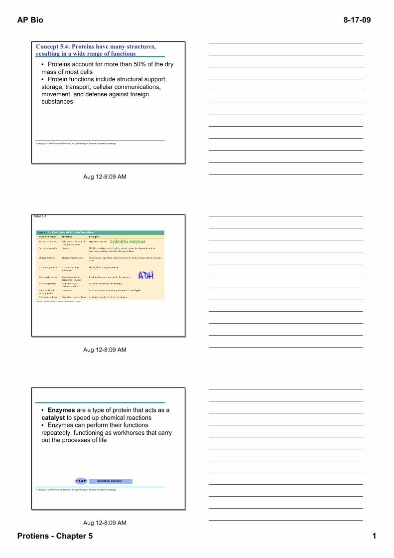
Table 51
hydrolytic enzymes
Aug 128:09 AM
• Enzymes are a type of protein that acts as a catalyst to speed up chemical reactions• Enzymes can perform their functions repeatedly, functioning as workhorses that carry out the processes of life
Animation: Enzymes
Copyright © 2008 Pearson Education, Inc., publishing as Pearson Benjamin Cummings
AP Bio
Protiens Chapter 5 2
81709
Aug 128:09 AM
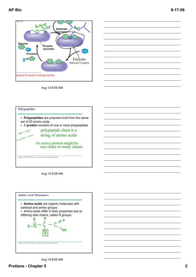
Fig. 516
Enzyme(sucrase)
Substrate(sucrose)
Fructose
Glucose
OH
H O
H2O
Induced fit model of enzyme activity
EnzymeSubstrate Complex
Aug 128:09 AM
Polypeptides
• Polypeptides are polymers built from the same set of 20 amino acids• A protein consists of one or more polypeptides
Copyright © 2008 Pearson Education, Inc., publishing as Pearson Benjamin Cummings
polypeptide chain is astring of amino acids
An active protein might beone chain or many chains
ADH = l ch
ain
Hemoglobi
n = 4 chain
s
Aug 128:09 AM
Amino Acid Monomers
• Amino acids are organic molecules with carboxyl and amino groups• Amino acids differ in their properties due to differing side chains, called R groups
Copyright © 2008 Pearson Education, Inc., publishing as Pearson Benjamin Cummings
AP Bio
Protiens Chapter 5 3
81709
Aug 128:09 AM
Fig. 5UN1
Aminogroup
Carboxylgroup
α carbon
Aug 128:09 AM
Fig. 517Nonpolar
Glycine(Gly or G)
Alanine(Ala or A)
Valine(Val or V)
Leucine(Leu or
L)
Isoleucine
(Ile or I)
Methionine
(Met or M)
Phenylalanine
(Phe or F)
Trypotphan(Trp or W)
Proline(Pro or P)
Polar
Serine(Ser or
S)
Threonine(Thr or T)
Cysteine(Cys or C)
Tyrosine(Tyr or Y)
Asparagine
(Asn or N)
Glutamine
(Gln or Q)Electricall
ychargedAcidi
cBasic
Aspartic acid
(Asp or D)
Glutamic acid
(Glu or E)
Lysine(Lys or K)
Arginine(Arg or R)
Histidine(His or H)
Aug 138:47 AM

AP Bio
Protiens Chapter 5 4
81709
Aug 128:09 AM
Fig. 517a
Nonpolar
Glycine (Gly or G)
Alanine (Ala or A)
Valine (Val or V)
Leucine (Leu or L)
Isoleucine (Ile or I)
Methionine (Met or M)
Phenylalanine (Phe or F)
Tryptophan (Trp or W)
Proline (Pro or P)
Aug 128:09 AM
Fig. 517c
Acidic
Arginine (Arg or R)
Histidine (His or H)
Aspartic acid (Asp or D)
Glutamic acid (Glu or E)
Lysine (Lys or K)
Basic
Electricallycharged
Aug 128:09 AM
Amino Acid Polymers
• Amino acids are linked by peptide bonds• A polypeptide is a polymer of amino acids• Polypeptides range in length from a few to more than a thousand monomers • Each polypeptide has a unique linear sequence of amino acids
Copyright © 2008 Pearson Education, Inc., publishing as Pearson Benjamin Cummings
AP Bio
Protiens Chapter 5 5
81709
Aug 128:09 AM
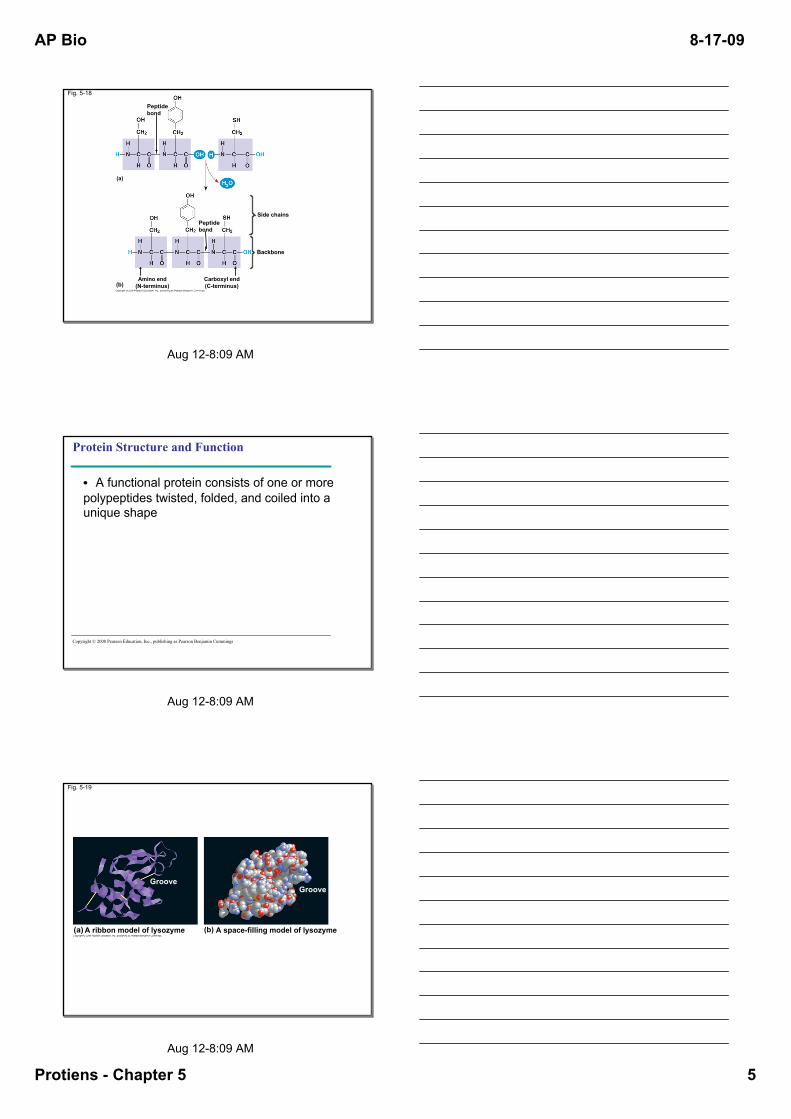
Peptidebond
Fig. 518
Amino end(Nterminus)
Peptidebond
Side chains
Backbone
Carboxyl end(Cterminus)
(a)
(b)
Aug 128:09 AM
Protein Structure and Function
• A functional protein consists of one or more polypeptides twisted, folded, and coiled into a unique shape
Copyright © 2008 Pearson Education, Inc., publishing as Pearson Benjamin Cummings
Aug 128:09 AM
Fig. 519
A ribbon model of lysozyme(a) (b) A spacefilling model of lysozyme
GrooveGroove

AP Bio
Protiens Chapter 5 6
81709
Aug 128:09 AM
Fig. 519a
A ribbon model of lysozyme(a)
Groove
Aug 128:09 AM
Fig. 519b
(b) A spacefilling model of lysozyme
Groove
Aug 128:09 AM
• The sequence of amino acids determines a protein’s threedimensional structure• A protein’s structure determines its function
Copyright © 2008 Pearson Education, Inc., publishing as Pearson Benjamin Cummings

AP Bio
Protiens Chapter 5 7
81709
Aug 128:09 AM
Fig. 520
Antibody protein Protein from flu virus
Aug 128:09 AM
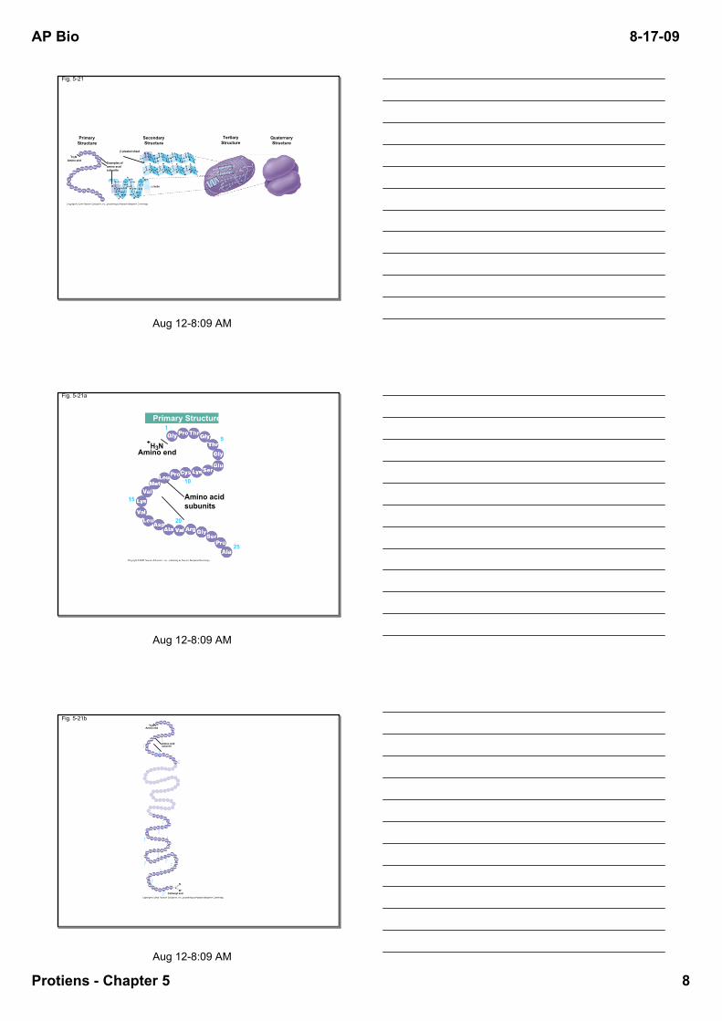
Four Levels of Protein Structure
• The primary structure of a protein is its unique sequence of amino acids• Secondary structure, found in most proteins, consists of coils and folds in the polypeptide chain• Tertiary structure is determined by interactions among various side chains (R groups)• Quaternary structure results when a protein consists of multiple polypeptide chains
Animation: Protein Structure Introduction
Copyright © 2008 Pearson Education, Inc., publishing as Pearson Benjamin Cummings
Aug 128:09 AM
• Primary structure, the sequence of amino acids in a protein, is like the order of letters in a long word • Primary structure is determined by inherited genetic information
Animation: Primary Protein Structure
Copyright © 2008 Pearson Education, Inc., publishing as Pearson Benjamin Cummings
AP Bio
Protiens Chapter 5 8
81709
Aug 128:09 AM
Fig. 521
PrimaryStructure
SecondaryStructure
TertiaryStructure
β pleated sheet
Examples ofamino acidsubunits
+H3N Amino end
α helix
QuaternaryStructure
Aug 128:09 AM
Fig. 521a
Amino acidsubunits
+H3N Amino end
25
20
15
10
5
1Primary Structure
Aug 128:09 AM
Fig. 521b
Amino acidsubunits
+H3N Amino end
Carboxyl end125
120
115
110
10510
0
95
908
5
80
75
20
25
15
10
5
1
AP Bio
Protiens Chapter 5 9
81709
Aug 128:09 AM
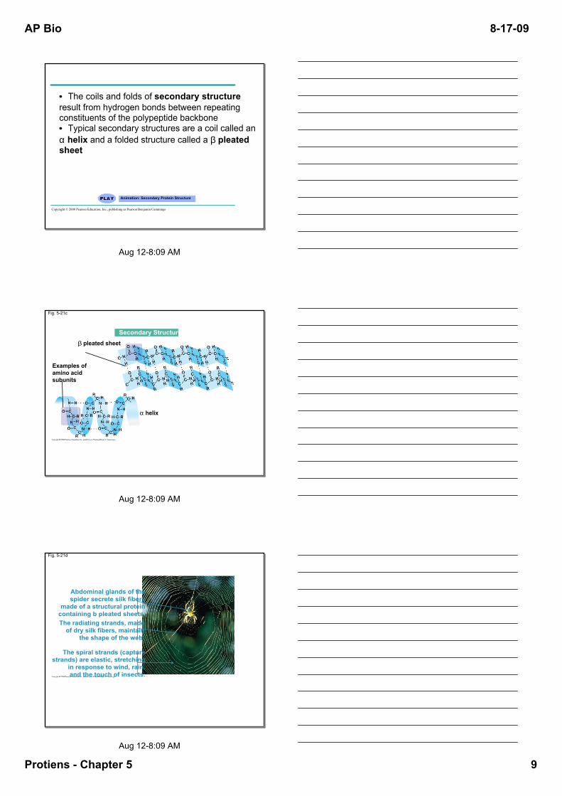
• The coils and folds of secondary structure result from hydrogen bonds between repeating constituents of the polypeptide backbone• Typical secondary structures are a coil called an α helix and a folded structure called a β pleated sheet
Animation: Secondary Protein Structure
Copyright © 2008 Pearson Education, Inc., publishing as Pearson Benjamin Cummings
Aug 128:09 AM
Fig. 521c
Secondary Structureβ pleated sheet
Examples ofamino acidsubunits
α helix
Aug 128:09 AM
Fig. 521d
Abdominal glands of thespider secrete silk fibers
made of a structural proteincontaining b pleated sheets.The radiating strands, madeof dry silk fibers, maintain
the shape of the web.
The spiral strands (capturestrands) are elastic, stretching
in response to wind, rain,and the touch of insects.
AP Bio
Protiens Chapter 5 10
81709
Aug 128:09 AM
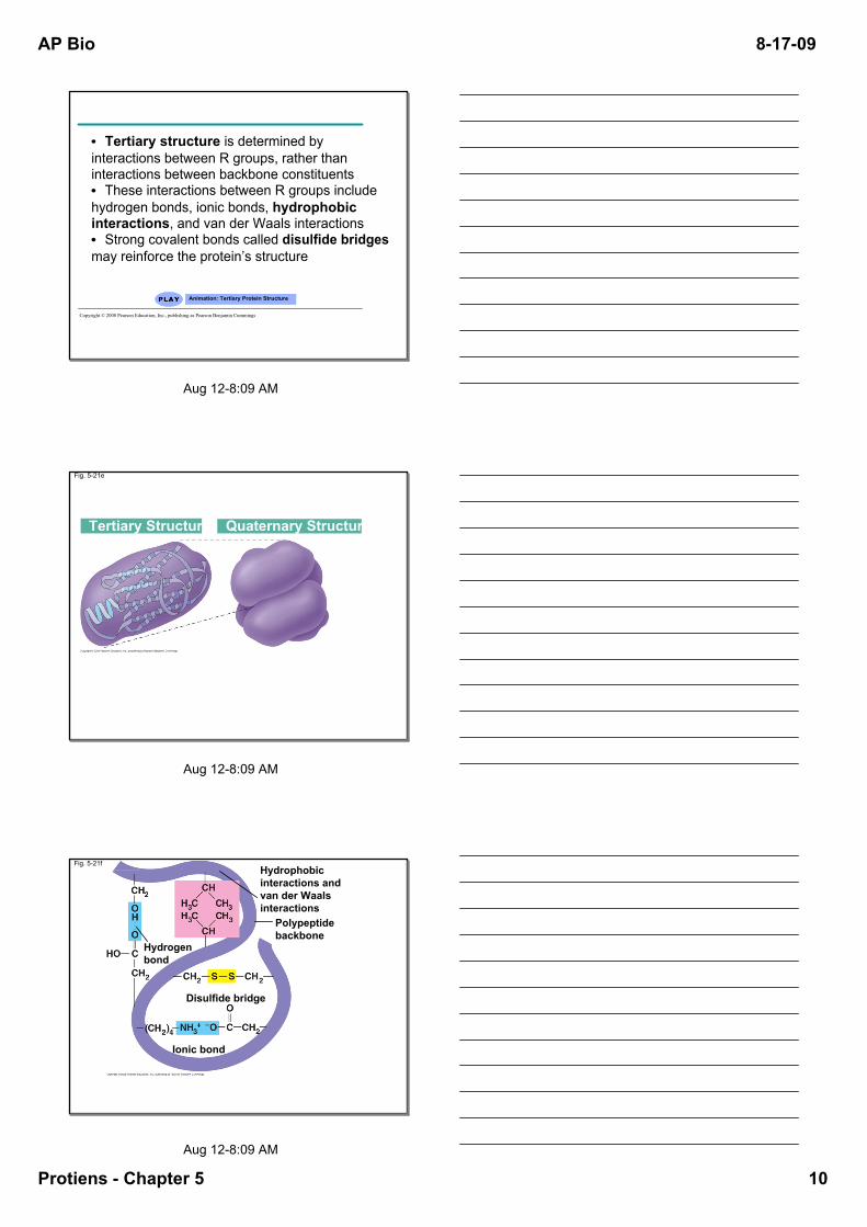
• Tertiary structure is determined by interactions between R groups, rather than interactions between backbone constituents• These interactions between R groups include hydrogen bonds, ionic bonds, hydrophobic interactions, and van der Waals interactions• Strong covalent bonds called disulfide bridges may reinforce the protein’s structure
Animation: Tertiary Protein Structure
Copyright © 2008 Pearson Education, Inc., publishing as Pearson Benjamin Cummings
Aug 128:09 AM
Fig. 521e
Tertiary Structure Quaternary Structure
Aug 128:09 AM
Fig. 521f
Polypeptidebackbone
Hydrophobicinteractions andvan der Waalsinteractions
Disulfide bridge
Ionic bond
Hydrogenbond
AP Bio
Protiens Chapter 5 11
81709
Aug 128:09 AM
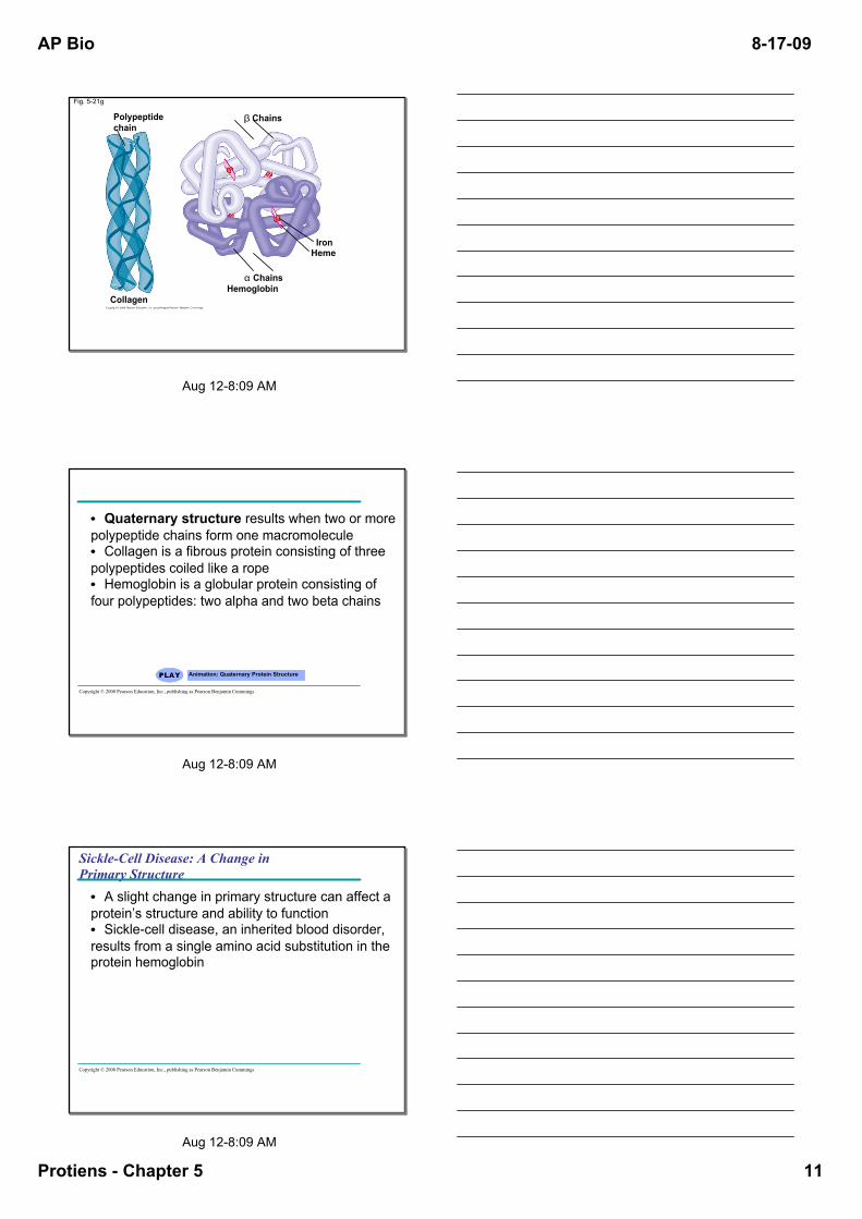
Fig. 521g
Polypeptidechain
β Chains
HemeIron
α Chains
CollagenHemoglobin
Aug 128:09 AM
• Quaternary structure results when two or more polypeptide chains form one macromolecule• Collagen is a fibrous protein consisting of three polypeptides coiled like a rope• Hemoglobin is a globular protein consisting of four polypeptides: two alpha and two beta chains
Animation: Quaternary Protein Structure
Copyright © 2008 Pearson Education, Inc., publishing as Pearson Benjamin Cummings
Aug 128:09 AM
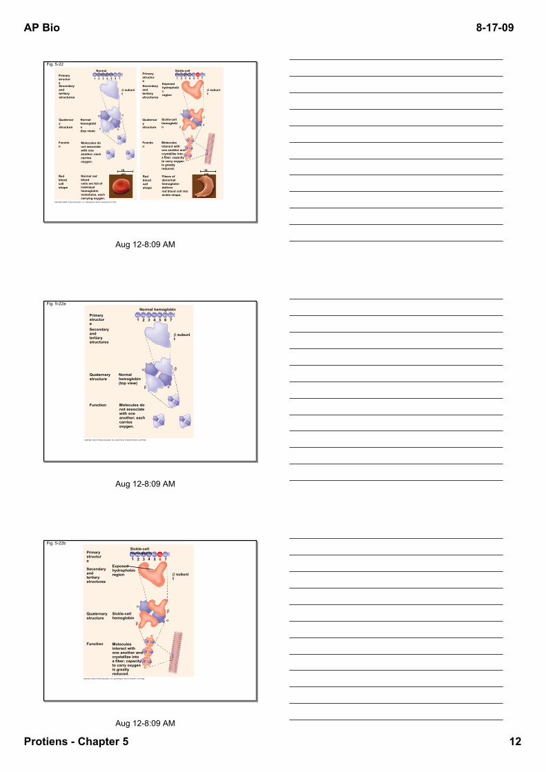
SickleCell Disease: A Change in Primary Structure
• A slight change in primary structure can affect a protein’s structure and ability to function • Sicklecell disease, an inherited blood disorder, results from a single amino acid substitution in the protein hemoglobin
Copyright © 2008 Pearson Education, Inc., publishing as Pearson Benjamin Cummings
AP Bio
Protiens Chapter 5 12
81709
Aug 128:09 AM
Fig. 522
PrimarystructureSecondaryand tertiarystructures
Quaternarystructure
Normalhemoglobin(top view)
PrimarystructureSecondaryand tertiarystructures
Quaternarystructure
Function
Function
β subunit
Molecules donot associatewith oneanother; eachcarries oxygen.
Red bloodcell shape
Normal red bloodcells are full ofindividualhemoglobinmoledules, eachcarrying oxygen.
10 µm
Normal hemoglobin
β
β
α
α
1 2 3 4 5 6 7Val His LeuThr Pro GluGlu
Red bloodcell shape
β subunit
Exposedhydrophobicregion
Sicklecellhemoglobin β
α
Moleculesinteract withone another andcrystallize intoa fiber; capacityto carry oxygenis greatly reduced.
β
α
Fibers of abnormalhemoglobin deformred blood cell intosickle shape.
10 µm
Sicklecell hemoglobin GluProThrLeuHisVal Val1 2 3 4 5 6 7
Aug 128:09 AM
Fig. 522a
PrimarystructureSecondaryand tertiarystructures
Function
Quaternarystructure
Molecules donot associatewith oneanother; eachcarries oxygen.
Normalhemoglobin(top view)
β subunit
Normal hemoglobin
7654321
β
α
α
β
GluVal
His Leu
Thr
Pro
Glu
Aug 128:09 AM
Fig. 522b
Primarystructure
Secondaryand tertiarystructures
Function
Quaternarystructure
Molecules interact with one another andcrystallize into a fiber; capacity to carry oxygenis greatly reduced.
Sicklecellhemoglobin
β subunit
Sicklecell hemoglobin
7654321
β
α
α
β
Val
Val
His Leu
Thr
Pro
Glu
Exposedhydrophobicregion

AP Bio
Protiens Chapter 5 13
81709
Aug 128:09 AM
Fig. 522c
Normal red bloodcells are full ofindividualhemoglobinmolecules, each carrying oxygen.
Fibers of abnormalhemoglobin deformred blood cell intosickle shape.
10 µm 10 µm
Aug 128:09 AM
What Determines Protein Structure?
• In addition to primary structure, physical and chemical conditions can affect structure• Alterations in pH, salt concentration, temperature, or other environmental factors can cause a protein to unravel• This loss of a protein’s native structure is called denaturation• A denatured protein is biologically inactive
Copyright © 2008 Pearson Education, Inc., publishing as Pearson Benjamin Cummings
Aug 128:09 AM
Fig. 523
Normal protein Denatured protein
Denaturation
Renaturation

AP Bio
Protiens Chapter 5 14
81709
Aug 128:09 AM
Protein Folding in the Cell
• It is hard to predict a protein’s structure from its primary structure• Most proteins probably go through several states on their way to a stable structure• Chaperonins are protein molecules that assist the proper folding of other proteins
Copyright © 2008 Pearson Education, Inc., publishing as Pearson Benjamin Cummings