Embed Size (px)
Citation preview
11
Cutting Edge Technology for Aortic
Aneurysms
Cutting Edge Technology for Aortic
Aneurysms
UnityPoint Health Methodist, Peoria, IL [email protected]
Aortic AneurysmsHow Big is the Problem?
♥ 1 - 5 % of general population affected
� Incidence is increasing
♥ AAA: 100,000 – 250,000 new cases each year in the U.S.
♥ TAA: approximately 15, 000 new cases each year
♥ 43,000 – 47,000 deaths per year (CDC)
� Twice as many deaths from thoracic aortic dissection and rupture than abdominal
Aortic AneurysmsHow Big is the Problem?
♥ 10th – 18th leading cause of death in the USA
♥ 2/3 of patients who suffer a ruptured aneurysm will die before even reaching the hospital.
♥ 90% mortality with ruptured AAA
Source: Society of Thoracic Surgeons
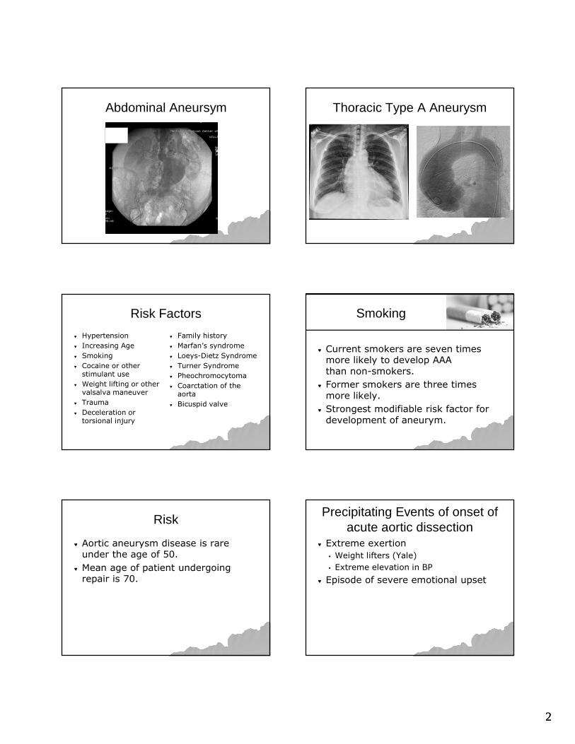
Normal Size of Aorta
Size in CM
Root 3.5–3.91
Ascending 2.86
Mid Descending
2.39–2.64
Diaphragmatic 2.43-2.69
Source: J Vasc Surg 1991:13:452-8 and 2010 Guidelines TAD.
Aortic Aneurysm (AA)
♥ Abnormal dilation of the aortic wall that alters the vessel shape and blood flow� 50% increase in the diameter of a
vessel in comparison of it’s expected normal
♥ With gradual enlargement, the aorta becomes increasingly weakened, leading to possible dissection and rupture.
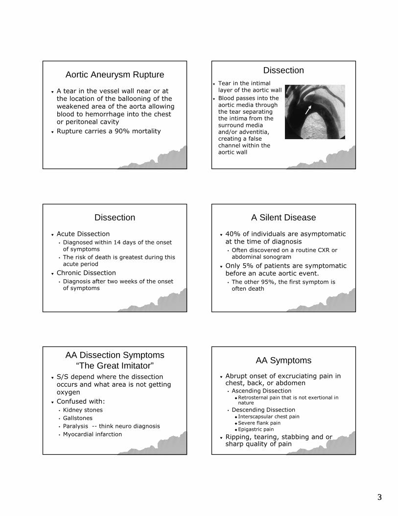
Aortic Aneurysm (AA)
ThoracicTAA
ThoracicTAA
AbdominalAAA
AbdominalAAA
22
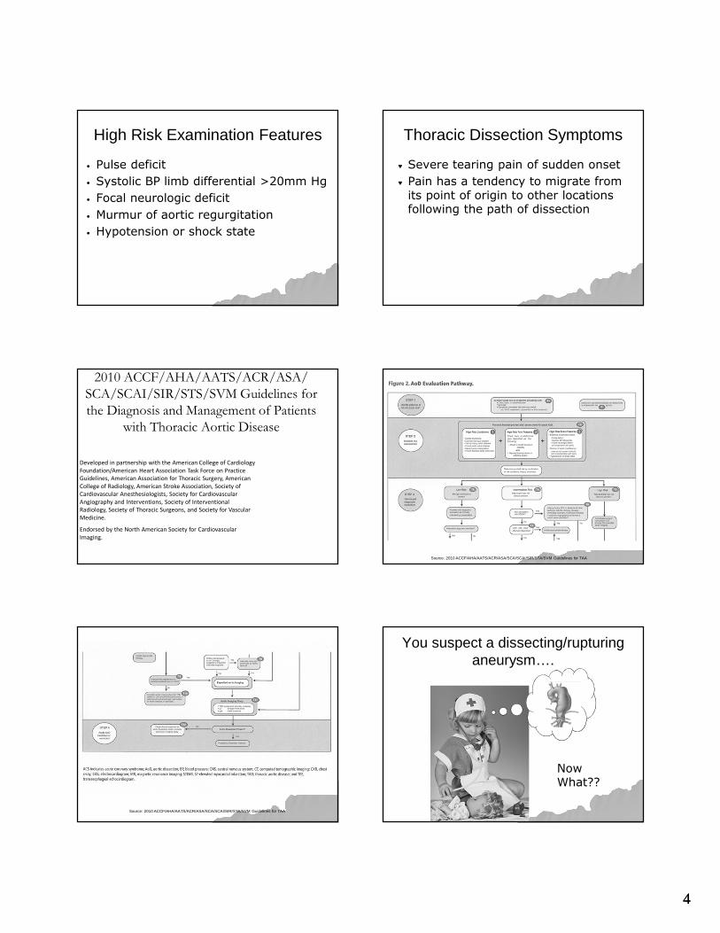
Abdominal Aneursym Thoracic Type A Aneurysm
Risk Factors
♥ Hypertension
♥ Increasing Age
♥ Smoking
♥ Cocaine or other stimulant use
♥ Weight lifting or other valsalva maneuver
♥ Trauma
♥ Deceleration or torsional injury
♥ Family history
♥ Marfan’s syndrome
♥ Loeys-Dietz Syndrome
♥ Turner Syndrome
♥ Pheochromocytoma
♥ Coarctation of the aorta
♥ Bicuspid valve
Smoking
♥ Current smokers are seven times more likely to develop AAA than non-smokers.
♥ Former smokers are three times more likely.
♥ Strongest modifiable risk factor for development of aneurym.
Risk
♥ Aortic aneurysm disease is rare under the age of 50.
♥ Mean age of patient undergoing repair is 70.
Precipitating Events of onset of acute aortic dissection
♥ Extreme exertion
� Weight lifters (Yale)
� Extreme elevation in BP
♥ Episode of severe emotional upset
33
Aortic Aneurysm Rupture
♥ A tear in the vessel wall near or at the location of the ballooning of the weakened area of the aorta allowing blood to hemorrhage into the chest or peritoneal cavity
♥ Rupture carries a 90% mortality
Dissection♥ Tear in the intimal
layer of the aortic wall
♥ Blood passes into the aortic media through the tear separating the intima from the surround media and/or adventitia, creating a false channel within the aortic wall
Dissection
♥ Acute Dissection
� Diagnosed within 14 days of the onset of symptoms
� The risk of death is greatest during this acute period
♥ Chronic Dissection
� Diagnosis after two weeks of the onset of symptoms
A Silent Disease
♥ 40% of individuals are asymptomatic at the time of diagnosis
� Often discovered on a routine CXR or abdominal sonogram
♥ Only 5% of patients are symptomatic before an acute aortic event.
� The other 95%, the first symptom is often death
AA Dissection Symptoms“The Great Imitator”
♥ S/S depend where the dissection occurs and what area is not getting oxygen
♥ Confused with:
� Kidney stones
� Gallstones
� Paralysis -- think neuro diagnosis
� Myocardial infarction
AA Symptoms
♥ Abrupt onset of excruciating pain in chest, back, or abdomen � Ascending Dissection
�Retrosternal pain that is not exertional in nature
� Descending Dissection � Interscapsular chest pain
�Severe flank pain
�Epigastric pain
♥ Ripping, tearing, stabbing and or sharp quality of pain
44
High Risk Examination Features
• Pulse deficit
• Systolic BP limb differential >20mm Hg
• Focal neurologic deficit
• Murmur of aortic regurgitation
• Hypotension or shock state
Thoracic Dissection Symptoms
♥ Severe tearing pain of sudden onset
♥ Pain has a tendency to migrate from its point of origin to other locations following the path of dissection
2010 ACCF/AHA/AATS/ACR/ASA/
SCA/SCAI/SIR/STS/SVM Guidelines for
the Diagnosis and Management of Patients
with Thoracic Aortic Disease
Developed in partnership with the American College of Cardiology
Foundation/American Heart Association Task Force on Practice
Guidelines, American Association for Thoracic Surgery, American
College of Radiology, American Stroke Association, Society of
Cardiovascular Anesthesiologists, Society for Cardiovascular
Angiography and Interventions, Society of Interventional
Radiology, Society of Thoracic Surgeons, and Society for Vascular
Medicine.
Endorsed by the North American Society for Cardiovascular
Imaging.
Source: 2010 ACCF/AHA/AATS/ACR/ASA/SCA/SCAI/SIR/STA/SVM Guidelines for TAA
Source: 2010 ACCF/AHA/AATS/ACR/ASA/SCA/SCAI/SIR/STA/SVM Guidelines for TAA
You suspect a dissecting/rupturing aneurysm….
Now What??
55
Rapid Triage & Treatments ♥ Aortic Center Aortic Pathway
Methodist Hospital Houston. TX
Diagnostics
♥ 12 Lead EKG to r/o STEMI
♥ Chest x-ray – not very helpful as no abnormalities noted
♥ CT scan
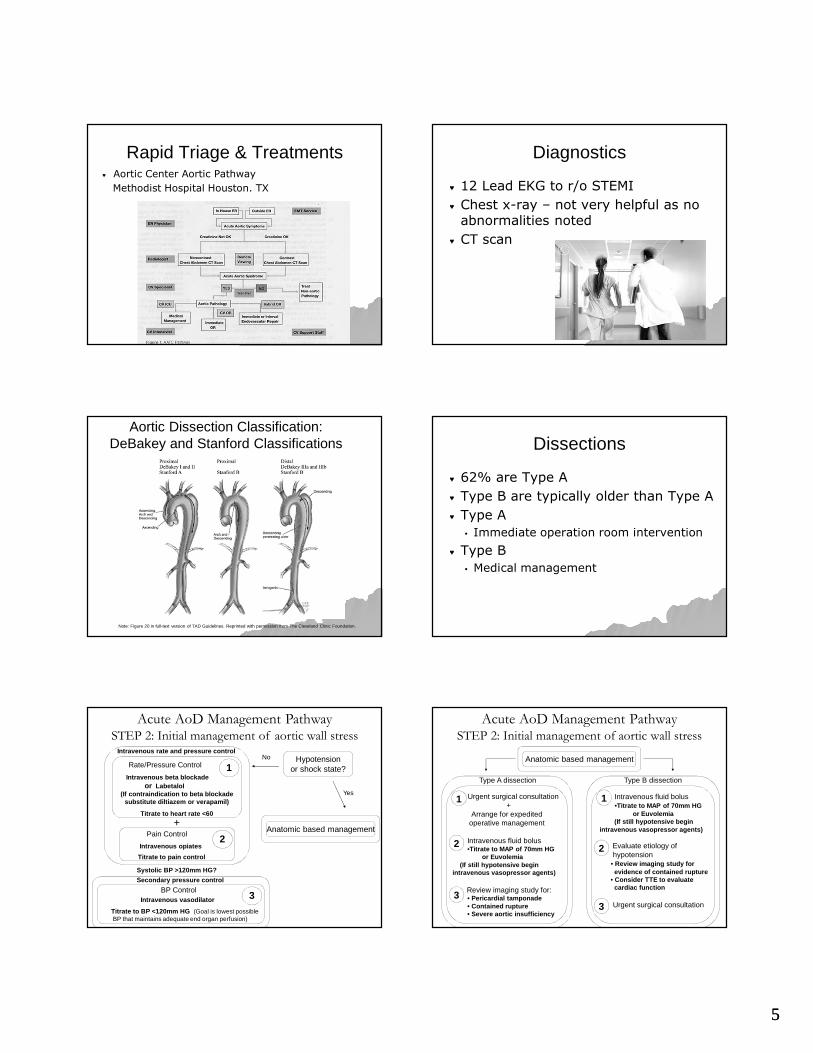
Aortic Dissection Classification: DeBakey and Stanford Classifications
Note: Figure 20 in full-text version of TAD Guidelines. Reprinted with permission from The Cleveland Clinic Foundation.
Dissections
♥ 62% are Type A
♥ Type B are typically older than Type A
♥ Type A
� Immediate operation room intervention
♥ Type B
� Medical management
Rate/Pressure Control
Intravenous beta blockadeor Labetalol
(If contraindication to beta blockadesubstitute diltiazem or verapamil)
Titrate to heart rate <60
1
Pain Control
Intravenous opiates
Titrate to pain control
Intravenous rate and pressure control
2
+
Hypotensionor shock state?
No
Yes
Systolic BP >120mm HG?
BP ControlIntravenous vasodilator
Titrate to BP <120mm HG (Goal is lowest possible BP that maintains adequate end organ perfusion)
Secondary pressure control
3
Anatomic based management
Acute AoD Management PathwaySTEP 2: Initial management of aortic wall stress
Acute AoD Management PathwaySTEP 2: Initial management of aortic wall stress
Anatomic based management
Urgent surgical consultation+
Arrange for expeditedoperative management
Intravenous fluid bolus•Titrate to MAP of 70mm HG
or Euvolemia(If still hypotensive begin
intravenous vasopressor agents)
Review imaging study for:• Pericardial tamponade• Contained rupture• Severe aortic insufficiency
1
2
3
Type A dissection
Intravenous fluid bolus•Titrate to MAP of 70mm HG
or Euvolemia(If still hypotensive begin
intravenous vasopressor agents)
Evaluate etiology of hypotension
• Review imaging study forevidence of contained rupture
• Consider TTE to evaluatecardiac function
Urgent surgical consultation
2
3
Type B dissection
1
66
Indications for AA repair
Thoracic♥ Symptomatic
♥ Diameter 5.5 - 6 cm
♥ Diameter 4.4 - 5 cm associated with genetic disorder(Marfan’s syndrome)
♥ Symptoms suggesting expansion or compression of surrounding structures
Indications for AA repair
Abdominal
♥ Diameter > 5 cm
♥ Diameter < 4 cm needs regular follow up
♥ Diameter 4 – 5 cm, management is controversial
Indications for AA repair
Both: TAA & AAA♥ Rapidly expanding aneurysms
� growth rate > 0.5 cm/year
♥ Symptomatic aneurysm regardless of size
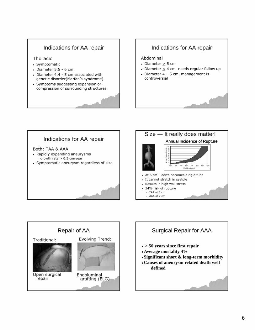
Size --- It really does matter!
♥ At 6 cm – aorta becomes a rigid tube
♥ It cannot stretch in systole
♥ Results in high wall stress
♥ 34% risk of rupture
� TAA at 6 cm
� AAA at 7 cm
Annual Incidence of RuptureAnnual Incidence of Rupture
Repair of AA
Traditional:
Open surgical repair
Evolving Trend:
Endoluminal grafting (ELG)
Surgical Repair for AAA
♥ > 50 years since first repair♥Average mortality 4%♥Significant short & long-term morbidity♥Causes of aneurysm related death well
defined
77
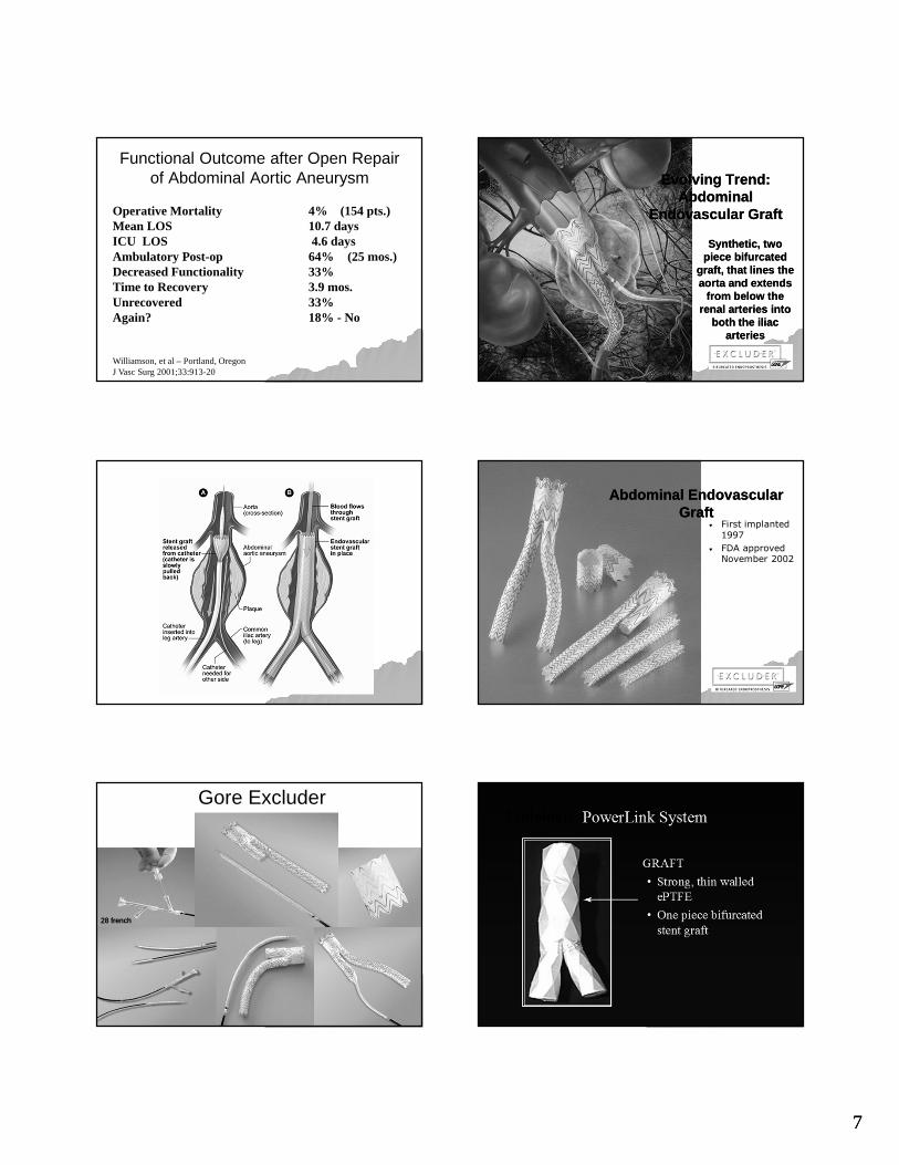
Functional Outcome after Open Repair of Abdominal Aortic Aneurysm
Operative Mortality 4% (154 pts.)Mean LOS 10.7 daysICU LOS 4.6 daysAmbulatory Post-op 64% (25 mos.)Decreased Functionality 33%Time to Recovery 3.9 mos.Unrecovered 33%Again? 18% - No
Williamson, et al – Portland, OregonJ Vasc Surg 2001;33:913-20
Evolving Trend: Abdominal
Endovascular Graft
Evolving Trend: Abdominal
Endovascular Graft
Synthetic, two piece bifurcated
graft, that lines the aorta and extends
from below the renal arteries into
both the iliac arteries
Synthetic, two piece bifurcated
graft, that lines the aorta and extends
from below the renal arteries into
both the iliac arteries
Abdominal Endovascular Graft
Abdominal Endovascular Graft
♥ First implanted
1997
♥ FDA approved November 2002
Gore Excluder
28 french28 french
Endologix
88
AneurRx Talent
Medtronic
Endurant
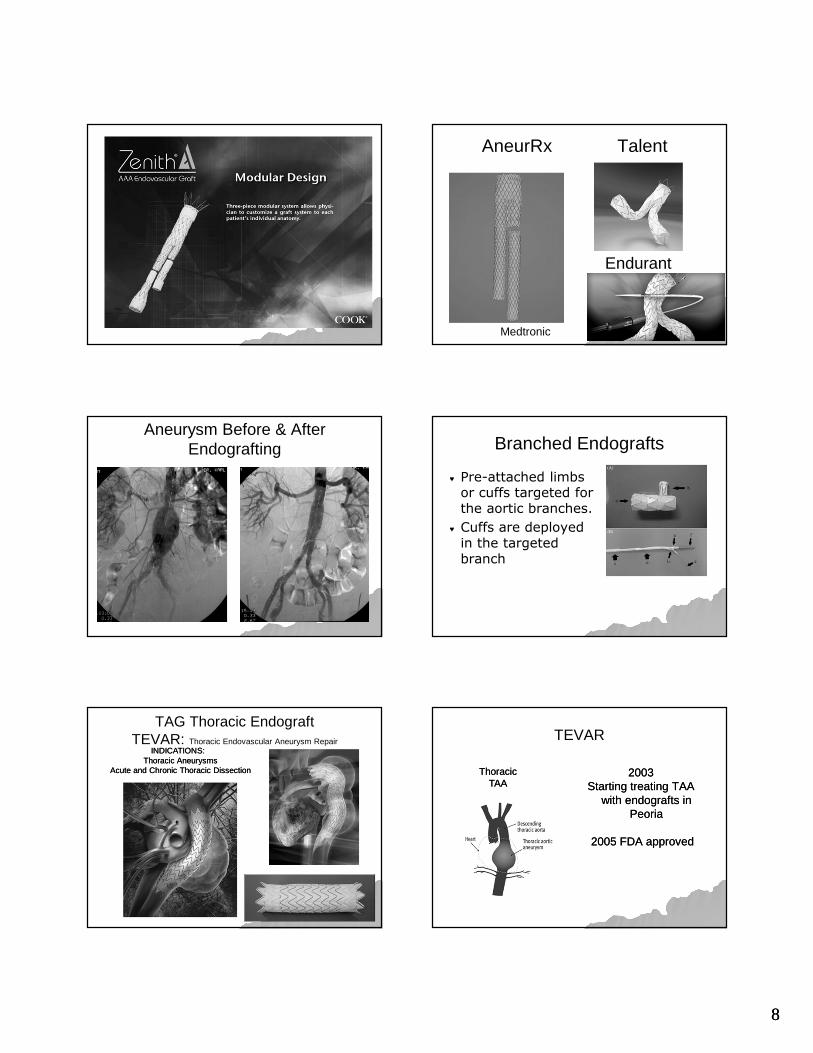
Aneurysm Before & After Endografting Branched Endografts
♥ Pre-attached limbs or cuffs targeted for the aortic branches.
♥ Cuffs are deployed in the targeted branch
TAG Thoracic EndograftTEVAR: Thoracic Endovascular Aneurysm Repair
INDICATIONS: Thoracic Aneurysms
Acute and Chronic Thoracic Dissection
INDICATIONS: Thoracic Aneurysms
Acute and Chronic Thoracic Dissection
TEVAR
ThoracicTAA
ThoracicTAA
2003 Starting treating TAA
with endografts in Peoria
2005 FDA approved
2003 Starting treating TAA
with endografts in Peoria
2005 FDA approved
99
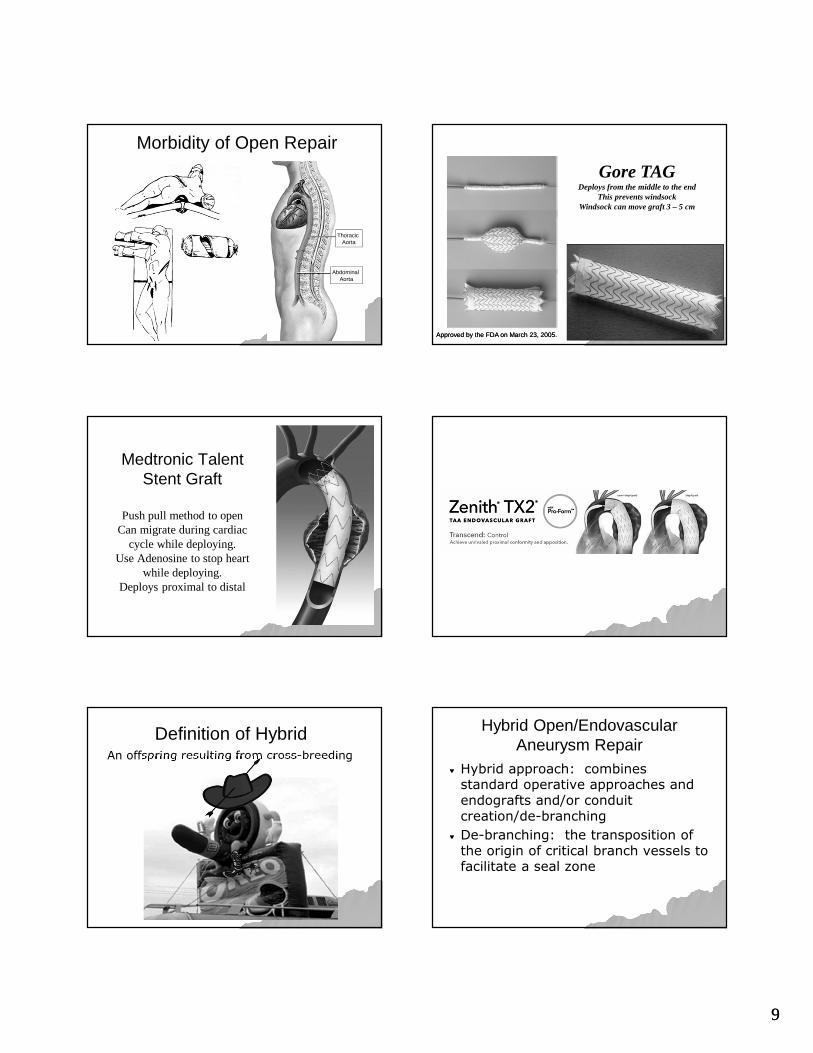
Morbidity of Open Repair
Thoracic Aorta
Abdominal Aorta
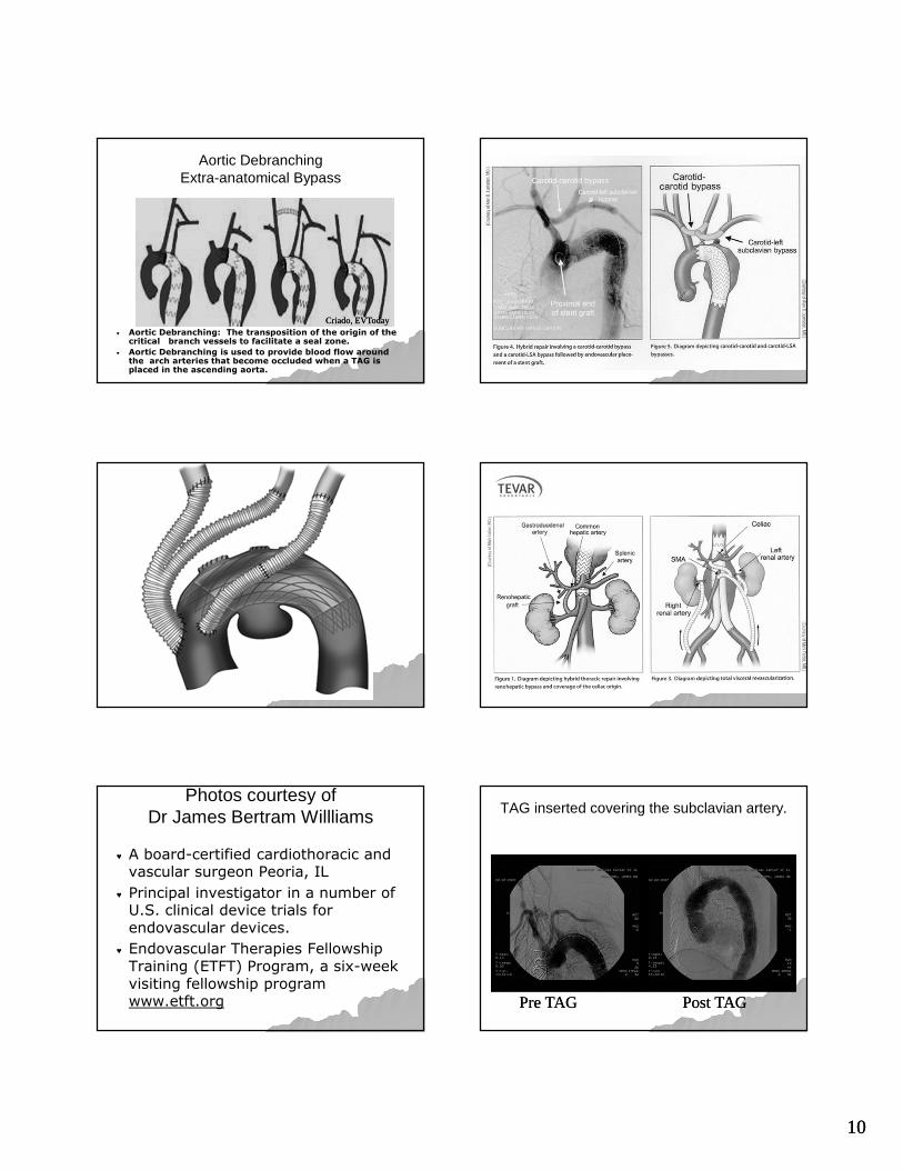
Gore TAGDeploys from the middle to the end
This prevents windsockWindsock can move graft 3 – 5 cm
Approved by the FDA on March 23, 2005.Approved by the FDA on March 23, 2005.
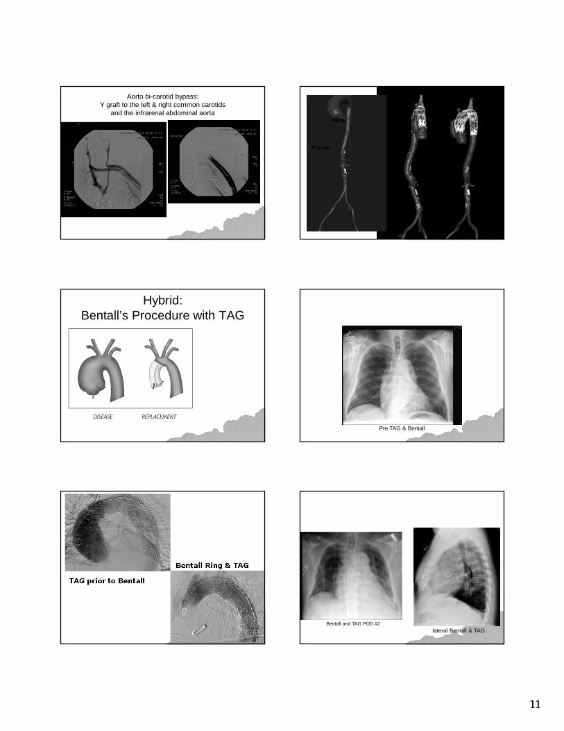
Medtronic Talent Stent Graft
Push pull method to openCan migrate during cardiac
cycle while deploying.Use Adenosine to stop heart
while deploying.Deploys proximal to distal
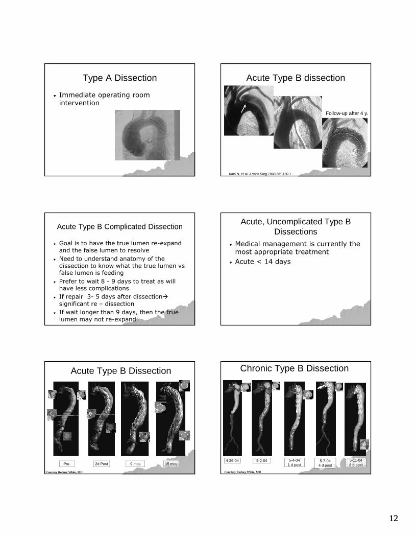
Definition of HybridAn offspring resulting from cross-breeding
Hybrid Open/Endovascular Aneurysm Repair
♥ Hybrid approach: combines standard operative approaches and endografts and/or conduit creation/de-branching
♥ De-branching: the transposition of the origin of critical branch vessels to facilitate a seal zone
1010
Aortic DebranchingExtra-anatomical Bypass
Criado, EVTodayCriado, EVToday♥ Aortic Debranching: The transposition of the origin of the
critical branch vessels to facilitate a seal zone.
♥ Aortic Debranching is used to provide blood flow around the arch arteries that become occluded when a TAG is placed in the ascending aorta.
Photos courtesy of Dr James Bertram Willliams
♥ A board-certified cardiothoracic and vascular surgeon Peoria, IL
♥ Principal investigator in a number of U.S. clinical device trials for endovascular devices.
♥ Endovascular Therapies Fellowship Training (ETFT) Program, a six-week visiting fellowship program www.etft.org
TAG inserted covering the subclavian artery.
Pre TAGPre TAG Post TAGPost TAG
1111
Aorto bi-carotid bypass: Y graft to the left & right common carotids
and the infrarenal abdominal aorta
Pre-op
Post-op
Hybrid:Bentall’s Procedure with TAG
Pre TAG & Bentall
TAG prior to Bentall
Bentall Ring & TAG
Bentall and TAG POD #2
lateral Bentall & TAG
1212
Type A Dissection
♥ Immediate operating room intervention
Acute Type B dissection
Follow-up after 4 y.
Kato N, et al. J Vasc Surg 2003;38:1130-1
Acute Type B Complicated Dissection
♥ Goal is to have the true lumen re-expand and the false lumen to resolve
♥ Need to understand anatomy of the dissection to know what the true lumen vs false lumen is feeding
♥ Prefer to wait 8 - 9 days to treat as will have less complications
♥ If repair 3- 5 days after dissection�
significant re – dissection
♥ If wait longer than 9 days, then the true lumen may not re-expand
Acute, Uncomplicated Type B Dissections
♥ Medical management is currently the most appropriate treatment
♥ Acute < 14 days
Pre- 2d Post 9 mos 15 mos
Acute Type B Dissection
Courtesy Rodney White, MD
4-26-04 5-2-04 5-4-041 d post
5-11-048 d post
5-7-044 d post
Chronic Type B Dissection
Courtesy Rodney White, MD
1313
Nursing Care
Preoperative Care
♥ Usually AM admit
♥ Hydrate with NS at 125 -150 ml/hour
♥ If Creatinine > 1.6 may give Mucomyst or Bicarbonate infusion (3 amps Bicarb/1000ml D5W at 3 ml/hr x 6 hours --- start 1 hour preop)
♥ Permit to include possible resection of aortic aneurysm
♥ Teaching
Preop DiagnosticsTo measure length & diameter of the arteries
♥ Duplex scan
♥ CT (without contrast)
♥ Aortogram (with calibrated catheter)
♥ Spiral CT
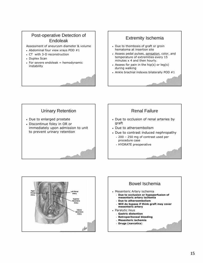
♥ Intravascular ultrasound
♥ 3-D CT Reconstruction
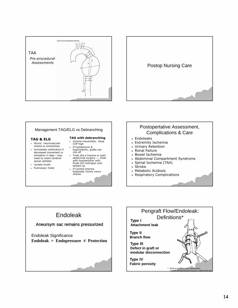
Numerous Pre-procedural Assessments
Celiac axis
SMA
Cephalad neck
Caudad neck
Length
Femoral artery/runoff
Int. iliac artery
Iliac artery
Lumbars
IMA
Kinking
Diameter
Intravascular UltrasoundProximal Neck
1414
TAA
Pre-procedural
AssessmentsPostop Nursing Care
Management TAG/ELG vs Debranching
TAG & ELG♥ Hourly neurovascular
checks to extremities
♥ Immediate notification if
decreased movement or
sensation in legs-- may need to insert cerebral
spinal catheter
♥ Lactate levels
♥ Pulmonary Toilet
TAG with debranching♥ Volume resuscitate. Keep
CVP high
♥ If hypotensive & hypovolemic, grafts can clot off
♥ Treat like a trauma or open abdominal surgery --- treat with hypotentsion with fluids not inotropes until tanked up.
♥ If Carotid arteries bypassed, hourly neuro checks
Postopertative Assessment, Complications & Care
♥ Endoleaks♥ Extremity Ischemia♥ Urinary Retention♥ Renal Failure ♥ Bowel Ischemia♥ Abdominal Compartment Syndroms♥ Spinal Ischemia (TAA)♥ Stroke♥ Metabolic Acidosis♥ Respiratory Complications
Endoleak
Endoleak SignificanceEndoleak = Endopressure ≠ Protection
Aneursym sac remains pressurizedAneursym sac remains pressurized
Perigraft Flow/Endoleak: Definitions*
* White et.al., Endoleak Classification, Journal of Endovascular Surgery, 1998;5:305-309
Type IAttachment leak
Type IIBranch flow
Type IIIDefect in graft ormodular disconnection
Type IVFabric porosity
1515
Post-operative Detection of Endoleak
Assessment of aneurysm diameter & volume
♥ Abdominal four view xrays POD #1
♥ CT with 3-D reconstruction
♥ Duplex Scan
♥ For severe endoleak = hemodynamic instability
Extremity Ischemia
♥ Due to thombosis of graft or groin hematoma at insertion site
♥ Assess pedal pulses, sensation, color, and temperature of extremities every 15 minutes x 4 and then hourly.
♥ Assess for pain in the hip(s) or leg(s) during walking
♥ Ankle brachial indexes bilaterally POD #1
Urinary Retention
♥ Due to enlarged prostate
♥ Discontinue foley in OR or immediately upon admission to unit to prevent urinary retention
Renal Failure
♥ Due to occlusion of renal arteries by graft
♥ Due to atheroembolism
♥ Due to contrast induced nephropathy
� 200 – 250 mg of contrast used per procedure case
� HYDRATE preoperative
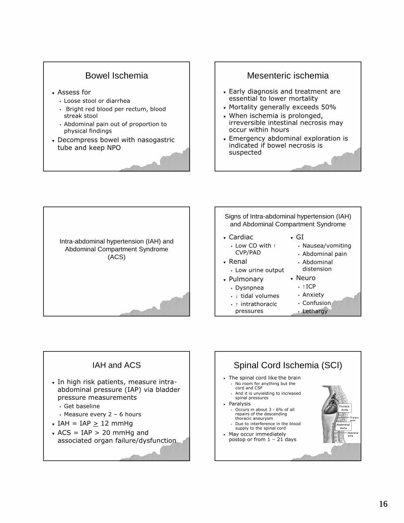
Right Renal Artery
Right Renal Artery
Left Renal Artery
Left Renal Artery
Superior Mesenteric
Artery
Superior Mesenteric
Artery
Inferior Mesenteric
Artery
Inferior Mesenteric
Artery
Bowel Ischemia
♥ Mesenteric Artery ischemia � Due to occlusion or hypoperfusion of
mesenteric artery ischemia
� Due to atheroembolism
� Will do bypass if think graft may cover mesenteric artery
♥ Paralytic ileus� Gastric distention
� Retroperitoneal bleeding
� Mesenteric ischemia
� Drugs (narcotics)
1616
Bowel Ischemia
♥ Assess for
� Loose stool or diarrhea
� Bright red blood per rectum, blood streak stool
� Abdominal pain out of proportion to physical findings
♥ Decompress bowel with nasogastric tube and keep NPO
Mesenteric ischemia
♥ Early diagnosis and treatment are essential to lower mortality
♥ Mortality generally exceeds 50%
♥ When ischemia is prolonged, irreversible intestinal necrosis may occur within hours
♥ Emergency abdominal exploration is indicated if bowel necrosis is suspected
Intra-abdominal hypertension (IAH) and Abdominal Compartment Syndrome
(ACS)
Signs of Intra-abdominal hypertension (IAH) and Abdominal Compartment Syndrome
♥ Cardiac
� Low CO with ↑CVP/PAD
♥ Renal
� Low urine output
♥ Pulmonary
� Dysnpnea
� ↓ tidal volumes
� ↑ intrathoracicpressures
♥ GI
� Nausea/vomiting
� Abdominal pain
� Abdominal distension
♥ Neuro
� ↑ICP
� Anxiety
� Confusion
� Lethargy
IAH and ACS
♥ In high risk patients, measure intra-abdominal pressure (IAP) via bladder pressure measurements
� Get baseline
� Measure every 2 – 6 hours
♥ IAH = IAP > 12 mmHg
♥ ACS = IAP > 20 mmHg and associated organ failure/dysfunction
Spinal Cord Ischemia (SCI)♥ The spinal cord like the brain
� No room for anything but the cord and CSF
� And it is unyielding to increasedspinal pressures
♥ Paralysis � Occurs in about 3 - 6% of all
repairs of the descending thoracic aneurysm
� Due to interference in the blood supply to the spinal cord
♥ May occur immediately postop or from 1 – 21 days
Thoracic Aorta
Abdominal Aorta
1717
Spinal Cord Ischemia
♥ Ischemia to the cord � Leads to cord edema
� Can cause the lumbar ICP to rise & impede normal flow of CSF within the spinal cana
♥ Thoracic or lumbar spinal cord damage causes paraplegia
♥ Similar to muscular ‘compartment syndrome’
Spinal Cord Ischemia (SCI)
♥ The mechanisms leading to SCI:
� The interruption of multiple branch vessels that provide spinal cord perfusion.
♥ Hypotension - MAP < 70 - 90
� Periop &/or postop
� Can be a precipitating factor causing SCI
At risk for permanent and transient paraplegia
♥ Complicated Type B dissection
♥ Hybrid aortic procedures
♥ Aortic transection
♥ Chronic renal failure
♥ Smoking
Prevention of Spinal Cord Ischemia
♥ Prevent Hypotension MAP < 70 - 90
� Treat with fluids to keep CVP > 6
Treatment Spinal Cord Ischemia
♥ Drainage of the lumbar CSF can reduce the risk of cord damage when reducing pressure to < 7 – 10 mmHg
♥ Keep MAP > 90 - 99 mmHg
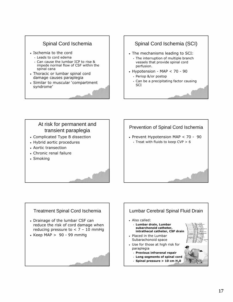
Lumbar Cerebral Spinal Fluid Drain
♥ Also called:
� Lumbar drain, Lumbar subarchonoid catheter, intrathecal catheter, CSF drain
♥ Placed in the Lumbar Subarachonoid space
♥ Use for those at high risk for paraplegia
� Previous infrarenal repair
� Long segments of spinal cord
� Spinal pressure > 10 cm H20
1818
Lumbar Cerebral Spinal Fluid Drain
The “Zero” reference point must be positioned at the level of the
catheter insertion site
The “Zero” reference point must be positioned at the level of the
catheter insertion site
Adjust drip chamber height until black level is
at the proper drainage resistance. Frequently 10
cmH 20
Adjust drip chamber height until black level is
at the proper drainage resistance. Frequently 10
cmH 20
PurposePrevent
paraplegia
PurposePrevent
paraplegia
Lumbar CSF Drain Safety
♥ Place CSF transducer on opposite side of bed as hemodynamic pressure monitoring
♥ Must be a nonflush pressure system
♥ Turn drainage system off when getting patient up to chair
♥ Level after repositioning patient
♥ Remember to unclamp
♥ Aseptic technique is a must!
Spinal Ischemia Assessment
♥ Record CSF output hourly
♥ Notify MD if CSF drainage is > 20 –30 ml/hr
♥ Note color of CSF
♥ Hourly spinal cord assessment for changes in sensation and/or movement
CSF Drainage
♥ Maintain CSF pressure 10 – 15 mmHg for the first 24 hours
♥ Then let rise to 15 mmHG
♥ If CSF pressure goes up above normal, blood flow to the spinal cord goes down, resulting in cord ischemia
Complications of CSF Drain
♥ Infection
♥ Overdrainage� Subdural hematoma
� Herniation
♥ Spinal cord hematoma
♥ Headache
♥ Pneumocranium (from air entering system)
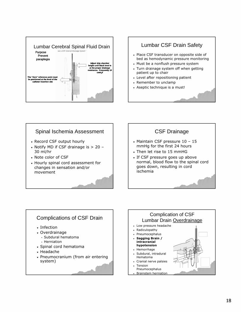
Complication of CSFLumbar Drain Overdrainage
♥ Low pressure headache
♥ Radiculopathy
♥ Pneumocephalus
♥ Sagging Brain / intracranial hypotension
♥ Hemorrhage
♥ Subdural, intradural Hematoma
♥ Cranial nerve palsies
♥ Tension Pneumocephalus
♥ Brainstem herniation
1919
Post removal of Lumbar drain
♥ Cap 24 hours prior to removal
♥ Assess for lower extremity weakness or loss of sensation
♥ SCI can occur up to 30 days post op.
♥ Teach patients to come to ED immediately for aggressive treatment if they notice any change, numbness, or weakness in their legs.
Stroke
♥ 4- 7% risk
♥ Routine neuro checks
Respiratory Complications♥ Due to general anesthesia and
smoking� Incentive Spirometry every 1- 2 hours
while awake
� Aggressive Activity�HOB 30o
�Chair when stable
�Ambulate 200’ evening of surgery
�Then Ambulate 4- 6 times per day
♥ Left Pleural Effusion� Something may be bleeding
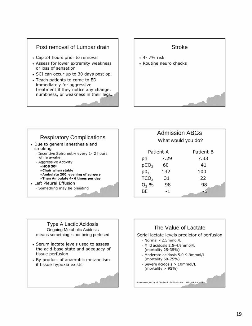
Admission ABGsWhat would you do?
Patient A Patient B
ph 7.29 7.33
pCO2 60 41
p02 132 100
TCO2 31 22
O2 % 98 98
BE -1 -6
Type A Lactic AcidosisOngoing Metabolic Acidosis
means something is not being perfused
♥ Serum lactate levels used to assess the acid-base state and adequacy of tissue perfusion
♥ By product of anaerobic metabolism if tissue hypoxia exists
The Value of LactateSerial lactate levels predictor of perfusion
� Normal <2.5mmol/L
� Mild acidosis 2.5-4.9mmol/L (mortality 25-35%)
� Moderate acidosis 5.0-9.9mmol/L (mortality 60-75%)
� Severe acidosis > 10mmol/L (mortality > 95%)
Shoemaker, WC et al. Textbook of critical care. 1995. WB Saunders
2020
Serum Lactate levels
♥ Serum Lactate levels every 4 hours x 24 hours
♥ Level will be around 4 – 5mmol/L on admission
♥ Lactate levels need to decrease
♥ May be the first indication that something is wrong
Discharge
♥ Abdominal – POD #1 from CVICU
♥ Thoracic -- POD #2 or 3
♥ Teaching
♥ 10 days post procedure the patient should be back to normal activities
♥ MRI conditional up to 3 Tesla
Follow-up
♥ CT scan at 1, 2, 6, and 12 months and then annually to assess for aortic growth
♥ Teaching
� Avoidance of exertional activities
�Betablockers blunt pressure spikes
� Avoidance of extreme emotional upsets
References• Desai ND, Pochettino A, Szeto W, et al. Thoracic endovascular aortic repair:
Evolution of therapy, patterns of use, and results in a 10 year experience. J Thorac Cardiovasc Surg. 2011;142(3):587-594.
• Elefreriades JA, Farakas EA. Thoracic aortic aneurysm: Clinically pertinent controversies and uncertainties. J Am Coll Cardiol 2010;55:841-856.
• Gallager H. (2010) Intra-Abdomonial Hypertension. AACN Adv Crit Care: 21(2):205-217.
• Hiratzka LF, Bakris GL, Beckman JA et al. 2010 ACCF/AHA/AATS/ACR/ASA/SCA/SCAI/SIR/STA/SVM guidelines for the diagnosis and management of patients with thoracic aortic disease: a report of the American College of Thoracic Surgery, American College of Radiology, American Stroke Association, Society of Cardiovascular Anesthesiologists, Society for Cardiovascular Angiography and Interventions, Society of Interventional Radiology, Society of Thoracic Surgeons, and Society for Vascular Medicine. Circ 2010;121:e
• Methodist DeBakey Heart & Vascular Center. DeBakey Cardiovascular Journal. July – Sept 2011;7(3):1-56. (17 articles in this journal relating to endovascular stenting)
• Moser D, Riegal B. (2008). Cardiac Nursing: A companion to Braunwald’s Heart Disease. St Louis MO: Sanders Elsevier.
• Steuer J, Eriksson MO, Nyman R, Bjorck M, Wanhainen A. Early and long-term outcome after thoracic endovascular aortic repair (TEVAR) for acute complicated type B aortic dissection. 2001;41:318-323.
• Svensson LG, Kouchoukos NT, Miller C et al. Expert consensus document on the treatment of descending thoracic aortic disease using endovascular stent-grafts. Ann Thorac Surg 2008;85:S1-41.
• Ullery BW, Cheung AT, Fairman RM, et al. Risk factor, outcomes, and clinical manifestations of spinal cord ischemia following thoracic endovascular aortic repair. J Vasc Surg 2011;54:677-684.