Embed Size (px)
Citation preview
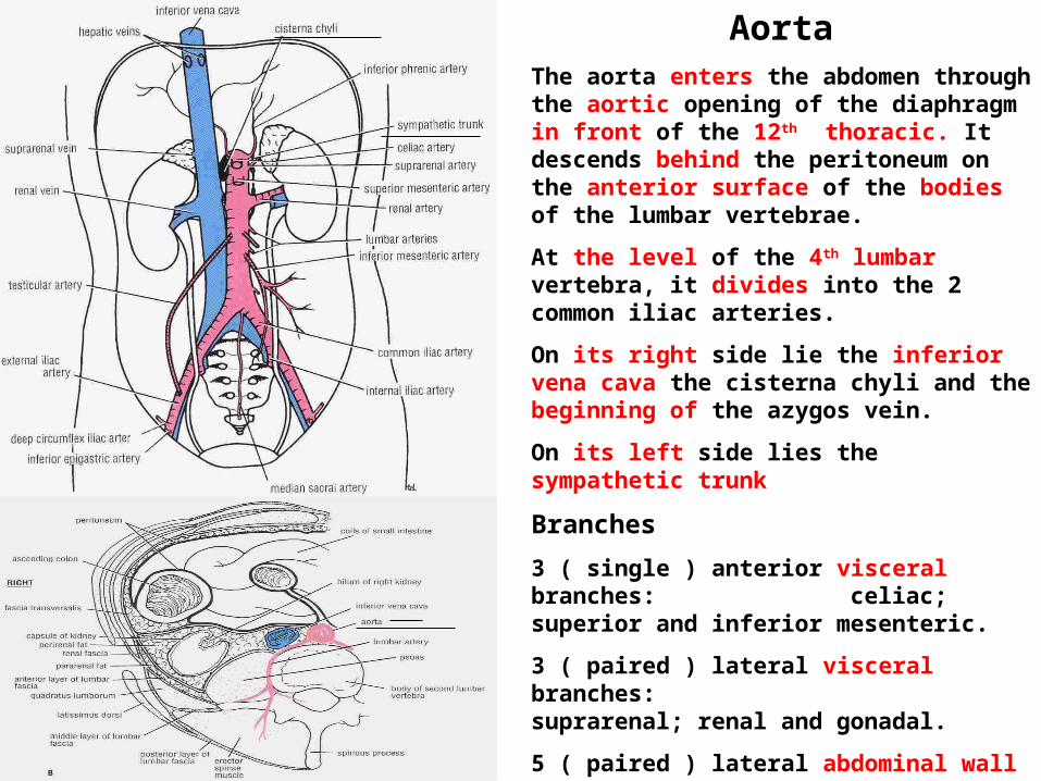
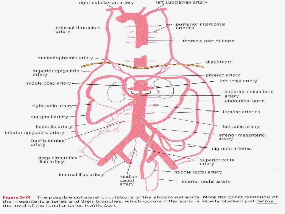
AortaThe aorta enters the abdomen through the aortic opening of the diaphragm in front of the 12th thoracic. It descends behind the peritoneum on the anterior surface of the bodies of the lumbar vertebrae.
At the level of the 4th lumbar vertebra, it divides into the 2 common iliac arteries.
On its right side lie the inferior vena cava the cisterna chyli and the beginning of the azygos vein.
On its left side lies the sympathetic trunk
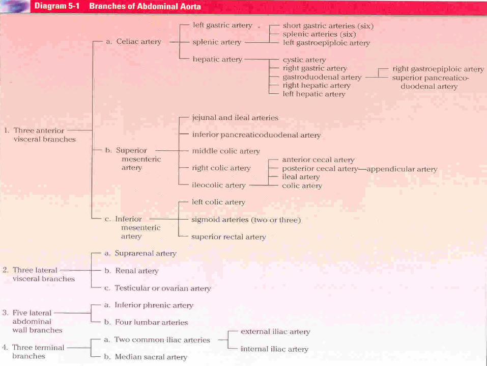
Branches
3 ( single ) anterior visceral branches: celiac; superior and inferior mesenteric.
3 ( paired ) lateral visceral branches: suprarenal; renal and gonadal.
5 ( paired ) lateral abdominal wall branches:
inferior phrenic and 4 lumbar arteries.
3 ( single ) terminal branches: 2 common iliac and the median sacral arteries
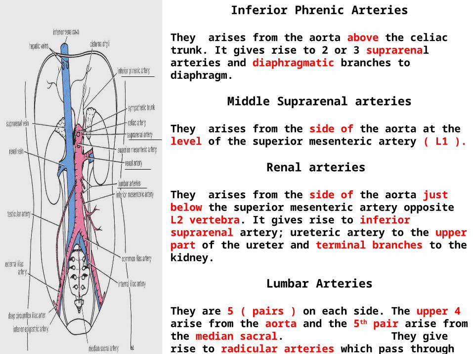
Inferior Phrenic Arteries
They arises from the aorta above the celiac trunk. It gives rise to 2 or 3 suprarenal arteries and diaphragmatic branches to diaphragm.
Middle Suprarenal arteries
They arises from the side of the aorta at the level of the superior mesenteric artery ( L1 ).
Renal arteries
They arises from the side of the aorta just below the superior mesenteric artery opposite L2 vertebra. It gives rise to inferior suprarenal artery; ureteric artery to the upper part of the ureter and terminal branches to the kidney.
Lumbar Arteries
They are 5 ( pairs ) on each side. The upper 4 arise from the aorta and the 5th pair arise from the median sacral. They give rise to radicular arteries which pass through the intervertebral foramina into the vertebral canal to supply the menings of the spinal cord. Also, they supply the muscles of the posterior and anterior abdominal wall.
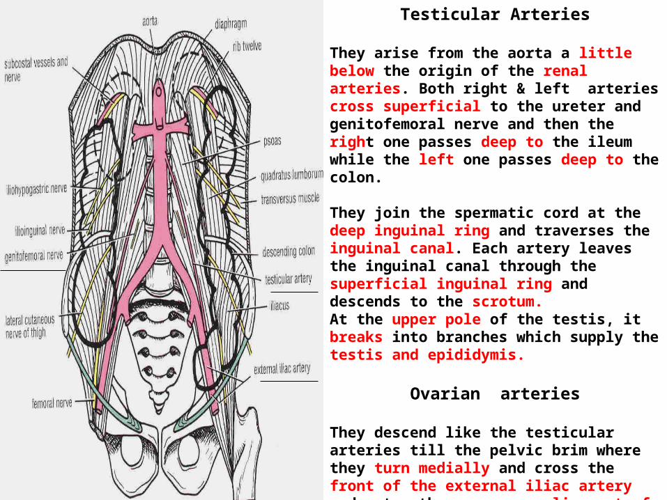
Testicular Arteries
They arise from the aorta a little below the origin of the renal arteries. Both right & left arteries cross superficial to the ureter and genitofemoral nerve and then the right one passes deep to the ileum while the left one passes deep to the colon.
They join the spermatic cord at the deep inguinal ring and traverses the inguinal canal. Each artery leaves the inguinal canal through the superficial inguinal ring and descends to the scrotum. At the upper pole of the testis, it breaks into branches which supply the testis and epididymis.
Ovarian arteries
They descend like the testicular arteries till the pelvic brim where they turn medially and cross the front of the external iliac artery and enter the suspensory ligament of the ovary.
They supply the ureter; ovary; uterus and uterine tubes.
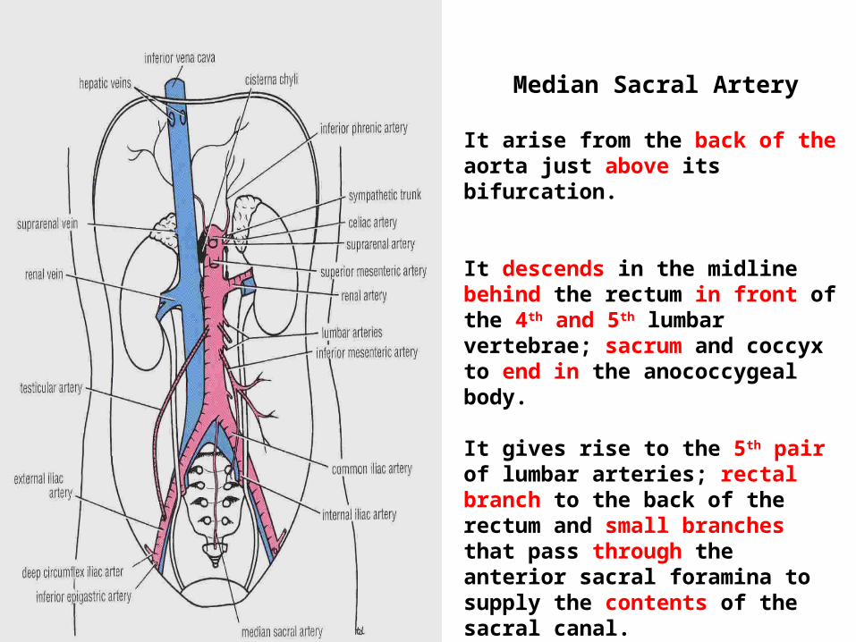
Median Sacral Artery
It arise from the back of the aorta just above its bifurcation.
It descends in the midline behind the rectum in front of the 4th and 5th lumbar vertebrae; sacrum and coccyx to end in the anococcygeal body.
It gives rise to the 5th pair of lumbar arteries; rectal branch to the back of the rectum and small branches that pass through the anterior sacral foramina to supply the contents of the sacral canal.
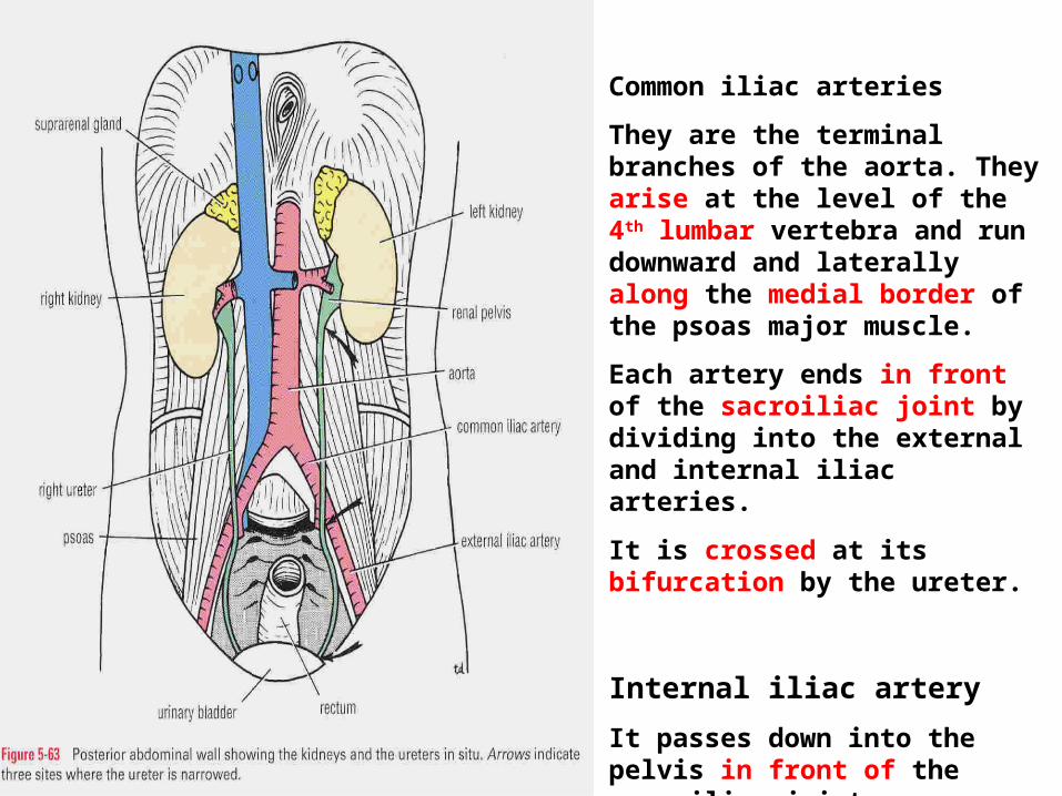
Common iliac arteries
They are the terminal branches of the aorta. They arise at the level of the 4th lumbar vertebra and run downward and laterally along the medial border of the psoas major muscle.
Each artery ends in front of the sacroiliac joint by dividing into the external and internal iliac arteries.
It is crossed at its bifurcation by the ureter.
Internal iliac artery
It passes down into the pelvis in front of the sacroiliac joint.
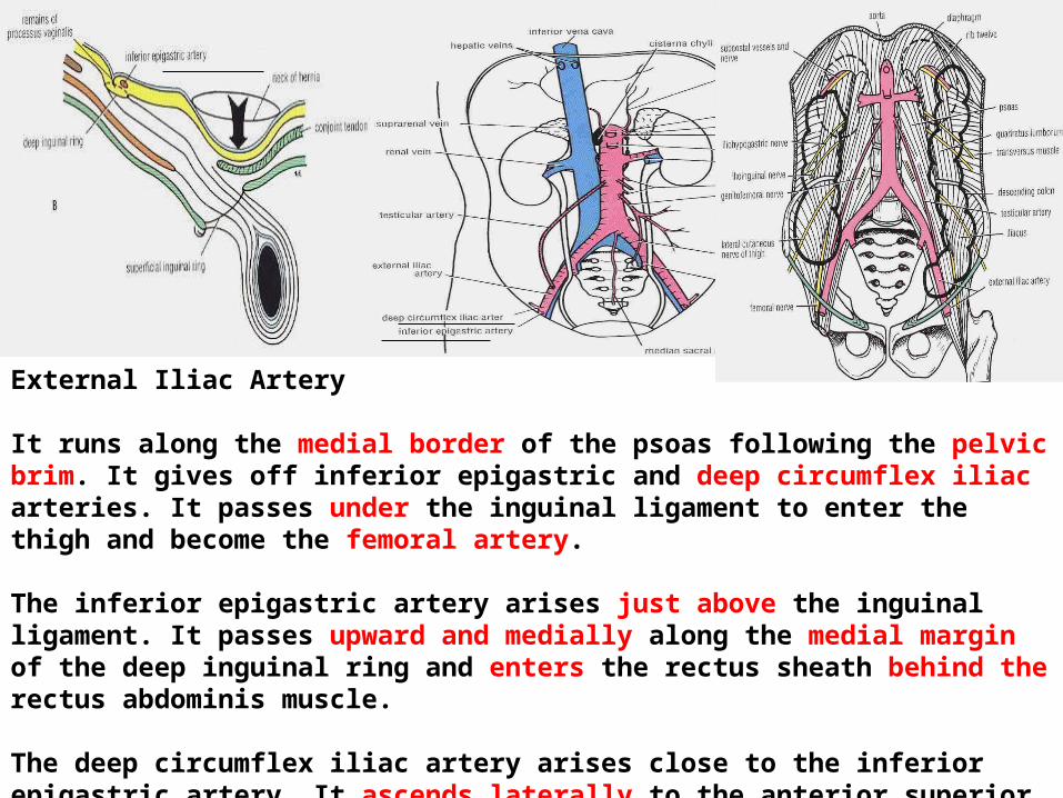
External Iliac Artery
It runs along the medial border of the psoas following the pelvic brim. It gives off inferior epigastric and deep circumflex iliac arteries. It passes under the inguinal ligament to enter the thigh and become the femoral artery.
The inferior epigastric artery arises just above the inguinal ligament. It passes upward and medially along the medial margin of the deep inguinal ring and enters the rectus sheath behind the rectus abdominis muscle.
The deep circumflex iliac artery arises close to the inferior epigastric artery. It ascends laterally to the anterior superior iliac spine and iliac crest supplying the muscles of the anterior abdominal wall.

Surface Anatomy of Aorta
Point 1 in the median plane one inch above the transpyloric plane.
Point 2 lies half inch below and to the left of the umbilicus ( at the level of the supracristal plane) which connects the highest parts of the 2 iliac crests.
Connect point 1 by 2 vertical lines 2 cm apart.
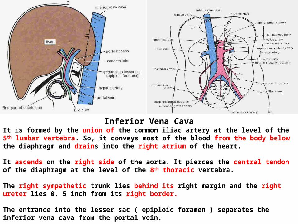
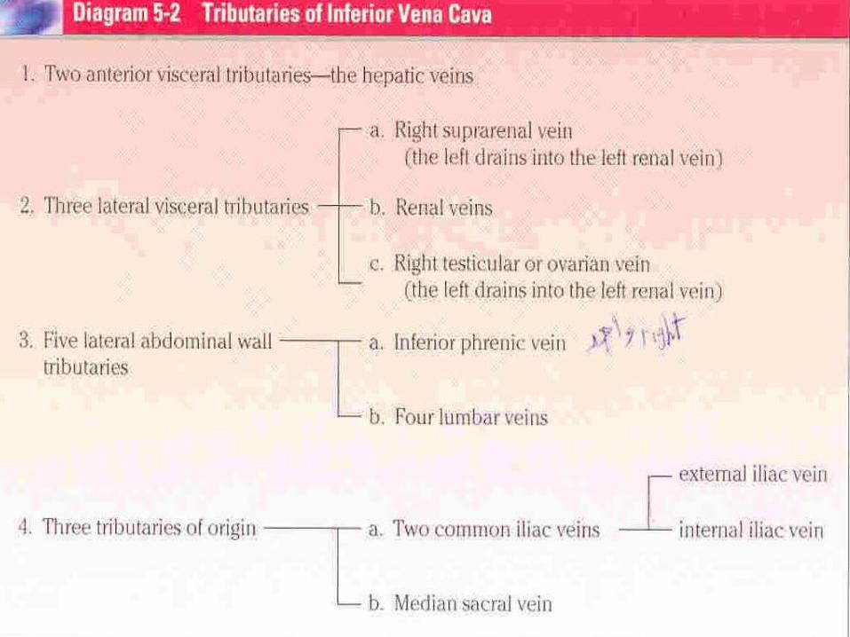
Inferior Vena CavaIt is formed by the union of the common iliac artery at the level of the 5th lumbar vertebra. So, it conveys most of the blood from the body below the diaphragm and drains into the right atrium of the heart.
It ascends on the right side of the aorta. It pierces the central tendon of the diaphragm at the level of the 8th thoracic vertebra.
The right sympathetic trunk lies behind its right margin and the right ureter lies 0. 5 inch from its right border.
The entrance into the lesser sac ( epiploic foramen ) separates the inferior vena cava from the portal vein.
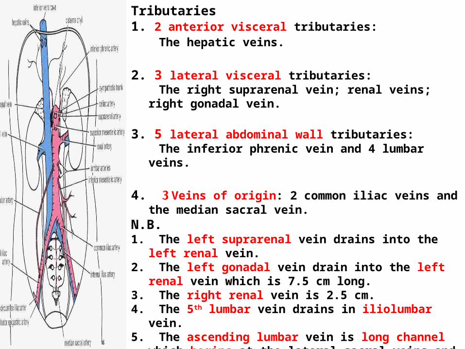
Tributaries 1. 2 anterior visceral tributaries: The hepatic veins.
2. 3 lateral visceral tributaries: The right suprarenal vein; renal veins; right gonadal
vein. 3. 5 lateral abdominal wall tributaries: The inferior phrenic vein and 4 lumbar veins.
4. 3 Veins of origin: 2 common iliac veins and the median sacral vein.
N.B. 1. The left suprarenal vein drains into the left renal vein. 2. The left gonadal vein drain into the left renal vein which
is 7.5 cm long.3. The right renal vein is 2.5 cm. 4. The 5th lumbar vein drains in iliolumbar vein.5. The ascending lumbar vein is long channel which
begins at the lateral sacral veins and ends in the azygos vein on the right side and in the inferior hemiazygos vein on the left side.

Superior Mesenteric Vein
It begins at the ileocecal junction. It runs upward on the posterior abdominal wall within the root of the mesentery on the right side of the superior mesenteric artery.
It passes in front of the 3rd part of the duodenum and behind the neck of the pancreas where it joins the splenic vein to form the portal vein.
It receives tributaries that correspond to the branches of the superior mesenteric artery.
Also, receives the inferior pancreaticoduodenal vein and the right gastroepiploic vein.
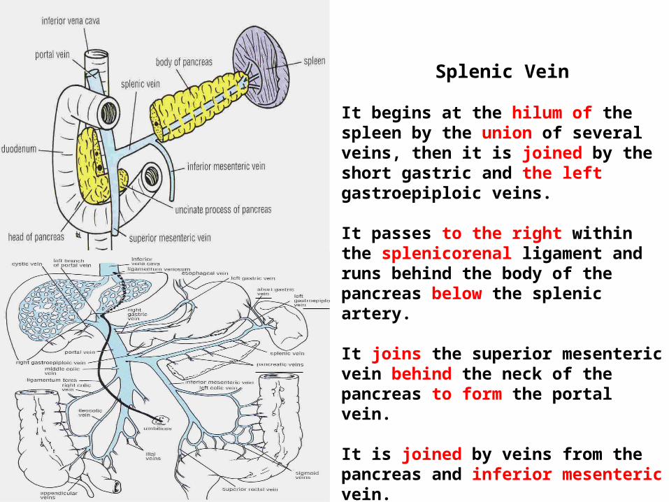
Splenic Vein
It begins at the hilum of the spleen by the union of several veins, then it is joined by the short gastric and the left gastroepiploic veins.
It passes to the right within the splenicorenal ligament and runs behind the body of the pancreas below the splenic artery.
It joins the superior mesenteric vein behind the neck of the pancreas to form the portal vein.
It is joined by veins from the pancreas and inferior mesenteric vein.

Inferior Mesenteric Vein
It begins halfway down the anal canal as the superior rectal vein.
It passes up the posterior abdominal wall on the left side of the inferior mesenteric artery and the duodenojejunal flexure.
It joins the splenic vein behind the pancreas.
It receives tributaries that correspond to the branches of the artery.
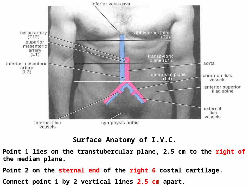
Surface Anatomy of I.V.C.
Point 1 lies on the transtubercular plane, 2.5 cm to the right of the median plane.
Point 2 on the sternal end of the right 6 costal cartilage.
Connect point 1 by 2 vertical lines 2.5 cm apart.
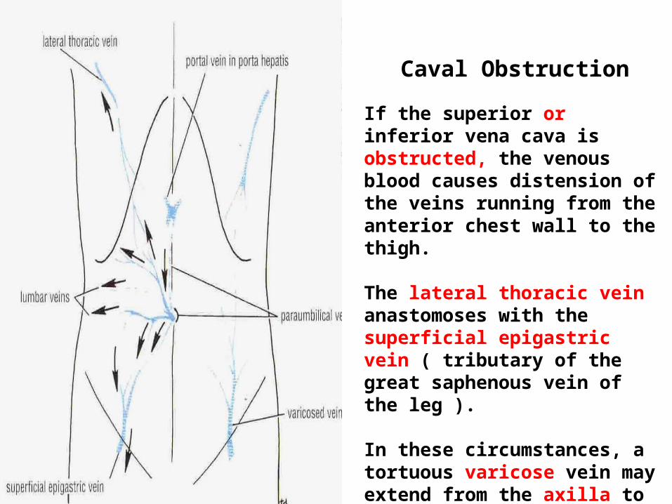
Caval Obstruction
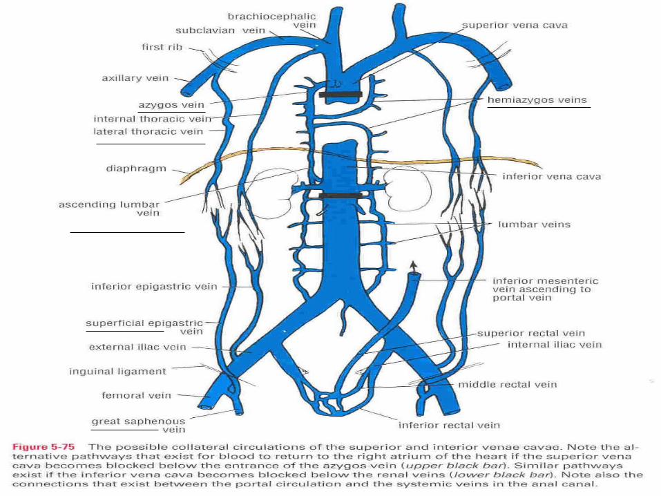
If the superior or inferior vena cava is obstructed, the venous blood causes distension of the veins running from the anterior chest wall to the thigh.
The lateral thoracic vein anastomoses with the superficial epigastric vein ( tributary of the great saphenous vein of the leg ).
In these circumstances, a tortuous varicose vein may extend from the axilla to the lower abdomen.