Embed Size (px)
Citation preview

AOFAS Resident Review Course September 28, 2013
Constantine A. Demetracopoulos, MD
Hospital for Special Surgery
AOFAS Resident Review
Disclosure Nothing to disclose

AOFAS Resident Review
Anatomy
Etiology
Evaluation
Non-operative treatment
Operative treatment
Overview
AOFAS Resident Review
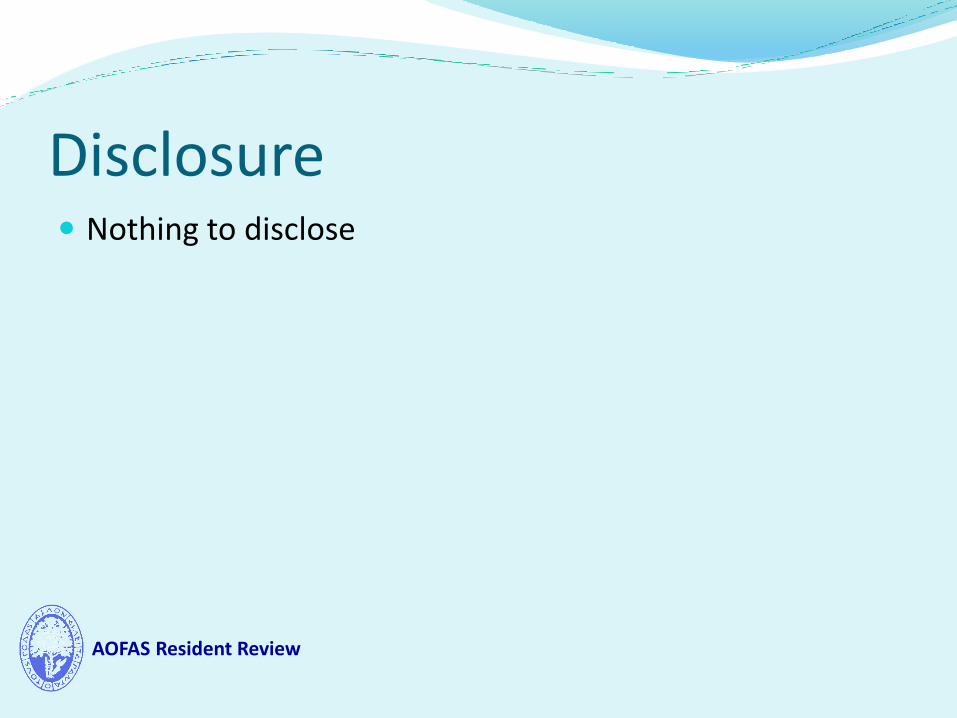
Anatomy Muscle Insertion Innervation Function
TA Med cun & base of 1st MT L4-S1; L4; DPN Dorsiflex & Invert
EDL Extensor aponeurosis of
lesser toes
L4-S1; L5; DPN Ext toes & Dorsiflex
EHL Base of distal phalanx
hallux
L4-S1; L5; DP Ext toe & Dorsiflex
PL Base of 1st MT & med cun L4-S1; S1; SPN PF 1st ray & Evert
PB Base of 5th MT L4-S1; S1; SPN Evert
GCS Calc L5-S2; S1; Tib PF & Invert
FDL/FHL Distal phalanx toes L5-S2; S1; Tib Flex toe, PF & invert
TP Navicular, midfoot, forefoot L5-S1; Tib Invert & PF

AOFAS Resident Review
Etiology of Paralytic Foot CNS – ex. CVA, head trauma
Spasticity, hyperreflesia
Spine Radiculopathy Spondylolysis Spinal Stenosis
Peripheral nerve injury Traumatic – Penetrating or blunt, knee dislocation, compartment
syndrome Iatrogenic – THA or TKA
Injury to Sciatic nerve during THA more likely to affect CPN Valgus and flexion contracture increase risk of injury during
TKA. Neoplastic / Mass effect

AOFAS Resident Review
Assessment of Foot Drop Steppage gait, “Slap foot gait”
Excessive hip and knee flexion during swing phase of gait to allow the foot and toes to clear the ground
Swing phase Supination deformity -> CPN injury affecting Extensors & Peroneals
Stance phase Walk on the lateral border of the foot
Assess range of motion. Flexible vs. Fixed Muscle strength testing
Beware of secondary recruitment Walk on their heels
Sensory exam L4 radiculopathy versus Common Peroneal Neuropathy
Reflexes Upper MN versus Lower MN
AOFAS Resident Review
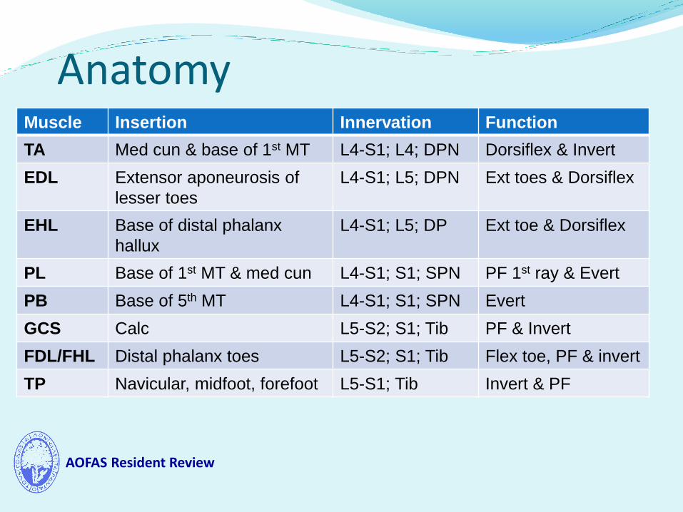
Assessment of Foot Drop Weight bearing radiographs
MRI Lumbar radiculopathy
Knee dislocation
TA rupture (dx evident by exam)
Neoplasm / mass effect
EMG/NCV EMG – Sharp waves & fibrillations at 3-5 wks, rest activity
NCV – Motor and sensory latency Prolonged in compression neuropathy, absent in nerve laceration
distal to injury.
Baseline and f/u to assess recovery

AOFAS Resident Review
Treatment PT for heel cord stretching AFO
Plantar flexion stop hinge Dorsiflexion-assist Flaccid paralysis -> fixed AFO
Nerve Decompression Lumbar decompression
Nerve repair / grafting Knee dislocation
< 6cm 70% 6 – 12cm 43% 13 – 24cm 25%
AOFAS Resident Review
Treatment Timing
Acute nerve laceration -> Acute repair
CVA -> 12 to 18 months of rehab to determine motor recovery 25% regain normal ambulation, 75% some level of ambulation
Closed head injury -> 12 to 18 months of rehab to determine motor recovery
Knee dislocation/CPN crush/stretch injury Evidence to suggest that early tendon transfer time of nerve graft
may improve outcomes
Ferraresi et al. Neurosurg Rev 2003
AOFAS Resident Review
Treatment Tendon Transfer
Should not be performed if nerve function may recover
Flexible deformities
Muscle will lose one grade of strength after transfer
In-phase / Out-of-phase (swing or stance phase)
In-phase transfer functions in a dynamic manner
Out-of-phase transfer is a static restraint to deformity
?phase conversion
Goal – Walk without a brace
AOFAS Resident Review
Treatment Posterior tibial tendon transfer to the dorsum of the foot (out-
of-phase)
Interosseous membrane
PTT in direct line from its muscle through IOM to lateral cuneiform
Anchor point is lateral cuneiform - slightly lateral of midline to promote DF and Eversion
PTT may be constricted and stenosed within window in IOM
Anteromedial tibia
PTT is not in direct line from its origin to anchor point
Anchor point is middle cuneiform, smaller bone, greater risk of fracture
Does not stenose at the IOM and glides smoothly around tibia

AOFAS Resident Review
Treatment PTT transfer
Tension with ankle at 10 degrees of DF
May require Achilles lengthening
FDL transfer to the navicular to oppose P. brevis
Botulinum toxin injections into the gastrocnemius-soleus complex to protect the tendon transfer post-op
Early active immobilization has no added risk for tendon pullout and has similar functional outcomes compared with immobilization
Rath et al. CORR 2010 – RCT Level I study
AOFAS Resident Review
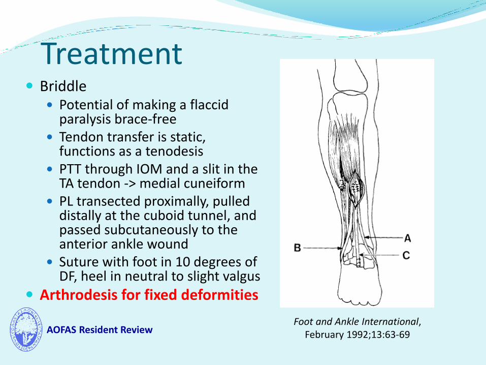
Treatment Briddle
Potential of making a flaccid paralysis brace-free
Tendon transfer is static, functions as a tenodesis
PTT through IOM and a slit in the TA tendon -> medial cuneiform
PL transected proximally, pulled distally at the cuboid tunnel, and passed subcutaneously to the anterior ankle wound
Suture with foot in 10 degrees of DF, heel in neutral to slight valgus
Arthrodesis for fixed deformities
Foot and Ankle International, February 1992;13:63-69

AOFAS Resident Review
Take home points Identify the cause
Assess deformity
Thorough assessment of what is missing and what is left
Timing of intervention
Tendon transfer only when there is no recovery
Tendon transfer in a flexible deformity, arthrodesis in a fixed deformity
Low threshold for Achilles lengthening
Tension transfer in 10 degrees of DF

AOFAS Resident Review
Image Source Rodriguez, R. The bridle procedure in the treatment of
paralysis of the foot. Foot Ankle Int. February 1992;13:63-69.

AOFAS Resident Review
Thank You