Embed Size (px)
Citation preview
Antiviral Activity and Toxicity of Fialuridine in the WoodchuckModel of Hepatitis B Virus Infection
BUD C. TENNANT,1 BETTY H. BALDWIN,1 LOU ANN GRAHAM,1 MARY A. ASCENZI,1 WILLIAM E. HORNBUCKLE,1
PETER H. ROWLAND,1 ILIA A. TOCHKOV,1 AMY E. YEAGER,1 HOLLIS N. ERB,1 JOSEPH M. COLACINO,2 CARLOS LOPEZ,2
JEFFERY A. ENGELHARDT,2 RONALD R. BOWSHER,2 FRANK C. RICHARDSON,2 WILLIAM LEWIS,3 PAUL J. COTE,4
BRENT E. KORBA,4 AND JOHN L. GERIN4
Woodchucks were used to study the antiviral activity andtoxicity of fialuridine (FIAU; 1,-28deoxy-28fluoro-1-b-D-arabinofuranosyl-5-iodo-uracil). In an initial experiment,groups of six chronic woodchuck hepatitis virus (WHV)carrier woodchucks received daily doses of FIAU by intra-peritoneal injection for 4 weeks. At 0.3 mg/kg/d, theantiviral effect was equivocal, but at 1.5 mg/kg/d, FIAU hadsignificant antiviral activity. No evidence of drug toxicitywas observed during the 4-week period of treatment orduring posttreatment follow-up. In a second experiment,groups of nine WHV carriers or uninfected woodchuckswere given 1.5 mg/kg/d of FIAU orally for 12 weeks, and theresults compared with placebo-treated controls. After 4weeks, the serum WHV-DNA concentration in the FIAU-treated carrier group was two to three logs lower than thatin the placebo-treated group. After 12 weeks of FIAUtreatment, serum WHV DNA was not detectable by conven-tional dot-blot analysis, hepatic WHV-DNA replicative inter-mediates (RI) had decreased 100-fold, and hepatic expres-sion of WHV core antigen was remarkably decreased. Noevidence of toxicity was observed after 4 weeks, but, after 6to 7 weeks, food intake decreased and, after 8 weeks, themean body weights of woodchucks treated with FIAU weresignificantly lower than controls. Anorexia, weight loss,muscle wasting, and lethargy became progressively severe,
and all FIAU-treated woodchucks died or were euthanized78 to 111 days after treatment began. Hepatic insufficiency(hyperbilirubinemia, decreased serum fibrinogen, elevatedprothrombin time), lactic acidosis, and hepatic steatosiswere characteristic findings in the final stages of FIAUtoxicity in woodchucks. The syndrome of delayed toxicityin woodchucks was similar to that observed previously inhumans treated with FIAU, suggesting that the woodchuckshould be valuable in future investigations of the molecularmechanisms of FIAU toxicity in vivo and for preclinicaltoxicological evaluation of other nucleoside analogs beforeuse in patients. (HEPATOLOGY 1998;28:179-191.)
Fialuridine (FIAU; 1,-28deoxy-28fluoro-1-b-D-arabinofura-nosyl-5-iodo-uracil) is a pyrimidine nucleoside analog withantiviral activity against herpes viruses1,2 and hepatitis B virus(HBV).3,4 During the summer of 1993, patients with chronicHBV infection that had been treated with FIAU for 9.5 to 13weeks developed signs of severe and, in some cases, fatalhepatotoxicity associated with lactic acidosis.5,6 The observedtoxicity had not been anticipated on the basis of earlier 2- and4-week clinical trials of FIAU in human patients or on thebasis of preclinical toxicological studies using conventionallaboratory animals.5
The toxicological studies that preceded the FIAU clinicaltrials7,8 and the studies that followed9 used healthy animalswith normal hepatic morphology and function. FIAU toxicity,however, had been observed in patients with chronic HBVinfection, and it became necessary to know if HBV-inducedliver disease had been an essential factor in the pathogenesisof the observed human drug toxicity.10 To investigate thisquestion, we used woodchucks chronically infected with thewoodchuck hepatitis virus (WHV) as a model of HBVinfection. WHV belongs to the genus Orthohepadnavirus(family: Hepadnaviridae), of which HBV is the prototypemember.11 Woodchucks with naturally acquired12,13 or experi-mental14,15 chronic WHV infection develop progressivelysevere chronic hepatitis and hepatocellular carcinoma (HCC),similar to the diseases observed in humans with chronic HBVinfection.11-15
The experiments reported here were designed to assess theantiviral activity of FIAU against WHV, and to determine thepossible influence of hepadnavirus infection on toxicity bycomparing the effects of FIAU in normal woodchucks and inchronic WHV carriers.
Abbreviations: FIAU, fialuridine (1,-28deoxy-28fluoro-1-b-D-arabinofuranosyl-5-iodo-uracil); HBV, hepatitis B virus; WHV, woodchuck hepatitis virus; HCC, hepatocellularcarcinoma; AST, aspartate transaminase; ALT, alanine transaminase; AP, alkalinephosphatase; GGT, g-glutamyl transpeptidase; BUN, blood urea nitrogen; SDH, sorbitoldehydrogenase; WHsAg, woodchuck hepatitis virus surface antigen; anti-WHc, anti-body to woodchuck hepatitis virus core antigen; anti-WHs, antibody to woodchuckhepatitis virus surface antigen; WHcAg, woodchuck hepatitis virus core antigen; RI,replicative intermediates; mtDNA, mitochondrial DNA.
From the 1College of Veterinary Medicine, Cornell University, Ithaca, NY; 2LillyResearch Laboratories, Eli Lilly and Company, Indianapolis, IN; 3Department ofPathology and Laboratory Medicine, College of Medicine, University of Cincinnati,Cincinnati, OH; 4Division of Molecular Virology and Immunology, GeorgetownUniversity School of Medicine, Rockville, MD.
Received June 23, 1997; accepted February 17, 1998.Supported by NO1-AI-35164 to Cornell University (B.C.T.) and NO1-AI-45179 to
Georgetown University (J.L.G.) from the National Institute of Allergy and InfectiousDiseases.
Dr. Rowland’s current address is: Histopath Consulting, Albany, NY.Dr. Richardson’s current address is: NeXstar Pharmaceuticals, Inc., Boulder, CO.Address reprint requests to: Bud C. Tennant, D.V.M., Gastrointestinal Unit, Depart-
ment of Clinical Sciences, College of Veterinary Medicine, Cornell University, Ithaca,NY 14853. Fax: (607) 253-3289.
Copyright r 1998 by the American Association for the Study of Liver Diseases.0270-9139/98/2801-0024$3.00/0
179
MATERIALS AND METHODS
Experimental Animals. The woodchucks used in these studies werethe offspring of WHV-negative females from a breeding colonymaintained at Cornell University. WHV-infected woodchucks wereinoculated subcutaneously at 3 days of age with 100 µL of inoculumcontaining 5 million woodchuck infectious doses of WHV. Infectiv-ity of the standardized infectious serum pool (WHV7P1) had beenestablished previously by titration in adult woodchucks. Approxi-mately 70% of woodchucks inoculated as neonates in this mannerwith the WHV7P1 pool became chronic WHV carriers. Pups fromWHV-infected litters and uninfected control litters were weaned at 6to 8 weeks of age. The diet consisted of laboratory animal chowformulated for rabbits (Agway Big Red Rabbit Food, Syracuse, NY),but specially pelleted in blocks for woodchucks. Diet and water wereprovided ad libitum.
Experimental Procedures. FIAU was provided as a dry powder (LillyResearch Laboratories, Indianapolis, IN). Two separate experimentsthat involved the administration of FIAU to woodchucks wereconducted. The objective of Experiment 1 was to determine therelationship of dose to antiviral activity in a 4-week treatmentprotocol. Woodchucks in Experiment 1 were 10 months of age whenthe study was begun, and approximately equal numbers of malesand females were used in each of three experimental groups. Onegroup of six chronic WHV carriers received FIAU at a dosage of 0.3mg/kg/d by intraperitoneal injection, a second group of six WHVcarriers received FIAU at a dosage of 1.5 mg/kg/d intraperitoneally,and a group of six chronic WHV carriers received the isotonic salinevehicle intraperitoneally as placebo. All groups were treated for 4weeks. FIAU treatment in Experiment 1 was initiated December 9,1992, and a 12-week posttreatment follow-up period was completedMarch 30, 1993. After learning during the final week of June, 1993,of the toxicity caused by FIAU treatment in HBV patients,5,6 thewoodchucks of Experiment 1 were re-examined on July 2, 1993, andsubsequently were monitored for evidence of drug-related toxicityfor 38 additional weeks, when the study was terminated.
Experiment 2 was designed specifically to assess the possibleinfluence of chronic hepadnavirus infection on FIAU toxicity.Thirty-six 24- to 26-month-old adult woodchucks of both sexeswere used. One group of 9 woodchucks, seronegative for markers ofWHV infection, received FIAU at a dosage of 1.5 mg/kg/d orally for12 weeks. A second group of 9 WHV-negative woodchucks receivedwater vehicle daily for 12 weeks as placebo. A third group of 9chronic WHV carrier woodchucks received FIAU at a dosage of 1.5mg/kg/d orally for 12 weeks, and a fourth group of 9 chronic WHVcarrier woodchucks received water vehicle as placebo for 12 weeks.A dose syringe was used to administer aqueous FIAU solution orplacebo into the cheek, and this was followed by administration of 4to 5 mL of semipurified liquid diet (Liquid Woodchuck ControlDiet, Dyets, Inc., Bethlehem, PA) to ensure consumption of drug orplacebo. A posttreatment follow-up period of 12 weeks was origi-nally planned. Because of FIAU toxicity, however, the longestposttreatment survival was 27 days. Experiment 2 was initiatedFebruary 8, 1994.
Woodchucks in Experiments 1 and 2 were observed daily at thetime they received food and water, at the time drug or placebo wasadministered, and each time they were anesthetized to obtain bloodsamples or liver biopsies. Abnormalities in appearance or behavior,including alterations in food and water intake, were recorded. Whenexperimental woodchucks became cachectic or moribund, theywere euthanized (ketamine/xylazine anesthesia, followed by pento-barbital overdose). In Experiment 2, each time an FIAU-treatedwoodchuck died or was euthanized, a placebo-treated woodchuckmatched by sex and WHV status also was euthanized. Experimentswere conducted in accordance with the Guide for the Care and Use forLaboratory Animals (National Academy Press, revised 1996) andwere reviewed and approved by the Cornell University InstitutionalAnimal Care and Use Committee.
Hematology and Clinical Biochemistry. Blood samples were obtainedwith woodchucks under ketamine (50 mg/kg) and xylazine (5mg/kg) anesthesia before initiation of treatment, during the periodof FIAU treatment, and during posttreatment follow-up. Bloodsamples were obtained routinely from the femoral vein or artery.Venous blood samples used to measure blood pH and PCO2
(bicarbonate) were obtained from the external jugular vein. Com-plete blood counts were performed using an electronic cell-countingsystem (Model S1IV, Coulter Corporation, Hialeah, FL), except fordifferential leukocyte counts that were performed manually. Anautomatic chemical analyzer was used for the measurement ofaspartate transaminase (AST), alanine transaminase (ALT), alkalinephosphatase (AP), g-glutamyl transpeptidase (GGT), bilirubin,sodium, potassium, chloride, calcium, inorganic phosphorus, glu-cose, blood urea nitrogen (BUN), creatinine, creatine kinase, lipase,and amylase (DACOS, Coulter Corporation). Automated proce-dures also were used for measurement of prothrombin time andfibrinogen (BioQuest FibroSystem, Becton-Dickinson and Co., Cock-eysville, MD). Sorbitol dehydrogenase (SDH) was determined usingfructose as substrate, and the conversion of NADH to NADquantitated spectrophotometrically (Sigma Diagnostics, St. Louis,MO). Blood lactate was determined enzymatically by the conversionof lactate to pyruvate by lactate dehydrogenase and by measuring theconversion of NAD to NADH spectrophotometrically (Sigma Diag-nostics).
In Experiment 2, urine specimens were collected after 8, 10, and12 weeks of treatment from anesthetized woodchucks, and analysesof urine were performed (Multistix, Bayer Corp., DiagnosticsDivision, Elkhart, IN).
Serological Tests for WHV Markers and Nucleic Acid Analyses of Liver.Tests for WHV surface antigen (WHsAg), antibody to WHV coreantigen (anti-WHc), and antibody to WHV surface antigen (anti-WHs) were performed using WHV-specific enzyme-linked immuno-sorbent assays.16 Test serum was diluted 1:100 so that WHsAg,anti-WHc, and anti-WHs were determined under saturating condi-tions. Sample/negative ratios greater than 3.1 were considered positive.
The influence of FIAU on WHV replication was determined bycomparing the levels of serum WHV DNA and the hepatic WHVnucleic acids of FIAU-treated chronic WHV carriers with those ofplacebo-treated controls before, during, and following treatment.The WHV-DNA levels of serum were determined by dot-blothybridization analysis. Serum samples were centrifuged at 14,000gfor 2 minutes. Aliquots (10 µL or 100 µL) were denatured with anequal volume of 2.0 mol/L NaOH/203 SSC for 30 minutes at roomtemperature and immediately applied to nitrocellulose (presoakedin sterile 203 SSC) under vacuum. After entry into the membrane,samples were washed with 0.4 mL sterile 1.0 mol/L Tris-HCl (pH7.4)/2 mol/L NaOH, and then with 0.4 mL 203 SSC. The membranewas then rinsed with sterile 23 SSC (2 minutes), air-dried (5minutes), and baked for 30 minutes at 80°C under vacuum. Themembranes were then hybridized with a full-length (3.2-kb),gel-purified WHV-DNA probe as previously described.3,15 Thesensitivity cut-off of this assay is approximately 1.0 pg WHV DNA(3.3 3 105 WHV genome equivalents) per milliliter of serum. WHVnucleic acids (replicative intermediates [RI], monomeric WHVDNA, WHV RNA) were measured by Southern and Northern blotanalysis15 using liver specimens obtained at biopsy or at postmor-tem, immediately following euthanasia.
Histopathological and Ultrastructural Studies of Liver. Liver biopsyspecimens were obtained while animals were under general anesthe-sia (ketamine/xylazine) using 14-gauge disposable biopsy needles(Biopty-Cut, C.R. Bard, Inc., Covington, GA) directed by ultrasoundimaging. The needle was inserted at a site near the ventral midlinejust caudal to the xiphoid cartilage and directed dorsolaterally andsomewhat cranially into the margin of the left lateral lobe of theliver. One specimen was placed immediately in liquid nitrogen andstored at 270°C before nucleic acid analysis. A second specimen wasfixed in phosphate-buffered formalin, embedded in paraffin, sec-tioned, and stained with hematoxylin-eosin for conventional light
180 TENNANT ET AL. HEPATOLOGY July 1998
microscopic examination. Sections of paraffin-embedded tissue alsowere stained immunohistochemically for WHsAg and WHV coreantigen (WHcAg) using a peroxidase method and polyclonal rabbitantibodies raised against the respective WHV antigens. A thirdspecimen obtained from woodchucks of Experiment 2 was choppedinto 1-mm cubes, fixed in diluted Karnovsky’s solution, embeddedin plastic, thin-sectioned, and examined by electron microscopy. Afourth specimen was placed in OCT medium (Tissue-Tek, SakuraFinetek USA, Inc., Torrance, CA) and snap-frozen in a pentane/solidCO2 slurry. Frozen sections (some of which were fixed in formalinbut not otherwise processed) were stained for fat with Oil Red O.
Hematoxylin-eosin–stained liver biopsy sections were examinedunder code without reference to treatment. The severity of portalhepatitis, parenchymal hepatitis, bile duct proliferation, and fattychange was scored using a scale of 0 to 4. Steatosis also was classifiedqualitatively as microvesicular, macrovesicular, or mixed. Oil RedO–stained sections were used to confirm the presence and severity ofsteatosis. Histological specimens stained for WHsAg or WHcAgwere scored (scale of 0 to 4) for intensity of staining and thepercentage of hepatocytes stained, and the primary intracellularlocalization of the antigen was recorded.
Postmortem examinations were performed on all woodchucksthat died or were euthanized. In Experiment 2, the postmortemprocedures for placebo-treated woodchucks were identical to thoseused for woodchucks treated with FIAU. Immediately followingeuthanasia, five separate specimens of liver were obtained andprocessed and examined in a manner similar to that used for liverbiopsies.
Analyses of FIAU in Serum and Liver. In Experiment 2, serumsamples were collected for FIAU analysis during the treatment phase14.5 to 18 hours following FIAU administration and in survivingwoodchucks during posttreatment follow-up. The serum concentra-tion of FIAU was determined using a competitive binding radioim-munoassay based on a rabbit antibody raised against FIAU (58-0-hemisuccinate conjugated to keyhole limpet hemocyanin).17 Thesensitivity (0.2 ng/mL), specificity (negligible interference fromknown FIAU metabolites and endogenous nucleosides), and interas-say variability (5.0% to 19.7%) of this procedure have beendescribed.17
For determination of hepatic FIAU incorporated into cellularDNA, specimens of liver were obtained immediately followingeuthanasia and processed by a previously described procedure.9
Briefly, DNA was isolated from whole liver homogenate using theASAP Genomic DNA Isolation Kit (Boehringer Mannheim, Corp.,Indianapolis, IN). DNA extracted from liver homogenate wasdenatured by heating to 95°C for 5 minutes, and the solution placedon ice. DNA was hydrolyzed with snake venom phosphodiesteraseand bacterial AP, FIAU measured by radioimmunoassay,17 and theresults expressed as picomoles of FIAU per micromoles of thymidinedetermined by high-pressure liquid chromatography.
Statistical Methods. The Student’s t test was used for comparisonsof continuous data (solute concentrations, enzyme activity, FIAUcontent of hepatic DNA) between WHV-infected and uninfectedwoodchucks, and between FIAU-treated and placebo-treated groups.Each test included only samples taken at the same time point. Meanvalues were expressed 6 SD. For comparisons of ordinal databetween groups (histopathological scores), Wilcoxon’s rank sumtest was used. All these tests used a 5 0.05 and were one-tailed.Preliminary tests between WHV-negative and WHV-positive groupstreated either with FIAU or placebo were performed using two-tailedtests, and when P $ .50, results of WHV-positive and negativegroups were combined for further analysis.
RESULTS
Antiviral Activity of FIAU. In Experiment 1, intraperitonealtreatment with FIAU at 1.5 mg/kg/d was associated with asignificant 10-fold decrease in serum WHV DNA after 1 weekof treatment (P 5 .01). After 4 weeks of treatment, the
average decrease was 100-fold versus placebo-treated con-trols (P 5 .001). Serum WHV DNA gradually increased afterdrug withdrawal, but at all time points examined throughoutthe 12-week posttreatment period, the values were signifi-cantly lower than those of the controls (P , .001). Inwoodchucks treated with FIAU at 0.3 mg/kg/d, mean serumWHV-DNA levels were not significantly different from thosein the placebo-treated controls, although some treated wood-chucks temporarily had lower WHV-DNA levels during drugtreatment.
Hepatic WHV-DNA RI were measured in Experiment 1before treatment, at the end of the 4-week treatment period,and at 4 and 12 weeks following drug withdrawal. Inwoodchucks treated with FIAU at 1.5 mg/kg, mean hepatic RIat the end of the 4-week treatment period were threefold lessthan those in placebo-treated controls, and the difference washighly significant (P 5 .002). Levels of hepatic WHV RIincreased in all individuals within 4 weeks after completionof treatment at the 1.5-mg/kg/d FIAU dosage, but remainedsignificantly below control values (P 5 .02). Twelve weeksafter treatment ended, the mean levels of hepatic WHV RIwere less than those in the control group, but the differencewas not significant (P 5 .06). In woodchucks treated withFIAU at 0.3 mg/kg/d, levels of hepatic WHV RI were notaltered. No significant differences were observed betweencontrol woodchucks and woodchucks treated with FIAU ateither 0.3 or 1.5 mg/kg/d in hepatic episomal (3.2-kb)WHV-DNA genomes or hepatic WHV RNA. There was noserological evidence of resolution of WHV infection, and allFIAU- and placebo-treated woodchucks remained test-positive for WHsAg and anti-WHc, and test-negative foranti-WHs at the end of the study.
Histological examination of hepatic biopsies from theFIAU- and placebo-treated woodchucks before beginning thestudy demonstrated mild to moderate degrees of portal andparenchymal hepatitis. Biopsies of liver obtained at the end ofFIAU treatment and at 4 and 12 weeks after drug treatmentended were examined, and neither the severity of hepaticinflammation nor the expression of either WHsAg or WHcAgwere influenced by treatment.
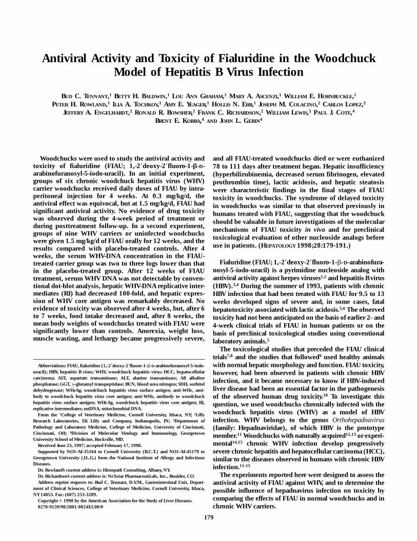
In Experiment 2, treatment with FIAU at an oral dose of1.5 mg/kg/d had a prompt and marked effect on viralreplication. Within 2 weeks, the mean serum WHV-DNAlevel in FIAU-treated woodchucks was 10-fold less than thatin the placebo-treated woodchucks (P , .001) (Fig. 1).Within 4 weeks, the mean WHV-DNA level in the FIAU-treated group was two to three logs less than that in theplacebo group (P , .001) and, after 10 weeks, was below therange detectable by quantitative blot hybridization analysis.The serum WHV-DNA levels of carriers receiving placebowere unchanged during the study (Fig. 1).
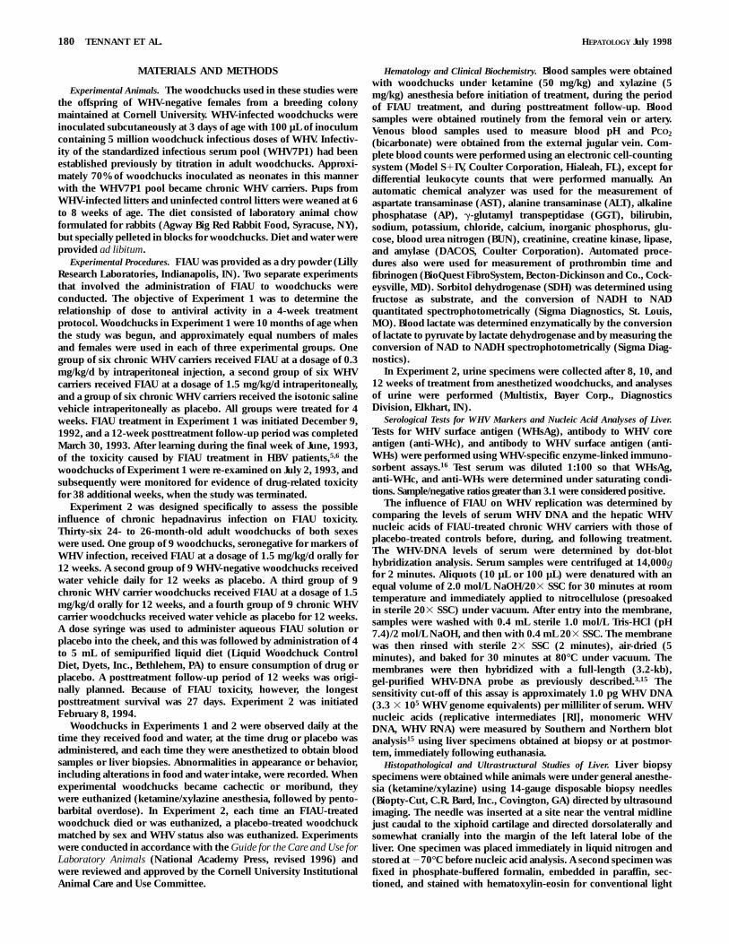
In Experiment 2, two- to threefold reductions of hepaticWHV-DNA RI occurred after 4 weeks of FIAU treatment and,after 12 weeks, WHV-DNA RI had decreased 10-fold (bothP , .001 vs. placebo) (Fig. 2). Reductions of episomalWHV-DNA genomes and WHV RNA were not observed after4 weeks of treatment, but had declined an average of twofoldafter 12 weeks of treatment (data not shown).
At the end of the 12-week treatment, percutaneous liverbiopsies were obtained from six surviving chronic WHVcarriers that had received FIAU and from six that receivedplacebo. The median scores for WHcAg expression beforeinitiation of therapy were 3/4 in the placebo group and 3.5/4
HEPATOLOGY Vol. 28, No. 1, 1998 TENNANT ET AL. 181
in the FIAU-treated group. After 4 weeks of treatment, themedian scores for WHcAg expression were 3/4 in both theplacebo- and FIAU-treated groups. At 12 weeks, the medianWHcAg expression score was 3/4 in the placebo group and1/4 in the FIAU-treated group (P , .001). The median scoresfor expression of WHsAg in hepatocyte plasma membraneswere 2/4 and 1/4 in the placebo-treated and FIAU-treatedgroups, respectively, after 12 weeks of treatment (P 5 .12).The median scores for cytoplasmic expression of WHsAg insingle hepatocytes were 1/4 in both the placebo-treated andFIAU-treated groups.
Toxicity of FIAU. In Experiment 1, there were no physicalsigns of FIAU toxicity in woodchucks treated at either 0.3mg/kg/d or 1.5 mg/kg/d for 4 weeks. No significant differ-ences between the mean body weights of FIAU-treatedwoodchucks and placebo-treated controls were observedduring treatment or the follow-up period. There were noFIAU-related hematological or clinical biochemical changesduring treatment or during posttreatment follow-up. Inbiopsies of liver obtained at the end of treatment and at 4 and12 weeks after treatment ended, there were no histologicaldifferences between FIAU- and placebo-treated woodchucksin severity of hepatitis, steatosis, or in the expression of WHcand WHsAg.
At the end of the original 12-week posttreatment period(March 30, 1993), the mean body weight of woodchucks ofthe FIAU high-dose group (2.5 6 0.3 kg) was lower than thecorresponding mean body weight of placebo-treated controls(2.9 6 0.4 kg; P 5 .11). By the time follow-up studies inExperiment 1 were reinitiated on July 2, 1993, all three
groups of woodchucks had gained weight. The mean bodyweight of the FIAU high-dose group (3.4 6 0.3 kg) at thistime was significantly lower, however, than that of theplacebo-treated controls (3.8 6 0.3 kg; P 5 .03), and for theremainder of the posttreatment study (9 months), mean bodyweights of the FIAU high-dose group were lower than thoseof the controls (data not shown).
From initiation of FIAU treatment on December 9, 1992,until termination of follow-up studies on March 1, 1994, noFIAU treatment–related alterations were observed in serumtests for hepatic injury or hepatic function (GGT, SDH, ALT,AST, AP, bilirubin) or renal function (BUN and creatinine).Serum WHV DNA of the FIAU high-dose group had returnedto pretreatment levels by the time posttreatment follow-upwas reinitiated 25 weeks after drug administration ended.
Between September 30 and December 2, 1993, two placebo-treated controls and two low-dose, FIAU-treated woodchuckswere euthanized because of the development of HCC (neo-plasms considered to be a direct result of infection withWHV). Between February 16 and March 1, 1994, the finalsurviving woodchucks from the study were euthanized.Postmortem examinations were performed on five of six ofthe original FIAU high-dose group, three of six of the FIAUlow-dose group, and four of six of the placebo-treatedcontrols. In woodchucks from all three groups, varyingdegrees of chronic hepatitis were found. The severity wasconsidered to be that expected for chronic WHV carriers thatwere approximately 2 years of age. Similar degrees of hepaticneoplasia were observed in FIAU-treated and control groups.There were no gross or histopathological lesions attributableto FIAU treatment.
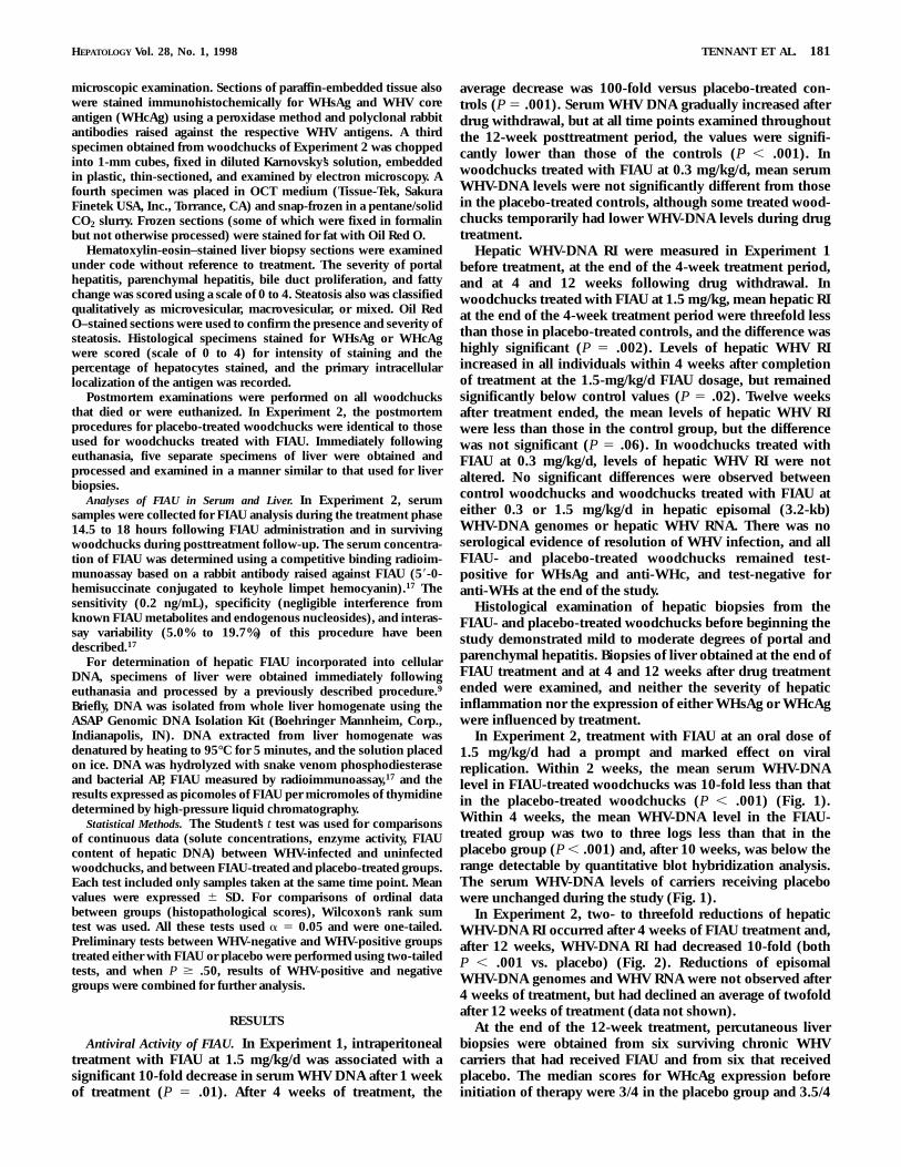
FIG. 1. Serum WHV DNA (pg/mL) of chronic WHV carrier woodchuckstreated orally for 12 weeks with FIAU (1.5 mg/kg/d; n 5 9) or with placebo(n 5 9). After 4 weeks, there was a 100- to 1,000-fold reduction of serumWHV DNA in the FIAU-treated group compared with controls (P , .001),and the difference was sustained during the remainder of the study.
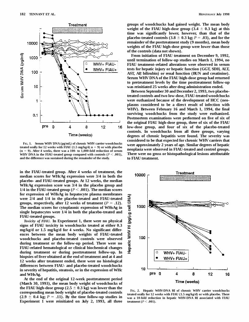
FIG. 2. Hepatic WHV-DNA RI of chronic WHV carrier woodchuckstreated orally for 12 weeks with FIAU (1.5 mg/kg/d) or with placebo. Therewas a 10-fold reduction in hepatic WHV-DNA RI associated with FIAUtreatment (P , .001).
182 TENNANT ET AL. HEPATOLOGY July 1998
In Experiment 2, there was no clinical, hematological, orbiochemical evidence of FIAU toxicity during the first 4weeks of oral drug treatment. Histological examination ofhepatic biopsies obtained after 4 weeks demonstrated nodifferences between groups of uninfected and WHV carrierwoodchucks treated with FIAU and corresponding groups ofplacebo-treated controls. These initial observations weresimilar to those of Experiment 1 in which there was noconclusive evidence of toxicity after intraperitoneal adminis-tration of FIAU for 4 weeks.
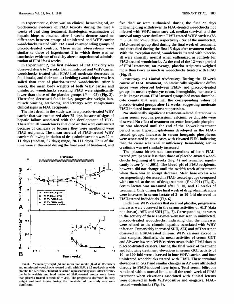
In Experiment 2, the first evidence of FIAU toxicity wasobserved after 6 to 7 weeks. Both uninfected and WHV carrierwoodchucks treated with FIAU had moderate decreases infood intake, and their contact bedding (wood chips) was lesssoiled than that of placebo-treated woodchucks. After 8weeks, the mean body weights of both WHV carrier anduninfected woodchucks receiving FIAU were significantlylower than those of the placebo groups (P 5 .05) (Fig. 3).Thereafter, decreased food intake, progressive weight loss,muscle wasting, weakness, and lethargy were conspicuousclinical signs in FIAU recipients.
The first death in the study was in a placebo-treated WHVcarrier that was euthanized after 75 days because of signs ofhepatic failure associated with the development of HCC.Thereafter, all woodchucks that died or that were euthanizedbecause of cachexia or because they were moribund wereFIAU recipients. The mean survival of FIAU-treated WHVcarriers following initiation of drug administration was 90 611 days (median, 87 days; range, 78-111 days). Four of thenine were euthanized during the final week of treatment, and
five died or were euthanized during the first 27 daysfollowing drug withdrawal. In FIAU-treated woodchucks notinfected with WHV, mean survival, median survival, and thesurvival range were similar to FIAU-treated WHV carriers (856 5, 84, and 79-99 days, respectively). Six of the uninfected,FIAU-treated group died during the final week of treatment,and three died during the first 15 days after treatment ended.With the exception noted, woodchucks treated with placeboall were clinically normal when euthanized as controls forFIAU-treated woodchucks. At the end of the 12-week periodof FIAU treatment, on average, placebo recipients weighedmore than twice as much as woodchucks treated with FIAU(Fig. 3).
Hematology and Clinical Biochemistry. During the 12-weekcourse of FIAU treatment, no statistically significant differ-ences were observed between FIAU- and placebo-treatedgroups in mean erythrocyte count, hemoglobin, hematocrit,or leukocyte count. FIAU-treated groups had mean thrombo-cyte counts that were half the corresponding values ofplacebo-treated groups after 12 weeks, suggesting moderateFIAU-induced bone marrow suppression.
No statistically significant treatment-related alterations inmean serum sodium, potassium, calcium, or chloride wereobserved. No effect of treatment on serum inorganic phospho-rus was observed until the end of the 12-week treatmentperiod when hyperphosphatemia developed in the FIAU-treated groups. Increases in serum inorganic phosphoruswere associated in most cases with elevated BUN, indicatingthat the cause was renal insufficiency. Remarkably, serumcreatinine was not similarly increased.
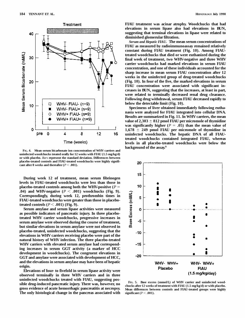
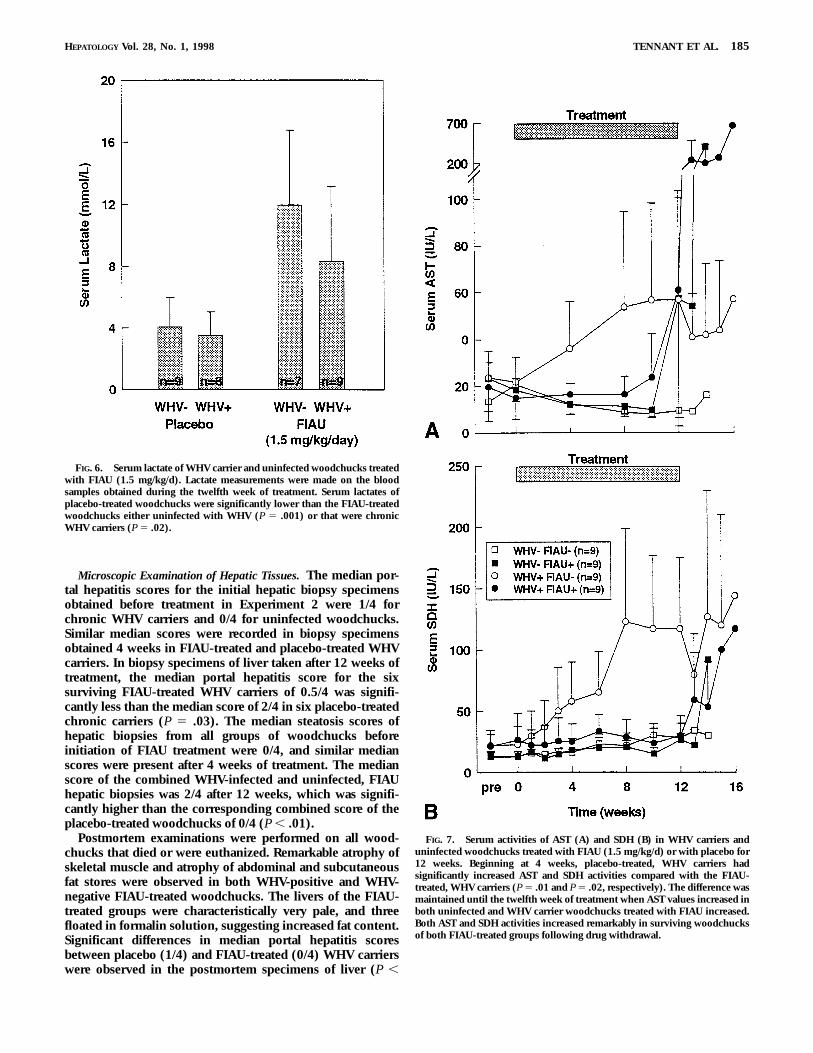
The plasma bicarbonate concentrations of both FIAU-treated groups were less than those of placebo-treated wood-chucks beginning at 8 weeks (Fig. 4) and remained signifi-cantly lower (P , .001). The blood pH of FIAU recipients,however, did not change until the twelfth week of treatmentwhen there was an abrupt decrease. Mean base excess wascorrespondingly decreased in FIAU-treated groups comparedwith controls at the end of drug treatment (P , .001) (Fig. 5).Serum lactate was measured after 8, 10, and 12 weeks oftreatment. Only during the final week of drug administrationwere increases in serum lactate of 3- to 10-fold observed inFIAU-treated individuals (Fig. 6).
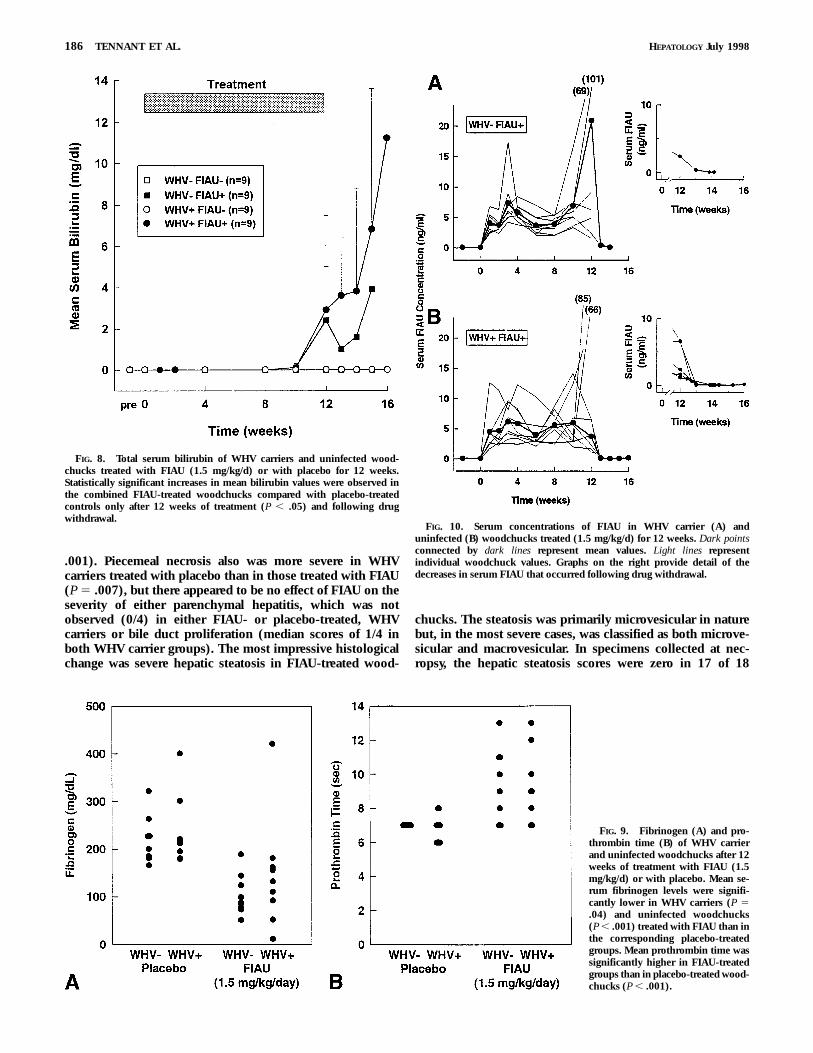
In chronic WHV carriers that received placebo, progressiveincreases were observed in the serum activities of ALT (datanot shown), AST, and SDH (Fig. 7). Corresponding increasesin the activity of these enzymes were not seen in uninfected,placebo-treated woodchucks, indicating that the increaseswere related to the chronic hepatitis associated with WHVinfection. Remarkably, increased SDH, ALT, and AST were notobserved in FIAU-treated chronic WHV carriers except infinal samples. Similarly, the mean activities of serum GGTand AP were lower in WHV carriers treated with FIAU than inplacebo-treated carriers. During the final week of treatmentand following treatment, elevations in serum GGT activity of10- to 100-fold were observed in four WHV carriers and fouruninfected woodchucks treated with FIAU. These terminalelevations in GGT and similar changes in AP were attributeddirectly to FIAU-induced liver injury. Total serum bilirubinremained within normal limits until the tenth week of FIAUtreatment when elevations associated with clinical icteruswere observed in both WHV-positive and -negative, FIAU-treated woodchucks (Fig. 8).
FIG. 3. Mean body weight (A) and mean food intake (B) of WHV carriersand uninfected woodchucks treated orally with FIAU (1.5 mg/kg/d) or withplacebo for 12 weeks. Standard deviation represented by bars. After 8 weeks,the body weights and food intake of FIAU-treated groups were lowerthan placebo-treated controls (P 5 .05). The progressive decreases in bodyweight and food intake during the remainder of the study also weresignificant.
HEPATOLOGY Vol. 28, No. 1, 1998 TENNANT ET AL. 183
A
B
During week 12 of treatment, mean serum fibrinogenlevels in FIAU-treated woodchucks were less than those inplacebo-treated controls among both the WHV-positive (P 5.04) and WHV-negative (P , .001) woodchucks (Fig. 9).Correspondingly, during week 12, prothrombin times inFIAU-treated woodchucks were greater than those in placebo-treated controls (P , .001) (Fig. 9).
Serum amylase and serum lipase activities were measuredas possible indicators of pancreatic injury. In three placebo-treated WHV carrier woodchucks, progressive increases inserum amylase were observed during the course of treatment,but similar elevations in serum amylase were not observed inplacebo-treated, uninfected woodchucks, suggesting that theelevations in WHV carriers receiving placebo were part of thenatural history of WHV infection. The three placebo-treatedWHV carriers with elevated serum amylase had correspond-ing increases in serum GGT activity (a marker of HCCdevelopment in woodchucks). The congruent elevations inGGT and amylase were associated with development of HCC,and the elevations in serum amylase may have been of hepaticorigin.
Elevations of four- to fivefold in serum lipase activity wereobserved terminally in three WHV carriers and in threeuninfected woodchucks treated with FIAU, suggesting pos-sible drug-induced pancreatic injury. There was, however, nogross evidence of acute hemorrhagic pancreatitis at necropsy.The only histological change in the pancreas associated with
FIAU treatment was acinar atrophy. Woodchucks that hadelevations in serum lipase also had elevations in BUN,suggesting that terminal elevations in lipase were related todiminished glomerular filtration.
Serum and Hepatic FIAU. The mean serum concentrations ofFIAU as measured by radioimmunoassay remained relativelyconstant during FIAU treatment (Fig. 10). Among FIAU-treated woodchucks that died or were euthanized during thefinal week of treatment, two WHV-negative and three WHVcarrier woodchucks had marked elevations in serum FIAUconcentration, and one of these individuals accounted for thesharp increase in mean serum FIAU concentration after 12weeks in the uninfected group of drug-treated woodchucks(Fig. 10). In four of the five, the marked elevations in serumFIAU concentration were associated with significant in-creases in BUN, suggesting that the increases, at least in part,were related to terminally decreased renal drug clearance.Following drug withdrawal, serum FIAU decreased rapidly tobelow the detectable limit (Fig. 10).
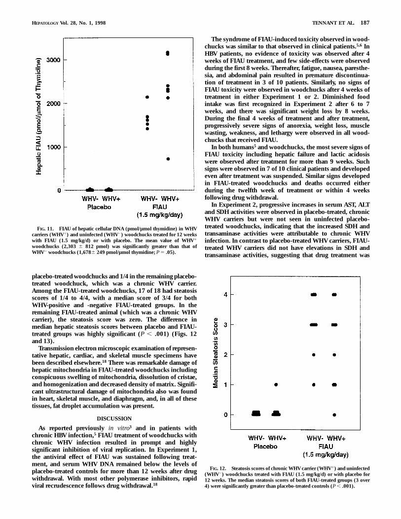
Specimens of liver obtained immediately following eutha-nasia were analyzed for FIAU integrated into cellular DNA.Results are summarized in Fig. 11. In WHV carriers, the meanvalue of 2,303 6 812 pmol FIAU per micromole of thymidinewas significantly higher (P 5 .05) than the mean value of1,678 6 249 pmol FIAU per micromole of thymidine inuninfected woodchucks. The hepatic DNA of all FIAU-treated woodchucks contained integrated FIAU, whereaslevels in all placebo-treated woodchucks were below thebackground of the assay.9
FIG. 4. Mean serum bicarbonate ion concentration of WHV carriers anduninfected woodchucks treated orally for 12 weeks with FIAU (1.5 mg/kg/d)or with placebo. Bars represent the standard deviation. Differences betweenplacebo-treated controls and FIAU-treated woodchucks were highly signifi-cant after 8 weeks and thereafter (P , .001).
FIG. 5. Base excess (mmol/L) of WHV carrier and uninfected wood-chucks after 12 weeks of treatment with FIAU (1.5 mg/kg/d) or with placebo.Mean differences between controls and FIAU-treated groups were highlysignificant (P , .001).
184 TENNANT ET AL. HEPATOLOGY July 1998
Microscopic Examination of Hepatic Tissues. The median por-tal hepatitis scores for the initial hepatic biopsy specimensobtained before treatment in Experiment 2 were 1/4 forchronic WHV carriers and 0/4 for uninfected woodchucks.Similar median scores were recorded in biopsy specimensobtained 4 weeks in FIAU-treated and placebo-treated WHVcarriers. In biopsy specimens of liver taken after 12 weeks oftreatment, the median portal hepatitis score for the sixsurviving FIAU-treated WHV carriers of 0.5/4 was signifi-cantly less than the median score of 2/4 in six placebo-treatedchronic carriers (P 5 .03). The median steatosis scores ofhepatic biopsies from all groups of woodchucks beforeinitiation of FIAU treatment were 0/4, and similar medianscores were present after 4 weeks of treatment. The medianscore of the combined WHV-infected and uninfected, FIAUhepatic biopsies was 2/4 after 12 weeks, which was signifi-cantly higher than the corresponding combined score of theplacebo-treated woodchucks of 0/4 (P , .01).
Postmortem examinations were performed on all wood-chucks that died or were euthanized. Remarkable atrophy ofskeletal muscle and atrophy of abdominal and subcutaneousfat stores were observed in both WHV-positive and WHV-negative FIAU-treated woodchucks. The livers of the FIAU-treated groups were characteristically very pale, and threefloated in formalin solution, suggesting increased fat content.Significant differences in median portal hepatitis scoresbetween placebo (1/4) and FIAU-treated (0/4) WHV carrierswere observed in the postmortem specimens of liver (P ,
FIG. 7. Serum activities of AST (A) and SDH (B) in WHV carriers anduninfected woodchucks treated with FIAU (1.5 mg/kg/d) or with placebo for12 weeks. Beginning at 4 weeks, placebo-treated, WHV carriers hadsignificantly increased AST and SDH activities compared with the FIAU-treated, WHV carriers (P 5 .01 and P 5 .02, respectively). The difference wasmaintained until the twelfth week of treatment when AST values increased inboth uninfected and WHV carrier woodchucks treated with FIAU increased.Both AST and SDH activities increased remarkably in surviving woodchucksof both FIAU-treated groups following drug withdrawal.
FIG. 6. Serum lactate of WHV carrier and uninfected woodchucks treatedwith FIAU (1.5 mg/kg/d). Lactate measurements were made on the bloodsamples obtained during the twelfth week of treatment. Serum lactates ofplacebo-treated woodchucks were significantly lower than the FIAU-treatedwoodchucks either uninfected with WHV (P 5 .001) or that were chronicWHV carriers (P 5 .02).
HEPATOLOGY Vol. 28, No. 1, 1998 TENNANT ET AL. 185
.001). Piecemeal necrosis also was more severe in WHVcarriers treated with placebo than in those treated with FIAU(P 5 .007), but there appeared to be no effect of FIAU on theseverity of either parenchymal hepatitis, which was notobserved (0/4) in either FIAU- or placebo-treated, WHVcarriers or bile duct proliferation (median scores of 1/4 inboth WHV carrier groups). The most impressive histologicalchange was severe hepatic steatosis in FIAU-treated wood-
chucks. The steatosis was primarily microvesicular in naturebut, in the most severe cases, was classified as both microve-sicular and macrovesicular. In specimens collected at nec-ropsy, the hepatic steatosis scores were zero in 17 of 18
FIG. 8. Total serum bilirubin of WHV carriers and uninfected wood-chucks treated with FIAU (1.5 mg/kg/d) or with placebo for 12 weeks.Statistically significant increases in mean bilirubin values were observed inthe combined FIAU-treated woodchucks compared with placebo-treatedcontrols only after 12 weeks of treatment (P , .05) and following drugwithdrawal.
FIG. 9. Fibrinogen (A) and pro-thrombin time (B) of WHV carrierand uninfected woodchucks after 12weeks of treatment with FIAU (1.5mg/kg/d) or with placebo. Mean se-rum fibrinogen levels were signifi-cantly lower in WHV carriers (P 5.04) and uninfected woodchucks(P , .001) treated with FIAU than inthe corresponding placebo-treatedgroups. Mean prothrombin time wassignificantly higher in FIAU-treatedgroups than in placebo-treated wood-chucks (P , .001).
FIG. 10. Serum concentrations of FIAU in WHV carrier (A) anduninfected (B) woodchucks treated (1.5 mg/kg/d) for 12 weeks. Dark pointsconnected by dark lines represent mean values. Light lines representindividual woodchuck values. Graphs on the right provide detail of thedecreases in serum FIAU that occurred following drug withdrawal.
186 TENNANT ET AL. HEPATOLOGY July 1998
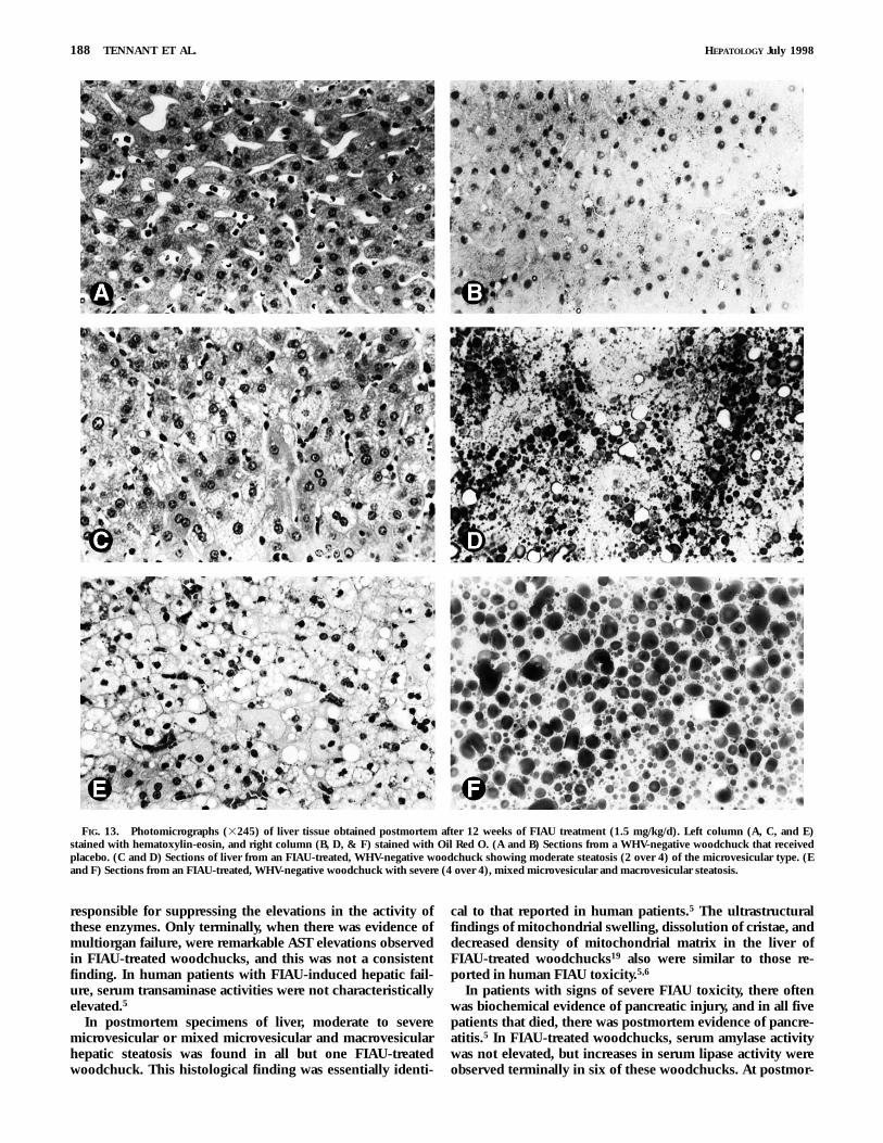
placebo-treated woodchucks and 1/4 in the remaining placebo-treated woodchuck, which was a chronic WHV carrier.Among the FIAU-treated woodchucks, 17 of 18 had steatosisscores of 1/4 to 4/4, with a median score of 3/4 for bothWHV-positive and -negative FIAU-treated groups. In theremaining FIAU-treated animal (which was a chronic WHVcarrier), the steatosis score was zero. The difference inmedian hepatic steatosis scores between placebo and FIAU-treated groups was highly significant (P , .001) (Figs. 12and 13).
Transmission electron microscopic examination of represen-tative hepatic, cardiac, and skeletal muscle specimens havebeen described elsewhere.18 There was remarkable damage ofhepatic mitochondria in FIAU-treated woodchucks includingconspicuous swelling of mitochondria, dissolution of cristae,and homogenization and decreased density of matrix. Signifi-cant ultrastructural damage of mitochondria also was foundin heart, skeletal muscle, and diaphragm, and, in all of thesetissues, fat droplet accumulation was present.
DISCUSSION
As reported previously in vitro3 and in patients withchronic HBV infection,5 FIAU treatment of woodchucks withchronic WHV infection resulted in prompt and highlysignificant inhibition of viral replication. In Experiment 1,the antiviral effect of FIAU was sustained following treat-ment, and serum WHV DNA remained below the levels ofplacebo-treated controls for more than 12 weeks after drugwithdrawal. With most other polymerase inhibitors, rapidviral recrudescence follows drug withdrawal.18
The syndrome of FIAU-induced toxicity observed in wood-chucks was similar to that observed in clinical patients.5,6 InHBV patients, no evidence of toxicity was observed after 4weeks of FIAU treatment, and few side-effects were observedduring the first 8 weeks. Thereafter, fatigue, nausea, paresthe-sia, and abdominal pain resulted in premature discontinua-tion of treatment in 3 of 10 patients. Similarly, no signs ofFIAU toxicity were observed in woodchucks after 4 weeks oftreatment in either Experiment 1 or 2. Diminished foodintake was first recognized in Experiment 2 after 6 to 7weeks, and there was significant weight loss by 8 weeks.During the final 4 weeks of treatment and after treatment,progressively severe signs of anorexia, weight loss, musclewasting, weakness, and lethargy were observed in all wood-chucks that received FIAU.
In both humans5 and woodchucks, the most severe signs ofFIAU toxicity including hepatic failure and lactic acidosiswere observed after treatment for more than 9 weeks. Suchsigns were observed in 7 of 10 clinical patients and developedeven after treatment was suspended. Similar signs developedin FIAU-treated woodchucks and deaths occurred eitherduring the twelfth week of treatment or within 4 weeksfollowing drug withdrawal.
In Experiment 2, progressive increases in serum AST, ALTand SDH activities were observed in placebo-treated, chronicWHV carriers but were not seen in uninfected placebo-treated woodchucks, indicating that the increased SDH andtransaminase activities were attributable to chronic WHVinfection. In contrast to placebo-treated WHV carriers, FIAU-treated WHV carriers did not have elevations in SDH andtransaminase activities, suggesting that drug treatment was
FIG. 11. FIAU of hepatic cellular DNA (pmol/µmol thymidine) in WHVcarriers (WHV1) and uninfected (WHV2) woodchucks treated for 12 weekswith FIAU (1.5 mg/kg/d) or with placebo. The mean value of WHV1
woodchucks (2,303 6 812 pmol) was significantly greater than that ofWHV2 woodchucks (1,6786 249 pmol/µmol thymidine; P 5 .05).
FIG. 12. Steatosis scores of chronic WHV carrier (WHV1) and uninfected(WHV2) woodchucks treated with FIAU (1.5 mg/kg/d) or with placebo for12 weeks. The median steatosis scores of both FIAU-treated groups (3 over4) were significantly greater than placebo-treated controls (P , .001).
HEPATOLOGY Vol. 28, No. 1, 1998 TENNANT ET AL. 187
responsible for suppressing the elevations in the activity ofthese enzymes. Only terminally, when there was evidence ofmultiorgan failure, were remarkable AST elevations observedin FIAU-treated woodchucks, and this was not a consistentfinding. In human patients with FIAU-induced hepatic fail-ure, serum transaminase activities were not characteristicallyelevated.5
In postmortem specimens of liver, moderate to severemicrovesicular or mixed microvesicular and macrovesicularhepatic steatosis was found in all but one FIAU-treatedwoodchuck. This histological finding was essentially identi-
cal to that reported in human patients.5 The ultrastructuralfindings of mitochondrial swelling, dissolution of cristae, anddecreased density of mitochondrial matrix in the liver ofFIAU-treated woodchucks19 also were similar to those re-ported in human FIAU toxicity.5,6
In patients with signs of severe FIAU toxicity, there oftenwas biochemical evidence of pancreatic injury, and in all fivepatients that died, there was postmortem evidence of pancre-atitis.5 In FIAU-treated woodchucks, serum amylase activitywas not elevated, but increases in serum lipase activity wereobserved terminally in six of these woodchucks. At postmor-
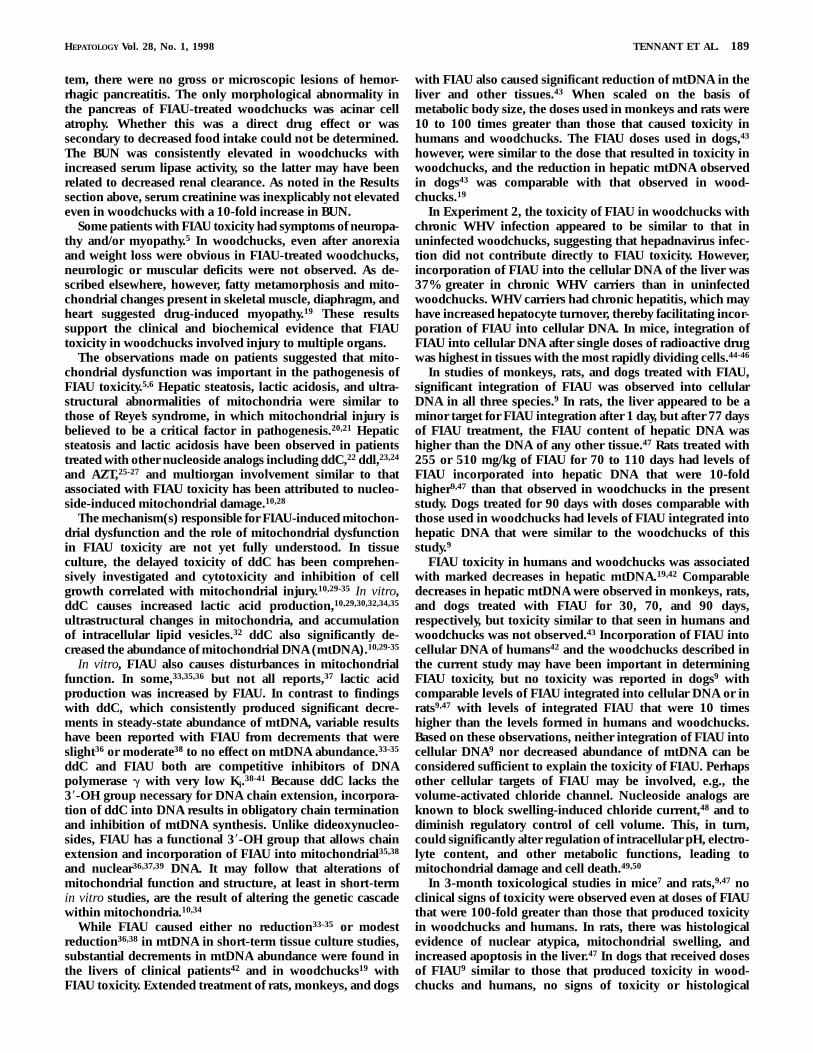
FIG. 13. Photomicrographs (3245) of liver tissue obtained postmortem after 12 weeks of FIAU treatment (1.5 mg/kg/d). Left column (A, C, and E)stained with hematoxylin-eosin, and right column (B, D, & F) stained with Oil Red O. (A and B) Sections from a WHV-negative woodchuck that receivedplacebo. (C and D) Sections of liver from an FIAU-treated, WHV-negative woodchuck showing moderate steatosis (2 over 4) of the microvesicular type. (Eand F) Sections from an FIAU-treated, WHV-negative woodchuck with severe (4 over 4), mixed microvesicular and macrovesicular steatosis.
188 TENNANT ET AL. HEPATOLOGY July 1998
tem, there were no gross or microscopic lesions of hemor-rhagic pancreatitis. The only morphological abnormality inthe pancreas of FIAU-treated woodchucks was acinar cellatrophy. Whether this was a direct drug effect or wassecondary to decreased food intake could not be determined.The BUN was consistently elevated in woodchucks withincreased serum lipase activity, so the latter may have beenrelated to decreased renal clearance. As noted in the Resultssection above, serum creatinine was inexplicably not elevatedeven in woodchucks with a 10-fold increase in BUN.
Some patients with FIAU toxicity had symptoms of neuropa-thy and/or myopathy.5 In woodchucks, even after anorexiaand weight loss were obvious in FIAU-treated woodchucks,neurologic or muscular deficits were not observed. As de-scribed elsewhere, however, fatty metamorphosis and mito-chondrial changes present in skeletal muscle, diaphragm, andheart suggested drug-induced myopathy.19 These resultssupport the clinical and biochemical evidence that FIAUtoxicity in woodchucks involved injury to multiple organs.
The observations made on patients suggested that mito-chondrial dysfunction was important in the pathogenesis ofFIAU toxicity.5,6 Hepatic steatosis, lactic acidosis, and ultra-structural abnormalities of mitochondria were similar tothose of Reye’s syndrome, in which mitochondrial injury isbelieved to be a critical factor in pathogenesis.20,21 Hepaticsteatosis and lactic acidosis have been observed in patientstreated with other nucleoside analogs including ddC,22 ddl,23,24
and AZT,25-27 and multiorgan involvement similar to thatassociated with FIAU toxicity has been attributed to nucleo-side-induced mitochondrial damage.10,28
The mechanism(s) responsible for FIAU-induced mitochon-drial dysfunction and the role of mitochondrial dysfunctionin FIAU toxicity are not yet fully understood. In tissueculture, the delayed toxicity of ddC has been comprehen-sively investigated and cytotoxicity and inhibition of cellgrowth correlated with mitochondrial injury.10,29-35 In vitro,ddC causes increased lactic acid production,10,29,30,32,34,35
ultrastructural changes in mitochondria, and accumulationof intracellular lipid vesicles.32 ddC also significantly de-creased the abundance of mitochondrial DNA (mtDNA).10,29-35
In vitro, FIAU also causes disturbances in mitochondrialfunction. In some,33,35,36 but not all reports,37 lactic acidproduction was increased by FIAU. In contrast to findingswith ddC, which consistently produced significant decre-ments in steady-state abundance of mtDNA, variable resultshave been reported with FIAU from decrements that wereslight36 or moderate38 to no effect on mtDNA abundance.33-35
ddC and FIAU both are competitive inhibitors of DNApolymerase g with very low Ki.38-41 Because ddC lacks the38-OH group necessary for DNA chain extension, incorpora-tion of ddC into DNA results in obligatory chain terminationand inhibition of mtDNA synthesis. Unlike dideoxynucleo-sides, FIAU has a functional 38-OH group that allows chainextension and incorporation of FIAU into mitochondrial35,38
and nuclear36,37,39 DNA. It may follow that alterations ofmitochondrial function and structure, at least in short-termin vitro studies, are the result of altering the genetic cascadewithin mitochondria.10,34
While FIAU caused either no reduction33-35 or modestreduction36,38 in mtDNA in short-term tissue culture studies,substantial decrements in mtDNA abundance were found inthe livers of clinical patients42 and in woodchucks19 withFIAU toxicity. Extended treatment of rats, monkeys, and dogs
with FIAU also caused significant reduction of mtDNA in theliver and other tissues.43 When scaled on the basis ofmetabolic body size, the doses used in monkeys and rats were10 to 100 times greater than those that caused toxicity inhumans and woodchucks. The FIAU doses used in dogs,43
however, were similar to the dose that resulted in toxicity inwoodchucks, and the reduction in hepatic mtDNA observedin dogs43 was comparable with that observed in wood-chucks.19
In Experiment 2, the toxicity of FIAU in woodchucks withchronic WHV infection appeared to be similar to that inuninfected woodchucks, suggesting that hepadnavirus infec-tion did not contribute directly to FIAU toxicity. However,incorporation of FIAU into the cellular DNA of the liver was37% greater in chronic WHV carriers than in uninfectedwoodchucks. WHV carriers had chronic hepatitis, which mayhave increased hepatocyte turnover, thereby facilitating incor-poration of FIAU into cellular DNA. In mice, integration ofFIAU into cellular DNA after single doses of radioactive drugwas highest in tissues with the most rapidly dividing cells.44-46
In studies of monkeys, rats, and dogs treated with FIAU,significant integration of FIAU was observed into cellularDNA in all three species.9 In rats, the liver appeared to be aminor target for FIAU integration after 1 day, but after 77 daysof FIAU treatment, the FIAU content of hepatic DNA washigher than the DNA of any other tissue.47 Rats treated with255 or 510 mg/kg of FIAU for 70 to 110 days had levels ofFIAU incorporated into hepatic DNA that were 10-foldhigher9,47 than that observed in woodchucks in the presentstudy. Dogs treated for 90 days with doses comparable withthose used in woodchucks had levels of FIAU integrated intohepatic DNA that were similar to the woodchucks of thisstudy.9
FIAU toxicity in humans and woodchucks was associatedwith marked decreases in hepatic mtDNA.19,42 Comparabledecreases in hepatic mtDNA were observed in monkeys, rats,and dogs treated with FIAU for 30, 70, and 90 days,respectively, but toxicity similar to that seen in humans andwoodchucks was not observed.43 Incorporation of FIAU intocellular DNA of humans42 and the woodchucks described inthe current study may have been important in determiningFIAU toxicity, but no toxicity was reported in dogs9 withcomparable levels of FIAU integrated into cellular DNA or inrats9,47 with levels of integrated FIAU that were 10 timeshigher than the levels formed in humans and woodchucks.Based on these observations, neither integration of FIAU intocellular DNA9 nor decreased abundance of mtDNA can beconsidered sufficient to explain the toxicity of FIAU. Perhapsother cellular targets of FIAU may be involved, e.g., thevolume-activated chloride channel. Nucleoside analogs areknown to block swelling-induced chloride current,48 and todiminish regulatory control of cell volume. This, in turn,could significantly alter regulation of intracellular pH, electro-lyte content, and other metabolic functions, leading tomitochondrial damage and cell death.49,50
In 3-month toxicological studies in mice7 and rats,9,47 noclinical signs of toxicity were observed even at doses of FIAUthat were 100-fold greater than those that produced toxicityin woodchucks and humans. In rats, there was histologicalevidence of nuclear atypica, mitochondrial swelling, andincreased apoptosis in the liver.47 In dogs that received dosesof FIAU9 similar to those that produced toxicity in wood-chucks and humans, no signs of toxicity or histological
HEPATOLOGY Vol. 28, No. 1, 1998 TENNANT ET AL. 189
lesions in the liver were observed. Because toxicity of FIAU inwoodchucks was not influenced by WHV infection, it wasconcluded that species-specific differences in the toxicity ofFIAU exist between mice, rats, and dogs on one hand andhumans and woodchucks on the other. FIAC,51 a prodrug ofFIAU, and the closely related 5-methyl51 and 5-ethyl52
analogs all have been reported to have delayed toxicity inwoodchucks similar to that observed with FIAU in this study.The similarity of the syndromes of FIAU toxicity in wood-chucks and humans suggests that the woodchuck could be avaluable animal model for the recommended8 further investi-gation of the pathogenesis of FIAU toxicity and for thepreclinical toxicological assessment of related nucleosideanalogs undergoing development as potential human drugs.
Acknowledgment: The authors thank Carol Smith, CarolRoneker, Joanne Valentino, Bonnie Harrison, Marvin Moore,Linda Chapman, Brone Griniuviene, Kevin Tankersley, andthe Staff of the Division of Laboratory Animal Services fortheir technical support in the conduct of the studies; CarolAnn Kuklo for preparation of the manuscript; Mary Royer foreditorial assistance; and Dr. Leslye D. Johnson and Dr. Jay H.Hoofnagle for valuable advice during the course of thesestudies and for review of the manuscript.
REFERENCES
1. McLaren C, Chen MS, Barbhaiya RH, Buroker RA, Oleson FB. Preclinicalinvestigations of FIAU, an anti-herpes agent. In: Kono R, ed. HerpesViruses and Viral Chemotherapy. New York: Elsevier Science Publishers,1985:57-61.
2. Schinazi RF, Fox JJ, Watanabe KA, Nahmias AJ. Activities of 1-(2-deoxy-2-fluoro-b-D-arabinofuranosyl)-5-iodocytosine and its metabolites againstherpes simplex virus types 1 and 2 in cell culture and in mice infectedintracerebrally with herpes simplex virus type 2. Antimicrob AgentsChemother 1986;29:77-84.
3. Korba BE, Gerin JL. Use of a standardized cell culture assay to assessactivities of nucleoside analogs against hepatitis B virus replication.Antiviral Res 1992;19:55-70.
4. Staschke KA, Colacino JM, Mabry TE, Jones CD. The in vitro anti-hepatitis B virus activity of FIAU [1-(28-deoxy-28-fluoro-1-b-D-arabinofuranosyl-5-iodo)uracil] is selective, reversible, and determined,at least in part, by the host cell. Antiviral Res 1994;23:45-61.
5. McKenzie R, Fried MW, Sallie R, Conjeevaram H, Di Bisceglie AM, ParkY, Savarese B, et al. Hepatic failure and lactic acidosis due to fialuridine(FIAU), an investigational nucleoside analogue for chronic hepatitis B.N Engl J Med 1995;333:1099-1105.
6. Stevenson W, Gaffey M, Ishitani M, McCullough C, Dickson R, CaldwellS, Lobo P, et al. Clinical course of four patients receiving the experimen-tal antiviral agent fialuridine for the treatment of chronic hepatitis Binfection. Transplant Proc 1995;27:1219-1221.
7. Witt A, Williams R, Pierce R. Report of an FDA Task Force. Fialuridine:Hepatic and Pancreatic Toxicity. Washington, DC: Food and DrugAdministration, November 12, 1993:1-91.
8. Manning FJ, Swartz M, eds. Review of the Fialuridine (FIAU) ClinicalTrials. Institute of Medicine. Washington, DC: National Academy Press,1995:1-269.
9. Richardson RC, Engelhardt JA, Bowsher RR. Fialuridine accumulates inDNA of dogs, monkeys, and rats following long-term oral administra-tion. Proc Natl Acad Sci U S A 1994;91:12003-12007.
10. Parker WB, Cheng YC. Mitochondrial toxicity of antiviral nucleosideanalogs. J NIH Res 1994;6:57-61.
11. Tennant BC, Gerin JL. The woodchuck model of hepatitis B virusinfection. In: Arias IM, Boyer J, Fausto N, Jakoby WB, Schachter D,Shafritz DA, eds. The Liver: Biology and Pathobiology. 3rd ed. New York:Raven, 1994:1455-1466.
12. Summers J, Smolec JM, Snyder R. A virus similar to human hepatitis Bvirus associated with hepatitis and hepatoma in woodchucks. Proc NatlAcad Sci U S A 1978;75:4533-4537.
13. Roth L, King JM, Hornbuckle WE, Harvey HJ and Tennant BC. Chronichepatitis and hepatocellular carcinoma associated with persistent wood-chuck hepatitis virus infection. Vet Pathol 1985;22:338-343.
14. Popper H, Roth L, Purcell RH, Tennant BC, Gerin JL. Hepatocarcinoge-nicity of the woodchuck hepatitis virus. Proc Natl Acad Sci U S A1987;84:866-870.
15. Korba BE, Wells FV, Baldwin B, Cote PJ, Tennant BC, Popper H, Gerin JL.Hepatocellular carcinoma in woodchuck hepatitis virus-infected wood-chucks. Presence of viral DNA in tumor tissue from chronic carriers andanimals serologically recovered from acute infections. HEPATOLOGY
1989;9:461-470.16. Cote PJ, Roneker C, Cass K, Schodel F, Peterson D, Tennant B, de
Noronha F, et al. New enzyme immunoassays for the serologic detectionof woodchuck hepatitis virus infection. Viral Immunol 1993;6:161-169.
17. Bowsher RR, Compton JA, Kirkwood JA, Place GD, Jones CD, Mabry TE,Hyslop DL, et al. Sensitive and specific radioimmunoassay for fialuri-dine: initial assessment of pharmacokinetics after single oral doses tohealthy volunteers. Antimicrob Agents Chemother 1994;38:2134-2142.
18. Tennant BC, Baldwin BH, Hornbuckle WE, Korba BE, Cote PJ, Gerin JL.Animal models in the preclinical assessment of therapy for viralhepatitis. Antiviral Ther 1996;1(suppl 4):47-52.
19. Lewis W, Griniuviene B, Tankersley KO, Levine ES, Montione R,Engelman L, de Courten-Myers G, et al. Depletion of mitochondrialDNA, destruction of mitochondria, and accumulation of lipid dropletsresult from fialuridine treatment in woodchucks (Marmota monax). LabInvest 1997;76:77-87.
20. Corkey BE, Hale DE, Glennon MC, Kelly RI, Coates PM, Kilpatrick L,Stanley CA. Relationship between unusual hepatic acyl coenzyme Aprofiles and the pathogenesis of Reye’s syndrome. J Clin Invest 1988;82:782-788.
21. Heubi JE, Partin JC, Partin JS, Schubert WK. Reye’s syndrome: currentconcepts. HEPATOLOGY 1987;7:155-164.
22. Chattha G, Arieff AI, Cummings C, Tierney LM Jr. Lactic acidosiscomplicating the acquired immunodeficiency syndrome. Ann InternMed 1993;118:37-39.
23. Lai KK, Gang DL, Zawacki JK, Cooley TP. Fulminant hepatic failureassociated with 28,38-dideoxyinosine (ddI). Ann Intern Med 1991;115:283-284.
24. Bissuel F, Bruneel F, Habersetzer F, Chassard D, Cotte L, Chevallier M,Bernuau J, et al. Fulminant hepatitis with severe lactate acidosis inHIV-infected patients on didanosine therapy. J Int Med 1994;235:367-371.
25. Freiman JP, Helfert KE, Hamrell MR, Stein DS. Hepatomegaly with severesteatosis in HIV-seropositive patients. AIDS 1993;7:379-385.
26. Jolliet P, Widmann JJ. Reye’s syndrome in adults with AIDS. Lancet1990;335:1457.
27. Gopinath R, Hutcheon M, Cheema-Dhadli S, Halperin M. Chronic lacticacidosis in a patient with acquired immunodeficiency syndrome andmitochondrial myopathy: biochemical studies. J Am Soc Nephrol1992;3:1212-1219.
28. Lewis W, Dalakas MC. Mitochondrial toxicity of antiviral drugs. NatMed 1995;1:417-422.
29. Chen C-H, Cheng Y-C. Delayed cytotoxicity and selective loss ofmitochondrial DNA in cells treated with the anti-human immunodefi-ciency virus compound 28,38-dideoxycytidine. J Biol Chem 1989;264:11934-11937.
30. Chen C-H, Vasquez-Padua M, Cheng Y-C. Effect of anti-human immuno-deficiency virus nucleoside analogs on mitochondrial DNA and itsimplication for delayed toxicity. Mol Pharmacol 1991;39:625-628.
31. Chen C-H, Cheng Y-C. The role of cytoplasmic deoxycytidine kinase inthe mitochondrial effects of the anti-human immunodeficiency viruscompound, 28,38-dideoxycytidine. J Biol Chem 1992;267:2856-2859.
32. Medina DJ, Tsai C-H, Hsiung GD, Cheng Y-C. Comparison of mitochon-drial morphology, mitochondrial DNA content, and cell viability incultured cells treated with three anti-human immunodeficiency virusdideoxynucleosides. Antimicrob Agents Chemother 1994;38:1824-1828.
33. Bridges EG, Kukhanova M, Pai SB, Gullen E, Zhu YL, Cheng YC.Potential action of 1(2-deoxy-2-fluoro-b-D-arabinofuranosyl)-5-iodoura-cil on mitochondrial DNA in human cells [Abstract]. Am Assoc CancerRes 1994;35:311.
34. Colacino JM, Malcolm SK, Jaskunas SR. Effect of fialuridine onreplication of mitochondrial DNA in CEM cells and in human hepatoblas-toma cells in culture. Antimicrob Agents Chemother 1994;38:1997-2002.
35. Martin JL, Brown CE, Matthews-Davis N, Reardon JE. Effects of antiviralnucleoside analogs on human DNA polymerases and mitochondrialDNA synthesis. Antimicrob Agents Chemother 1994;38:2743-2749.
36. Klecker RW, Katki AG, Collins JM. Toxicity, metabolism, DNA incorpo-ration with lack of repair, and lactate production for 1-(28-fluoro-28-
190 TENNANT ET AL. HEPATOLOGY July 1998
deoxy-b-D-arabinofuranosyl)-5-iodouracil in U-937 and MOLT-4 cells.Mol Pharmacol 1994;46:1204-1209.
37. Cui L, Yoon S, Schinazi RF, Sommadossi J-P. Cellular and molecularevents leading to mitochondrial toxicity of 1-(2-deoxy-2-fluoro-1-b-D-arabinofuranosyl)-5-iodouracil in human liver cells. J Clin Invest1995;95:555-563.
38. Lewis W, Levine ES, Griniuviene B, Tankersley KO, Colacino JM,Sommadossi J-P, Watanabe KA, et al. Fialuridine and its metabolitesinhibit DNA polymerase g at sites of multiple adjacent analog incorpora-tion, decrease mtDNA abundance, and cause mitochondrial structuraldefects in cultured hepatoblasts. Proc Natl Acad Sci U S A 1996;93:3592-3597.
39. Colacino JM, Horn JW, Horn DM, Richardson FC. Incorporation offialuridine (FIAU) into mitochondrial DNA and effects of FIAU on themorphology of mitochondria in human hepatoblastoma cells. Toxicol InVitro 1996;10:297-303.
40. Lewis W, Meyer RR, Simpson JF, Colacino JM, Perrino FW. MammalianDNA polymerases alpha, beta, gamma, delta, and epsilon incorporatefialuridine (FIAU) monophosphate into DNA and are inhibited competi-tively by FIAU triphosphate. Biochemistry 1994;33:14620-14624.
41. Cherrington JM, Allen SJ, McKee BH, Chen MS. Kinetic analysis of theinteraction between the diphosphate of (S)-1-(3-hydroxy-2-phosphonyl-methoxypropyl)cytosine, ddCTP, AZTTP, and FIAUTP with humanDNA polymerases b and g. Biochem Pharmacol 1994;48:1986-1988.
42. Sallie R, Kleiner D, Richardson F, Conjeevaram H, Zullo S, Mutimer D,Hoover S, et al. Mechanisms of FIAU induced hepatotoxicity [Abstract].HEPATOLOGY 1994;20:209A.
43. Helvering LM, Richardson KA, Englehardt JA, Richardson FC. Fialuri-dine (FIAU) depletes mitochondrial DNA in the liver and heart of rat,dog and monkey during long-term oral administration [Abstract]. AmAssoc Cancer Res 1995;36:358.
44. Chou T-C, Feinberg A, Grant AJ, Vidal P, Reichman U, Watanabe KA,
Fox JJ, et al. Pharmacological disposition and metabolic fate of 28-fluoro-5-iodo-1-b-D-arabinofuranosylcytosine in mice and rats. Cancer Res1981;41:3336-3342.
45. Grant AJ, Feinberg A, Chou TC, Watanabe KA, Fox JJ, Philips FS.Incorporation of metabolites of 28-fluoro-5-iodo-1-b-D-arabinofuranosyl-cytosine into deoxyribonucleic acid of neoplastic and normal mamma-lian tissues. Biochem Pharmacol 1982;31:1103-1108.
46. Chen MS, Van Nostrand M, Oshana SC. Quantitative determination ofantiviral nucleoside analog in DNA. Anal Biochem 1986;156:300-304.
47. Richardson FC, Horn DM, Scheuring JC, Huffman DM, Oakes DW,Englehardt JA, Kirkwood JA, et al. Kinetics of accumulation of fialuri-dine (FIAU) in F-344 rat during long-term oral administration [Ab-stract]. Am Assoc Cancer Res 1995;36:358.
48. Gschwentner M, Susanna A, Woll E, Ritter M, Nagl UO, Schmarda A,Laich A, et al. Antiviral drugs from the nucleoside analog family blockvolume-activated chloride channels. Mol Med 1995;1:407-417.
49. Aguilar HI, Botla R, Arora AS, Bronk S, Gores GJ. Induction of themitochondrial permeability transition by protease activity in rats: amechanism of hepatocyte necrosis. Gastroenterology 1996;110:558-566.
50. Carini R, Autelli R, Bellomo G, Dianzani MU, Albano E. Sodium-mediated cell swelling is associated with irreversible damage in isolatedhepatocytes exposed to hypoxia or mitochondrial toxins. BiochemBiophys Res Commun 1995;206:180-185.
51. Fourel I, Hantz O, Watanabe KA, Jacquet C, Chomel B, Fox JJ, Trepo C.Inhibitory effects of 28-fluorinated arabinosyl-pyrimidine nucleosides onwoodchuck hepatitis virus replication in chronically infected wood-chucks. Antimicrob Agents Chemother 1990;34:473-475.
52. Korba BE, Cote PJ, Tennant BC, Gerin JL. Woodchuck hepatitis virusinfection as a model for the development of antiviral therapies againstHBV. In: Hollinger FB, Lemon SM, Margolis H, eds. Viral Hepatitis andLiver Disease. Baltimore: Williams & Wilkins, 1991:663-665.
HEPATOLOGY Vol. 28, No. 1, 1998 TENNANT ET AL. 191