Embed Size (px)
Citation preview

Dynamic Article LinksC<Food & Function
Cite this: Food Funct., 2012, 3, 1118
www.rsc.org/foodfunction REVIEW
Dow
nloa
ded
by U
nive
rsity
of
Illin
ois
- U
rban
a on
12
Mar
ch 2
013
Publ
ishe
d on
06
Aug
ust 2
012
on h
ttp://
pubs
.rsc
.org
| do
i:10.
1039
/C2F
O10
279J
View Article Online / Journal Homepage / Table of Contents for this issue
Antitumor activity of mushroom polysaccharides: a review
Lu Ren, Conrad Perera and Yacine Hemar*
Received 16th December 2011, Accepted 1st July 2012
DOI: 10.1039/c2fo10279j
Mushrooms were considered as a special delicacy by early civilizations and valued as a credible source
of nutrients including considerable amounts of dietary fiber, minerals, and vitamins (in particularly,
vitamin D). Mushrooms are also recognized as functional foods for their bioactive compounds offer
huge beneficial impacts on human health. One of those potent bioactives is b-glucan, comprising a
backbone of glucose residues linked by b-(1/3)-glycosidic bonds with attached b-(1/6) branch
points, which exhibits antitumor and immunostimulating properties. The commercial pharmaceutical
products from this polysaccharide source, such as schizophyllan, lentinan, grifolan, PSP
(polysaccharide–peptide complex) and PSK (polysaccharide–protein complex), have shown evident
clinical results. The immunomodulating action of mushroom polysaccharides is to stimulate natural
killer cells, T-cells, B-cells, neutrophils, and macrophage dependent immune system responses via
differing receptors involving dectin-1, the toll-like receptor-2 (a class of proteins that play a role in the
immune system), scavengers and lactosylceramides. b-Glucans with various structures present distinct
affinities toward these receptors to trigger different host responses. Basically, their antitumor abilities
are influenced by the molecular mass, branching configuration, conformation, and chemical
modification of the polysaccharides. This review aims to integrate the information regarding
nutritional, chemical and biological aspects of polysaccharides in mushrooms, which will possibly be
employed to elucidate the correlation between their structural features and biological functions.
1. Introduction
The fossil record has proven the long existence of fungi as far
back in time as the Paleozoic era (408–438 million years ago) in
the Silurian period.1 Mushrooms, as part of the fungal diversity
for around 300 million years, might probably have been collected
by prehistoric humans as food and possibly with medicinal aims.2
As the civilization of mankind progressed, mushrooms have been
valued as edible and medicinal resources based on the long
existing history in some Asian countries like China and Japan.
Asian people have collected, cultivated and consumed mush-
rooms for over two thousand years due to their pleasant flavor
and texture. Traditional knowledge defines mushrooms as fleshy,
aerial umbrella-shaped, fruiting bodies of macrofungi.3 In the
literature, mushrooms are acceptably defined as macrofungi
comprising distinctive and visible fruiting bodies which can be
hypogeous or epigeous.4
Mushrooms can be considered as a functional food for their
great nutritional and medicinal values as dietary supplements,
which has been sparked by the concerns about health and
nutrition matters of consuming natural foods.2 The bioactivities
of mushrooms have been confirmed by extensive studies. Zhang
et al.3 stated that in 1957, Lucas discovered the bioactivity of
School of Chemical Sciences, The University of Auckland, Auckland, NewZealand. E-mail: [email protected]
1118 | Food Funct., 2012, 3, 1118–1130
Basidiomycetes mushrooms for the first time by isolating a
substance from Boletus edulis which demonstrated a significant
inhibitory effect against Sarcoma S-180 tumor cells. Since then,
numerous polysaccharides showing antitumor activity have been
extracted from a variety of mushrooms. Recently, a huge amount
of compounds isolated from mushrooms have been greatly
highlighted for their sound pharmaceutical applications. These
compounds, including lectins, polysaccharides, polysaccharide–
peptides, and polysaccharide–protein complexes, have been
proven to possess effective functions such as: immunomodula-
tory, anticancer,5 anti-inflammatory,6 and antioxidant7,8 effects,
along with lowering blood cholesterol levels effects.9 In partic-
ular, the commercialization of several polysaccharides and
polysaccharide conjugates has made patients benefit from such
anticancer therapies. They are schizophyllan, lentinan, grifolan,
PSP (polysaccharide–peptide complex) and krestin (poly-
saccharide–protein complex).3
2. Mycological terms
The basic terminology used for the fruiting body of mushrooms
is represented in Fig. 1. The gathered edible mushrooms are
commonly described as higher fungi or macrofungi. The fruiting
body (carpophore, mycocarp) in higher fungi is found mostly
above ground. A fruiting body grows from spacious under-
ground mycelia (hyphae) by the process of fructification. The
This journal is ª The Royal Society of Chemistry 2012
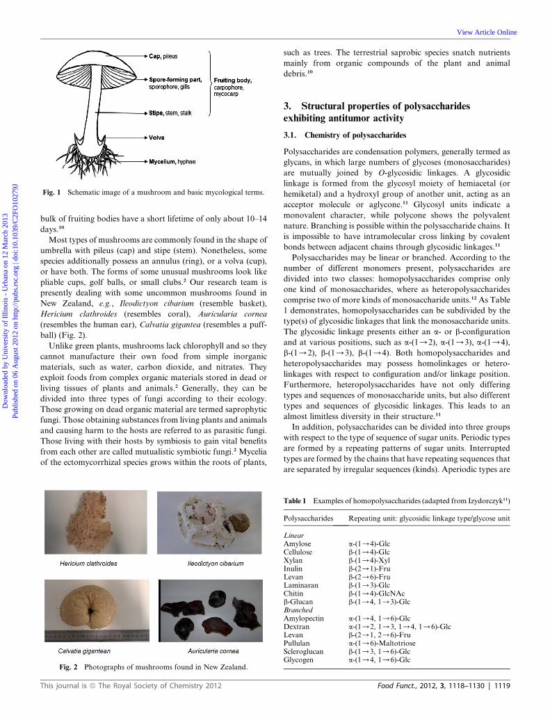
Fig. 1 Schematic image of a mushroom and basic mycological terms.
Dow
nloa
ded
by U
nive
rsity
of
Illin
ois
- U
rban
a on
12
Mar
ch 2
013
Publ
ishe
d on
06
Aug
ust 2
012
on h
ttp://
pubs
.rsc
.org
| do
i:10.
1039
/C2F
O10
279J
View Article Online
bulk of fruiting bodies have a short lifetime of only about 10–14
days.10
Most types of mushrooms are commonly found in the shape of
umbrella with pileus (cap) and stipe (stem). Nonetheless, some
species additionally possess an annulus (ring), or a volva (cup),
or have both. The forms of some unusual mushrooms look like
pliable cups, golf balls, or small clubs.2 Our research team is
presently dealing with some uncommon mushrooms found in
New Zealand, e.g., Ileodictyon cibarium (resemble basket),
Hericium clathroides (resembles coral), Auricularia cornea
(resembles the human ear), Calvatia gigantea (resembles a puff-
ball) (Fig. 2).
Unlike green plants, mushrooms lack chlorophyll and so they
cannot manufacture their own food from simple inorganic
materials, such as water, carbon dioxide, and nitrates. They
exploit foods from complex organic materials stored in dead or
living tissues of plants and animals.2 Generally, they can be
divided into three types of fungi according to their ecology.
Those growing on dead organic material are termed saprophytic
fungi. Those obtaining substances from living plants and animals
and causing harm to the hosts are referred to as parasitic fungi.
Those living with their hosts by symbiosis to gain vital benefits
from each other are called mutualistic symbiotic fungi.2 Mycelia
of the ectomycorrhizal species grows within the roots of plants,
Fig. 2 Photographs of mushrooms found in New Zealand.
This journal is ª The Royal Society of Chemistry 2012
such as trees. The terrestrial saprobic species snatch nutrients
mainly from organic compounds of the plant and animal
debris.10
3. Structural properties of polysaccharidesexhibiting antitumor activity
3.1. Chemistry of polysaccharides
Polysaccharides are condensation polymers, generally termed as
glycans, in which large numbers of glycoses (monosaccharides)
are mutually joined by O-glycosidic linkages. A glycosidic
linkage is formed from the glycosyl moiety of hemiacetal (or
hemiketal) and a hydroxyl group of another unit, acting as an
acceptor molecule or aglycone.11 Glycosyl units indicate a
monovalent character, while polycone shows the polyvalent
nature. Branching is possible within the polysaccharide chains. It
is impossible to have intramolecular cross linking by covalent
bonds between adjacent chains through glycosidic linkages.11
Polysaccharides may be linear or branched. According to the
number of different monomers present, polysaccharides are
divided into two classes: homopolysaccharides comprise only
one kind of monosaccharides, where as heteropolysaccharides
comprise two of more kinds of monosaccharide units.12 As Table
1 demonstrates, homopolysaccharides can be subdivided by the
type(s) of glycosidic linkages that link the monosaccharide units.
The glycosidic linkage presents either an a- or b-configuration
and at various positions, such as a-(1/2), a-(1/3), a-(1/4),
b-(1/2), b-(1/3), b-(1/4). Both homopolysaccharides and
heteropolysaccharides may possess homolinkages or hetero-
linkages with respect to configuration and/or linkage position.
Furthermore, heteropolysaccharides have not only differing
types and sequences of monosaccharide units, but also different
types and sequences of glycosidic linkages. This leads to an
almost limitless diversity in their structure.11
In addition, polysaccharides can be divided into three groups
with respect to the type of sequence of sugar units. Periodic types
are formed by a repeating patterns of sugar units. Interrupted
types are formed by the chains that have repeating sequences that
are separated by irregular sequences (kinds). Aperiodic types are
Table 1 Examples of homopolysaccharides (adapted from Izydorczyk11)
Polysaccharides Repeating unit: glycosidic linkage type/glycose unit
LinearAmylose a-(1/4)-GlcCellulose b-(1/4)-GlcXylan b-(1/4)-XylInulin b-(2/1)-FruLevan b-(2/6)-FruLaminaran b-(1/3)-GlcChitin b-(1/4)-GlcNAcb-Glucan b-(1/4, 1/3)-GlcBranchedAmylopectin a-(1/4, 1/6)-GlcDextran a-(1/2, 1/3, 1/4, 1/6)-GlcLevan b-(2/1, 2/6)-FruPullulan a-(1/6)-MaltotrioseScleroglucan b-(1/3, 1/6)-GlcGlycogen a-(1/4, 1/6)-Glc
Food Funct., 2012, 3, 1118–1130 | 1119

Dow
nloa
ded
by U
nive
rsity
of
Illin
ois
- U
rban
a on
12
Mar
ch 2
013
Publ
ishe
d on
06
Aug
ust 2
012
on h
ttp://
pubs
.rsc
.org
| do
i:10.
1039
/C2F
O10
279J
View Article Online
characterized by irregular sequences of monosaccharide units,
linkage positions, and configurations.11
Polysaccharides have various degrees of polymerization (DP)
which is determined by the number of monosaccharide units in a
chain. Only a few polysaccharides are found having DPs below
100. Polysaccharides are secondary gene products, which is
different with proteins. Therefore, the products from various
biosynthetic enzymes (glycosyl transferases) in nature are not
under strict and direct genetic control in their synthesis. The
mechanisms controlling certain biosynthetic events, such as the
density and distribution of branches along the polysaccharide
chain or the chain’s length, are not fully elucidated, although the
general biosynthetic pathways of many polysaccharides have
been well studied.11 There is a general agreement that different
transferases are demanded for the addition of each mono-
saccharide unit to the growing chain. The backbone growth most
likely happens by adding new sugar residues to the nonreducing
end (tailward growth). Whereas, the addition of new residues
happens at the reducing end (headward growth) when involving
lipid intermediates. Most polysaccharides are considered to
undergo precise synthesis. Its backbone growth is concurrent
with the addition of side chains. The synthesis and transport of
the plant cell wall polysaccharides are conducted by the
membrane systems of the endoplasmic reticulum, Golgi bodies,
and plasma lemma. The rate of transfer and deposition of the
newly synthesized polysaccharide in the target tissue are
considered to play some role in determining its chain length.11
Post-polymerization modification can be performed via ester-
ification and/or etherification of chains. These modifications, in
some cases, may take place simultaneously with polymerization
of the backbone chains. Lacking strict genetic control during the
synthesis of each polysaccharide chain results in a great degree of
heterogeneity of those polymers based on their molecular mass
and DP, and certain aspects of molecular structure, such as ratio
of different monosaccharides, linkage distribution, degree and
distribution of branches. Hence, polysaccharides are regarded as
polydispersed polymers. Nonetheless, not all structural charac-
teristics of the polysaccharide are heterogeneous. For instance,
the most conservative is the configuration of glycosidic bonds in
polysaccharides, while the most variable characteristic is the
molecular weight.11
In other words, polysaccharides belong to a structurally
diverse class of macromolecules. The monosaccharide units in
polysaccharides can interconnect at several points to produce
various branched or linear structures.13 This vast potential
variability in polysaccharide structure could offer possibilities to
the precise regulatory mechanisms of various cell–cell interac-
tions in higher organisms.14
3.2. Mushroom polysaccharides showing antitumor activity
Polysaccharides showing antitumor activity have been isolated
from the fruiting bodies, cultured mycelia and culture filtrates of
basidiomycetes. These polysaccharides showing antitumor
activity have a great variety of chemical composition, structure
and antitumor activity.15 Since Lucas3 first reported the poly-
saccharides extracted from mushrooms indicating antitumor
activity in 1957, a great deal of polysaccharides that show anti-
tumor activity have been isolated from mushrooms and their
1120 | Food Funct., 2012, 3, 1118–1130
antitumor activities have been extensively studied. Some studies
on those polysaccharides are shown in Table 2.
Furthermore, although mushroom polysaccharides exhibit
remarkable antitumor activity, their tumor inhibition ability
varies greatly. For example, some reported polysaccharides
present different antitumor activities in vivo through screening
studies using sarcoma-180 in mice (Table 3).
The main polysaccharides in mushrooms are glucans with
different types of glycosidic linkages, such as (1/3)-, (1/6)-b-
glucans and (1/3)-a-glucans. Also, there are some hetero-
glycans present in mushrooms. The others are PSP complexes
which are mostly bound to protein residues.39 Although, the
fungal cell wall is the main source of polysaccharides demon-
strating antitumor activity, chitin and chitosan (fungal chitin)
show no antitumor activity.40
Particularly, polysaccharides that exhibit strong antitumor
action are greatly different in their chemical structures. Anti-
tumor activity is demonstrated by a wide range of glycans which
extend from homopolymers to highly complex heteropolymers.41
Some monosaccharide types of the polysaccharides showing
antitumor activity consist of glucose, galactose, mannose, xylose,
arabinose, fucose, ribose and glucuronic acid. In some mush-
room species, polysaccharides binding with proteins or peptides
as a polysaccharide–protein or polysaccharide–peptide complex
indicate higher potent antitumor activity.3
Apart from the well-known antitumor (1/3)-b-glucans, those
biologically active glucans are linear or branched molecules
which contain a backbone composed of a- or b-linked glucose
units; some of them have side chains attached at different posi-
tions. Heteroglucan side chains hold glucuronic acid, xylose,
galactose, mannose, arabinose, or ribose, which may be in
different combinations. Heteroglycans are another large group
of bioactive polysaccharides that are classified as galactans,
fucans, xylans, and mannans by individual sugar components in
the backbone. Likewise, heteroglycan side chains may hold
arabinose, mannose, fucose, galactose, xylose, glucuronic acid,
and a glucose moiety as a main component.3
In the seventies and eighties, three antitumor agents of poly-
saccharide nature, namely lentinan, schizophyllan and protein-
bound polysaccharide (PSK, Krestin), were isolated from
Lentinus edodes, Schizophyllum commune and Coriolus versicolor,
respectively. They have since become large market items in
Japan.40 Lentinan and schizophyllan belong to pure b-glucans,
while PSK is a protein-bound b-glucan. In China, a poly-
saccharopeptide (PSP) has been isolated and employed as an
anti-cancer and immunomodulatory agent in clinical
treatments.15
Lentinan is a representative mushroom b-glucan, which shows
effective antitumor and immunopotentiating activity. Its primary
structure is a (1/3)-b-glucan containing five (1/3)-b-glucose
residues in a linear linkage and two (1/6)-b-glucopyranoside
branches in side chains (Fig. 3A). This leads to a right-handed
triple helical structure. The molecular weight of lentinan is about
400–800 � 103 Da.15,42
Schizophyllan is also a (1/3)-b-glucan containing a b-glu-
copyranosyl group joined by a b-(1/6) linkage to every third or
fourth residue of the main chain (Fig. 3B). It has a similar triple
helix structure and biological activity to lentinan, and possesses a
molecular weight of about 450 � 103 Da.15
This journal is ª The Royal Society of Chemistry 2012

Table 2 Reported polysaccharides showing antitumor activity from mushrooms
Mushroom species Polysaccharide source Antitumor type Reference
Cordyceps militaris Fruiting body Melanoma 16Lung cancer 17
Phellinus gilvus Fruiting body Lung cancer 18Phellinus linteus Mycelial culture Melanoma 19 and 20Pleurotus ostreatus Fruiting body Melanoma 21Ganoderma lucidum Fruiting body Breast cancer 22
Lung cancer 23Prostate cancer 24Cervical cancer 25
Lentinula edodes Fruiting body Breast cancer 26Pleurotus geesteranus Fruiting body Breast cancer 27Pleurotus tuber-regium Sclerotia Breast cancer 28Clitocybe alexandri Fruiting body Lung cancer 29Lepista inversa Fruiting body Lung cancer 29Sparassis crispa Fruiting body Lung cancer 30Agaricus blazei Fruiting body Prostate cancer 31Grifola frondosa Fruiting body Prostate cancer 32Trametes versicolor Fruiting body Prostate cancer 33Angelica sinensis Mycelia Cervical cancer 34
Table 3 Antitumor activities of extracts of mushroom species (againstsarcoma-180 in mice)
Mushroom speciesTumor inhibition(%) Reference
AgaricaceaeAgaricus bisporus 2 35AuriculariaceaeAuricularia auricula-judae 42 35CorticiaceaeLaetisaria arvalis 95 36PleurotaceaePleurotus tuber-regium: carboxymethylated hot alkali extracts (CMHZE)CMHZE-1 64 37CMHZE-2 48 37CMHZE-3 75 37CMHZE-4 53 37CMHZE-5 46 37CMHZE-6 43 37PolyporaceaeGanoderma tsugae 77 35Coriolus versicolor 77 35Trametes gibbosa 49 35Fomes fomentarios 5 35RussulaceaeRussula lepida 67.6 38StrophariaceaePholiota nameko 86 35TricholomataceaeLentinus edodes 80 35Flammulina velutipes 81 35Pleurotus ostreatus 75 35Tricholoma matsutake 91 35
Fig. 3 Structure units of polysaccharides showing antitumor activity:
lentinan (A), schizophyllan (B).
Dow
nloa
ded
by U
nive
rsity
of
Illin
ois
- U
rban
a on
12
Mar
ch 2
013
Publ
ishe
d on
06
Aug
ust 2
012
on h
ttp://
pubs
.rsc
.org
| do
i:10.
1039
/C2F
O10
279J
View Article Online
PSK (Krestin) is a b-glucan–protein complex consisting of 25–
38% protein residues. Its average molecular weight, measured by
ultracentrifuge analysis, is about 94 � 103 Da. It mainly contains
acidic amino acids, such as aspartic acid and glutamic acid, and
neutral amino acids, such as valine and leucine, and small
amounts of basic amino acid, such as lysine and arginine. The
main constituent monosaccharide is glucose with small amounts
of other sugar residues like mannose, fucose, xylose and galac-
tose. PSK has a (1/4)-b-glucan with (1/6)-b-glucopyranosidic
This journal is ª The Royal Society of Chemistry 2012
side chains for every fourth glucose unit. It possesses branches at
the 3- and 6-positions in a proportion of one every several
residual groups of (1/4) bonds.43 A polysaccharopeptide (MW
100 � 103 Da) that was isolated from a strain of Coriolus versi-
color in China has a similar glucan structure to PSK in Japan.44
A polysaccharide–protein complex (PSPC), that was extracted
from the culture filtrates of Tricholoma lobayense, consists of
54.3% polysaccharides containing galactose, glucose, arabinose,
xylose, rhamnose, fucose and mannose, and 35.9% protein con-
taining majorly aspartic acid, glutamic acid, serine, glycine,
lysine and threonine. PSPC shows the characteristics of a poly-
saccharide and intermolecular hydrogen bonds by inspection of
the infrared spectra. The polysaccharide moiety of PSPC belongs
to a unique heteroglycan with a molecular weight of about
154 � 103 Da.45
Ganoderan (MW 20 kDa) was isolated from Ganoderma
lucidum and classified as a Ganoderma species which are the most
Food Funct., 2012, 3, 1118–1130 | 1121
Dow
nloa
ded
by U
nive
rsity
of
Illin
ois
- U
rban
a on
12
Mar
ch 2
013
Publ
ishe
d on
06
Aug
ust 2
012
on h
ttp://
pubs
.rsc
.org
| do
i:10.
1039
/C2F
O10
279J
View Article Online
well known medicinal fungi in the Orient. It is an immuno-
modulatory b-glucan, which induces potent antitumor immunity
in tumour-bearing mice. It mostly contains glucose and 4%
protein.46 Moreover, the fruiting bodies and mycelia of Gano-
derma applanatum comprise b-glucan, heteroglycans and glycan-
protein complexes. These polysaccharides indicating antitumor
activity have the molecular weights ranging from 30 � 103 to
10 � 105 Da. Their basic chemical structure is (1/3)-b-gluco-
pyranan with 1–15 (1/6)-b-monoglucosyl side chains.47 Seven
potent antitumor polysaccharide–protein complexes have been
extracted from Ganoderma tsugae. Two of them were protein-
containing glucogalactans related to mannose and fucose. And
five are protein-containing (1/3)-b-glucans.15
4. Connection between structure and antitumoractivities of mushroom polysaccharides
Polysaccharides possess a huge variety of chemical compositions
and configurations and physical properties. The antitumor
activity of the polysaccharides can be influenced by the size of the
molecules, degree of branching, form, and solubility in water.14
Generally, the greater the molecular weight and the higher the
water solubility of these polysaccharides, the higher the anti-
tumor activity. In the study based on seven potent antitumor
polysaccharide–protein complexes from Ganoderma tsugae, it
was found that polysaccharides showing antitumor activity with
high activity derived from fruiting bodies were mainly hetero-
polysaccharides that had molecular weights of about 10 � 103
Da, containing galactose, glucose, mannose and fucose.
However, highly active polysaccharides isolated form mycelia
were mostly protein-containing glucans with molecular weights
of around 10 � 103 Da.40,48
Most polysaccharides exhibiting antitumor activity have been
reported to have the same basic b-glucan structure with different
types of glycosidic linkages. Hence, the antitumor action requires
structural features such as b-(1/3) linkages in the main chain of
the glucan and additional b-(1/6) branch points. b-Glucan,
comprising mainly (1/6) linkages possesses less activity.49
Nonetheless, there are other obvious variations in those poly-
saccharides. Polysaccharides that show antitumor activity may
contain other chemical structures, such as hetero-b-glucans,40
heteroglycan,50 b-glucan–protein,51 a-manno-b-glucan,40
a-glucan–protein40 and heteroglycan–protein complexes.52 For
instance, PSK and PSP have a b-glucan–protein, while PSPC
isolated from the Tricholoma species are a heteroglycan–protein
complex.
4.1. The influence of molecular mass
A high molecular mass is necessary for extensively enhancing
immunological and antitumor activities. Four fractions of PSK
have been successively separated. The highest molecular mass
fraction shows the strongest immunomodulatory activity. A
(1/3)-b-glucan, isolated from the cultured mycelium of Grifola
frondosa, indicates changes in biological activities with various
molecular masses, which was obtained by heat treatment for
different lengths of time at 150 �C. The fraction with the highest
molecular mass (800� 103 Da) shows the most potent antitumor
and immunomodulatory activities. These studies highlight the
1122 | Food Funct., 2012, 3, 1118–1130
possibility that polysaccharides showing antitumor activity may
not always be multiple enhancers of the host defense system, and
that a high molecular mass is needed for extensive enhancement
of immunological and antitumor activities. However, some low
molecular weight polysaccharides, such as lentinan and schizo-
phyllan, present the same antitumor activity against Sarcoma
180 as those with higher molecular weights. The divergent results
remain to be clarified.15
4.2. The effect of the branching configuration
If b-glucans are mostly linear, containing branches that are not
excessively long, they will exhibit antitumor activity. For
instance, pachyman, which is separated from Poria cocos, is
inactive although it is a branched b-glucan. Nonetheless,
pachymaran, that is formed by debranching pachyman using
periodate oxidatiton and mild hydrolysis shows pronounced
antitumor activity. Miyazaki et al.53 suggested that the optimal
branching frequency is from 0.2 (1 in 5 backbone residues) to
0.33 (1 in 3 backbone residues). Lentinan (2/5) is a b-1,3-D-glucan
possessing two branches for every five D-glucopyranosyl residues.
Schizophyllan (1/3) is also a b-1,3-D-glucan having one branch
for every three D-glucopyranosyl residues. The polysaccharide of
PSK (1/5) is a (1/3)-b,(1/4)-D-glucan of one branch for every
five D-glucopyranosyl residues. Their antitumor activities are not
apparently different even though the degree of their branches
differs. However, the debranched lentinan indicates a more
effective antitumor activity than the native lentinan at a dose of
2.0 mg kg�1 for five days in mice.15
4.3. The impact of conformation
The conformations of polysaccharides demonstrating antitumor
activity include single helix, triple helix and random coil. Len-
tinan, schizophyllan and PSK all consist of a triple helix struc-
ture.15 It is known that a triple-helical tertiary conformation of
medicinal mushroom (1/3)-b-glucans is crucial for their
immune-stimulating activity. The tertiary structure of lentinan is
lost while its primary structure is not affected when it is dena-
tured with dimethyl sulfoxide, urea, or sodium hydroxide. Its
tumor inhibition properties are reduced during progressive
denaturation.54 Pachyman isolated from Poria cocos is a b-1,3-
D-glucan containing a single helix conformer, which is not bio-
logically active against tumor growth. However, when it becomes
pachymaran by periodate oxidation and mild hydrolysis, the
newly formed conformer shows pronounced antitumor activity.55
Schizophyllan–OH with a single helix structure, that is obtained
from the alkaline-treated schizophyllan, exhibits a reduced
ability to inhibit tumor growth as compared with the native
schizophyllan.42
Yadomae56 explained that many biological and immuno-
pharmacological activities demonstrated by mushroom
b-(1/3)-glucan, such as macrophage nitrogen oxide synthesis
and limulus factor G activation, are determined by the triple-
helix conformation, whereas others are not dependent on this
conformation, e.g., synthesis of interferon-g and colony stimu-
lating factor. Hence, it can be seen that the antitumor activity of
polysaccharide is dependent on the helical conformation.
The correlation between conformation and antitumor activity of
This journal is ª The Royal Society of Chemistry 2012
Dow
nloa
ded
by U
nive
rsity
of
Illin
ois
- U
rban
a on
12
Mar
ch 2
013
Publ
ishe
d on
06
Aug
ust 2
012
on h
ttp://
pubs
.rsc
.org
| do
i:10.
1039
/C2F
O10
279J
View Article Online
the polysaccharides or polysaccharide–protein complexes
implies that the existence of biological systems within the
host body recognize the configurational structure of
polysaccharide.15
4.4. The effect of solubility
The solubility of b-glucans is affected by their degree of poly-
merization and thus their physical organization.57 When the
alkali-insoluble, branched (1/3)-b-D-glucan, isolated from
Auricularia auricular-judae, was modified by controlled periodate
oxidation, borohydride reduction, and mild acid hydrolysis, its
water solubility was increased by having covalently linked
D-glucosyl residues, which demonstrated significantly potent
antitumor activity, whereas in the native state it had no such
activity.58
4.5. Enhancement of antitumor activity by chemical
modification
The improvement of the biological activity of polysaccharides
that show antitumor activity can be achieved by chemical
modification. Various carboxymethylated (CM), hydroxylated,
formylmethylated, aminethylated and sulfated products have
been designed.
The successful schemes for chemical improvement of mush-
room polysaccharides have been designed for Ganoderma luci-
dum, Grifola frondosa and Leucopaxillus giganteus. Two main
procedures are involved in these schemes: modification of
mushroom polysaccharides by Smith degradation (oxydo-
reducto-hydrolysis) and activation by the method of for-
molysis.49 During the Smith degradation modification, original
polysaccharide solutions are first oxidized to polyaldehydes by
0.1 M NaIO4 in darkness. They are then are reduced into poly-
alcohols by NaBH4 in an alkaline medium adjusted to pH 8 with
2 M NaOH, and hydrolyzed by 1 M H2SO4 at room tempera-
ture.59 Chemical activation of the mushroom polysaccharides by
means of formolysis consists of degradation of the poly-
saccharides by formic acid in 99% HCOOH solution. Fractions
are obtained by alcohol (99% EtOH) precipitation.59 The two
original polysaccharides do not possess activity, however, their
polyaldehyde polyol, formylated, and formolysis derivatives
exhibit significant activity. Furthermore, polyaldehyde, and
polyol–polysaccharides obtained from a polysaccharide that has
low antitumor activity indicates activity that is stronger than the
original polysaccharide.59
The method of carboxymethylation can be employed to
transform b-glucans into a water-soluble form. For instance, the
fruit bodies of Pleurotus ostreatus are treated with 0.15 MNaOH
solution at 95 �C for 2 h. The residue is collected and washed with
water until neutral. It is then suspended in 0.06% NaCl solution,
adjusted to pH 4.5 with acetic acid, and stirred for 6 h at 50 �C.The polysaccharide thus obtained is a b-(1/3)-linked glucan
with every fourth glucopyranosyl residue substituted at 0–6 with
single D-glucopyranosyl groups. Carboxymethylated glucan P.
ostreatus shows immunomodulatory effects, and elevated
phagocytic activity.60,61 The linear (1/3)-a-glucans derived
from Amanitamuscaria and Agrocybe cylindracea show little
antitumor activity. After modification, the carboxymethylated
This journal is ª The Royal Society of Chemistry 2012
linear (1/3)-a-glucans indicate strong antitumor activity
against Sarcoma 180 and immunomodulating activity in mice.15
Chemical modification of branched mushroom poly-
saccharides resulting in side-chain reduction can be performed
not only by Smith degradation but also by enzymatic reactions.
Debranched pachymaran and CM-pachynmaran is a b-1,3-
D-glucan showing more effective antitumor activity.15 Linear low
molecular weight a-(1/4)-glucans that are prepared after
enzymatic reduction of the side chains and protein component
(active hexose correlated compounds – AHCC) show immuno-
modulatory and anticancer properties.62
The antitumor activity of the formylmethylated and amino-
ethylated derivatives of schizophyllan against Sarcoma 180 solid
tumor in mice is largely augmented compared with the native
schizophyllan.63 The sulfated lentinan and schizophyllan prod-
ucts show potent anti-human immunodeficiency virus activity
though with reduced antitumor effect. These investigations give
the direction that the improvement of the biological activities of
polysaccharides may be effectively approached by chemical
modifications.15
5. Immunomodulating activities and mechanisms ofantitumor activity by mushroom polysaccharides
5.1. Cancer
A neoplasm is defined as an abnormal mass or colony of cells
formed by a relatively autonomous new growth of tissue.64 Most
of the neoplasms originate from the clonal expansion of a single
cell which has undergone neoplastic transformation. The trans-
formation of a normal cell to a neoplastic cell can be triggered by
chemical, physical, or biological agent (or event), which directly
and irreversibly changes the cell genome. Neoplastic cells have
the characteristics exhibited by the loss of some specialized
functions and the acquisition of new biological properties, such
as self-sufficiency in growth signals, insensitivity to growth-
inhibitory signals, evasion of apoptosis, limitless replicative
potential, sustained angiogenesis, and tissue invasion and
metastasis. Neoplastic cells deliver their heritable biological
characteristics to progeny cells.64
Cancer is a generic term used for malignant neoplasms.64 A
characteristic property of cancer cells is anaplasia, which denotes
a lack of normal structural and functional characteristics.
Literally, a tumor describes a swelling of any type, such as an
inflammation of other swelling. Generally, the development of a
cancer includes three stages. In the initial stage, a mutagen binds
to the cell DNA and results in damage. Usually, the initiation is
not sufficient to trigger tumor production by itself. The second
stage is called activation, in which a tumor promoter is activated
to cause the formation of small benign tumors. In the third stage
of progression, the loss of the normal tight control over the cell
cycle leads to uncontrolled cell proliferation.65
Furthermore, the biological behavior or clinical course of
neoplasm can be divided into benign or malignant. A malignant
neoplasm, that presents a greater degree of autonomy, is capable
of invasion and metastatic activity, which may be resistant to
treatment and leads to death. A benign neoplasm, with a lesser
degree of autonomy, is usually not invasive, does not metasta-
size. It usually generates no great harm if treated adequately.64
Food Funct., 2012, 3, 1118–1130 | 1123

Dow
nloa
ded
by U
nive
rsity
of
Illin
ois
- U
rban
a on
12
Mar
ch 2
013
Publ
ishe
d on
06
Aug
ust 2
012
on h
ttp://
pubs
.rsc
.org
| do
i:10.
1039
/C2F
O10
279J
View Article Online
5.2. Antitumor activity by mushroom polysaccharides
Mushroom polysaccharides perform their antitumor action
mainly via activation of the immune response of the host
organism. These substances are considered as biological response
modifiers.66 Basically, this suggests that: (1) they cause no harm
and exert no additional stress on the body; (2) they assist the
body to adapt to a variety of environmental and biological
stresses; and (3) they place a nonspecific action on the body,
supporting some or all of the major systems, such as nervous,
hormonal, and immune systems, as well as regulatory func-
tions.67 Substances that are capable of interacting with the
immune system can either upregulate or downregulate specific
aspects of the host response. Whether certain substances enhance
or suppress immune responses are dependent on many factors
including dose, route of administration, and timing of adminis-
tration of the substances in question. The type of activity can also
be determined by their mechanism of action or the site of
activity.68
Mushroom polysaccharides have been proven in a wide range
of antitumor activities. Numerous studies have reported that the
compounds, particularly b-D-glucan derivatives, nonspecifically
activate cellular and humoral components of the host immune
system so that they raise functional activity of macrophages,
monomuclear cells, and neutrophils.69 b-D-Glucans are recog-
nized by the human immune systems as foreign molecules since
they are not synthesized by humans. These compounds can cause
both innate and adaptive immune responses.70 The defence of the
body against microbial attack and against spontaneously
occurring malignant tumor cells consists of a dynamic orches-
trated interplay of innate and acquired immune responses
(Fig. 4). Innate immunity, containing macrophages, neutrophils,
natural killer cells (NKs) and dendritic cells (DCs) as gate-
keepers, is regulated by chemical-messengers or cytokines and by
activating inflammatory and acute phase responses.42 The
mononuclear phagocyte system (e.g., macrophages and mono-
cytes), DCs and certain lymphocytes (e.g., NK cells) exact
numerous important functions involving the recognition and
destruction of abnormal cells. Specific immunity to abnormal
Fig. 4 Immune responses stimulated by fungal b-glucans.
1124 | Food Funct., 2012, 3, 1118–1130
cells or tissues contains humoral (e.g., generates antibodies) and
cell-mediated immunity (also enhances inflammatory responses
and ultimately kills infected or abnormal cells). Therefore, an
adequately functional immune response is critical to the recog-
nition and removal of tumor cells.71 For instance, the activated
phagocytes can eliminate pathogens by phagocytosis.72 The
macrophages target and remove dead cells and intracellular
pathogens.73 NKs circulating in blood lyse cancer and virus-
infected cells. Neutrophils attack pyogenic bacteria.74
The adaptive immune system works on the despondence to the
introduction of foreign antigens, involving both B and T cells.
B cells generate antibodies to mediate humoral immunity, while
T cells trigger cell-mediated immunity.73 Cytokines potentiate
T cell differentiation to helper T cells 1 (Th1) and 2 (Th2), which
mediate cell and humoral immunities, respectively.75 DCs,
derived from monocytes, are involved in the adaptive immune
response, presenting antigens to T cells to activate immune
responses.73 Multicellular organisms contain receptors which are
called ‘pattern recognition receptors’ (PRRs), detecting innately
foreign structures like pathogen-associated molecular patterns
(PAMPs). Fungal b-glucans are considered as PAMPs and are
recognized by appropriate cell surface receptors to initiate
immune responses. Some receptors have been identified in
humans, such as dectin-1, complement receptor 3 (CR3), scav-
enger receptors, lactosylceramide (LacCer), and the toll-like
receptor (TLR).70
Dectin-1 is a lectin which contains four components, namely
an extracellular carbohydrate-recognition domain (CRD), a
stalk, a transmembrane region, and an intracellular cytoplasmic
tail.76 Dectin-1 is evidenced to be of the most importance in
activating innate immune responses in macrophages,77 since the
abolition of all macrophage-mediated responses can be caused
when blocked with an anti-dectin-1 antibody and knockout of
the detin-1 gene.78 Several signaling pathways contributed to by
the dectin-1 binding with the ligand, promoting innate immune
responses through the activation of phagocytosis, reactive
oxygen species (ROS) production, and induction of inflamma-
tory cytokines, have been identified. One pathway is that dectin-1
works synergistically with TLR to generate strong inflammatory
responses by stimulating cytokines such as tumor necrosis factor-
alpha (TNF-a), interleukine-2 (IL-2) and IL-12.79 The second
pathway is independent of TLR, which is mediated via spleen
tyrosine kinase (Syk) to yield other cytokines, such as the
macrophage inflammatory protein-2 (MIP2, CXC2) and IL-2
and IL-10 in mice DC cells.80 In addition, another independent
signaling pathway that is activated by the dectin-1 receptor is
phagocytosis in macrophages.79
The CR3 receptor, that comprises CD11b and CD18 domains,
recognizes a large range of microbial cells and acts as an adhesion
molecule. It is presented primarily on neutrophils, monocytes
and NK cells, but not macrophages.81 CD11b contains two
binding sites. The one located within the C terminus is for
b-glucans, whereas the other which is located within the
N-terminus, is for iC3b (cleaved component 3 fragment of serum
complement system).82 When b-glucans are bound to CR3, the
adhesion to microbial cells is increased and the iC3b pathway is
activated to result in tumor cytotoxicity.82 It is essential that both
binding sites are occupied to trigger this activation, since cyto-
toxicity is blocked by an anti-CR3 antibody.83
This journal is ª The Royal Society of Chemistry 2012

Dow
nloa
ded
by U
nive
rsity
of
Illin
ois
- U
rban
a on
12
Mar
ch 2
013
Publ
ishe
d on
06
Aug
ust 2
012
on h
ttp://
pubs
.rsc
.org
| do
i:10.
1039
/C2F
O10
279J
View Article Online
Scavenger receptors that are located in myeloid and endothe-
lial cells consist of a heterogeneous group of proteins with two
transmembrane domains, two intracellular domains and one
extracellular domain, recognizing a range of foreign cells, low-
density lipoprotein (LDL), high-density lipoprotein (HDL) and
selected polyanionic ligands.84 The Src receptor activates
multiple signaling pathways involving Src family kinase(s),
phosphatidylinositol-3 kinase (P13K), Akt kinase, and p38
mitogen-activated protein kinase (MAPK), and an endothelial
nitric oxide synthase (eNOS).84 However, Chen and Seviour74
think that they are not important due to the lack of sufficient
evidence to understand the biological effects mediated by fungal
b-glucans.
LacCer, located in neutrophil and endothelial cells, which is
a glycolipid, possessing a hydrophobic ceramid lipid and
hydrophilic sugar moiety, recognizes both microbial cells and
b-(1/3)-glucans.85 TLRs are transmembrane receptors of a
novel protein family, responding to the presence of a diverse
group of microbes, such as fungi, bacteria, viruses and
protozoa.86 Nonetheless, further investigation is required to
clarify the pathway that the immune responses are activated by
b-(1/3)-glucans via these receptors.
Polysaccharides derived from mushrooms do not attack
cancer cells directly, while they generate their antitumor effects
via the activation of different immune responses in the host.
Many experiments have confirmed this. For example, the anti-
tumor effect of polysaccharides is lost in neonatal thymectom-
ized mice, or is decreased significantly after administration of
anti-lymphocyte serum.87 The results imply that the antitumor
action of polysaccharides needs an intact T cell component and
that the activity is mediated through a thymus-dependent
immune mechanism.42 The pathway of the possible immune
mechanism shows that the administration of lentinan can
promote potentiation of the responses of precursor T cells and
macrophages to cytokines that are produced by certain groups of
lymphocytes after specific recognition of tumor cells.42 The
induction of the marked rise in the amount of TNF-a, IL-1, IL-3
and interferon (IFN) by lentinan causes maturation, differenti-
ation, and proliferation of immunocompetent cells for host
defence mechanisms.42 Lentinan is also able to restore the sup-
pressed activity of helper T cells in the tumor bearing host to
their normal state, resulting in the complete restoration of
humoral immune responses.54 Furthermore, lentinan-induced
delayed-type hypersensitivity response at tumor sites plays a role
in eradicating tumors by regulating infiltration of activated
immune effector cells, such as natural killer cells and cytotoxic T
lymphocytes.88 Compared with lentinan, schizophyllan has a
similar composition, antitumor activity, as well as a mechanism
for antitumor action. Grifolan derived from Grifola frondosa is
similar to schizophyllan in primary structure. It is a novel
macrophage activator enhancing mPNA levels of IL-6, IL-1, and
TNF-a macrophages.89,90
Direct tumor inhibition activity of mushroom polysaccharides
has also been documented. Despite the mechanism of anti-
proliferation of polysaccharides towards tumor lines in vitro
being unclear, some researchers have demonstrated that the
expression of signals within tumor cells could be changed by the
incubation of polysaccharides together with tumor cells. This
could arrest the cell cycle and produce apoptosis,which
This journal is ª The Royal Society of Chemistry 2012
elucidates the in vitro anti-proliferative effect of poly-
saccharides.91 A polysaccharide–peptide complex (PSP) isolated
from Trametes versicolar was reported to significantly decrease
proliferation of MAD-MB-231 breast cancer cells.92 These
results suggest that mushroom polysaccharides not only stimu-
late the proliferation of T lymphocytes and the immune function
through the immunopotentiation, but also exact a direct action
on the tumor cells. However, little is known regarding the direct
effect of polysaccharides on cancer cells.3
5.3. Mechanisms of antitumor by mushroom polysaccharides
The proliferation of tumor cells can be prevented through diverse
mechanisms, including cell cycle arrest, induction of tumor cell
death by apoptosis and secondary necrosis, together with stim-
ulation of the antitumor activity of macropahges.93 In the
eukaryotic cell cycle, cyclins and cyclin-dependent kinases (Cdks)
are critical regulators. Cell cycle progression is regulated at
several irreversible transition points. The passage is controlled by
the activity of Cdks.94 At least three differing mechanisms,
namely binding of cyclin proteins, phosphorylation, and binding
of the cyclin-dependent kinase inhibitors (CKIs), have been
discovered in the activity of Cdks. The accumulation of cyclins
D, E, and A, which bind to and activate different Cdk catalytic
subunits, is able to promote the progression from G1 to S phase
in mammalian cells.95 The research group of Hsieh96 found that
ethanol–water C. versicolor extracts could induce G0/G1 phase
arrest in tumor cells.
The death of tumor cells undergoing antitumor therapy can be
caused by apoptosis and/or necrosis. Apoptosis is a form of cell
death in which a programmed sequence of events results in the
ingestion of cell remains by surrounding cells without releasing
harmful substances.93 Apoptosis is tightly controlled by a
number of gene products that either promote or block cell death
at different stages of the cell cycle.28 One of the major gene
groups that regulate apoptosis is the Bcl-2 family, which is
composed of a large number of proteins. They all belong to three
sub-families based on the number of Bcl-2-homology (BH)
domains present in these proteins; (i) a subfamily consisting of
Bcl-2, Bcl-xL and Bcl-w performs anti-apoptotic activities and
shares sequence homology, especially in four regions, BH1
through BH4; (ii) a subfamily including Bax, Bad and Bak shares
sequence homology at BH1, BH2 and BH3, exerting pro-
apoptotic activity; (iii) a subfamily containing Bik and Bid shares
sequence homology within the BH3 domain only, which shows
pro-apoptotic activity.97 Bax is a 21 kD protein of 192 amino
acids98 that shares homology with Bcl-2 in conserved regions,
including BH1 and BH2. Hence, Bax may heterodimerize with
Bcl-2 or other proteins and/or hodimerize.99 Bax is a nuclear-
encoded protein present in higher eukaryotes which can pierce
the mitochondrial outer membrane to mediate cell death by
apoptosis.100 During apoptosis, Bax may form oligodimers that
are considered to cause the permeabilization of the mitochon-
drial membranes, either by forming channels,101 by interacting
with components of the permeability transition pore,101 or by
altering fission and fusion processes.102 For instance, the water
extract of Cordyceps militaris (WECM) induced apoptosis in
human lung carcinoma A549 cells. The data revealed that there
was a concentration-dependent increase of Bax expression in
Food Funct., 2012, 3, 1118–1130 | 1125

Dow
nloa
ded
by U
nive
rsity
of
Illin
ois
- U
rban
a on
12
Mar
ch 2
013
Publ
ishe
d on
06
Aug
ust 2
012
on h
ttp://
pubs
.rsc
.org
| do
i:10.
1039
/C2F
O10
279J
View Article Online
WECM treated A549 cells, but a decrease of Bcl-2.17 In another
study, the treatment with a novel polysaccharide isolated from
Angelica sinensis can reduce Bcl-2 and Bcl-X1 expression, and
raise Bax and Bak expression in HeLa cells, triggering
apoptosis.34
On the other hand, polysaccharides are known to induce
necrosis.20,103 Necrosis is accidental cell death without a precise
mechanism, leading to the break-down of the cell membrane and
release of intracellular compounds into surrounding tissue.104
TNF is a multifunctional cytokine which is a protein produced
by many cell types such as monocytes, macrophages, T cells, and
B cells with appropriate stimulation. The human TNF protein is
expressed as a 26 kDa (233 amino acid long) integral trans-
membrane precursor protein. A 17 kDa (157 amino acids)
mature TNF protein can be released from the precursor protein
into the medium by proteolytic cleavage, possibly involving a
serine protease.105 TNF is not only cytotoxic or cytostatic to
some tumor cell lines in vitro106 but also destroys actively
proliferating endothelial cells in primary culture.107 The hot
water extract from Polyporus rhinocerus has been proved to
increase TNF-a production.103
Macrophages defend the host by playing critical roles, con-
sisting of phagocytosis of pathogens and apoptotic cells,
production of cytokines, and proteolytic processing and presen-
tation of foreign antigens. Macrophages can be stimulated by
polysaccharides to release a broad spectrum of cytokines like
interleukins, TNF-a, and nitric oxide (NO), which are referred to
as the inhibitory factors of cancer.22 Two fractions of poly-
saccharides purified from Ganoderma lucidum were reported to
have a proliferative effect on macrophages up to 160% of the
control cells.22 NO has been studied in the last few years and is
recognized as a crucial messenger that indicates diverse patho-
physiological functions, such as neuronal transmission, vascular
relaxation, immune modulation, and cytotoxicity against tumor
cells.108 NO has been proved to be a main effector molecule
destructing tumor cells by activated macrophages.109 Basically,
macrophages that are stimulated by TNF-a to generate NO
through the expression of the iNOSgene.Moreover, the induction
of NO and TNF-a production and gene expression by activated
macrophages can have a cytotoxic impact on malignant cells.110
The toxic effects ofNOand its derivatives on target cells are based
on several mechanisms, which include (i) inactivating iron–sulfur
cluster-containing enzymes through loss of iron from cells; (ii)
inhibiting DNA-binding activity of zinc finger-type transcrip-
tional factors by the induction of the release of zinc from zinc-
containing proteins; and (iii) destructing the mitochondrial
membrane potential by affecting the activity of ion channels.111 It
was found that NO and TNF-a elicited by acidic polysaccharides
isolated from Phellinus linteus may contribute in vivo to its
immunomodulatory and anti-tumoricidal activities.20
The antitumor activity of an anionic sulfated polysaccharide
was performed by binding to positively charged DNA-binding
locus of enzymes via electrostatic interaction on the cell surface
of human cancer cells, inhibiting its binding to DNA.112 Like-
wise, carboxymethylated polysaccharides are proposed to prob-
ably bind non-specifically to DNA-interacting enzymes. Despite
that non-specific binding by individual carboxymethylated
groups might be weaker than the specific binding by DNA. The
large polysaccharidic molecules might be able to cover the locus
1126 | Food Funct., 2012, 3, 1118–1130
of the enzymes due to the multivalent nature of carboxymethy-
lated polysaccharides, blocking their reaction with the DNA
molecules.28
In addition, recent studies have reported that anti-angiogen-
esis might be one of the important mechanisms of antitumor
activity. Angiogenesis is based on several aspects that the endo-
thelial cells must proliferate to offer the necessary number of cells
for the growing vessels, and the cells are able to migrate.23
Angiogenesis can be tightly controlled by a balance of endoge-
nous inducers and inhibitors of angiogenesis. Primary tumors
cannot grow greater than 2–3 mm without eliciting neo-
vascularisation. In the initial stage of tumor progression, an
imbalance of angiogenesis regulators takes place, which favours
an angiogenic environment. This angiogenic change leads to
the oversecretion of angiogenesis inducers, such as vascular
endothelial growth factor (VEGF), and the subsequent neo-
vascularisation and growth of the tumor.113 Several endogenous
inhibitors of angiogenesis, such as angiostatin, endostatin,
interferons, thrombospondin-1 (TSP-1), tissue inhibitor of met-
alloproteinases (TIMP), and tumstatin, have been identified. The
understanding of the basic science of angiogenesis involving
those inducers and inhibitors has resulted in the development of
anti-angiogenic therapies.113 Ganoderma lucidum has been found
to suppress capillary morphogenesis of aortic endothelial cells.
This is affected by the mediation through the inhibition of
secretion of angiogenic factors like VEGF and transforming
growth factor-b 1 (TGF-b 1) from prostate cancer cells PC-3. G.
lucidum inhibits functions of kinases Erk1/2 and Akt resulting in
the inhibiton of AP-1, which causes the down-regulation of
expression of VEGF and TGF-b 1.24 Similarly, Cao and Lin23
reported that G. lucidum polysaccharide peptides exerted the
inhibitory actions not only on vascular cell proliferation, but also
on the secretion of VEGF by human lung carcinoma cells in
hypoxia.
Most of the reported antitumor mechanisms of poly-
saccharides were raised from the cytological studies, such as cell
cycle arrest, apoptosis, necrosis, and stimulation of immune
responses. However, each mechanism does not occur solely in a
one-way lane, which can connect with other mechanisms simul-
taneously in a complicated matrix. For example, in the theory of
necrosis, the functioning cytokine TNF can be produced by
numerous cell types like monocytes, macrophages, T cells, and B
cells under appropriate stimulation. This suggests that the death
of tumor cells during necrosis can be triggered by different
passages together at the same time, if the polysaccharides are
able to stimulate a number of those types of cells simultaneously.
Polysaccharides derived from mushrooms can perform their
antitumor activities under different mechanisms. A good
example is the extracts from Ganoderma lucidum, which have
been reported to demonstrate antitumor abilities by stimulation
of macrophages22 or by anti-angiogenesis.23 Note however, that
as these two studies were carried out separately, it is not known if
the effective compounds are identical or not. Anti-angiogenesis
can be regarded as a different approach, even though it can still
be placed in the biological scope.
However, except in the case of nitric oxide, which is produced
through the stimulation of the cell by mushroom poly-
saccharides, the exact mechanisms related to structural chemistry
and chemical interactions have not been fully researched. More
This journal is ª The Royal Society of Chemistry 2012
Dow
nloa
ded
by U
nive
rsity
of
Illin
ois
- U
rban
a on
12
Mar
ch 2
013
Publ
ishe
d on
06
Aug
ust 2
012
on h
ttp://
pubs
.rsc
.org
| do
i:10.
1039
/C2F
O10
279J
View Article Online
studies are needed to develop theories clarifying why and how the
specific conformations of polysaccharides are related to their
antitumor properties.
5.4. Methods used to quantify antitumor activity of mushroom
polysaccharides
Quantitative assessments of antitumor activity that relies on
analyzing the changes of entities of cells cultured in different
conditions can be divided into two groups, as in vitro and in vivo
analyses. The in vitro analysis is mainly performed by modern
colorimetric cell-based proliferation or toxicity assays using
compounds that stain the cells directly or that are metabolized
into coloured products,114 followed by quantification approaches
including spectrophotometric and fluorimetric techniques to
numerate appropriately labelled cells.115 Technically, cancer cells
are cultured in flasks with medium and antibiotics. Once their
growth reaches the desired density they are harvested. To
determine the total amount of the cells, an aliquot is taken and
stained, and then counted by a hemocytometer. For the anti-
tumor activity test, the cells at a certain concentration are seeded
in multiwall plates and cultured overnight. The following day,
the cells are drugged with cytotoxic compounds such as poly-
saccharides or anticancer medicines and further cultured for 2–5
culture doubling times. At the end of incubation, the cells are
treated with color reagents which are indicator dyes. These color
reagents include 3-(4,5-dimethylthiazol-2-yl)-2,5-diphenyl tetra-
zolium bromide (MTT), sodium 30-[1-(phenylaminocarbonyl)-
3,4-tetrazolium]-bis(4-methoxy-6-nitro) benzene sulfonic acid
hydrate (XTT), 3-(4,5-dimethylthiazol-2-yl)-5-(3-carboxy-
methoxyphenyl)-2-(4-sulfophenyl)-2H-tetrazolium (MTS), 4-[3-
(4-iodophenyl)-2-(4-nitrophenyl)-2H-5-tetrazolio]-1,3-benzene
disulfonate (WST-1), 7-hydroxy-3H-phenoxazin-3-one 10-oxide
(alamarBlue), and sulforhodamine B [2-(3-diethylamino-6-
diethylazaniumylidene-xanthen-9-yl)-5-sulfo-benzenesulfonate]
(SRB). Examples of the application of these indicator dyes are
given below. The last step of the in vitro method is the quantifi-
cation of cells in the control and drugged cells using colorimetric
devices such as plate readers. The inhibition rate is calculated
according to the formula116 below:
inhibition rate ð%Þ ¼ 1� absorbance of sample
absorbance of control� 100 (1)
Using similar cell culture procedures as stated above, many
standard assays have been developed to measure the cell prolif-
eration or the cell viability, which are normally termed according
to the names of the indicator dye used in the detection stage, e.g.,
MTT assay, XTT assay, and so on. Most of the indicator dyes
work on the principle that they can be metabolized by living cells.
Mitochondrial reduction of dyes has been developed as an
assessment of lymphocyte growth. Metabolism of tetrazolium
salts, such as MTT, XTT, MTS andWST-1, produce the basis of
colorimetric assays.117 MTT has been most widely used, as it can
be cleaved by functional mitochondria to produce formazan in
viable cells,118 resulting in measurable colour changes in the
culture. Nonetheless, it is impossible to follow-up cell cultures as
the MTT determination necessitates destruction of the cells.115
WST-1 has a similar working principle to MTT, through the
reaction with the mitochondrial succinate-tetrazolium reductase
This journal is ª The Royal Society of Chemistry 2012
to generate the formazan dye. The WST-1 reagent forms a
water-soluble formazan rather than the water-insoluble
product formed by MTT.119 This makes the WST-1 assay a
convenient and common tetrazolium salt technique in microplate
format.
In contrast, the alamarBlue assay involves a colorimetric and
fluorometric growth indicator that can be used to detect the
metabolic activity of cells.120 Basically, the native, oxidized form
of resazurin can be taken up readily by viable cells, which is
reduced intracellularly by oxidoreductases and the mitochon-
drial electron transport chain.121 The system incorporates
an oxidation–reduction (REDOX) indicator that leads to a
corresponding shift in its absorbance and fluorescence.122 The
alamarBlue assay has been considered superior to classical tests,
such as theMTT test, due to its advantages of high stability, non-
toxicity to the cells, and the possibility of continuous monitoring
of cultures over time.123,124
Another major technique for measuring cytotoxicity is the
SRB protein staining assay determining the cellular protein
content of adherent and suspension cultures, which is adopted
for routine use in the U.S. National Cancer Institute in vitro
anticancer screen.125 This assay depends on binding of the dye to
basic amino acids of cellular proteins. Its colorimetric evaluation
offers an estimate of total protein mass, which is directly
proportional to the cell mass.126 SRB staining is independent of
cell metabolic activity, thus cannot distinguish between viable
and dead cells.127
MTT and SRB assays have been successfully used to test the
antitumor activity of polysaccharides derived from mushrooms.
For instance, MTT assays were performed to determine the
cytotoxicity of the polysaccharides isolated from Cordyceps
militaris on B16-F10 melanoma cells.16 Similarly, the anticancer
ability of polysaccharides from Phellinus linteus against B16-F10
cells was evaluated utilizing a SRB assay.19 However, the appli-
cation of WST-1 and alamarBlue assays to investigate the cyto-
toxic effect of mushroom polysaccharides on cancer cells has
rarely been reported. Despite the MTT assay being dominantly
used on this kind of study in the past, more recent techniques,
namely WST-1 and alamarBlue have a potential to replace the
MTT assay.
Three cytotoxicity testing methods, including SRB, WST-1
and alamarBlue assays, have been employed in our research for
screening mushroom polysaccharides that can present antitumor
activities. Overall, the SRB assay does not demand time-sensitive
measurements and possesses a practical advantage for large-scale
screening compared to WST-1 and alamarBlue assays. On the
other hand, both WST-1 and alamarBlue assays are faster,
easier, and are less technique-sensitive than the SRB assay, which
involves multiple manual washing and drying steps. More
importantly, plates in WST-1 and alamarBlue tests can be read
and returned several times to the incubator for further color
development.
The in vivo methods used to study the antitumor activity are
conducted on animals, such as mice,128 dogs,129 and pigs.130 The
commonly used procedures involve implanting tumor cells into
animals, then administrating animals with anticancer
compounds such as polysaccharides for a period of time, and
detecting tumor changes compared to the control animals. The
inhibition ratio is calculated by the formula128 below:
Food Funct., 2012, 3, 1118–1130 | 1127
Dow
nloa
ded
by U
nive
rsity
of
Illin
ois
- U
rban
a on
12
Mar
ch 2
013
Publ
ishe
d on
06
Aug
ust 2
012
on h
ttp://
pubs
.rsc
.org
| do
i:10.
1039
/C2F
O10
279J
View Article Online
inhibition ratio ð%Þ ¼�A� B
A
�� 100 (2)
where A and B were the mean tumor weights of the negative
control and treated groups respectively.
In addition, the antitumor activity can be estimated by
calculating tumor volume (TV), which is based on the tumor size
measured with a calliper, using:34
TV ¼ LþW
2� �
L�W�� 0:5236 (3)
where L and W are the maximum diameter and the minimum
diameter of the tumor, respectively.
The in vivo analysis has been applied to determine the anti-
tumor activity of polysaccharides derived from mushrooms. For
example, Ding et al.128 successfully studied the antitumor activity
of a novel polysaccharide isolated from the Lactarius deliciosus
mushroom against S180 tumors in mice using the in vitro
procedures described above.
6. Conclusions
Mushrooms have been considered and consumed as a delicacy for
millenniums. Historic practices and scientific studies have also
highlighted that mushrooms are a bunch of highly recommended
dietary supplements due to their evidently nutritional values.
Polysaccharides found in mushrooms demonstrate a limitless
structural diversity that provides the largest capacity and
potential for creating biological functions. The structural vari-
ability makes the precise regulatory mechanisms of cell–cell
interactions flexible in higher organisms. These features have
been successfully exhibited in an excellent example of b-glucans
which act to recover the impaired immune systems of humans
and particularly against cancer and infectious diseases. The
antitumor abilities of polysaccharides from mushrooms have
been proven to work by activating different immune responses in
the host. The research data shows that the antitumor action of
polysaccharides is dependent on their capabilities to bind to cell
receptors such as dectin-1, CR3, LacCer, and scavenger recep-
tors, resulting in boosting of immune responses in affected cells
by activating multiple signal pathways. Although several
preliminary antitumor mechanisms have been reported, such as
cell cycle arrest, induction of tumor cell death by apoptosis and
secondary necrosis, stimulation of macrophages, and anti-
angiogenesis, more scientific insight is needed to build upon the
theories. In particularly, the structure and function relationship
is not fully understood. The biochemical affinities and passages
behind those reactions and functions are still unclear.
Therefore, further scientific studies are required to clarify the
mechanisms andcharacterize the responsible structural parameters
by utilizing chemical routes and biological molecular techniques.
After the map of structural features corresponding to specific
bioactive functions is elucidated by purification and screening of
polysaccharides, the predicted properties can be designed for the
synthesis of significantly pharmaceutical polysaccharides.
References
1 C. J. Alexopoulos, C. W. Mims and M. Blackwell, IntroductoryMycology, 4th edn, 1996.
1128 | Food Funct., 2012, 3, 1118–1130
2 S. T. Chang and P. G. Miles, Mushrooms: Cultivation, NutritionalValue, Medicinal Effect, and Environmental Impact, 2nd edn, CRCPress LLC, Boca Raton, 2004, pp. 1–7.
3 M. Zhang, S. W. Cui, P. C. Chueng and K. Q. Wang, Trends FoodSci. Technol., 2007, 18, 4–19.
4 P. G. Miles and S. T. Chang,Mushroom Biology: Concise Basics andCurrent Developments, World Scientific, Singapore, 1997, pp. 1–9.
5 M. F. Moradali, H. Mostafavi, S. Ghods and G. A. Hedjaroude, Int.Immunopharmacol., 2007, 7, 701–724.
6 M. K. Lu, J. J. Cheng, C. Y. Lin and C. C. Chang, Food Chem., 2010,118, 349–356.
7 S. Rout and R. Banerjee, Bioresour. Technol., 2007, 98, 3159–3163.8 J. L.Mau, G. R. Chao andK. T.Wu, J. Agric. Food Chem., 2001, 49,5461–5467.
9 P. C. K. Cheung, J. Nutr., 1998, 128, 1512–1516.10 P. Kala�c, Food Chem., 2009, 113, 9–16.11 M. Izydorczyk, in Food Carbohydrates, Chemistry, Physical
Properties, and Applications, ed. S.W. Cui, CRC Press, Taylor &Francis Group, Boca Raton, 2005, pp. 1–66.
12 G. O. Aspinall, Polysaccharides, Academic Press, Oxford UK, 1970,pp. 1–12.
13 N. Sharon and H. Lis, Sci. Am., 1993, 268, 74–81.14 S. P. Wasser, Appl. Microbiol. Biotechnol., 2002, 60, 256–274.15 V. E. C. Ooi and F. Liu, Curr. Med. Chem., 2000, 7, 715–729.16 J. S. Lee and E. K. Hong, Int. Immunopharmacol., 2011, 11, 1226–
1233.17 S. E. Park, H. S. Yoo, C.-Y. Jin, S. H. Hong, Y.-W. Lee, B. W. Kim,
S. H. Lee, W.-J. Kim, C. K. Cho and Y. H. Choi, Food Chem.Toxicol., 2009, 47, 1667–1675.
18 J. S. Bae, K. H. Jang, H. Yim and H. K. Jin, Cancer Lett., 2005, 218,43–52.
19 S. B. Han, C. W. Lee, J. S. Kang, Y. D. Yoon, K. H. Lee, K. Lee,S. K. Park and H. M. Kim, Int. Immunopharmacol., 2006, 6, 697–702.
20 G. Y. Kim, G. S. Choi, S. H. Lee and Y. M. Park, J.Ethnopharmacol., 2004, 95, 69–76.
21 M. Shamtsyan, V. Konusova, Y. Maksimova, A. Goloshchev,A. Panchenko, A. Simbirtsev, N. Petrishchev and N. Denisova, J.Biotechnol., 2004, 113, 77–83.
22 L. Y. Zhao, Y. H. Dong, G. T. Chen and Q. H. Hu, Carbohydr.Polym., 2010, 80, 783–789.
23 Q.-Z. Cao and Z.-B. Lin, Life Sci., 2006, 78, 1457–1463.24 G. Stanley, K. Harvey, V. Slivova, J. Jiang and D. Sliva, Biochem.
Biophys. Res. Commun., 2005, 330, 46–52.25 X.-P. Chen, Y. Chen, S.-B. Li, Y.-G. Chen, J.-Y. Lan and L.-P. Liu,
Carbohydr. Polym., 2009, 77, 389–393.26 C. Israilides, D. Kletsas, D. Arapoglou, A. Philippoussis,
H. Pratsinis, A. Ebringerov�a, V. H�r�ıbalov�a and S. E. Harding,Phytomedicine, 2008, 15, 512–519.
27 M. Zhang, L. Zhu, S. W. Cui, Q. Wang, T. Zhou and H. S. Shen, Int.J. Biol. Macromol., 2011, 48, 5–12.
28 M. Zhang, P. C.-K. Cheung, L. C.-M. Chiu, E. Y.-L. Wong andV. E.-C. Ooi, Carbohydr. Polym., 2006, 66, 455–462.
29 J. A. Vaz, S. A. Heleno, A. Martins, G. M. Almeida,M. H. Vasconcelos and I. C. F. R. Ferreira, Food Chem. Toxicol.,2010, 48, 2881–2884.
30 K. Yamamoto, T. Kimura, A. Sugitachi and N. Matsuura, Biol.Pharm. Bull., 2009, 32, 259–263.
31 C.-H. Yu, S.-F. Kan, C.-H. Shu, T.-J. Lu, L. Sun-Hwang andP. S. Wang, J. Nutr. Biochem., 2009, 20, 753–764.
32 S. A. Fullerton, A. A. Samadi, D. G. Tortorelis, M. S. Choudhury,C. Mallouh, H. Tazaki and S. Konno, Mol. Urol., 2000, 4, 7–13.
33 J. Slaton, M. Verneris, H. Lu and C. Wenner, J. Urol., 2011, 185,e295.
34 W. Cao, X.-Q. Li, X. Wang, H.-T. Fan, X.-N. Zhang, Y. Hou,S.-B. Liu and Q.-B. Mei, Phytomedicine, 2010, 17, 598–605.
35 S. Rajarathnam and M. N. Sashirekha, in Encyclopedia of FoodScience, Food Technology and Nutrition, ed. L. Trugo and P. M.Finglas, 2nd edn, 2003, pp. 4048–4054.
36 S. Aouadi, A. Heyraud, F. Seigle-Murandi, R. Steiman, J. Krausand G. Franz, Carbohydr. Polym., 1991, 16, 155–165.
37 M. Zhang, P. C. K. Cheung, L. Zhang, C. M. Chiu and V. E. C. Ooi,Carbohydr. Polym., 2004, 57, 319–325.
38 G. Zhang, J. Sun, H. Wang and T. B. Ng, Phytomedicine, 2010, 17,775–781.
This journal is ª The Royal Society of Chemistry 2012
Dow
nloa
ded
by U
nive
rsity
of
Illin
ois
- U
rban
a on
12
Mar
ch 2
013
Publ
ishe
d on
06
Aug
ust 2
012
on h
ttp://
pubs
.rsc
.org
| do
i:10.
1039
/C2F
O10
279J
View Article Online
39 P. A. J. Gorin and E. Barreto-Berger, in The Polysaccharides, ed. G.O. Aspinall, Academic Press, Orlando, 1983, vol. 2, pp. 365–409.
40 T. Mizuno, H. Saito, T. Nishitoba and H. Kawagashi, Food Rev.Int., 1995, 11, 23–61.
41 V. E. C. Ooi and F. Liu, Int. J. Med. Mushrooms, 1999, 1, 195–206.42 G. Chihara, Int. J. Orient. Med., 1992, 17, 57–77.43 S. Tsukagoshi, Y. Hashimoto, G. Fujii, H. Kobayashi, K. Nomoto
and K. Orita, Cancer Treat. Rev., 1984, 11, 131–155.44 Q. Y. Yang, S. C. Jong, X. Y. Li, J. X. Zhou, R. T. Chen and
L. Z. Xu, J. Immunol. Immunopharmacol., 1992, 12, 29–34.45 F. Liu, V. E. C. Ooi and S. T. Chang, World J. Microbiol.
Biotechnol., 1995, 11, 486–490.46 M. D. Han, H. Jeong, J. W. Lee, S. J. Back, S. U. Kim and
K. H. Yoon, Korean J. Mycol., 1995, 23, 285–297.47 T. Mizuno, Food Rev. Int., 1995, 11, 173–178.48 J. Zhang, G. Wang, H. Li, C. Zhuang, T. Mizuno, H. Ito,
H. Mayuzumi, H. Okamoto and J. Li, Biosci., Biotechnol.,Biochem., 1994, 58, 1202–1205.
49 T. Mizuno, Foods Food Ingredients J. Jpn., 1996, 167, 69–85.50 Q. P. Gao, R. Seljelid, H. Q. Chen and R. Jiang, Carbohydr. Res.,
1996, 288, 135–142.51 H. Kawagishi, T. Kanao, R. Inagaki, T. Mizuno, K. Shimura,
H. Ito, T. Hagiwara and T. Hakamura, Carbohydr. Polym., 1990,12, 393–404.
52 C. Zhuang, T. Mizuno, A. Shimada, H. Ito, C. Suzuki,Y. Mayuzumi, H. Okamoto, Y. Ma and J. Li, Biosci., Biotechnol.,Biochem., 1993, 57, 901–906.
53 T. Miyazaki, N. Oikawa, T. Yadomae, H. Yamada, Y. Yamada,H. Y. Hsu and H. Ito, Carbohydr. Res., 1979, 69, 165–170.
54 Y. Y. Maeda, S. T. Watanabe, C. Chihara and C. Rokutanda,Cancer Res., 1988, 48, 671–675.
55 G. Chihara, J. Hamuro, Y. Y. Maeda, Y. Arai and F. Fukuoka,Nature, 1970, 225, 943–944.
56 T. Yadomae, Yakugaku Zasshi, 2000, 120, 413–431.57 D. B. Zekovic, S. Kwiatkowski, M. M. Vrvic, D. Jakovljevic and
C. A. Moran, Crit. Rev. Biotechnol., 2005, 25, 205–230.58 A. Misaki, M. Kakuta, T. Sasaki, M. Tanaka and H. Miyaji,
Carbohydr. Res., 1981, 92, 115–29.59 C. Zhuang, T. Mizuno, H. Ito, K. Shimura and T. Sumiya, Nippon
Shokuhin Kogyo Gakkaishi, 1994, 41, 733–740.60 L. Kuniak, S. Kar�acsonyi, J. Augusti, A. Ginterov�a, S. Sz�ech�enyl,
D. Kravarik, J. Dubaj, and J. Varj�u, WO Pat., 9312243, 1993.61 S. Paulik, �Svr�cec, J. Moji�sov�a, A. Durove, Z. Beni�sek andM. H�uska,
J. Vet. Med., Ser. B, 1996, 43, 129–135.62 M. Ghoneum, M. Wimbley, F. Salem, A. McKlain, N. Attallah and
G. Gill, Int. J. Immunother., 1995, 11, 23–28.63 S. Usui, Y. Tomono, M. Sakai, T. Kiho and S. Ukai, Biol. Pharm.
Bull., 1995, 18, 1630–1636.64 B. Z. Zaidman, M. Yassin, J. Mahajna and S. P. Wasser, Appl.
Microbiol. Biotechnol., 2005, 67, 453–468.65 A. T. Borchers, C. L. Keen and M. E. Gershwin, Exp. Biol. Med.,
2004, 229, 393–406.66 S. P. Wasser and A. L.Weis, Int. J. Med.Mushrooms, 1999, 1, 31–62.67 I. I. Brekhman, Man and Biologically Active Substances, Pergamon
Press Ltd., Oxford UK, 1980, pp. 1–83.68 A. O. Tzianabos, Clin. Microbiol. Rev., 2000, 13, 523–33.69 E. Wakshull, D. Brunke-Reese, J. Lindermuth, L. Fisette and
R. S. Nathans, et al., Immunopharmacology, 1999, 41, 89–107.70 G. D. Brown and S. Gordon, Cell. Microbiol., 2005, 7, 471–479.71 M.-F. Moradali, H. Mostafavi, S. Ghods and G.-A. Hedjaroude,
Int. Immunopharmacol., 2007, 7, 701–724.72 G. D. Brown, P. R. Taylor, D. M. Reid, J. A. Willment,
D. L. Williams, L. Martinez-Pomares, S. Y. Wong and S. Gordon,J. Exp. Med., 2002, 196, 407–412.
73 C. Munz, R.M. Steinman and S. Fujii, J. Exp. Med., 2005, 202, 203–207.
74 J. Z. Chen and R. Seviour, Mycol. Res., 2007, 111, 635–652.75 G. Trinchieri, Nat. Rev. Immunol., 2003, 3, 133–146.76 G. D. Brown and S. Gordon, Nature, 2001, 413, 36–37.77 J. A. Willment, A. S. Marshall, D. M. Reid, D. L. Williams,
S. Y. Wong, S. Gordon and G. D. Brown, Eur. J. Immunol., 2005,35, 1539–1547.
78 P. R. Taylor, S. V. Tsoni, J. A. Willment, K.M. Dennehy,M. Rosas,H. Findon, K. Haynes, C. Steele, M. Botto, S. Gordon andG. D. Brown, Nat. Immunol., 2007, 8, 31–38.
This journal is ª The Royal Society of Chemistry 2012
79 G. D. Brown, Nat. Rev. Immunol., 2006, 6, 33–43.80 N. C. Rogers, E. C. Slack, A. D. Edwards, M. A. Nolte, O. Schulz,
E. Schweighoffer, D. L. Williams, S. Gordon, V. L. Tybulewicz,G. D. Brown and C. Reis e Sousa, Immunity, 2005, 22, 507–517.
81 G. D. Ross, Crit. Rev. Immunol., 2000, 20, 197–222.82 Y. Xia and G. D. Ross, J. Immunol., 1999, 162, 7285–7293.83 B. Li, D. J. Allendorf, R. Hansen, J. Marroquin, C. Ding,
D. E. Cramer and J. Yan, J. Immunol., 2006, 177, 1661–1669.84 C. Assanasen, C.Mineo, D. Seetharam, I. S. Yuhanna, Y. L.Marcel,
M. A. Connelly, D. L. Williams, M. de la Llera-Moya, P. W. Shauland D. L. Silver, J. Clin. Invest., 2005, 115, 969–977.
85 J. W. Zimmerman, J. Lindermuth, P. A. Fish, G. P. Palace,T. T. Stevenson and D. E. DeMong, J. Biol. Chem., 1998, 273,22014–22020.
86 A. Roeder, C. J. Kirschning, R. A. Rupec, M. Schaller, G. Weindland H. C. Korting, Med. Mycol., 2004, 42, 485–498.
87 Y. Y. Maeda and G. Chihara, Nature, 1971, 229, 634–647.88 M. Suzuki, M. Iwashiro, F. Takatsuki, K. Kuribayashi and
J. Hamuro, Cancer Sci., 1994, 85, 409–417.89 S. C. Jong, J. M. Birmingham and S. H. Pai, J. Immunol.
Immunopharmacol., 1991, 11, 115–122.90 Y. Adachi, M. Okazaki, N. Ohno and T. Yadomae, Biol. Pharm.
Bull., 1994, 17, 1554–60.91 Y. Y. Chen and H. M. Chang, Food Chem. Toxicol., 2004, 42, 759–
769.92 L. Chow, W. Lo, C. S. Loo, W. Hu and X. J. Sham, Am. J. Chin.
Med., 2003, 31, 1–9.93 Lj. Harhaji, S. Mijatovi�c, D. Maksimovi�c-Ivani�c, I. Stojanovi�c,
M. Mom�cilovi�c, V. Maksimovi�c, S. Tufegd�zi�c, Z. Marjanovi�c,M. Mostarica-Stojkovi�c, �Z. Vu�cini�c and S. Sto�si�c-Gruji�ci�c, FoodChem. Toxicol., 2008, 46, 1825–1833.
94 C. J. Sherr, Science, 1996, 274, 1672–1677.95 G. F. Draetta, Curr. Biol., 1997, 7, 50–52.96 T. C. Hsieh, J. Kunicki, Z. Darzynkiewicz and J. M. Wu, J. Altern.
Complementary Med., 2002, 8, 591–602.97 D. T. Chao and S. J. Korsmeyer, Annu. Rev. Immunol., 1998, 16,
395–419.98 M. Suzuki, R. J. Youle and N. Tjandra, Cell, 2000, 103, 645–654.99 Z. N. Oltvai, C. L. Milliman and S. J. Korsmeyer, Cell, 1993, 74,
609–619.100 D.Westphal, G. Dewson, P. E. Czabotar and R.M. Kluck, Biochim.
Biophys. Acta, 2011, 1813, 521–531.101 K. W. Kinnally and B. Antonsson, Apoptosis, 2007, 12, 857–868.102 A. Nechushtan, C. L. Smith, I. Lamensdorf, S. H. Yoon and
R. J. Youle, J. Cell Biol., 2001, 153, 1265–1276.103 C. Guo, K. H.Wong and P. C. Cheung, Int. J. Med. Mushrooms,
2011, 13, 237–244.104 D. Miljkovic, M. Markovic, N. Bogdanovic, S. M. Mostarica and
V. Trajkovic, Cell. Immunol., 2002, 215, 72–77.105 P. Scuderi, J. Immunol., 1989, 143, 168–173.106 G. B. Dealtry, M. S. Naylor, W. Fiers and F. R. Balkwill, Exp. Cell
Res., 1987, 170, 428–438.107 B. Robaye, R. Mosselmans, W. Fiers, J. E. Dumont and P. Galand,
Am. J. Pathol., 1991, 138, 447–453.108 C. J. Lowenstein, J. L. Dinerman and S. H. Snyder, Ann. Intern.
Med., 1994, 120, 227–237.109 S. Moncada, R. M. Palmer and E. A. Higgs, Pharmacol. Rev., 1991,
43, 109–142.110 P. J. Duerksen-Hughes, D. Day, S. M. Laster, N. A. Zachariades,
L. Aquino and L. R. Gooding, J. Immunol., 1992, 149, 2114–2122.111 J. C. Drapier and J. B. Hibbs Jr, J. Immunol., 1988, 140, 2829–38.112 K. Umemura, K. Yanase, M. Suzuki, K. Okutani, T. Yamori and
T. Andoh, Biochem. Pharmacol., 2003, 66, 481–487.113 J. A. Jim�enez, C.-H. Kao, S. Raikwar and T. A. Gardner, Urologic
Oncology: Seminars and Original Investigations, Elsevier, Orlando,2006, vol. 24, pp. 260–268.
114 J. Weyermann, D. Lochmann and A. Zimmer, Int. J. Pharm., 2005,288, 369–376.
115 S. Al-Nasiry, N. Geusens, M. Hanssens, C. Luyten andR. Pijnenborg, Hum. Reprod., 2007, 22, 1304–1309.
116 D. Gan, L.-P.Ma, C.-X. Jiang, R.-J. Xu and X.-X. Zeng,Carbohydr.Polym., 2011, 84, 997–1003.
117 F. Nedel, F. N. Soki, M. C. M. Conde, B. D. Zeitlin,S. B. C. Tarquinio, J. E. N€or, F. K. Seixas and F. F. Demarco,Int. Endod. J., 2011, 44, 59–64.
Food Funct., 2012, 3, 1118–1130 | 1129

Dow
nloa
ded
by U
nive
rsity
of
Illin
ois
- U
rban
a on
12
Mar
ch 2
013
Publ
ishe
d on
06
Aug
ust 2
012
on h
ttp://
pubs
.rsc
.org
| do
i:10.
1039
/C2F
O10
279J
View Article Online
118 T. Mosmann, J. Immunol. Methods, 1983, 65, 55–63.119 J. H. Kau, C. G. Lin, H. H. Huang, H. L. Hsu, K. C. Chen, Y. P. Wu
and H. C. Lin, Curr. Microbiol., 2002, 44, 106–111.120 C. N. Baker and F. C. Tenover, J. Clin. Microbiol., 1996, 34, 2654–
2659.121 P. Goegan, G. Johnson and R. Vincent, Toxicol. in Vitro, 1995, 9,
257–266.122 M. V. Lancaster and R. D. Fields, US Pat., 5 501 959, 1996.123 R. D. Fields and M. V. Lancaster, Am. Biotechnol. Lab., 1993, 11,
48–50.124 S. A. Ahmed, R. M. Gogal Jr and J. E. Walsh, J. Immunol. Methods,
1994, 170, 211–224.
1130 | Food Funct., 2012, 3, 1118–1130
125 G. J. Kerkvliet, J. Natl. Cancer Inst., 1990, 82, 1087.126 L. V. Rubinstein, R. H. Shoemaker, K. D. Paull, R. M. Simon,
S. Tosini, P. Skehan, D. A. Scudiero, A. Monks and M. R. Boyd,J. Natl. Cancer Inst., 1990, 82, 1113.
127 Y. P. Keepers, P. E. Pizao, G. J. Peters, J. V. Ark-Otte, B. Winogradand H. M. Pinedo, Eur. J. Cancer Clin. Oncol., 1991, 27, 897–900.
128 X. Ding, Y.-L. Hou and W.-R. Hou, Carbohydr. Polym., 2012, 89,397–402.
129 T. J. Palker and T. J. Yang, J. Comp. Pathol., 1985, 95, 247–258.130 D. L. Klein, P. Cote, W. Garner, R. M. Wallen and J. W. Pearson,
Leuk. Res., 1980, 4, 185–195.
This journal is ª The Royal Society of Chemistry 2012


![A Review on General Nutritional Compounds and ...2014/03/01 · Shiitake mushroom intake and the frequency of mutations, are still needed . Basically, their antitumor abili[52] -](https://img.pdfslide.us/doc/110x75/5f719393d9eab820a163e5e4/a-review-on-general-nutritional-compounds-and-20140301-shiitake-mushroom.jpg)