Embed Size (px)
Citation preview
109
Introduction
Cereals, vegetables, fruits, pulses, spices, and other plant foods contain many microconstituents other than vitamins and minerals that are known to be biologically active (Krishnaswamy and Polasa, 2001). Among these macronutrients include fibers, carotenoids, allium com-pounds, dithiolthiones/glucosinolates, isothiocynates, terpenoids, phytoestrogens, protease inhibitors, phytic acids, flavonoids, phenolic compounds, plant sterols, and saponin (Krishnaswamy and Polasa, 2001). These phytochemicals induce drug-metabolizing enzymes in the body and act by detoxifying substances capable of producing harmful effects. The antineoplastic effects of
inducing and inhibiting agents in foods focus on specific monooxygenases, such as aryl hydrocarbon hydroxy-lase (AHH), uridine diphosphate (UDP) glucuronyl transferase (UDPGT), and glutathione S-transferases (Krishnaswamy, 1996).
Hibiscus sabdariffa L. (Malvaceae), a local drink com-monly called “zobo,” is taken all through Nigeria as a refreshment drink during ceremonial gatherings. The drinking of zobo is now common in the major cities in Nigeria. It has been shown to protect against t-BHP–induced oxidative damage in primary cultured rat hepatocytes (Tseng et al., 1997) and was also suggested to have the protective effects of protocatechuic acid and
RESEARCH ARTICLE
Antioxidant and drug detoxification potentials of Hibiscus sabdariffa anthocyanin extract
Taofeek O. Ajiboye,1 Nasir A. Salawu,2 Musa T. Yakubu,1 Adenike T. Oladiji1 Musbau A. Akanji,1 and Joseph I. Okogun3
1Phytomedicine and Toxicological Research Unit, Biochemistry Department, University of Ilorin, Ilorin, Nigeria, 2Quality Control-Chemistry Unit, Xechem Pharmaceutical Nigeria Limited, Sheda Science and Technology Complex (SHETSCO), Sheda-Abuja, Nigeria, and 3Medicinal Plant Research and Traditional Medicine Department, National Institute for Pharmaceutical Research and Development, Idu-Abuja, Nigeria
AbstractThe antioxidant and drug metabolizing potentials of Hibiscus anthocyanin extract in CCl
4- induced oxidative damage
of rat liver was investigated. Hibiscus anthocyanin extract effectively scavenge α-diphenyl-β-picrylhydrazyl (DPPH) radical, superoxide ion, and hydrogen peroxide. It produced a 92% scavenging effect of DPPH radical at a concentration of 2.0 mg/mL. Hibiscus anthocyanin extract produced a 69 and 90% scavenging effect on superoxide ion and hydrogen peroxide, respectively, at 1.0 mg/mL, which compared favorably with the synthetic antioxidant (butylated hydroanisole and α-tocopherol). A reducing power of this anthocyanin was examined using K
3Fe(CN)
6. Hibiscus anthocyanin extract
has reducing power that is approximately 2-fold that of the synthetic antioxidant, butylated hydroanisole. Hibiscus anthocyanin extract produced a significantly increase and completely attenuated the CCl
4-mediated decrease in
antioxidant enzymes (e.g., catalase, superoxide dismutase, glutathione peroxidase, and glutathione reductase). However, the level of nonenzymic antioxidant molecules (i.e., vitamins C and E) were significant preserved by Hibiscus anthocyanin extract. There was an induction of phase II drug-detoxifying enzymes: glutathione S-transferase, NAD(H):quinone oxidoreductase, and uridyl diphosphoglucuronosyl transferase by 65, 45, and 57%, respectively. In view of these properties, Hibiscus sabdariffa anthocyanin extract can act as a prophylactic by intervening as a free radical scavenger both in vitro and in vivo as well as inducing the phase II drug detoxification enzymes.
Keywords: Antioxidant, enzymic antioxidant system, nonenzymic antioxidant, drug detoxification mechanisms, Hisbiscus sabdariffa anthocyanin extract
Address for Correspondence: Taofeek O. Ajiboye, Phytomedicine and Toxicological Research Unit, Biochemistry Department, University of Ilorin, P.M.B. 1515, Ilorin, Nigeria; E-mail: [email protected]
(Received 23 April 2009; revised 21 October 2010; accepted 29 October 2010)
Drug and Chemical Toxicology, 2011; 34(2): 109–115© 2011 Informa Healthcare USA, Inc.ISSN 0148-0545 print/ISSN 1525-6014 onlineDOI: 10.3109/01480545.2010.536767
Drug and Chemical Toxicology
2011
34
2
109
115
23 April 2009
21 October 2010
29 October 2010
0148-0545
1525-6014
© 2011 Informa Healthcare USA, Inc.
10.3109/01480545.2010.536767
LDCT
536767
Dru
g an
d C
hem
ical
Tox
icol
ogy
Dow
nloa
ded
from
info
rmah
ealth
care
.com
by
RM
IT U
nive
rsity
on
10/0
8/13
For
pers
onal
use
onl
y.

110 Taofeek O. Ajiboye et al.
Drug and Chemical Toxicology
anthocyanins (Figure 1) (Tseng et al., 1996. Wang et al., 2000). In view of the role flavonoids play as a prophylatic of many diseases, the antioxidant potentials, using the antioxidant and drug detoxification systems, were exam-ined in this study.
Materials and methods
MaterialsChemicalsButylated hydroanisole (BHA), hydrogen peroxide, nitro blue tetrazolium (NBT), α, α-diphenyl-β-picrylhydrazyl (DPPH), α-tocopherol, glutathione disulphide, epinephrine, glucose-6- phosphate, 1-chloro-2,4-dinitrobenzene, glutathione (reduced form), 2, 6-dichlo-rophenol-indophenol, β-NADH, β-NAD, and uridine 5′-diphosphoglucuronic acid were purchased from Sigma-Aldrich Chemical Co. (St. Louis, Missouri, USA). The glutathione peroxidase assay kit was procured from Randox (Crumlin, UK). All other reagents were of analyti-cal grade.
Plant materialDried H. sabdariffa L. calyxes were obtained at Ibrahim Babangida market (Suleja, Niger, Nigeria). Identification and authentication were done by Mrs. G. Ugbabe of the Medicinal Plant Research and Traditional Medicine Department, National Institue for Pharmaceutical Research Development (Idu-Abuja, Nigeria).
MethodsAnthocyanin extraction and purificationThe modified method of Takeda et al. (1994), which involves the use of a mixture of formic acid, ethanol, and distilled water (1:10:9) instead of 1 M of HCl, methanol, and water (1:10:9), was used for H. sabdariffa calyxes anthocyanin extraction. Fifty grams of calyxes were extracted in 170 mL of the modified solvent for 24 hours. The filtered extract was transferred into a separatory funnel and “washed” three times with equal volumes of ethylacetate to remove flavones. The third volume of the ethylacetate that was added and the extract were mixed thoroughly in the separatory funnel and left overnight. The ethylacetate-free layer, containing the partially purified anthocyanin, was obtained. Then, 50 mL of the ethylacetate-free extract and 50 mL of 0.5% neutral lead acetate solution were mixed and kept at 4ºC for 48 hours to ensure complete precipitation of anthocyanin. Approximately 90 mL of a blue supernatant were dis-carded. The precipitate was resuspended in the remain-ing supernatant and transferred to a test tube. The content of the test tube was thereafter centrifuged at 5,000 rpm for 5 minutes. A blue supernatant and a dark precipitate (anthocyanin) were obtained, and the supernatant was discarded. Five milliliters of 0.5% solution of sulfuric acid were added to the precipitate to remove lead as lead sulfate (PbSO
4), and the precipitate was simultaneously
resolubilized to give a red solution. The mixture was
filtered to remove the PbSO4 and the filtrate. The filtrate
was concentrated under a water bath to obtain H. sab-dariffa L. anthocyanin, which was stored in a dark plastic bottle in a referigerator until use.
UV-visible spectrometry analysisUV-visible absorbance spectra of H. sabdariffa anthocya-nin were scanned at 190–1,100 nm with a PerkinElmer UV-visible spectrometer (Lambda EZ201; PerkinElmer, Waltham, Massachusetts, USA). The spectra (result not shown) obtained agree with that of Wang et al. (2000).
Scavenging of DPPH radicalThe effect of H. sabdariffa L. anthocyanin extract on DPPH radical was estimated as described by Yen and Duh (1994). Anthocyanin extract (concentration rang-ing from 0.2 to 2.0 mg/mL) was added to 1 mL of 20 mM of DPPH radical in a methanolic solution. The mixtures were shaken and left to stand for 30 minutes at room temperature. The absorbance of the resulting solution was measured at 517 nm.
Scavenging of superoxide ionThe scavenging effect of H. sabdariffa L. anthocya-nin extract on superoxide ion was examined by the spectrophotometrical measurement of the product formed on reduction nitro blue tetrazolium (Yen and Chen, 1995). Briefly, superoxide ion was generated in a nonenzymic system. The reaction mixture contained 1 mL of H. sabdariffa L. anthocyanin extract (0.2–1.0 mg/ mL) in distilled water, 1 mL of 60 µM of phenazine methosul-fate (PMS) in phosphate buffer (0.1 M, pH 7.4), 1 mL of 468 µM of NADH in phosphate buffer, and 1 mL of 150 µM of NBT in phosphate buffer and was incubated at ambient temperature for 5 minutes, and the color was read at 560 nm against blank samples.
Scavenging of hydrogen peroxideThe ability of H. sabdariffa L. anthocyanin extract to scavenge hydrogen peroxide was determined accord-ing to the method of Ruch et al. (1989). Briefly, 4 mM of hydrogen peroxide was prepared in phosphate-buffered saline (PBS; pH 7.4). H. sabdariffa L. anthocyanin extract (0.2–1.0 mg/mL final concentration) in 4 mL of distilled water was added to 0.6 mL of hydrogen peroxide solu-tion. Absorbance of hydrogen peroxide at 230 nm was read 10 minutes later against a blank solution containing H. sabdariffa L. anthocyanin extract without hydrogen peroxide.
Reducing powerThe reducing power of H. sabdariffa anthocyanin extract was evaluated according to the method of Oyaizu (Oyaizu, 1986). Varying amounts of H. sabdariffa anthocyanin extract were suspended in 1 mL of distilled water and mixed with 2.5 µL of 0.2 M of phosphate buffer (pH 6.6) and 2.5 mL of 1% K
3Fe(CN)
6. The mixture was incubated
at 50°C for 20 minutes, then 2.5 µL of trichloroacetic acid
Dru
g an
d C
hem
ical
Tox
icol
ogy
Dow
nloa
ded
from
info
rmah
ealth
care
.com
by
RM
IT U
nive
rsity
on
10/0
8/13
For
pers
onal
use
onl
y.
Antioxidant and drug detoxification potentials of Hibiscus sabdariffa 111
© 2011 Informa Healthcare USA, Inc.
(TCA) were added to the mixture. Following centrifuga-tion at 3,000 rpm for 10 minutes, 2.5 µL of the superna-tant were mixed with an equal amount of distilled water and 0.5 mL of 0.1% FeCl
3. The absorbance of the resulting
solution was measured at 700 nm. Increased absorbance of the reaction mixture indicated increased reducing power.
Animal treatmentsThirty male albino rats with a mean weight of 175 ± 6.6 g were randomly grouped into six groups. The first group served as the control, receiving sterile, distilled water (vehicle), and was fed with normal rat chow. The second group of rats received 0.5 mL/kg body weight carbon tet-rachloride interperitoneally (i.p.) on the last day of treat-ment. Rats in the third group were treated with 200 mg/kg body weight of H. sabdariffa anthocyanin extract for 14 days, while the fourth, fifth, and sixth groups were pretreated with 200 mg/kg body weight of butylated hydroxyanisole, α-tocopherol, and H. sabdariffa antho-cyanin extract for 14 days before being challenged with 0.5 mL of carbon tetrachloride/kg body weight on the last day of treatment. Animals were maintained on a 12-hour light-dark cycle. The study was approved by the Departmental Ethical Committee on the Care and Use of Experimental Animals. The animals were used according to the National Institutes of Health (NIH) Guide for the Care and Use of Laboratory Animals (NIH, 1985) in accor-dance with the principles of Good Laboratory Procedure (GLP) (WHO, 1998).
Tissue and serum preparationThe rats were sacrificed 24 hours after the last day of treatments by placing them in a jar containing wool soaked in ether. Their jugular vein was sharply cut with a clean, sterile scapel blade after they were unconscious. The blood samples collected were allowed to clot for 1
hour and centrifuged at 300 x g for 5 minutes for serum preparation. The supernatant was used for the estima-tion of serum enzymes. Livers excised from the rats were cleaned free of blood and immersed in ice-cold 0.25 M of sucrose solution. It was blotted with tissue paper, cut thinly with a sterile scapel blade, and then homogenized in ice-cold 0.25 M of sucrose solution (1:5; w/v) (Akanji and Ngaha, 1989). The homogenates were centrifuged at 800 x g, then frozen at−20°C to ensure the maximum release of the enzymes located in the cell before being used for the enzyme assay.
Enzyme assaySerum vitamins C and E were assayed according to the method described by Omaye et al. (1979) and Desai (1984), respectively. Superoxide dismutase and catalase were asssayed as reported by Misra and Fridovich (1972) and Beers and Sizer (1952), respectively. Glutathione peroxidase and reductase were also done according to Rotruck et al. (1973) and Mavis and Stellwagen (1968), repectively. Microsomal UDPGT activity was assayed according to Fishman and Bernfeld (1955). Glutathione S-transferase was determined according to the method of Habig et al. (1974). NAD(H):quinone oxidoreductase was determined according to Brower and Woodbridge (1970).
Statistical analysisPercentage was used for the analysis of the in vitro study, while analysis of variance (ANOVA), followed by Duncan’s multiple range test, was used to detect any significant differences among means as well as the inter-actions between the variables used in this study, using SPSS (version 15.0; SPSS, Inc., Chicago, Illinois, USA). Differences were considered statistically significant at P < 0.05.
Results
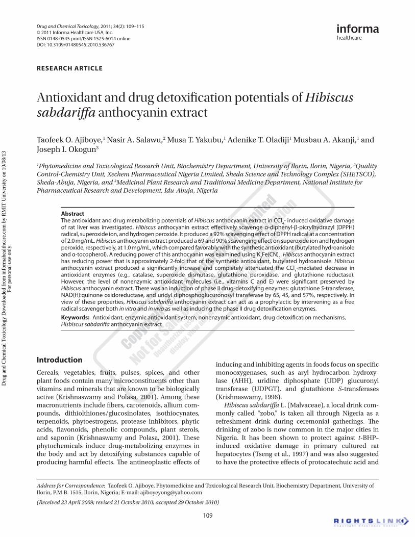
In vitro studyEffect of Hibiscus anthocyanin extract on DPPH radicalsFigure 2 shows the scavenging effect of Hibiscus antho-cyanin extract on DPPH radicals. The scavenging effect
1009080706050403020100
% In
hibi
tion
0.2 0.5 1 2Concentration (mg/ml)
H. sabdariffaAnthocyaninButylatedHydroanisoleα-Tocopherol
Figure 2. Scavenging effect of H. sabdariffa anthocyanins on DPPH radical.
R1 = R2:Pelargonidin
R1 = OH; R2= H:Cyanidin
R1 = R2 = OH:Delphinidin
R1 = OCH3; R2= OH:Petunidin
R1 = R2 = OCH3:Malvidin
R2
R1
O+
OH
OH
OH
HO
Figure 1. Anthocyanin.
Dru
g an
d C
hem
ical
Tox
icol
ogy
Dow
nloa
ded
from
info
rmah
ealth
care
.com
by
RM
IT U
nive
rsity
on
10/0
8/13
For
pers
onal
use
onl
y.
112 Taofeek O. Ajiboye et al.
Drug and Chemical Toxicology
of Hibiscus anthocyanin extract produced a marked scav-enging effect on DPPH radical in a dose-dependent man-ner, with the highest percentage (92.3%) observed for the highest dose. While α-tocopherol produced a scavenging effect, which was almost as effective as Hibiscus antho-cyanin extract, the scavenging effect of BHA could not compare with them, with its highest concencentration exhibiting only 81.4%.
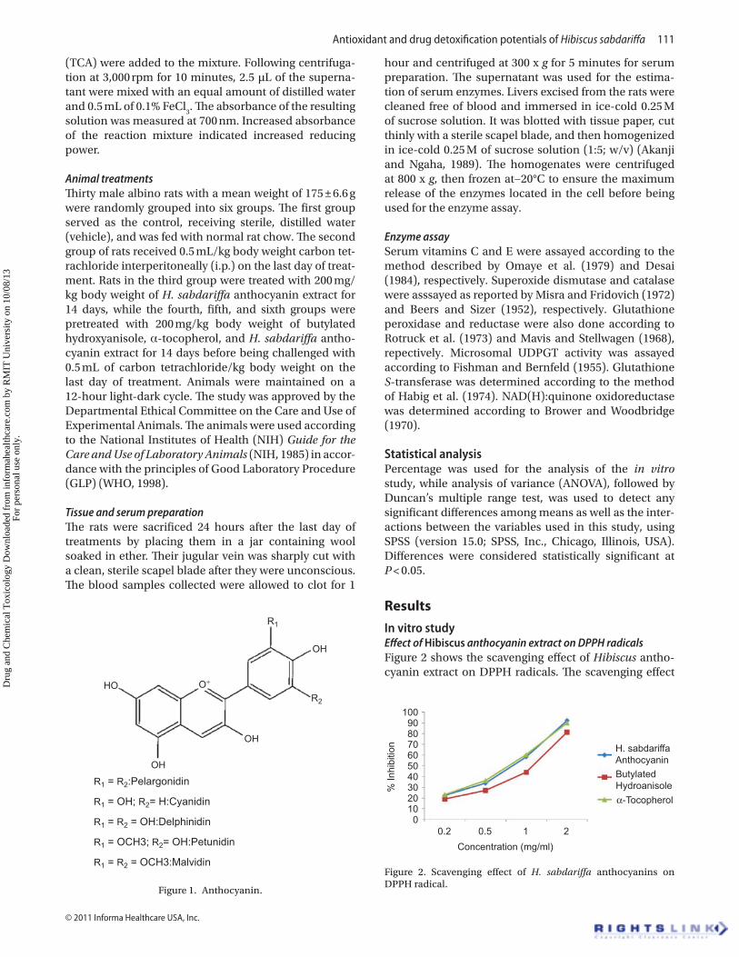
Effect of Hibiscus anthocyanin on superoxide ionHibiscus anthocyanin extract, BHA, and α-tocopherol demonstrated the ability to scavenge superoxide ion in vitro (Figure 3). The Hibiscus anthocyanin extract was the most effective of the three, with its highest concen-tration demonstrating a 68.5% scavenging effect, while α-tocopherol and BHA had their highest dose exhibit-ing a 66.5 and 57.7% scavenging effect, respectively, on superoxide ion.
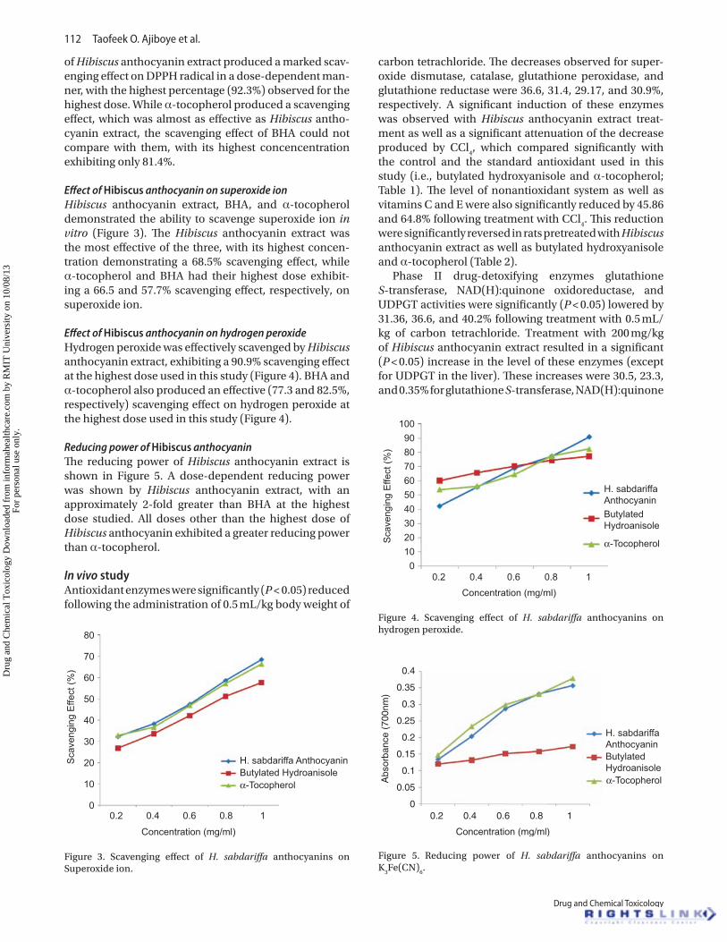
Effect of Hibiscus anthocyanin on hydrogen peroxideHydrogen peroxide was effectively scavenged by Hibiscus anthocyanin extract, exhibiting a 90.9% scavenging effect at the highest dose used in this study (Figure 4). BHA and α-tocopherol also produced an effective (77.3 and 82.5%, respectively) scavenging effect on hydrogen peroxide at the highest dose used in this study (Figure 4).
Reducing power of Hibiscus anthocyaninThe reducing power of Hibiscus anthocyanin extract is shown in Figure 5. A dose-dependent reducing power was shown by Hibiscus anthocyanin extract, with an approximately 2-fold greater than BHA at the highest dose studied. All doses other than the highest dose of Hibiscus anthocyanin exhibited a greater reducing power than α-tocopherol.
In vivo studyAntioxidant enzymes were significantly (P < 0.05) reduced following the administration of 0.5 mL/kg body weight of
carbon tetrachloride. The decreases observed for super-oxide dismutase, catalase, glutathione peroxidase, and glutathione reductase were 36.6, 31.4, 29.17, and 30.9%, respectively. A significant induction of these enzymes was observed with Hibiscus anthocyanin extract treat-ment as well as a significant attenuation of the decrease produced by CCl
4, which compared significantly with
the control and the standard antioxidant used in this study (i.e., butylated hydroxyanisole and α-tocopherol; Table 1). The level of nonantioxidant system as well as vitamins C and E were also significantly reduced by 45.86 and 64.8% following treatment with CCl
4. This reduction
were significantly reversed in rats pretreated with Hibiscus anthocyanin extract as well as butylated hydroxyanisole and α-tocopherol (Table 2).
Phase II drug-detoxifying enzymes glutathione S-transferase, NAD(H):quinone oxidoreductase, and UDPGT activities were significantly (P < 0.05) lowered by 31.36, 36.6, and 40.2% following treatment with 0.5 mL/kg of carbon tetrachloride. Treatment with 200 mg/kg of Hibiscus anthocyanin extract resulted in a significant (P < 0.05) increase in the level of these enzymes (except for UDPGT in the liver). These increases were 30.5, 23.3, and 0.35% for glutathione S-transferase, NAD(H):quinone
80
70
60
50
40
30
20
10
0
Sca
veng
ing
Effe
ct (%
)
0.2 0.4 0.6 0.8 1
Concentration (mg/ml)
H. sabdariffa AnthocyaninButylated Hydroanisoleα-Tocopherol
Figure 3. Scavenging effect of H. sabdariffa anthocyanins on Superoxide ion.
8090
100
706050403020100
Sca
veng
ing
Effe
ct (%
)
0.2 0.4 0.6 0.8 1Concentration (mg/ml)
H. sabdariffaAnthocyaninButylatedHydroanisole
α-Tocopherol
Figure 4. Scavenging effect of H. sabdariffa anthocyanins on hydrogen peroxide.
H. sabdariffaAnthocyaninButylatedHydroanisoleα-Tocopherol
0.4
0.35
0.3
0.25
0.2
0.15
0.1
0.05
0
Abs
orba
nce
(700
nm)
0.2 0.4 0.6 0.8 1
Concentration (mg/ml)
Figure 5. Reducing power of H. sabdariffa anthocyanins on K
3Fe(CN)
6.
Dru
g an
d C
hem
ical
Tox
icol
ogy
Dow
nloa
ded
from
info
rmah
ealth
care
.com
by
RM
IT U
nive
rsity
on
10/0
8/13
For
pers
onal
use
onl
y.
Antioxidant and drug detoxification potentials of Hibiscus sabdariffa 113
© 2011 Informa Healthcare USA, Inc.
oxidoreductase, and UDPGT, respectively. The decrease observed was significantly attenuated by pretreatment (Table 3).
Discussion
The consumption of dietary antioxidants plays a sig-nificant role in the reduction of the incidence of cancer and free radical–related complications associated with a variety of diseases, such as cardiovascular disease, liver disease, and diabetes, among many others (Ajiboye et al., 2010). This effect results from the interactions of the dietary antioxidant with one or more biomolecules at cellular and molecular levels or both, leading to the induction of certain enzymes as well as inhibition or inactivation of some enzymes (Ajiboye et al., 2010). This study thus provides the antioxidant potentials of Hibiscus anthocyanin extract using a wide range of in vitro assay protocols, the antioxidant systems, and drug detoxifi-cation mechanisms, with the aim toward providing an additional phytomedicine that can manage oxidative-stress–associated diseases.
Polyphenols have a well-recognized antioxidant capacity both in vitro and in vivo (Rice-Evans, 2001). This antioxidant activity has been attributed mainly to their capacity to scavenge oxygen and nitrogen active species (Bors et al., 1990) and to chelate redox-active metals (van Acker, 1998).
Free radical chain reaction, a common method of lipid peroxidation (LPO), causes some deleterious alterations to cellular molecules, ranging from the polyunsaturated fatty acid of the plasma membrane to the macromol-ecules located in the cells. Radical scavengers interfere in LPO by terminating the chain reaction (Bran-Williams et al., 1995). Hibiscus anthocyanin extract scavenges the DPPH radical by 92.1% at the highest dose, which proved to be more effective than the known antioxidant used in this study. This could be an electron-donating capability effect of the Hibiscus anthocyanin extract, thus forming a stable product and, consequently, terminating free radi-cal chain reaction.
Reactive oxygen species (ROS) hydroxyl (OH) and superoxide (O
2) radicals, and hydrogen peroxide (H
2O
2)
generated in biological systems are counteracted by the existing enzymic antioxidant system of bio-logical systems (e.g., catalase, superoxide dismutase, glutathione peroxidase, glutathione reductase, and
glucose-6-phosphate dehydrogenase), thus prevent-ing damage to cellular macromolecules (Ajiboye et al., 2010). This action is, however, exhaustive, and dietary antioxidants can play a significant role in preventing this damage upon absorption in vivo. Also, Dahl and Richardson (1978) have implicated singlet oxygen (O.), hydroxyl radical (Czapski, 1984), and hydrogen per-oxide (formed from superoxide) to initiate LPO. Thus, the strong scavenging effect of Hibiscus anthocyanin extract on superoxide ion as well as hydrogen perox-ide is an implication of its being possible in terminat-ing LPO, which could arise from superoxide (Yen and Duh, 1994) and the stronger ROS (e.g., singlet oxygen, hydroxyl radical, and hydrogen peroxide). If effectively absorbed into the blood system, Hibiscus anthocyanin extract could prevent cellular and oxidative damage arising from superoxide ion as well as its stronger, more active products.
Hibiscus anthocyanin extract displays a significant reducing power potential on K
3Fe(CN)
6. This shows it to
effectively halt the oxidation of cellular macromolecules by oxidizing molecules that could arise from the metabo-lism of either drugs or toxins.
Reactive species of oxygen and nitrogen are coun-teracted by the enzymic antioxidant system (such as superoxide dismutase, catalase, glutathione peroxidase, and glutathione reductase) in the body, thus preventing damage of cellular macromolecules (Ajiboye et al., 2010). These macromolecules are prevented from LPO, base modification, DNA cross-links, and covalent binding to protein scavenging the reactive species of oxygen (e.g., O
2, OH, and H
2O
2) generated. Superoxide dismutase
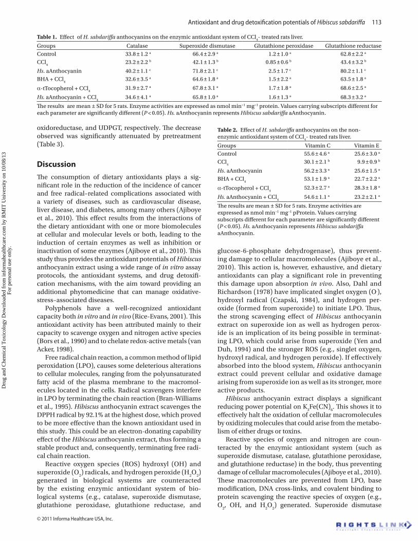
Table 1. Effect of H. sabdariffa anthocyanins on the enzymic antioxidant system of CCl4- treated rats liver.
Groups Catalase Superoxide dismutase Glutathione peroxidase Glutathione reductaseControl 33.8 ± 1.2 a 66.4 ± 2.9 a 1.2 ± 1.0 a 62.8 ± 2.2 a
CCl4
23.2 ± 2.2 b 42.1 ± 1.3 b 0.85 ± 0.6 b 43.4 ± 3.2 b
Hs. aAnthocyanin 40.2 ± 1.1 c 71.8 ± 2.1 c 2.5 ± 1.7 c 80.2 ± 1.1 c
BHA + CCl4
32.6 ± 3.5 a 64.6 ± 1.8 a 1.5 ± 2.2 a 63.5 ± 1.8 a
α-tTocopherol + CCl4
31.9 ± 2.7 a 67.8 ± 3.1 a 1.7 ± 1.8 a 68.6 ± 2.5 a
Hs. aAnthocyanin + CCl4
34.6 ± 4.1 a 65.8 ± 1.0 a 1.6 ± 1.3 a 68.3 ± 3.2 a
The results are mean ± SD for 5 rats. Enzyme activities are expressed as nmol min−1 mg−1 protein. Values carrying subscripts different for each parameter are significantly different (P < 0.05). Hs. aAnthocyanin represents Hibiscus sabdariffa aAnthocyanin.
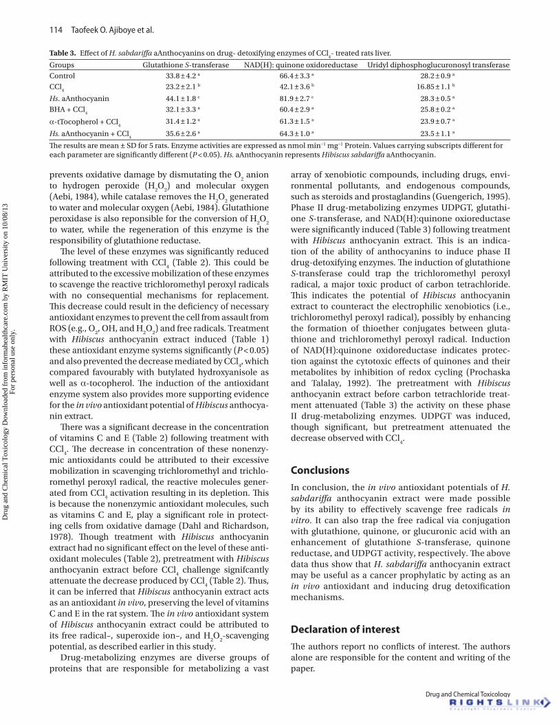
Table 2. Effect of H. sabdariffa anthocyanins on the non-enzymic antioxidant system of CCl
4- treated rats liver.
Groups Vitamin C Vitamin EControl 55.6 ± 4.6 a 25.6 ± 3.0 a
CCl4
30.1 ± 2.1 b 9.9 ± 0.9 b
Hs. aAnthocyanin 56.2 ± 3.3 a 25.6 ± 1.5 a
BHA + CCl4
53.1 ± 1.9 a 22.7 ± 2.2 a
α-tTocopherol + CCl4
52.3 ± 2.7 a 28.3 ± 1.8 a
Hs. aAnthocyanin + CCl4
54.6 ± 1.1 a 23.2 ± 2.1 a
The results are mean ± SD for 5 rats. Enzyme activities are expressed as nmol min−1 mg−1 pProtein. Values carrying subscripts different for each parameter are significantly different (P < 0.05). Hs. aAnthocyanin represents Hibiscus sabdariffa aAnthocyanin.
Dru
g an
d C
hem
ical
Tox
icol
ogy
Dow
nloa
ded
from
info
rmah
ealth
care
.com
by
RM
IT U
nive
rsity
on
10/0
8/13
For
pers
onal
use
onl
y.
114 Taofeek O. Ajiboye et al.
Drug and Chemical Toxicology
prevents oxidative damage by dismutating the O2 anion
to hydrogen peroxide (H2O
2) and molecular oxygen
(Aebi, 1984), while catalase removes the H2O
2 generated
to water and molecular oxygen (Aebi, 1984). Glutathione peroxidase is also reponsible for the conversion of H
2O
2
to water, while the regeneration of this enzyme is the responsibility of glutathione reductase.
The level of these enzymes was significantly reduced following treatment with CCl
4 (Table 2). This could be
attributed to the excessive mobilization of these enzymes to scavenge the reactive trichloromethyl peroxyl radicals with no consequential mechanisms for replacement. This decrease could result in the deficiency of necessary antioxidant enzymes to prevent the cell from assault from ROS (e.g., O
2, OH, and H
2O
2) and free radicals. Treatment
with Hibiscus anthocyanin extract induced (Table 1) these antioxidant enzyme systems significantly (P < 0.05) and also prevented the decrease mediated by CCl
4, which
compared favourably with butylated hydroxyanisole as well as α-tocopherol. The induction of the antioxidant enzyme system also provides more supporting evidence for the in vivo antioxidant potential of Hibiscus anthocya-nin extract.
There was a significant decrease in the concentration of vitamins C and E (Table 2) following treatment with CCl
4. The decrease in concentration of these nonenzy-
mic antioxidants could be attributed to their excessive mobilization in scavenging trichloromethyl and trichlo-romethyl peroxyl radical, the reactive molecules gener-ated from CCl
4 activation resulting in its depletion. This
is because the nonenzymic antioxidant molecules, such as vitamins C and E, play a significant role in protect-ing cells from oxidative damage (Dahl and Richardson, 1978). Though treatment with Hibiscus anthocyanin extract had no significant effect on the level of these anti-oxidant molecules (Table 2), pretreatment with Hibiscus anthocyanin extract before CCl
4 challenge signifcantly
attenuate the decrease produced by CCl4 (Table 2). Thus,
it can be inferred that Hibiscus anthocyanin extract acts as an antioxidant in vivo, preserving the level of vitamins C and E in the rat system. The in vivo antioxidant system of Hibiscus anthocyanin extract could be attributed to its free radical–, superoxide ion–, and H
2O
2-scavenging
potential, as described earlier in this study.Drug-metabolizing enzymes are diverse groups of
proteins that are responsible for metabolizing a vast
array of xenobiotic compounds, including drugs, envi-ronmental pollutants, and endogenous compounds, such as steroids and prostaglandins (Guengerich, 1995). Phase II drug-metabolizing enzymes UDPGT, glutathi-one S-transferase, and NAD(H):quinone oxioreductase were significantly induced (Table 3) following treatment with Hibiscus anthocyanin extract. This is an indica-tion of the ability of anthocyanins to induce phase II drug-detoxifying enzymes. The induction of glutathione S-transferase could trap the trichloromethyl peroxyl radical, a major toxic product of carbon tetrachloride. This indicates the potential of Hibiscus anthocyanin extract to counteract the electrophilic xenobiotics (i.e., trichloromethyl peroxyl radical), possibly by enhancing the formation of thioether conjugates between gluta-thione and trichloromethyl peroxyl radical. Induction of NAD(H):quinone oxidoreductase indicates protec-tion against the cytotoxic effects of quinones and their metabolites by inhibition of redox cycling (Prochaska and Talalay, 1992). The pretreatment with Hibiscus anthocyanin extract before carbon tetrachloride treat-ment attenuated (Table 3) the activity on these phase II drug-metabolizing enzymes. UDPGT was induced, though significant, but pretreatment attenuated the decrease observed with CCl
4.
Conclusions
In conclusion, the in vivo antioxidant potentials of H. sabdariffa anthocyanin extract were made possible by its ability to effectively scavenge free radicals in vitro. It can also trap the free radical via conjugation with glutathione, quinone, or glucuronic acid with an enhancement of glutathione S-transferase, quinone reductase, and UDPGT activity, respectively. The above data thus show that H. sabdariffa anthocyanin extract may be useful as a cancer prophylatic by acting as an in vivo antioxidant and inducing drug detoxification mechanisms.
Declaration of interest
The authors report no conflicts of interest. The authors alone are responsible for the content and writing of the paper.
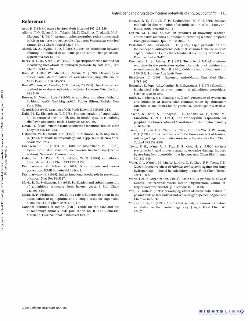
Table 3. Effect of H. sabdariffa aAnthocyanins on drug- detoxifying enzymes of CCl4- treated rats liver.
Groups Glutathione S-transferase NAD(H): quinone oxidoreductase Uridyl diphosphoglucuronosyl transferaseControl 33.8 ± 4.2 a 66.4 ± 3.3 a 28.2 ± 0.9 a
CCl4
23.2 ± 2.1 b 42.1 ± 3.6 b 16.85 ± 1.1 b
Hs. aAnthocyanin 44.1 ± 1.8 c 81.9 ± 2.7 c 28.3 ± 0.5 a
BHA + CCl4
32.1 ± 3.3 a 60.4 ± 2.9 a 25.8 ± 0.2 a
α-tTocopherol + CCl4
31.4 ± 1.2 a 61.3 ± 1.5 a 23.9 ± 0.7 a
Hs. aAnthocyanin + CCl4
35.6 ± 2.6 a 64.3 ± 1.0 a 23.5 ± 1.1 a
The results are mean ± SD for 5 rats. Enzyme activities are expressed as nmol min−1 mg−1 Protein. Values carrying subscripts different for each parameter are significantly different (P < 0.05). Hs. aAnthocyanin represents Hibiscus sabdariffa aAnthocyanin.
Dru
g an
d C
hem
ical
Tox
icol
ogy
Dow
nloa
ded
from
info
rmah
ealth
care
.com
by
RM
IT U
nive
rsity
on
10/0
8/13
For
pers
onal
use
onl
y.
Antioxidant and drug detoxification potentials of Hibiscus sabdariffa 115
© 2011 Informa Healthcare USA, Inc.
ReferencesAebi, H. (1984). Catalase in vitro. Meth Enzymol 105:121–126.Ajiboye, T. O., Salau, A. K., Yakubu, M. T., Oladiji, A. T., Akanji, M. A.,
Okogun, J. I. (2010). Acetaminophen perturbed redox homeostasis in Wistar rat liver: protective role of aqueous Pterocarpus osun leaf extract. Drug Chem Toxicol 33:77–87.
Akanji, M. A., Ngaha, E. O. (1989). Studies on correlation between chloroquine-induced tissue damage and serum changes in rats. Experientia 45:143–146.
Beers, R. F., Jr., Sizer, I. W. (1952). A spectrophotmetric method for measuring breakdown of hydrogen peroxide by catalase. J Biol Chem 195:133–140.
Bors, W., Heller, W., Michel, C., Saran, M. (1990). Flavonoids as antioxidants: determination of radical-scavenging efficiencies. Meth Enzymol 186:343–355.
Bran-Williams, W., Cuvelier, M. E., Berset, C. (1995). Use of free radical method to evaluate antioxidant activity. Lebensm Wiss Technol 28:25–30.
Brower, M., Woodbridge, J. (1970). A rapid determination of ethanol in blood. AACC Natl Mtg, AACC, Statler Hilton, Buffalo, New York, USA.
Czapski, G. (1984). Reaction of OH. Meth Enzymol 105:209–214.Dahl, M. K., Richardson, T. (1978). Photogeneration of superioxide
ion in serum of bovine milk and in model systems containing riboflavin and amino acids. J Dairy Sci 61:400–407.
Desai, I. D. (1984). Vitamin E analysis method for animal tissues. Meth Enzymol 105:138–143.
Fishmann, W. H., Bernfeld, P. (1955). In: Colowick, S. P., Kaplan, N. O. (Eds.), Methods in enzymology, vol. 1 (pp 262–264). New York: Academic Press.
Guengerich, F. P. (1995). In: Oritiz de Montellano, P. R. (Ed.), Cytochrome P450: structure, mechanism, biochemistry (second edition). New York: Plenum Press.
Habig, W. H., Pabst, M. J., Jakoby, W. B. (1974) Glutathione S-transferase. J Biol Chem 249:7130–7139.
Krishnaswamy, K., Polasa, K. (2001). Non-nutrients and cancer prevention. ICMR Bulletin Vol 31:No. 1.
Krishnaswamy, K. (1996). Indian functional foods: role in prevention of cancer. Nutr Rev 54:S127.
Mavis, R. D., Stellwagen, E. (1968). Purifcation and subunit structure of glutathione reductase from bakers’ yeast. J Biol Chem 243:809–814.
Misra, H. P., Fridovich, I. (1972). The role of superoxide anion in the autoxidation of epinephrine and a simple assay for superoxide dismutase. J Biol Chem 247:3170–3175.
National Institutes of Health. (1985). Guide for the care and use of laboratory animals. NIH publication no. 83–127. Bethesda, Maryland, USA: National Institutes of Health.
Omaye, S. T., Turbull, T. P., Sauberchich, H. C. (1979). Selected methods for determination of ascorbic acid in cells, tissues, and fluids. Meth Enzymol 6:3–11.
Oyaizu, M. (1986). Studies on products of browning reaction: antioxidative activities of product of browning reaction prepared from glucosamine. Jpn J Nut 44:307–315.
Pesh-Imam, M., Recknagel, R. O. (1977). Lipid peroxidation and the concept of antioxygenic potential: vitamin E change in acute experimental CCl4 and ethanol-induced liver injury. Toxicol Appl Pharmacol 42:463–475.
Prochaska, H. J., Talalay, P. (1992). The role of NAD(H):quinone reductase in the protection against the toxicity of quinine and related agents. In: Sies, H. (Ed.), Oxidants and antioxidants (pp 195–211). London: Academic Press.
Rice-Evans, C. (2001). Flavonoid antioxidants. Curr Med Chem 8:797–807.
Rotruck, J. T., Pope, A. L., Ganther, H. F., Swason, A. B. (1973). Selenium: biochemical role as a component of glutathione peroxidase. Science 179:588–590.
Ruch, K. J., Cheng, S. J., Klaunig, J. E. (1989). Prevention of cytotoxicity and inhibition of intercellular communication by antioxidant catechin isolated from Chinese green tea. Carcinogenesis 10:1003– 1008.
Takeda, K., Sato, S., Kobayashi, H., Kanaitsuka, V., Uene, M., Kinoshita, T., et al. (1994). The anthocyanin responsible for purplish blue flower colour of Aconitum chinense Phytochemistry 36:613–616.
Tseng, T. H., Kao, E. S., Chu, C. Y., Chou, F. P., Lin Wu, H. W., Wang, C. J. (1997). Protective effects of dried flower extracts of Hibiscus sabdariffa L. against oxidative stress in rat hepatocytes. Food Chem Toxicol 35:1159–1164.
Tseng, T. H., Wang, C. J., Kao, E. S., Chu, H. Y. (1996.) Hibiscus protocatechuic acid protects aagainst oxidative damage induced by tert-butylhydroperoxide in rat hepatocytes. Chem Biol Interact 101:137–148.
Wang, C. J., Wang, J. M., Lin, W. L., Chu, C. Y., Chou, F. P., Tseng, T. H. (2000). Protective effect of Hibiscus anthocyanin against tert-butyl hydroperoxide-induced hepatic injury in rats. Food Chem Toxicol 38:411–416.
World Health Organization. (1998). Basic OECD principles of GLP. Geneva, Switzerland: World Health Organization. Online at: http://www.who/int/tdr/publications 02-01-2008.
Yen, G., Duh, P. (1994). Scavenging effect of methanolic extract of peanut hulls on free radical and active oxygen species. J Agric Food Chem 42:629–632.
Yen, G., Chen, H. (1995). Antioxidant activity of various tea extract in relation to their antimutagenicity. J Agric Food Chem 43: 27–32.
Dru
g an
d C
hem
ical
Tox
icol
ogy
Dow
nloa
ded
from
info
rmah
ealth
care
.com
by
RM
IT U
nive
rsity
on
10/0
8/13
For
pers
onal
use
onl
y.


![Genetic Dissection of a Major Anthocyanin QTL Contributing ... · anthocyanin (pink) pigment was estimated as [(R + B)/2] 2 G. QTL affecting anthocyanin concentration in the backcross](https://img.pdfslide.us/doc/110x75/5e6421962a91715ff42dfa60/genetic-dissection-of-a-major-anthocyanin-qtl-contributing-anthocyanin-pink.jpg)