Embed Size (px)
Citation preview
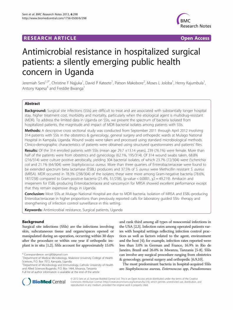
RESEARCH ARTICLE Open Access
Antimicrobial resistance in hospitalized surgicalpatients: a silently emerging public healthconcern in UgandaJeremiah Seni1,3*, Christine F Najjuka1, David P Kateete1, Patson Makobore2, Moses L Joloba1, Henry Kajumbula1,Antony Kapesa4 and Freddie Bwanga1
Abstract
Background: Surgical site infections (SSIs) are difficult to treat and are associated with substantially longer hospitalstay, higher treatment cost, morbidity and mortality, particularly when the etiological agent is multidrug-resistant(MDR). To address the limited data in Uganda on SSIs, we present the spectrum of bacteria isolated fromhospitalized patients, the magnitude and impact of MDR bacterial isolates among patients with SSIs.
Methods: A descriptive cross sectional study was conducted from September 2011 through April 2012 involving314 patients with SSIs in the obstetrics & gynecology, general surgery and orthopedic wards at Mulago NationalHospital in Kampala, Uganda. Wound swabs were taken and processed using standard microbiological methods.Clinico-demographic characteristics of patients were obtained using structured questionnaires and patients’ files.
Results: Of the 314 enrolled patients with SSIs (mean age 29.7 ±13.14 years), 239 (76.1%) were female. More thanhalf of the patients were from obstetrics and gynecology (62.1%, 195/314). Of 314 wound swabs taken, 68.8%(216/314) were culture positive aerobically, yielding 304 bacterial isolates; of which 23.7% (72/304) were Escherichiacoli and 21.1% (64/304) were Staphylococcus aureus. More than three quarters of Enterobacteriaceae were found tobe extended spectrum beta lactamase (ESBL) producers and 37.5% of S. aureus were Methicillin resistant S. aureus(MRSA). MDR occurred in 78.3% (238/304) of the isolates; these were more among Gram-negative bacteria (78.6%,187/238) compared to Gram-positive bacteria (21.4%, 51/238), (p-value < 0.0001, χ2 = 49.219). Amikacin andimepenem for ESBL-producing Enterobacteriacea and vancomycin for MRSA showed excellent performance exceptthat they remain expensive drugs in Uganda.
Conclusion: Most SSIs at Mulago National Hospital are due to MDR bacteria. Isolation of MRSA and ESBL-producingEnterobacteriaceae in higher proportions than previously reported calls for laboratory guided SSIs- therapy andstrengthening of infection control surveillance in this setting.
Keywords: Antimicrobial resistance, Surgical patients, Uganda
BackgroundSurgical site infections (SSIs) are the infections involvingskin, subcutaneous tissue and organs/spaces opened ormanipulated during an operation, occurring within 30 daysafter the procedure or within one year if orthopedic im-plant is in situ [1,2]. SSIs account for approximately 15.0%
and rank third among all types of nosocomial infections inthe USA [2,3]. Infection rates among operated patients var-ies with hospital settings reflecting infection control prac-tices as well as factors related to the agent, environmentand the host [4]; for example, infection rates reported wereless than 3.0% in German and France, 16.9% in Rio deJaneiro, Brazil and 26.0% in Mwanza, Tanzania [5-8]. SSIscan involve any surgical procedure ranging from obstetrics& gynecology, general surgery and orthopedic [6,9,10].The most predominant bacteria in hospital-acquired SSIs
are Staphylococcus aureus, Enterococcus spp, Pseudomonas
* Correspondence: [email protected] of Medical Microbiology, Makerere University College of HealthSciences, P.O. Box 7072, Kampala, Uganda3Department of Microbiology and Immunology, Catholic University of Healthand Allied Sciences-Bugando, P.O. Box 1464, Mwanza, TanzaniaFull list of author information is available at the end of the article
© 2013 Seni et al.; licensee BioMed Central Ltd. This is an Open Access article distributed under the terms of the CreativeCommons Attribution License (http://creativecommons.org/licenses/by/2.0), which permits unrestricted use, distribution, andreproduction in any medium, provided the original work is properly cited.
Seni et al. BMC Research Notes 2013, 6:298http://www.biomedcentral.com/1756-0500/6/298
aeruginosa, Escherichia coli, and other Enterobacteriaceae;of these, single bacterial isolates are common whereas 9.0%to 27.0% of bacterial isolates from different surgical sites areattributed to polymicrobial [7-9,11]. These infections posetherapeutic challenges and are associated with substantiallylonger duration of hospital stay, increased hospital cost,higher morbidity and mortality [5,12], particularly when theagents are Methicillin resistant S. aureus (MRSA), Extendedspectrum beta lactamase (ESBL) producing Enterobacte-riaceae and/or other agents collectively referred to asmultidrug-resistant (MDR) [11,13,14]. Studies from devel-oping countries have shown high level of resistance (ran-ging from 50 to 100%) to the commonly used antibioticslike ampicillin, trimethoprim – sulphamethoxazole, genta-micin, chloramphenicol and third generation cephalospo-rins among S. aureus, E. coli, and P. aeruginosa [8,15] asopposed to low rates of resistance ranging from 0-50%in developed countries [16]. In both settings however, sub-stantial rates of resistance to oxacillin, erythromycin andclindamycin reported for S. aureus, ranged from 10-60%[8,9,15,16] whereas vancomycin (for S. aureus and otherGram-positive bacteria), amikacin, piperacillin-tazobactamand imepenem (for E. coli, P. aeruginosa and other Gram-negative bacteria) showed resistant rates of less than 25%[17,18].It is well known that specific therapeutic options to
patients with SSIs are largely dependent on data fromantimicrobial sensitivity tests generated by clinical la-boratories or sound epidemiological data from ongoingnosocomial infection surveillance [6,11,19].In Uganda, about 10% of the surgical procedures be-
come septic accounting for an increasing morbidity andmortality, with the commonest organism isolated beingS. aureus [20-22]; however data on the spectrum of bac-teria isolated from hospitalized patients and their anti-microbial susceptibility patterns to guide SSI-therapy inMulago National Hospital remains scanty. Furthermore,the magnitude and impact of MDR bacteria from SSIsare unknown. Thus, this study aimed at addressing theseareas. Data herein will be crucial in guiding SSIs-therapyand will form a baseline for nosocomial SSIs surveillance.
MethodsStudy design and sampling processThis descriptive cross-sectional study was conducted atMulago National Hospital in Kampala, Uganda. The hos-pital is located on Mulago Hill in the northern part of thecity of Kampala and is the largest hospital in Uganda withan estimated 1,500 beds.The study was conducted for a period of 8 months
from September 2011 to April 2012 and involved 314patients with clinical SSIs who consented to participate.The patients were from obstetrics & gynecology, generalsurgery and orthopedic wards. All patients with SSIs
occurring within 30 days after the operative procedureor within one year if orthopedic implant was in situ wereincluded, whereas surgical patients with community-acquired pyogenic infections such as abscess, furuncle andcarbuncles; patients with infection of an episiotomy; andpatients with open fractures were excluded from the study.
Study clearance and ethical considerationsThe study got ethical clearance from the InstitutionalReview Board (IRB) of Makerere University College ofHealth Sciences (# REC REF 2011–183), Mulago Hos-pital Research Committee (MREC #125) and the UgandaNational Council for Science and Technology (UNCST)(REF # HS 1080). A written informed consent from eachpatient/caretaker and assent for minors (11 to 17years)were obtained whereas for each minor (<11 years), con-sent was obtained from his/her parent or caretaker. Allpatient information was kept confidential and anonym-ous using codes.
Data collection and laboratory proceduresDemographic and clinical characteristics from patients werecollected using structured questionnaire and from patients’files (see Additional file 1). The infected site was cleanedusing normal saline and sterile gauze then, from each pa-tient, two wound swabs were collected using sterile cottonswabs in Amies transport media (Biolab, HUNGARY®).
Isolate identificationWound swabs were processed in the bacteriology labora-tory of the Department of Medical Microbiology, MakerereUniversity College of Health Sciences, within 2 hours ofcollection. The first wound swab was used to make Gramstain smears while the second one was inoculated intoblood agar, MacConkey agar, and mannitol-salt agar andincubated at 35-37°C for 24–48 hours. Identification ofbacteria was based on conventional physiological and bio-chemical methods such as Gram stain, catalase reaction,coagulase test, DNase test, hemolytic activity on sheepblood agar plates, bacitracin, optochin and trimethoprim-sulphamethoxazole (SXT) antimicrobial identification disksand bile esculin test for Gram-positive bacteria. Gram-negative bacteria were identified based on colony morph-ology on blood agar and MacConkey agar, followed bybiochemical reactions namely oxidase, triple sugar iron(TSI), sulphur indole and motility (SIM), citrate, and ure-ase tests [23].
Drug susceptibility testsFollowing identification of the bacterial isolates, astandard disc diffusion technique for drug susceptibilitytest (DST) was performed as recommended by Clinicaland Laboratory Standard Institute (CLSI) [24]. For Gram-positive bacteria, discs (Biolab®, HUNGARY) tested were
Seni et al. BMC Research Notes 2013, 6:298 Page 2 of 7http://www.biomedcentral.com/1756-0500/6/298
ampicillin (10 μg), oxacillin (1 μg), trimethoprim-sul-phamethoxazole (1.25/23.75 μg), tetracycline (30 μg),ciprofloxacin (5 μg), chloramphenicol (30 μg), gentami-cin (10 μg) [high level gentamicin (120 μg) for Entero-coccus spp], erythromycin (15 μg), clindamycin (2 μg),and vancomycin (30 μg). For Gram-negative bacteriadiscs (Biolab®, HUNGARY) tested included ampicillin(10 μg), piperacillin(100 μg), piperacillin-tazobactam(100/10 μg), amoxicillin-clavulanic acid (20/10 μg),trimethoprim-sulphamethoxazole (1.25/23.75 μg), tetra-cycline (30 μg), ciprofloxacin (5 μg), chloramphenicol(30 μg), gentamicin (10 μg), amikacin (30 μg), ceftriax-one (30 μg), ceftazidime (30 μg), cefepime (30 μg), andimipenem (10 μg). These were incubated at 35-37°C for24 hours.Isolates which were not identifiable by the standard
conventional methods, colistin DST for all Acinetobacterspp and P. aeruginosa as well as vancomycin DST for allS. aureus were confirmed using the Phoenix Automatedinstrument® (Becton-Dickson, Sparks Maryland) as permanufacturer’s instruction.For determining inducible clindamycin resistance, clin-
damycin disk (2 μg) and erythromycin disk (15 μg) wereplaced side by side approximately 15-26 mm apart. Flat-tening of the zone of inhibition adjacent to the eryth-romycin disk was regarded as a positive D-test. Asrecommended by CLSI, isolates were screened for ESBLproduction using the double disc method and MRSA wasidentified by the use of cefoxitin disc (30 μg) [24,25].MDR was defined as an isolate with resistance to three ormore antimicrobial classes [26].Results on isolate identity and antimicrobial suscepti-
bility patterns were promptly reported to the attendingdoctor for patient care.
Quality controlReference strains S. aureus ATCC 25923 and Staphylo-coccus epidermidis ATCC 12228 for Gram-positive bac-teria and E. coli ATCC 25922 and P. aeruginosa ATCC27853 for Gram-negative bacteria were used to quality-control microbiological procedures such as Gram staining ,growth of bacteria on respective media, microscopy, bio-chemical identification tests and drug susceptibility testing.
Data analysisVariables from the clinical and demographic data in thequestionnaire and laboratory data were entered intoExcel®, cleaned and exported to STATA software version11 (College Station, Texas, USA) for analysis accordingto the objectives of the study. Continuous variables weredescribed as mean (± standard deviation). Categoricalvariables were described as proportion and were ana-lyzed to compare the significance of difference in distri-bution by using Chi square test or Fischer’s exact test
where appropriate. To determine factors associated withbacteria isolation from SSI, we used univariate followedby multivariate logistic regression analysis. At univariatelevel all factors which had a p-value of less than 0.05were subjected to multivariate analysis. The strength ofassociation between factors and outcome was measuredusing odds ratio with respective 95% confidence interval.Factors with p-value of less than 0.05 on multivariate lo-gistic regression analysis were considered as independentassociation of bacteria isolation from SSI.
ResultsThis study enrolled 314 patients with clinical SSIs. Amongthese, 239 (76.1%) were female. The overall mean age was29.7 ± 13.14 years (minimum 12 and maximum 83 years).More than half of the patients were from obstetrics andgynecology wards, 62.1% (195/314), whereas 33.1% (104/314) and 4.8% (15/314) were from general surgery andorthopedic wards respectively. The most common surgicalprocedures were caesarean section 46.2% (145/314) andlaparotomy 42.7% (134/314); open reduction and internalfixation (ORIF) accounted for 3.5% (11/314) while othersurgical procedures contributed 7.6% (24/314).Of the 314 non-repeat wound swabs collected, 216
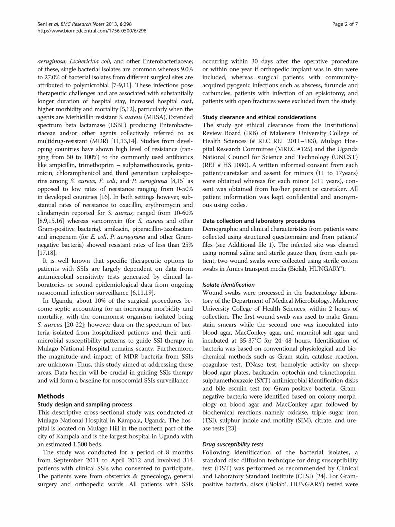
(68.8%) were culture positive aerobically. The most pre-dominant bacterial isolates were E. coli, 23.7% (72/304)and S. aureus, 21.1% (64/304) (see Figure 1). Single bac-terial isolates were recovered from 137 (63.4%) patientswhereas 79 (36.6%) had polymicrobial infections.All P. aeruginosa isolates were sensitive to colistin,
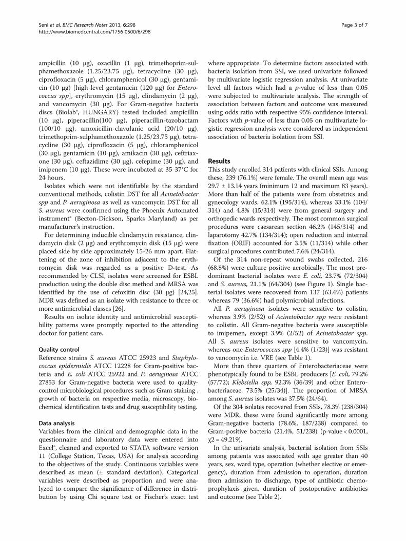
whereas 3.9% (2/52) of Acinetobacter spp were resistantto colistin. All Gram-negative bacteria were susceptibleto imipemen, except 3.9% (2/52) of Acinetobacter spp.All S. aureus isolates were sensitive to vancomycin,whereas one Enterococcus spp [4.4% (1/23)] was resistantto vancomycin i.e. VRE (see Table 1).More than three quarters of Enterobacteriaceae were
phenotypically found to be ESBL producers [E. coli, 79.2%(57/72); Klebsiella spp, 92.3% (36/39) and other Entero-bacteriaceae, 73.5% (25/34)]. The proportion of MRSAamong S. aureus isolates was 37.5% (24/64).Of the 304 isolates recovered from SSIs, 78.3% (238/304)
were MDR, these were found significantly more amongGram-negative bacteria (78.6%, 187/238) compared toGram-positive bacteria (21.4%, 51/238) (p-value < 0.0001,χ2 = 49.219).In the univariate analysis, bacterial isolation from SSIs
among patients was associated with age greater than 40years, sex, ward type, operation (whether elective or emer-gency), duration from admission to operation, durationfrom admission to discharge, type of antibiotic chemo-prophylaxis given, duration of postoperative antibioticsand outcome (see Table 2).
Seni et al. BMC Research Notes 2013, 6:298 Page 3 of 7http://www.biomedcentral.com/1756-0500/6/298
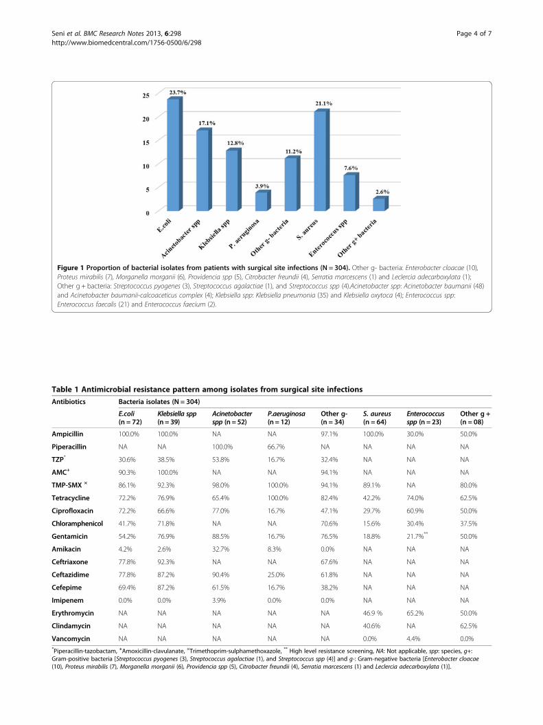
Figure 1 Proportion of bacterial isolates from patients with surgical site infections (N = 304). Other g- bacteria: Enterobacter cloacae (10),Proteus mirabilis (7), Morganella morganii (6), Providencia spp (5), Citrobacter freundii (4), Serratia marcescens (1) and Leclercia adecarboxylata (1);Other g + bacteria: Streptococcus pyogenes (3), Streptococcus agalactiae (1), and Streptococcus spp (4).Acinetobacter spp: Acinetobacter baumanii (48)and Acinetobacter baumanii-calcoaceticus complex (4); Klebsiella spp: Klebsiella pneumonia (35) and Klebsiella oxytoca (4); Enterococcus spp:Enterococcus faecalis (21) and Enterococcus faecium (2).
Table 1 Antimicrobial resistance pattern among isolates from surgical site infections
Antibiotics Bacteria isolates (N = 304)
E.coli(n = 72)
Klebsiella spp(n = 39)
Acinetobacterspp (n = 52)
P.aeruginosa(n = 12)
Other g-(n = 34)
S. aureus(n = 64)
Enterococcusspp (n = 23)
Other g +(n = 08)
Ampicillin 100.0% 100.0% NA NA 97.1% 100.0% 30.0% 50.0%
Piperacillin NA NA 100.0% 66.7% NA NA NA NA
TZP* 30.6% 38.5% 53.8% 16.7% 32.4% NA NA NA
AMC+ 90.3% 100.0% NA NA 94.1% NA NA NA
TMP-SMX × 86.1% 92.3% 98.0% 100.0% 94.1% 89.1% NA 80.0%
Tetracycline 72.2% 76.9% 65.4% 100.0% 82.4% 42.2% 74.0% 62.5%
Ciprofloxacin 72.2% 66.6% 77.0% 16.7% 47.1% 29.7% 60.9% 50.0%
Chloramphenicol 41.7% 71.8% NA NA 70.6% 15.6% 30.4% 37.5%
Gentamicin 54.2% 76.9% 88.5% 16.7% 76.5% 18.8% 21.7%** 50.0%
Amikacin 4.2% 2.6% 32.7% 8.3% 0.0% NA NA NA
Ceftriaxone 77.8% 92.3% NA NA 67.6% NA NA NA
Ceftazidime 77.8% 87.2% 90.4% 25.0% 61.8% NA NA NA
Cefepime 69.4% 87.2% 61.5% 16.7% 38.2% NA NA NA
Imipenem 0.0% 0.0% 3.9% 0.0% 0.0% NA NA NA
Erythromycin NA NA NA NA NA 46.9 % 65.2% 50.0%
Clindamycin NA NA NA NA NA 40.6% NA 62.5%
Vancomycin NA NA NA NA NA 0.0% 4.4% 0.0%*Piperacillin-tazobactam, +Amoxicillin-clavulanate, ×Trimethoprim-sulphamethoxazole, ** High level resistance screening, NA: Not applicable, spp: species, g+:Gram-positive bacteria [Streptococcus pyogenes (3), Streptococcus agalactiae (1), and Streptococcus spp (4)] and g-: Gram-negative bacteria [Enterobacter cloacae(10), Proteus mirabilis (7), Morganella morganii (6), Providencia spp (5), Citrobacter freundii (4), Serratia marcescens (1) and Leclercia adecarboxylata (1)].
Seni et al. BMC Research Notes 2013, 6:298 Page 4 of 7http://www.biomedcentral.com/1756-0500/6/298
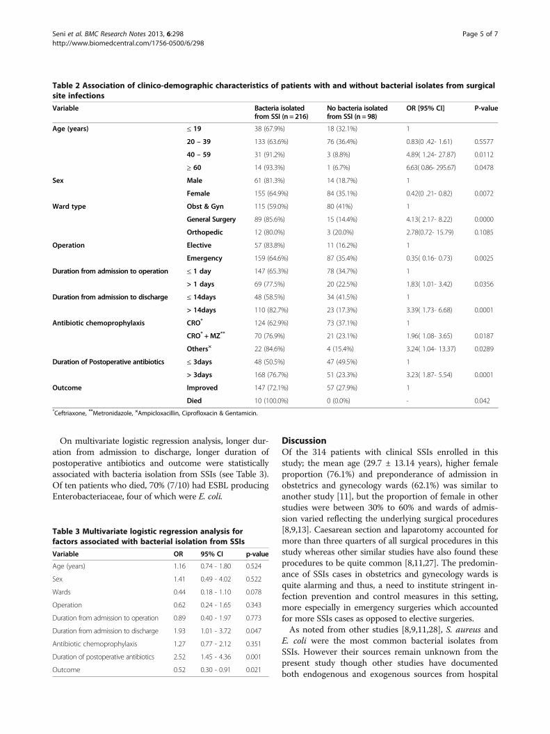
On multivariate logistic regression analysis, longer dur-ation from admission to discharge, longer duration ofpostoperative antibiotics and outcome were statisticallyassociated with bacteria isolation from SSIs (see Table 3).Of ten patients who died, 70% (7/10) had ESBL producingEnterobacteriaceae, four of which were E. coli.
DiscussionOf the 314 patients with clinical SSIs enrolled in thisstudy; the mean age (29.7 ± 13.14 years), higher femaleproportion (76.1%) and preponderance of admission inobstetrics and gynecology wards (62.1%) was similar toanother study [11], but the proportion of female in otherstudies were between 30% to 60% and wards of admis-sion varied reflecting the underlying surgical procedures[8,9,13]. Caesarean section and laparotomy accounted formore than three quarters of all surgical procedures in thisstudy whereas other similar studies have also found theseprocedures to be quite common [8,11,27]. The predomin-ance of SSIs cases in obstetrics and gynecology wards isquite alarming and thus, a need to institute stringent in-fection prevention and control measures in this setting,more especially in emergency surgeries which accountedfor more SSIs cases as opposed to elective surgeries.As noted from other studies [8,9,11,28], S. aureus and
E. coli were the most common bacterial isolates fromSSIs. However their sources remain unknown from thepresent study though other studies have documentedboth endogenous and exogenous sources from hospital
Table 2 Association of clinico-demographic characteristics of patients with and without bacterial isolates from surgicalsite infections
Variable Bacteria isolatedfrom SSI (n = 216)
No bacteria isolatedfrom SSI (n = 98)
OR [95% CI] P-value
Age (years) ≤ 19 38 (67.9%) 18 (32.1%) 1
20 – 39 133 (63.6%) 76 (36.4%) 0.83(0 .42- 1.61) 0.5577
40 – 59 31 (91.2%) 3 (8.8%) 4.89( 1.24- 27.87) 0.0112
≥ 60 14 (93.3%) 1 (6.7%) 6.63( 0.86- 295.67) 0.0478
Sex Male 61 (81.3%) 14 (18.7%) 1
Female 155 (64.9%) 84 (35.1%) 0.42(0 .21- 0.82) 0.0072
Ward type Obst & Gyn 115 (59.0%) 80 (41%) 1
General Surgery 89 (85.6%) 15 (14.4%) 4.13( 2.17- 8.22) 0.0000
Orthopedic 12 (80.0%) 3 (20.0%) 2.78(0.72- 15.79) 0.1085
Operation Elective 57 (83.8%) 11 (16.2%) 1
Emergency 159 (64.6%) 87 (35.4%) 0.35( 0.16- 0.73) 0.0025
Duration from admission to operation ≤ 1 day 147 (65.3%) 78 (34.7%) 1
> 1 days 69 (77.5%) 20 (22.5%) 1.83( 1.01- 3.42) 0.0356
Duration from admission to discharge ≤ 14days 48 (58.5%) 34 (41.5%) 1
> 14days 110 (82.7%) 23 (17.3%) 3.39( 1.73- 6.68) 0.0001
Antibiotic chemoprophylaxis CRO* 124 (62.9%) 73 (37.1%) 1
CRO* +MZ** 70 (76.9%) 21 (23.1%) 1.96( 1.08- 3.65) 0.0187
Others× 22 (84.6%) 4 (15.4%) 3.24( 1.04- 13.37) 0.0289
Duration of Postoperative antibiotics ≤ 3days 48 (50.5%) 47 (49.5%) 1
> 3days 168 (76.7%) 51 (23.3%) 3.23( 1.87- 5.54) 0.0001
Outcome Improved 147 (72.1%) 57 (27.9%) 1
Died 10 (100.0%) 0 (0.0%) - 0.042*Ceftriaxone, **Metronidazole, ×Ampicloxacillin, Ciprofloxacin & Gentamicin.
Table 3 Multivariate logistic regression analysis forfactors associated with bacterial isolation from SSIs
Variable OR 95% CI p-value
Age (years) 1.16 0.74 - 1.80 0.524
Sex 1.41 0.49 - 4.02 0.522
Wards 0.44 0.18 - 1.10 0.078
Operation 0.62 0.24 - 1.65 0.343
Duration from admission to operation 0.89 0.40 - 1.97 0.773
Duration from admission to discharge 1.93 1.01 - 3.72 0.047
Antibiotic chemoprophylaxis 1.27 0.77 - 2.12 0.351
Duration of postoperative antibiotics 2.52 1.45 - 4.36 0.001
Outcome 0.52 0.30 - 0.91 0.021
Seni et al. BMC Research Notes 2013, 6:298 Page 5 of 7http://www.biomedcentral.com/1756-0500/6/298
environment could be potential niches [29,30]. Similarto other related studies [9,18,31], high level of resistancewas found among commonly used antibiotics like ampi-cillin, trimethoprim-sulphamethoxazole, and tetracyclinein both Gram-positive and Gram-negative bacteria. Gram-negative bacteria showed more resistance to gentamicin,ciprofloxacin and chloramphenicol as compared to Gram-positive bacteria. With exception of Acinetobacter spp, allGram-negative bacteria displayed low resistance rates topiperacillin-tazobactam, amikacin and imipenem. Therewere also low resistance rates of Acinetobacter spp andP. aeruginosa to colistin. These findings are similar toanother study [17]. The present study has shown thatthe rates of resistance to erythromycin (46.9%) among S.aureus was relatively low compared to that of Entero-coccus spp (65.2%), with excellent performance of vanco-mycin on both S. aureus and Enterococcus spp. Thesefindings are in agreement to another study [18]. This studyfound more MDR among Gram-negative bacteria thanGram-positive bacteria; of these the proportion of MRSA(37.5%) among S. aureus isolates was more than previouslyreported (25.0% and 31.5%) from Uganda [9,21] and othercountries [8,16,18]. Thus, while β-lactamase-resistant anti-biotics such as cloxacillin could still be effective in thissetting, they are likely to be ineffective against the 38%of isolates that were confirmed as MRSA. The fact thatwe found no vancomycin resistance among S. aureus iso-lates shows that this drug remains the last resort in sys-temic infections caused by MRSA in this setting. Previousstudies [11,17] have shown low rates (14% to 22%) ofESBL-producers among Enterobacteriaceae isolates but thepresent study and another similar study in the same region[8] have shown ESBL-producers to account more thanthree quarter of Enterobacteriaceae. This can be attributedto the empirical use of third generation cephalosporins(usually in combination with another drug such as genta-micin) in almost all hospitalized patients and lack of anti-microbial resistance surveillance in surgical wards atMulago National Hospital. This is a major threat to patientcare as ESBL production renders use of these ceftriaxoneor ceftazidime useless. Absence of resistance to imepenemamong these isolates is however a good finding, except thatimepenem remains an expensive drug.Multivariate logistic regression analysis of clinical and
demographic characteristics of patients with SSIs in thisstudy showed that longer duration from admission to dis-charge, longer duration of postoperative antibiotics andoutcome (death) were associated with bacteria isolationfrom SSIs. These findings have also been shown in othersimilar studies [5,11,27].
LimitationThe study did not isolate strict anaerobes, which couldhave increased the number of bacterial isolates currently
reported as negative cultures. This was because of lack ofstandardized in-house detection methods and lack of an-aerobic detection panels in the Phoenix Automated instru-ment (Becton-Dickson, Sparks Maryland) that we used.
ConclusionMost SSIs at Mulago National Hospital are due to MDRbacteria, these are significantly more among Gram-negativethan Gram-positive bacteria. Isolation of MRSA and ESBL-producing Enterobacteriaceae in higher proportions thanpreviously reported calls for enhanced antibiotic steward-ship including laboratory guided SSIs-therapy and strength-ening of infection control surveillance by identifyingsources of these MDR isolates. In the light of these findings,there is a need to investigate whether there is clonal spreadof the predominant bacteria within/or among surgicalwards at Mulago National Hospital.
Additional file
Additional file 1: Appendix i. Questionnaire.
Competing interestsThe authors declare that they have no competing interests.
Authors’ contributionsConceived and designed the experiments: JS, DPK, and FB. Specimencollection: JS. Supervised the clinical component of research: PM. Performedthe experiments: JS. Supervised the laboratory component of research: CFN,DPK, and FB. Analyzed the data: JS, DPK, AK, and FB. Contributed reagents,materials and analysis tools: JS, CFN, HK, and MLJ. Wrote the manuscript: JS,CFN, DPK, PM, MLJ, HK, AK, and FB. All authors have read and approved thefinal manuscript.
AcknowledgementsThe authors would like to thank patients and health workers in all surgical wards;Emmanuel Aboce, Tonny Lugya, and Hannington Baluku for excellent technicalassistance; Willy Ssengooba for statistical inputs, and all staffs in the Departmentof Medical Microbiology for their support. This work was funded by CatholicUniversity of Health and Allied Sciences Bugando, Mwanza-Tanzania to JS.
Author details1Department of Medical Microbiology, Makerere University College of HealthSciences, P.O. Box 7072, Kampala, Uganda. 2Department of Surgery, MakerereUniversity College of Health Sciences, P.O. Box 7072, Kampala, Uganda.3Department of Microbiology and Immunology, Catholic University of Healthand Allied Sciences-Bugando, P.O. Box 1464, Mwanza, Tanzania. 4Departmentof Community Medicine, Catholic University of Health and Allied Sciences-Bugando, P.O. Box 1464, Mwanza, Tanzania.
Received: 23 November 2012 Accepted: 25 July 2013Published: 27 July 2013
References1. Horan TC, Gaynes RP, Martone WJ, Jarvis WR, Emori TG: CDC definitions of
nosocomial surgical site infections, 1992: a modification of CDCdefinitions of surgical wound infections. Am J Infect Control 1992,20(5):271–274.
2. Mangram AJ, Horan TC, Pearson ML, Silver LC, Jarvis WR: Guideline forprevention of surgical site infection, 1999. Centers for Disease Controland Prevention (CDC) Hospital Infection Control Practices AdvisoryCommittee. Am J Infect Control 1999, 27(2):97–132. quiz 133–134;discussion 196.
Seni et al. BMC Research Notes 2013, 6:298 Page 6 of 7http://www.biomedcentral.com/1756-0500/6/298
3. Emori TG, Gaynes RP: An overview of nosocomial infections, including therole of the microbiology laboratory. Clin Microbiol Rev 1993, 6(4):428–442.
4. Fabiano G, Pezzolla A, Filograna MA, Ferrarese F: [Risk factors of surgicalwound infection]. Ann Ital Chir 2004, 75(1):11–16.
5. Astagneau P, Rioux C, Golliot F, Brucker G: Morbidity and mortalityassociated with surgical site infections: results from the 1997–1999INCISO surveillance. J Hosp Infect 2001, 48(4):267–274.
6. Barwolff S, Sohr D, Geffers C, Brandt C, Vonberg RP, Halle H, Ruden H,Gastmeier P: Reduction of surgical site infections after Caesarean deliveryusing surveillance. J Hosp Infect 2006, 64(2):156–161.
7. Santos KR, Fonseca LS, Bravo Neto GP, Gontijo Filho PP: Surgical siteinfection: rates, etiology and resistance patterns to antimicrobialsamong strains isolated at Rio de Janeiro University Hospital. Infection1997, 25(4):217–220.
8. Mawalla B, Mshana SE, Chalya PL, Imirzalioglu C, Mahalu W: Predictors ofsurgical site infections among patients undergoing major surgery at BugandoMedical Centre in Northwestern Tanzania. BMC Surgery 2011, 11:21.
9. Anguzu JR, Olila D: Drug sensitivity patterns of bacterial isolates fromseptic post-operative wounds in a regional referral hospital in Uganda.Afr Health Sci 2007, 7(3):148–154.
10. Whitehouse JD, Friedman ND, Kirkland KB, Richardson WJ, Sexton DJ: Theimpact of surgical-site infections following orthopedic surgery at acommunity hospital and a university hospital: adverse quality of life,excess length of stay, and extra cost. Infect Control Hosp Epidemiol 2002,23(4):183–189.
11. Fehr J, Hatz C, Soka I, Kibatala P, Urassa H, Smith T, Mshinda H, Frei R,Widmer A: Risk factors for surgical site infection in a Tanzanian districthospital: a challenge for the traditional National Nosocomial InfectionsSurveillance system index. Infect Control Hosp Epidemiol 2006, 27(12):1401–1404.
12. Kirkland KB, Briggs JP, Trivette SL, Wilkinson WE, Sexton DJ: The impact ofsurgical-site infections in the 1990s: attributable mortality, excess lengthof hospitalization, and extra costs. Infect Control Hosp Epidemiol 1999,20(11):725–730.
13. Engemann JJ, Carmeli Y, Cosgrove SE, Fowler VG, Bronstein MZ, Trivette SL,Briggs JP, Sexton DJ, Kaye KS: Adverse clinical and economic outcomesattributable to methicillin resistance among patients withStaphylococcus aureus surgical site infection. Clin Infect Dis 2003,36(5):592–598.
14. Wassef MA, Hussein A, Abdul Rahman EM, El-Sherif RH: A prospectivesurveillance of surgical site infections: study for efficacy of preoperativeantibiotic prophylaxis. African Journal of Microbiology Research 2012,6(12):3072–3078.
15. Amare B, Abdurrahman Z, Moges B, Ali J, Muluken L, Alemayehu M, Yifru S,Sendek B, Belyhun YFM, et al: Postoperative surgical site bacterialinfections and drug susceptibility patterns at Gondar UniversityTeaching Hospital, Northwest Ethiopia. J Bacteriol Parasitol 2011, 2:126.
16. Throckmorton AD, Baddour LM, Hoskin TL, Boughey JC, Degnim AC:Microbiology of surgical site infections complicating breast surgery.Surg Infect (Larchmt) 2010, 11(4):355–359.
17. Hawser SP, Bouchillon SK, Hoban DJ, Badal RE: Epidemiologic trends,occurrence of extended-spectrum beta-lactamase production, andperformance of ertapenem and comparators in patients withintra-abdominal infections: analysis of global trend data from 2002–2007from the SMART study. Surg Infect (Larchmt) 2010, 11(4):371–378.
18. Kownhar H, Shankar EM, Vignesh R, Sekar R, Velu V, Rao UA: High isolationrate of Staphylococcus aureus from surgical site infections in an Indianhospital. J Antimicrob Chemother 2008, 61(3):758–760.
19. Krukerink M, Kievit J, de Mheen PJ M-v: Evaluation of routinely reportedsurgical site infections against microbiological culture results: a tool toidentify patient groups where diagnosis and treatment may beimproved. BMC Infect Dis 2009, 9:176.
20. Olaro C: Study to assess the risk factors for postoperative complicationsfollowing abdominal surgery in Mulago Hospital. Kampala: MakerereUniversity; 1999.
21. Ojulong J, Mwambu TP, Joloba M, Bwanga F, Kaddu-Mulindwa DH: Relativeprevalence of methicilline resistant Staphylococcus aureus and itssusceptibility pattern in Mulago Hospital, Kampala, Uganda. Tanzan JHealth Res 2009, 11(3):149–153.
22. Kitara DL, Kakande I, Mugisa BD, Obol JH: The postoperative complicationsprediction in Mulago Hospital using POSSUM scoring system. East andCentral African Journal of Surgery 2010, 15(2):90–96.
23. Koneman EW, Allen SD, Janda WM, Schreckenberger PC, Winn WC: Coloratlas and textbook of diagnostic microbiology. 5th edition. Philadelphia,Pa: Lippincott, Williams & Wilkins Publishers; 1997.
24. CLSI: Perfomance standars for antimicrobial susceptibility testing; twenty firstinformation supplement, vol. CLSI document M100-S21. Wayne, PA: Clinicaland Laboratory Standards Institute; 2011.
25. Livermore DM, Brown DF: Detection of beta-lactamase-mediatedresistance. J Antimicrob Chemother 2001, 48(Suppl 1):59–64.
26. D'Agata EM: Rapidly rising prevalence of nosocomial multidrug-resistant,F-negative bacilli: a 9-year surveillance study. Infect Control Hosp Epidemiol2004, 25(10):842–846.
27. Razavi SM, Ibrahimpoor M, Sabouri Kashani A, Jafarian A: Abdominalsurgical site infections: incidence and risk factors at an Iranian teachinghospital. BMC Surg 2005, 5:2.
28. Kasatpibal N, Jamulitrat S, Chongsuvivatwong V: Standardized incidencerates of surgical site infection: a multicenter study in Thailand. Am JInfect Control 2005, 33(10):587–594.
29. Wenzel RP, Perl TM: The significance of nasal carriage of Staphylococcusaureus and the incidence of postoperative wound infection. J Hosp Infect1995, 31(1):13–24.
30. Atata RF, Ibrahim YKE, Olurinola PF, Giwa A, Akanbi AA II, Sani AA: Clinicalbacterial isolates from hospital environment as agents of surgical woundnosocomial infections. Journal of Pharmacy & Bioresources 2010, 7(2):146–155.
31. Adegoke AA, Mvuyo T, Okoh AI, Steve J: Studies on multiple antibioticresistant bacterial isolated from surgical site infection. Scientific Researchand Essays 2010, 5(24):3876–3881.
doi:10.1186/1756-0500-6-298Cite this article as: Seni et al.: Antimicrobial resistance in hospitalizedsurgical patients: a silently emerging public health concern in Uganda.BMC Research Notes 2013 6:298.
Submit your next manuscript to BioMed Centraland take full advantage of:
• Convenient online submission
• Thorough peer review
• No space constraints or color figure charges
• Immediate publication on acceptance
• Inclusion in PubMed, CAS, Scopus and Google Scholar
• Research which is freely available for redistribution
Submit your manuscript at www.biomedcentral.com/submit
Seni et al. BMC Research Notes 2013, 6:298 Page 7 of 7http://www.biomedcentral.com/1756-0500/6/298