Embed Size (px)
Citation preview
www.wjpr.net Vol 5, Issue 5, 2016.
1441
Tripathi et al. World Journal of Pharmaceutical Research Tripathi et al. World Journal of Pharmaceutical Research
ANTIMICROBIAL PROPERTIES OF MORINGA OLIEFERA AGAINST
PATHOGENIC BACTERIA
1Saurabh Dixit,
2Astha Tripathi* and
3Prabhat Kumar
1DAV Dental College & Hospital, Tatul, Solan (H.P.).
2Shoolini University of Management Sciences and Biotechnology, Bajhol, Solan (H.P.).
3Tissue Culture Vaccine Lab, Indovax Pvt. Ltd., Siswala, Hisar.
ABSTRACT
The research work was investigated to compare the antimicrobial
effect of Moringa oliefera leaf, bark and seed on pathogenic bacteria.
Extracts were prepared in aqueous and organic solvents i.e. ethanol,
ethyl acetate, methanol and chloroform. Ethanol and ethyl acetate
extract of the leaf of M. oleifera were more effective while chloroform
showed no zone of inhibition with all bacterial strains except B.
subtilis. Leaf extract was selected for further study. With antibiotics,
maximum growth inhibition was observed in V. cholerae (98.8%) in
first dilution. MIC values demonstrate that in bacterial strains the most
sensitive strain with antibiotic was B. cereus (5 mg/ml) and with leaf
extract most sensitive strain was B. subtilis (16 mg/ml) however with antibiotics less sensitive
strains were S. typhi (18 mg/ml) and with leaf extract less sensitive strain was S. typhi (28
mg/ml).
KEYWORDS: Moringa oliefera, antimicrobial, MIC.
INTRODUCTION
Moringa oleifera is the most widely cultivated species of a monogeneric family, the
Moringaceae, that is native to the sub-Himalayan tracts of India, Pakistan, Bangladesh and
Afghanistan. Different parts of this plant are used in the indigenous systems of human
medicine for the treatment of a variety of human aliments. Ethanolic leaves extract of
Moringa oleifera used as hypotensive (Nikkon et al., 2003; Siddiqui and Khan, 1968;
Kirtikar and Basu, 1984). The leaves of M. oleifera are reported to be used as a
hypocholesterolemic agent, and hypoglycemic agent (Dangi et al., 2002; Ghasi et al., 2000;
World Journal of Pharmaceutical Research SJIF Impact Factor 6.805
Volume 5, Issue 5, 1441-1459. Research Article ISSN 2277– 7105
*Corresponding Author
Dr. Astha Tripathi
Shoolini University of
Management Sciences and
Biotechnology, Bajhol,
Solan (H.P.).
Article Received on
19 March 2016,
Revised on 10 April 2016,
Accepted on 01 May 2016
DOI: 10.20959/wjpr20165-6180
www.wjpr.net Vol 5, Issue 5, 2016.
1442
Tripathi et al. World Journal of Pharmaceutical Research Tripathi et al. World Journal of Pharmaceutical Research
Siddiqui and khan, 1968). M. oleifera is one of the best known medicinal plant. The Moringa
plant has been consumed by humans (Iqbal et al., 2006). It is one of the richest plant sources
of Vitamins A, B, C, D, E and K (Anwar and Bhanger, 2003; Babu 2000; Caceres et al.,
1992; Dayrit et al., 1990; Delisle et al., 1997). The vital minerals present in Moringa include
Calcium, Copper, Iron, Potassium, Magnesium, Manganese and Zinc. The antimicrobial
activities of M. oleifera leaves, roots, barks and seeds were investigated in vitro against
bacteria, yeast, dermatophytes and helminthes pathogenic to man. Antibacterial effect of
aqueous and ethanolic extracts of seeds of M. oleiferain the concentration of 1.5 unit and 1.10
unit in volumes 50, 100, 150 and 200 µl were examined against Staphylococcus aureus,
vibrio cholerea, Escherichia coli (isolated from the organism and the aquatic environment)
and Salmonella enteritidis. Antibacterial activity (inhibition halo> 13mm) against S. aureus,
V. cholera and E. coli isolated from the white leg shrimp, Litopenaeous vannmaei, was
detected in aqueous and ethanolic extract of moringa. E. coli isolated from tilapia fish and
Oreochrom isniloticus, were sensitive to the ethanolic extract of M. oleifera. During recent
years considerable work has been done to investigate the pharmacological actions of the
leaves and seeds of M. oleifera on scientific lines but only limited work has been reported so
far on antibacterial activity of M. oleifera root bark though it is reported to possess varied
medicinal properties. Therefore, it was considered worthy to investigate the antibacterial
activity of M. oleifera root bark. Bark used to cure Dental Caries/Toothache, Common cold,
External Sores/Ulcer, Anti-Tumor, Snakebite, Scorpion bite, Digestive, Headache,
Antinutrietional factors and Scurvy (Fahey, 2005).
The aims and objectives of the present work are to establish a well documented information
about the antimicrobial activity of M. oleifera extracts against 10 standard microorganisms
using reference antibiotics as experimental models.
MATERIAL AND METHODS
Material
1. Test organism
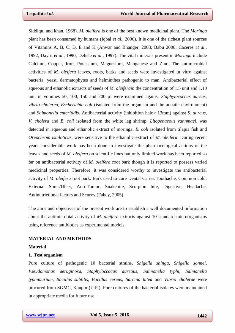
Pure culture of pathogenic 10 bacterial strains, Shigella shinga, Shigella sonnei,
Pseudomonas aeruginosa, Staphyloccocus aureous, Salmonella typhi, Salmonella
typhimurium, Bacillus subtilis, Bacillus cereus, Sarcina lutea and Vibrio cholerae were
procured from SGMC, Kanpur (U.P.). Pure cultures of the bacterial isolates were maintained
in appropriate media for future use.
www.wjpr.net Vol 5, Issue 5, 2016.
1443
Tripathi et al. World Journal of Pharmaceutical Research Tripathi et al. World Journal of Pharmaceutical Research
2.Chemicals: All chemicals and media used in this study were of analytical grade.
3. Antibiotic activity on bacterial strains
Octatadisk of different antibiotics were purchased for antibiotic susceptibility test of all ten
pathogenic bacterial strains.
4. Collection of plant material
The fresh leaves, seeds and bark of Moringa oleifera were collected from Agra and Jaipur.
The plant materials were air-dried in the laboratory for four weeks and then ground into
powdered form, using a mortar and pestel, and stored for future use.
Methods
Extraction
a) In aqueous solution
The air-dried leaves, bark and seed of M. oleifera (50 g) were dipped in 200 ml cold distilled
water in a conical flask stoppered with rubber cork and left for 7 days with occasional
shaking. Solution was filtered using sterile filter paper (Whattman No. 1) into a clean conical
flask and subjected to water bath evaporation where the aqueous solvent was evaporated at its
boiling temperature of 100°C and as well as by evaporation under vacuum. The standard
extracts obtained were stored in a refrigerator at 4°C for antibacterial activity test (Akueshi et
al., 2002).
b) In organic solvent
i. Ethanol and Ethyl acetate
Plant material of M. oleifera were collected and dried in shade. The dried materials were
ground to powder and suspended in petroleum ether and kept in refrigerator overnight. After
one week incubation, Solution was filtered using sterile filter paper (Whattman No. 1) into a
clean conical flask and subjected to water bath evaporation where the organic solvent was
evaporated at room temperature and the residue was dried at room temperature. The residue
was further divided into two parts and each part was suspended in ethanol and ethyl acetate
respectively in sterile 25 ml conical flasks and kept at 4ºC overnight. After overnight
incubation, the supernatant was filtered through Whatman No.1 filter paper and the filtrate
was dried to evaporate the organic solvent at room temperature. The standard extracts
obtained were then stored in a refrigerator at 4°C for antibacterial activity test (Valarmathy et
al., 2010).
www.wjpr.net Vol 5, Issue 5, 2016.
1444
Tripathi et al. World Journal of Pharmaceutical Research Tripathi et al. World Journal of Pharmaceutical Research
ii. Methanol and Chloroform
30 g of the plant material powder was thoroughly mixed in 450 ml methanol and chloroform
respectively and allowed to stand for one hour before filtering with the help of Whatman
filter paper No. 1. The filtrate was overnight dried in hot air oven (45°C). The extraction
yielded 6 g (methanol) and 3 g (chloroform). The standard extracts obtained were store in a
refrigerator at 4°C for antibacterial activity test (Thilza et al., 2010).
Growth inhibition: The percentage inhibition of bacterial growth was calculated by
comparing growth density with antibiotics and plant material extracts and without antibiotics
and plant material extracts. Inhibition (%) in growth density was calculated as follows
Minimum Inhibitory Concentration (MIC) (mg/l): Well labeled 9 tubes were taken. Add 2
ml of antibiotic solution (100 µg/ml) to the first tube then add 1 ml of sterile broth to all other
tubes. Transfer 1 ml from the first tube to the second tube. Using a separate pipette, mix the
contents of this tube and transfer 1 ml to the third tube. Continue dilutions in this manner to
tube number 8, being certain to change pipettes between tubes to prevent carryover of
antibiotic on the external surface of the pipette. Remove 1 ml from tube 8 and discard it. The
ninth tube, which serves as a control, receives no antibiotic. Suspend to an appropriate
turbidity several colonies of the culture to be tested in 5 ml of Mueller-Hinton broth to give a
slightly turbid suspension. Dilute this suspension by aseptically pipetting 0.2 ml of the
suspension into 40 ml of Mueller-Hinton broth. Add 1 ml of the diluted culture suspension to
each of the tubes. The final concentration of antibiotic is now one-half of the original
concentration in each tube. Incubate all tubes at 35oC overnight. Examine tubes for visible
signs of bacterial growth. The highest dilution without growth is the minimal inhibitory
concentration (MIC) (Murray, 2005).
RESULTS
Antibiotic activity on bacterial strains: Octatadisk of different antibiotics were used to
check antibiotic susceptibility of all ten pathogenic bacterial strains (Shigella shinga, Shigella
Sonnei, Pseudomonas aeruginosa, Staphyloccocus aureous, Salmonella typhi, Salmonella
typhimurium, Bacillus subtilis, Bacillus cereus, Sarcina lutea and Vibrio cholerae). All
www.wjpr.net Vol 5, Issue 5, 2016.
1445
Tripathi et al. World Journal of Pharmaceutical Research Tripathi et al. World Journal of Pharmaceutical Research
bacterial strains showed different antibiotic susceptibility with various antibiotics (Table-1;
Fig. 1).
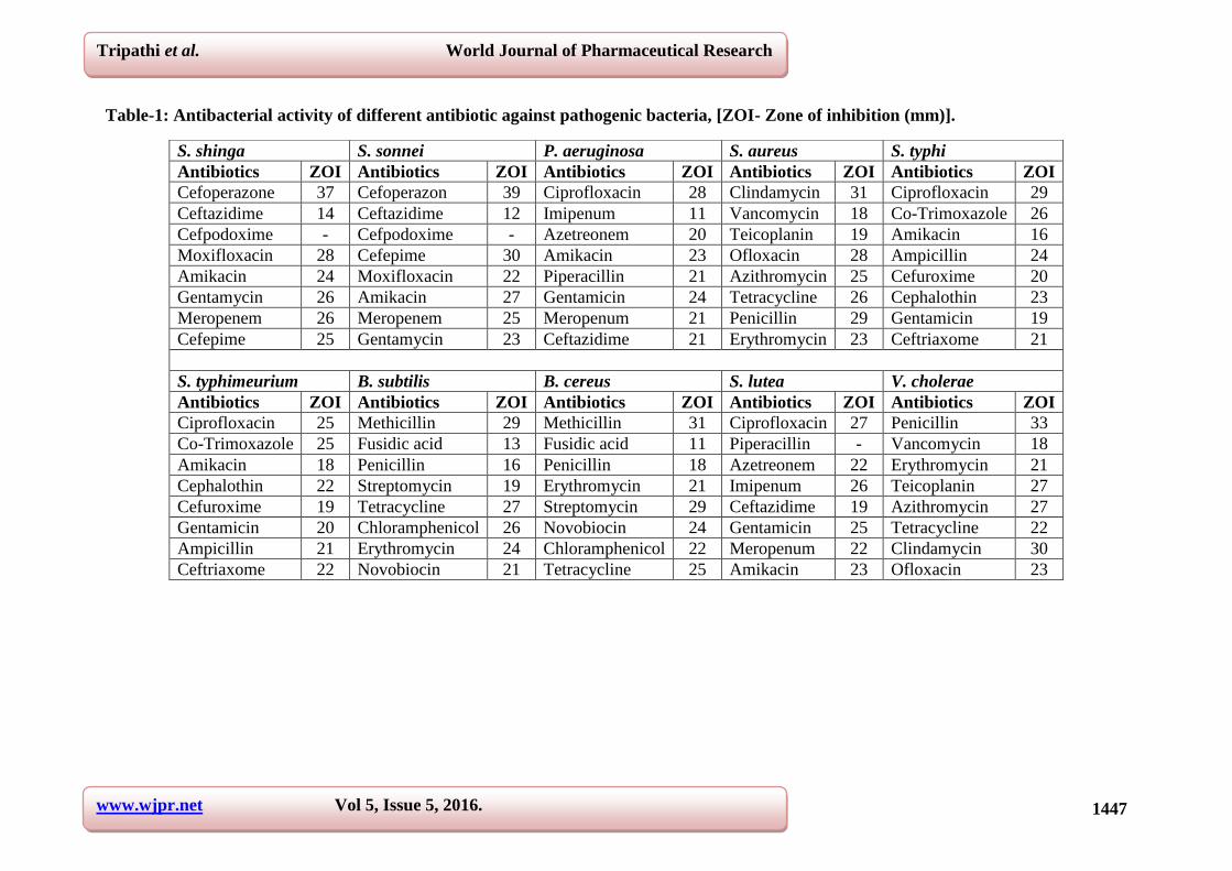
1. S. shinga
Cefoperazone showed maximum zone of inhibition of 37 mm while Ceftazidime shows
minimum zone of 14 mm however Cefpodoxime shows no zone against this bacteria. Other
antibiotics viz, Moxifloxacin, Amikacin, Gentamycin, Meropenem and Cefepime shows 28
mm, 24 mm, 26 mm, 26 mm, 25 mm zones of inhibition respectively.
2. S. sonnei
Cefoperazone showed maximum zone of inhibition of 39 mm while Ceftazidime shows
minimum zone of 12 mm however Cefpodoxime shows no zone against this bacteria. Other
antibiotics viz, Cefepime, Moxifloxacin, Amikacin, Meropenem and Gentamycin shows 30
mm, 22 mm, 27 mm, 25 mm, 23 mm zones of inhibition respectively.
3. P. aeruginosa
Ciprofloxacin shows maximum zone of 28 mm while Imipenum showed minimum zone of
11mm. Other antibiotics viz, Azetreonem, Amikacin, Piperacillin, Gentamicin, Meropenum
and Ceftazidime shows 20 mm, 23 mm, 21 mm, 24 mm, 21 mm, and 21 mm zone of
inhibition respectively.
4. S. aureus
Clindamycin show maximum inhibition zone of 31 mm while Vancomycin shows minimum
inhibition zone of 18 mm. Other antibiotics Teicoplanin, Ofloxacin, Azithromycin,
Tetracycline, Penicillin and Erythromycin shows 19 mm, 28 mm, 25 mm, 26 mm, 29 mm, 23
mm zone of inhibition respectively.
5. S. typhi
Ciprofloxacin showed maximum zone of inhibition of 29 mm followed by Co-Trimoxazole
shows maximum zone of 26 mm while Amikacin shows minimum zone of 16 mm. Other
antibiotics viz, Ampicillin, Cefuroxime, Cephalothin, Gentamicin, and Ceftriaxome shows 24
mm, 20 mm, 23 mm, 19 mm and 21 mm zone of inhibition.
6. S. typhimeurium
Ciprofloxacin and Co-Trimoxazole shows maximum zone of 25 mm while Amikacin shows
minimum zone of 18 mm. Other antibiotics viz, Cephalothin, Cefuroxime, Gentamicin,
www.wjpr.net Vol 5, Issue 5, 2016.
1446
Tripathi et al. World Journal of Pharmaceutical Research Tripathi et al. World Journal of Pharmaceutical Research
Ampicillin and Ceftriaxome shows 22 mm, 19 mm, 20 mm, 21 mm and 22 mm zone of
inhibition.
7. B. subtilis
Methicillin showed maximum zone of 29 mm while Fusidic acid showed minimum zone of
13 mm. Other antibiotics viz, Penicillin, Streptomycin, Tetracycline, Chloramphenicol,
Erythromycin and Novobiocin showed 16 mm, 19 mm, 27 mm, 26 mm, 24 mm and 21 mm
respectively.
8. B. cereus
Methicillin showed maximum zone of 31 mm while Fusidic acid showed minimum zone of
11 mm. Other antibiotics viz, Penicillin, Erythromycin, Streptomycin, Novobiocin,
Chloramphenicol and Tetracycline showed 18 mm, 21 mm, 29 mm, 24 mm, 22 mm and 25
mm respectively.
9. S. lutea
Ciprofloxacin showed maximum zone of 27 mm while Piperacillin showed minimum no
zone. Other antibiotics viz, Azetreonem, Imipenum, Ceftazidime, Gentamicin, Meropenum
and Amikacin showed 22 mm, 26 mm, 19 mm, 25 mm, 22 mm, and 23 mm zone of inhibition
respectively.
10. V. cholera
Penicillin showed maximum inhibition zone of 33 mm while Vancomycin showed minimum
inhibition zone of 18 mm. Other antibiotics Erythromycin, Teicoplanin, Azithromycin,
Tetracycline, Clindamycin and Ofloxacin showed 21 mm, 27 mm, 27 mm, 22 mm, 30 mm
and 23 mm zone of inhibition respectively.
www.wjpr.net Vol 5, Issue 5, 2016.
1447
Tripathi et al. World Journal of Pharmaceutical Research
Tripathi et al. World Journal of Pharmaceutical Research
Table-1: Antibacterial activity of different antibiotic against pathogenic bacteria, [ZOI- Zone of inhibition (mm)].
S. shinga S. sonnei P. aeruginosa S. aureus S. typhi
Antibiotics ZOI Antibiotics ZOI Antibiotics ZOI Antibiotics ZOI Antibiotics ZOI
Cefoperazone 37 Cefoperazon 39 Ciprofloxacin 28 Clindamycin 31 Ciprofloxacin 29
Ceftazidime 14 Ceftazidime 12 Imipenum 11 Vancomycin 18 Co-Trimoxazole 26
Cefpodoxime - Cefpodoxime - Azetreonem 20 Teicoplanin 19 Amikacin 16
Moxifloxacin 28 Cefepime 30 Amikacin 23 Ofloxacin 28 Ampicillin 24
Amikacin 24 Moxifloxacin 22 Piperacillin 21 Azithromycin 25 Cefuroxime 20
Gentamycin 26 Amikacin 27 Gentamicin 24 Tetracycline 26 Cephalothin 23
Meropenem 26 Meropenem 25 Meropenum 21 Penicillin 29 Gentamicin 19
Cefepime 25 Gentamycin 23 Ceftazidime 21 Erythromycin 23 Ceftriaxome 21
S. typhimeurium B. subtilis B. cereus S. lutea V. cholerae
Antibiotics ZOI Antibiotics ZOI Antibiotics ZOI Antibiotics ZOI Antibiotics ZOI
Ciprofloxacin 25 Methicillin 29 Methicillin 31 Ciprofloxacin 27 Penicillin 33
Co-Trimoxazole 25 Fusidic acid 13 Fusidic acid 11 Piperacillin - Vancomycin 18
Amikacin 18 Penicillin 16 Penicillin 18 Azetreonem 22 Erythromycin 21
Cephalothin 22 Streptomycin 19 Erythromycin 21 Imipenum 26 Teicoplanin 27
Cefuroxime 19 Tetracycline 27 Streptomycin 29 Ceftazidime 19 Azithromycin 27
Gentamicin 20 Chloramphenicol 26 Novobiocin 24 Gentamicin 25 Tetracycline 22
Ampicillin 21 Erythromycin 24 Chloramphenicol 22 Meropenum 22 Clindamycin 30
Ceftriaxome 22 Novobiocin 21 Tetracycline 25 Amikacin 23 Ofloxacin 23
www.wjpr.net Vol 5, Issue 5, 2016.
1448
Tripathi et al. World Journal of Pharmaceutical Research Tripathi et al. World Journal of Pharmaceutical Research
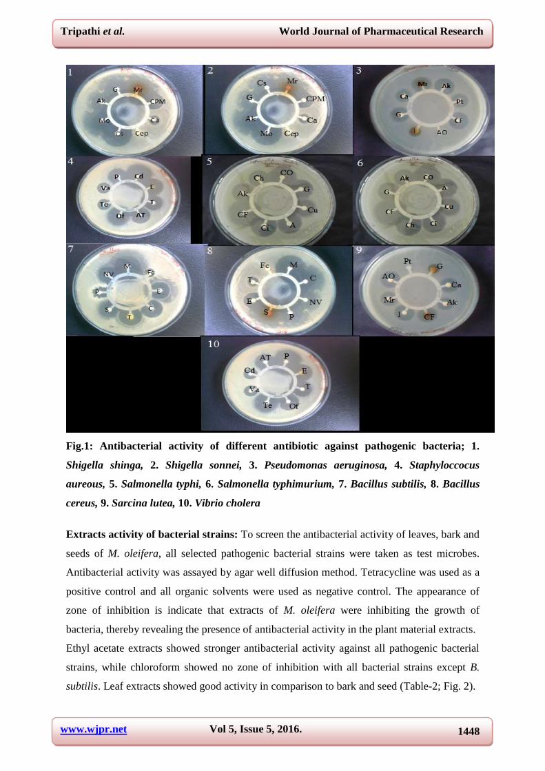
Fig.1: Antibacterial activity of different antibiotic against pathogenic bacteria; 1.
Shigella shinga, 2. Shigella sonnei, 3. Pseudomonas aeruginosa, 4. Staphyloccocus
aureous, 5. Salmonella typhi, 6. Salmonella typhimurium, 7. Bacillus subtilis, 8. Bacillus
cereus, 9. Sarcina lutea, 10. Vibrio cholera
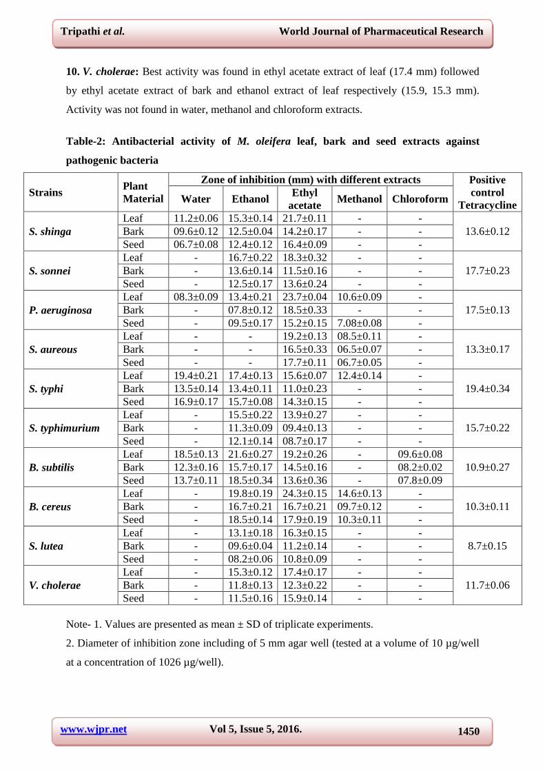
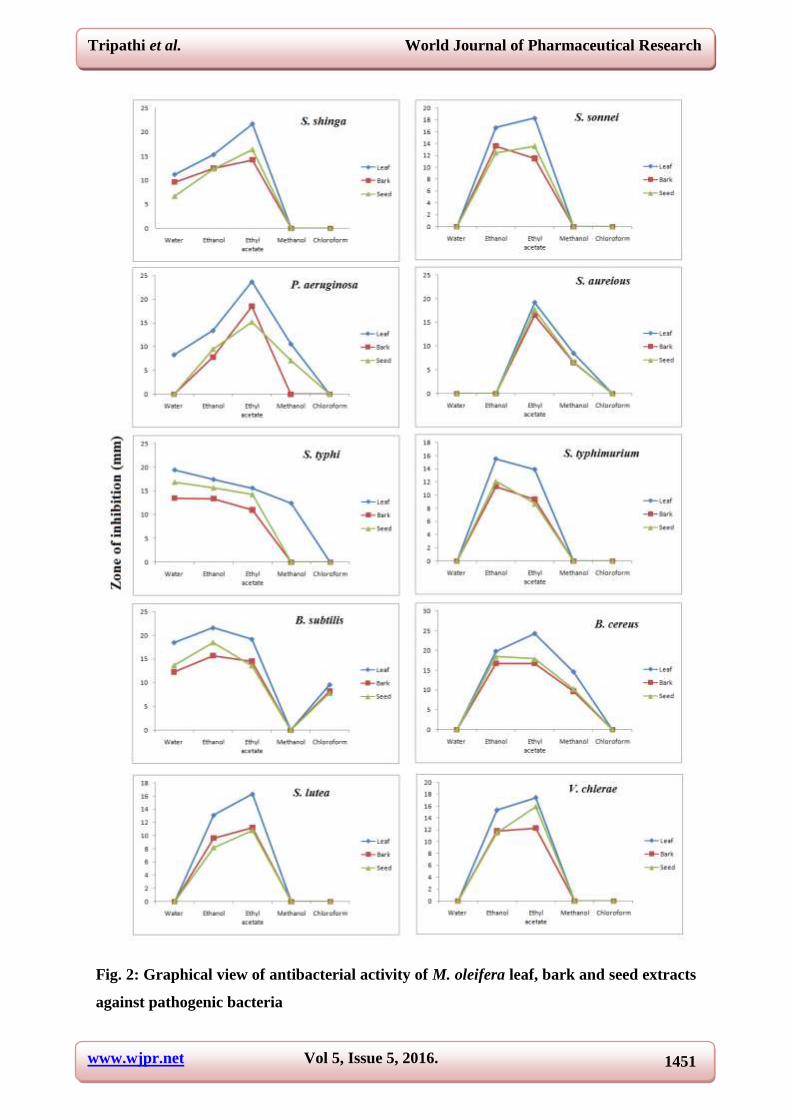
Extracts activity of bacterial strains: To screen the antibacterial activity of leaves, bark and
seeds of M. oleifera, all selected pathogenic bacterial strains were taken as test microbes.
Antibacterial activity was assayed by agar well diffusion method. Tetracycline was used as a
positive control and all organic solvents were used as negative control. The appearance of
zone of inhibition is indicate that extracts of M. oleifera were inhibiting the growth of
bacteria, thereby revealing the presence of antibacterial activity in the plant material extracts.
Ethyl acetate extracts showed stronger antibacterial activity against all pathogenic bacterial
strains, while chloroform showed no zone of inhibition with all bacterial strains except B.
subtilis. Leaf extracts showed good activity in comparison to bark and seed (Table-2; Fig. 2).

www.wjpr.net Vol 5, Issue 5, 2016.
1449
Tripathi et al. World Journal of Pharmaceutical Research Tripathi et al. World Journal of Pharmaceutical Research
1. S. shinga: Best activity was found in ethyl acetate extract of leaf (21.7 mm) followed by
ethanol and water extracts of leaf (15.3, 11.2 mm) respectively. Minimum zone of inhibition
was found with water extract of seed (6.7 mm) followed by water extract of bark (9.6 mm).
Activity was not found in methanol and chloroform extracts.
2. S. sonnei: Showed activity only in ethanol and ethyl acetate extracts. Best activity was
found in ethyl acetate extract of leaf (18.3 mm) followed by ethanol extract of leaf (16.7
mm). Activity was not found in water, methanol and chloroform extracts.
3. P. aeruginosa: Activity was found in all selected extracts except chloroform. Best activity
was found in ethyl acetate extract of leaf (23.7 mm) followed by bark and seed (18.5, 15.2
mm). In methanol extract activity was not found in bark while in water extract activity was
found only in leaf extract.
4. S. aureous: Activity was found in ethyl acetate and methanol extracts. Best activity was
found in ethyl acetate extract of leaf (19.2 mm) followed by seed and bark (17.7, 16.5 mm).
Activity was not found in water, methanol and chloroform extracts.
5. S. typhi: Activity was found in all selected extract except chloroform. While in methanol
extract activity was found only in leaf extract. Best activity was found in water extract of leaf
(19.4 mm) followed by ethanol extract of leaf and water extract of seed respectively (17.4,
16.9 mm).
6. S. typhimurium: Activity was not found in water, methanol and chloroform extracts. Best
activity was found in ethanol extract of leaf (15.5 mm) followed by ethyl acetate extracts of
leaf and ethanol extract of seed respectively (13.9, 12.1 mm).
7. B. subtilis: Except methanol extract, activity was found in all selected extracts. Best
activity was found in ethanol extract of leaf (21.6 mm) followed by ethyl acetate and water
extract of leaf and ethanol extract of seed respectively (19.2, 18.5, 18.5 mm).
8. B. cereus: Activity was not found in water and chloroform extract. Best activity was found
in ethyl acetate extract of leaf (24.3 mm) followed by ethanol extract of leaf and seed (19.8,
18.5 mm). Minimum zone of inhibition was found in methanol extract of bark (9.7 mm).
9. S. lutea: Activity was not found in water, methanol and chloroform extracts. Best activity
was found in ethyl acetate extract of leaf (16.3 mm) followed by ethanol extract of leaf and
ethyl acetate extract of bark respectively (13.1, 11.2 mm).
www.wjpr.net Vol 5, Issue 5, 2016.
1450
Tripathi et al. World Journal of Pharmaceutical Research Tripathi et al. World Journal of Pharmaceutical Research
10. V. cholerae: Best activity was found in ethyl acetate extract of leaf (17.4 mm) followed
by ethyl acetate extract of bark and ethanol extract of leaf respectively (15.9, 15.3 mm).
Activity was not found in water, methanol and chloroform extracts.
Table-2: Antibacterial activity of M. oleifera leaf, bark and seed extracts against
pathogenic bacteria
Strains Plant
Material
Zone of inhibition (mm) with different extracts Positive
control
Tetracycline Water Ethanol
Ethyl
acetate Methanol Chloroform
S. shinga
Leaf 11.2±0.06 15.3±0.14 21.7±0.11 - -
13.6±0.12 Bark 09.6±0.12 12.5±0.04 14.2±0.17 - -
Seed 06.7±0.08 12.4±0.12 16.4±0.09 - -
S. sonnei
Leaf - 16.7±0.22 18.3±0.32 - -
17.7±0.23 Bark - 13.6±0.14 11.5±0.16 - -
Seed - 12.5±0.17 13.6±0.24 - -
P. aeruginosa
Leaf 08.3±0.09 13.4±0.21 23.7±0.04 10.6±0.09 -
17.5±0.13 Bark - 07.8±0.12 18.5±0.33 - -
Seed - 09.5±0.17 15.2±0.15 7.08±0.08 -
S. aureous
Leaf - - 19.2±0.13 08.5±0.11 -
13.3±0.17 Bark - - 16.5±0.33 06.5±0.07 -
Seed - - 17.7±0.11 06.7±0.05 -
S. typhi
Leaf 19.4±0.21 17.4±0.13 15.6±0.07 12.4±0.14 -
19.4±0.34 Bark 13.5±0.14 13.4±0.11 11.0±0.23 - -
Seed 16.9±0.17 15.7±0.08 14.3±0.15 - -
S. typhimurium
Leaf - 15.5±0.22 13.9±0.27 - -
15.7±0.22 Bark - 11.3±0.09 09.4±0.13 - -
Seed - 12.1±0.14 08.7±0.17 - -
B. subtilis
Leaf 18.5±0.13 21.6±0.27 19.2±0.26 - 09.6±0.08
10.9±0.27 Bark 12.3±0.16 15.7±0.17 14.5±0.16 - 08.2±0.02
Seed 13.7±0.11 18.5±0.34 13.6±0.36 - 07.8±0.09
B. cereus
Leaf - 19.8±0.19 24.3±0.15 14.6±0.13 -
10.3±0.11 Bark - 16.7±0.21 16.7±0.21 09.7±0.12 -
Seed - 18.5±0.14 17.9±0.19 10.3±0.11 -
S. lutea
Leaf - 13.1±0.18 16.3±0.15 - -
8.7±0.15 Bark - 09.6±0.04 11.2±0.14 - -
Seed - 08.2±0.06 10.8±0.09 - -
V. cholerae
Leaf - 15.3±0.12 17.4±0.17 - -
11.7±0.06 Bark - 11.8±0.13 12.3±0.22 - -
Seed - 11.5±0.16 15.9±0.14 - -
Note- 1. Values are presented as mean ± SD of triplicate experiments.
2. Diameter of inhibition zone including of 5 mm agar well (tested at a volume of 10 µg/well
at a concentration of 1026 µg/well).
www.wjpr.net Vol 5, Issue 5, 2016.
1451
Tripathi et al. World Journal of Pharmaceutical Research Tripathi et al. World Journal of Pharmaceutical Research
Fig. 2: Graphical view of antibacterial activity of M. oleifera leaf, bark and seed extracts
against pathogenic bacteria

www.wjpr.net Vol 5, Issue 5, 2016.
1452
Tripathi et al. World Journal of Pharmaceutical Research Tripathi et al. World Journal of Pharmaceutical Research
Growth inhibition (%) in the presence of antibiotics and extracts
All selected pathogenic bacterial strains showed growth inhibition (%) in all dilutions of leaf
extract and antibiotic. However growth was reduced in higher to lower form in all dilutions
(Table- 3, 4; Fig. 3 a, b).
In leaf extract maximum growth inhibition was recorded in S. typhi (96.3%) followed by B.
cereus, P. aeruginosa and S. lutea respectively in first dilution (96.3, 93.6, 93.5%). Minimum
growth inhibition was recorded in S. typhimeurium and S. sonnei (5.3, 5.5%) followed by B.
subtilis and S. shinga respectively in last dilution (7.6, 8.9%).
With antibiotics, maximum growth inhibition was observed in V. cholerae (98.8%) followed
by S. typhimeurium and S. typhi in first dilution respectively (97.4, 96.7%). Minimum growth
inhibition was recorded in S. sonnei (5.2%) followed by S. lutea and S. aureus respectively in
last dilution (6.3, 6.4%).
Fig. 3 (a): Graphical view of growth inhibition (%) in leaf extract
Fig. 3 (b): Graphical view of growth inhibition (%) in antibiotics
www.wjpr.net Vol 5, Issue 5, 2016.
1453
Tripathi et al. World Journal of Pharmaceutical Research
Tripathi et al. World Journal of Pharmaceutical Research
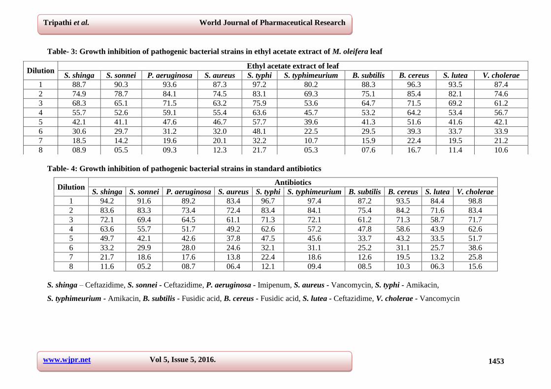
Table- 3: Growth inhibition of pathogenic bacterial strains in ethyl acetate extract of M. oleifera leaf
Table- 4: Growth inhibition of pathogenic bacterial strains in standard antibiotics
Dilution Antibiotics
S. shinga S. sonnei P. aeruginosa S. aureus S. typhi S. typhimeurium B. subtilis B. cereus S. lutea V. cholerae
1 94.2 91.6 89.2 83.4 96.7 97.4 87.2 93.5 84.4 98.8
2 83.6 83.3 73.4 72.4 83.4 84.1 75.4 84.2 71.6 83.4
3 72.1 69.4 64.5 61.1 71.3 72.1 61.2 71.3 58.7 71.7
4 63.6 55.7 51.7 49.2 62.6 57.2 47.8 58.6 43.9 62.6
5 49.7 42.1 42.6 37.8 47.5 45.6 33.7 43.2 33.5 51.7
6 33.2 29.9 28.0 24.6 32.1 31.1 25.2 31.1 25.7 38.6
7 21.7 18.6 17.6 13.8 22.4 18.6 12.6 19.5 13.2 25.8
8 11.6 05.2 08.7 06.4 12.1 09.4 08.5 10.3 06.3 15.6
S. shinga – Ceftazidime, S. sonnei - Ceftazidime, P. aeruginosa - Imipenum, S. aureus - Vancomycin, S. typhi - Amikacin,
S. typhimeurium - Amikacin, B. subtilis - Fusidic acid, B. cereus - Fusidic acid, S. lutea - Ceftazidime, V. cholerae - Vancomycin
Dilution Ethyl acetate extract of leaf
S. shinga S. sonnei P. aeruginosa S. aureus S. typhi S. typhimeurium B. subtilis B. cereus S. lutea V. cholerae
1 88.7 90.3 93.6 87.3 97.2 80.2 88.3 96.3 93.5 87.4
2 74.9 78.7 84.1 74.5 83.1 69.3 75.1 85.4 82.1 74.6
3 68.3 65.1 71.5 63.2 75.9 53.6 64.7 71.5 69.2 61.2
4 55.7 52.6 59.1 55.4 63.6 45.7 53.2 64.2 53.4 56.7
5 42.1 41.1 47.6 46.7 57.7 39.6 41.3 51.6 41.6 42.1
6 30.6 29.7 31.2 32.0 48.1 22.5 29.5 39.3 33.7 33.9
7 18.5 14.2 19.6 20.1 32.2 10.7 15.9 22.4 19.5 21.2
8 08.9 05.5 09.3 12.3 21.7 05.3 07.6 16.7 11.4 10.6
www.wjpr.net Vol 5, Issue 5, 2016.
1454
Tripathi et al. World Journal of Pharmaceutical Research
Tripathi et al. World Journal of Pharmaceutical Research
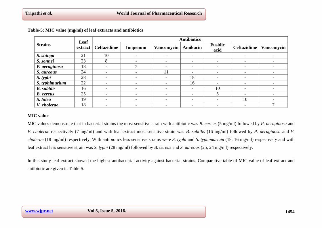
Table-5: MIC value (mg/ml) of leaf extracts and antibiotics
MIC value
MIC values demonstrate that in bacterial strains the most sensitive strain with antibiotic was B. cereus (5 mg/ml) followed by P. aeruginosa and
V. cholerae respectively (7 mg/ml) and with leaf extract most sensitive strain was B. subtilis (16 mg/ml) followed by P. aeruginosa and V.
cholerae (18 mg/ml) respectively. With antibiotics less sensitive strains were S. typhi and S. typhimurium (18, 16 mg/ml) respectively and with
leaf extract less sensitive strain was S. typhi (28 mg/ml) followed by B. cereus and S. aureous (25, 24 mg/ml) respectively.
In this study leaf extract showed the highest antibacterial activity against bacterial strains. Comparative table of MIC value of leaf extract and
antibiotic are given in Table-5.
Strains Leaf
extract
Antibiotics
Ceftazidime Imipenum Vancomycin Amikacin Fusidic
acid Ceftazidime Vancomycin
S. shinga 21 10 - - - - - -
S. sonnei 23 8 - - - - - -
P. aeruginosa 18 - 7 - - - - -
S. aureous 24 - - 11 - - - -
S. typhi 28 - - - 18 - - -
S. typhimurium 22 - - - 16 - - -
B. subtilis 16 - - - - 10 - -
B. cereus 25 - - - - 5 - -
S. lutea 19 - - - - - 10 -
V. cholerae 18 - - - - - - 7
www.wjpr.net Vol 5, Issue 5, 2016.
1455
Tripathi et al. World Journal of Pharmaceutical Research Tripathi et al. World Journal of Pharmaceutical Research
DISCUSSION
Our results demonstrate that the antimicrobial activity of the plant extracts of M. oleifera
affected predominantly pathogenic bacterial strains. The antimicrobial activity of extract
might be due to the presence of lipophilic compounds that might bind within or internal to the
cytoplasmic membrane (Jabeen et al., 2008). The extracts of M. oleifera leaves, bark and
seed showed antimicrobial activity with all selected bacterial strains. M. oleifera leaves
extracts worked in doze dependent manner, as the concentration of the extract decreased the
activity also decreased, indeed different MIC values were observed against different bacterial
strains. This is due to susceptibility of the species towards concentration of the extracts, after
which this extract damage, the species which is not tolerable for it (Ordonez et al., 2006).
In the last few years various study has been done for its antimicrobial activity from the
extract made using chloroform, ethanol. Where as in the present study antimicrobial activity
was observed in plant material extracts prepared with water, ethanol, ethyl acetate, methanol,
and chloroform. However there are no reports of antimicrobial activity against ethyl acetate
extract. In present study ethyl acetate extract show zone of inhibition against all selected
pathogenic bacterial strains S. shinga, S. sonnei, P. aeruginosa, S. aureous, S. typhi, S.
typhimurium, B. subtilis, B. cereus, S. lutea and V. cholerae.
The ethanolic extract of M. oleifera leaves has been demonstrated to exhibit anthelmintic
activity against Indian earthworm (Rastogi et al., 2009), antifungal activity against
dermatophytes (Chuang et al., 2007), antifertility (Prakash, 1998; Shukla et al., 1981) and
hypoglycemic potential (Jaiswal et al., 2009). A study on evaluation of M. oleifera leaves
extract on ovariectomy induced bone loss in rats records that the ethanolic extract of M.
olifera leaves possess osteoprotective effect comparable with estradiol (Burali et al., 2010)
and has been reported to reduce cyclophosphamide induced immunodepression by
stimulating cellular and humoral immunity in mice (Gupta et al., 2010; Siddarth and
Gupta, 2007).
The aqueous extract of M oleifera leaves have been demonstrated to exhibit protective effect
on ulcerated gastric tissue induced by aspirin, cerebral nodular lesion and cold stress in rats
(Patel et al., 2008), wound healing property in rats (Makkar and Becker, 1996) significant
hypoglycemic and antidiabetic potential (Jaiswal et al., 2009), antifertility activity (Prakash,
1998; Shukla et al., 1981) and the regulatory control on thyroid hormone status in adult Swiss
rats (Rathi et al., 2006).
www.wjpr.net Vol 5, Issue 5, 2016.
1456
Tripathi et al. World Journal of Pharmaceutical Research Tripathi et al. World Journal of Pharmaceutical Research
Folkard and Sutherland (2005) proposed utilization of Moringa seeds as food since it
sterilizes the food and destroys S. typhii which lives in the intestinal tracts of man. The
antibiotic nature of Moringa seeds is due to an oil it contains which on consumption forms a
thin film over the intestinal wall thus reducing or preventing the pathogen (by inhibition)
from penetrating the walls (Caceres and Lopez, 1991; Caceres et al.,1991; Nwosu and
Okafor, 1995). Other studies have also shown that M. oleifera seeds produce a gum that is
antityphoid in antibacterial activity (Fuglie, 1999; Harristoy et al., 2005). The antibacterial
activity of the plant has been demonstrated against both gram-negative and gram-positive
bacteria and this is in agreement with our findings (Siddhuraju and Becker, 2003; Vaghasiya
and Chanda, 2007; Mashiar et al., 2009). According to Madsen et al. (1987) Moringa have
been characterized as basic polypeptides which can bind suspended particles in water that
contain colonies of bacteria such as V. cholerae. The charged protein molecules can serve as
nontoxic natural polypeptides to settle mineral particles and organics in destruction of
specific bacteria (Aney et al., 2009).
CONCLUSION
When the obtained results were compared to antibiotics findings; it could be concluded that
the ethanol and ethyl acetate extract of the leaves obtained from M. oleifera was more
effective than the standard antibiotics used. According to high antimicrobial activity of the M.
oleifera leaf extracts further research work should be done using this plant. More studies are
needed to isolate and characterize the active compounds to be tested in vivo to determine the
toxicity and the optimum dose to be used as effective as antibiotics. Selected extracts could
be promising natural antimicrobial agents with potential applications in controlling bacteria
that cause diseases. The extracts can provide a cheap and sustainable method toward disease
reduction and can eventually improve the quality of life of the rural and peri-urban poor in
developing countries. However, Moringa extracts should not be regarded as a panacea for
reducing the disease incidences since issues of safety and toxicity need to be evaluated.
REFERENCES
1. Akueshi CO, Kadiri CO, Akueshi EU, Agina SE, Ngurukwem B. Antimicrobial potentials
of Hyptis sauvedens Poit (Lamiaccae). Journal of Botany, 2002; 15: 37-41.
2. Aney J, Rashmi T, Maushumi K, Kiran B. Pharmacological and pharmaceutical potential
of Moringa oleifera: A Review. Journal of Pharmaceutical Research, 2009; 2(9):
1424-1426.
www.wjpr.net Vol 5, Issue 5, 2016.
1457
Tripathi et al. World Journal of Pharmaceutical Research Tripathi et al. World Journal of Pharmaceutical Research
3. Anwar F, Bhanger MI. Analytical characterization of Moringa oleifera seed oil grown in
temperate regions of Pakistan. Journal of Agricultural and Food Chemistry, 2003; 51:
6558-6563.
4. Babu SC. Rural nutrition interventions with indigenous plant foods a case study of
vitamin deficiency in Malawi. International Food Policy Research Institute, Washington,
DC. Biotechnology, Agronomy Soc. Environment, 2000; 4(3): 169-179.
5. Burali SC, Kanagralkar V, Sravani OS, Patil SL. The beneficial effect of ethanolic extract
of Moringa oleifera on osteoporosis. International Journal of Pharmaceutical
Applications, 2010; 1(1): 50-58.
6. Caceres A, Lopez S. Pharmacocological properties of Moringa oleifera: 3. Effects of seed
extract in the treatment of experimental pyodermia. Fitoterapia, 1991; 62: 449-450.
7. Caceres A, Cabrera O, Morales O, Mollinedo P, Mendia P. Pharmacologic properties of
Moringa oleifera: Preliminary screening for antibacterial activity. Journal of
Ethnopharmacology, 1991; 33: 213-216.
8. Caceres A, Saravia A, Rizzo S, Zabala L, De Leon E, Nave F. Pharmacologic properties
of Moringa oleifera; Screening for antispasmodic, antiinflammatory and diuretic activity.
Journal of Ethnopharmacology, 1992; 36(2): 233-237.
9. Chuang PH, Lee CW, Chou JY, Murugan M, Shieh BJ, Chen HM. Anti-fungal activity of
crude extracts and essential oil of Moringa oleifera Lam. Biosource Technology, 2007;
98: 232-236.
10. Dangi SY, Jolly CI, Narayanan S. Antihypertensive activity of the total alkaloids from the
leaves of Moringa oleifera. Journal of pharmaceutical biology, 2002; 40(2): 144-148.
11. Dayrit FM, Alcantar AD, Villasenor IM. Studies on Moringa oleifera seeds, the antibiotic
compound and its deactivation in aqueous solution. Philippine Journal of Science, 1990;
119: 23-32.
12. Delisle H and Bakari S et al. Provitamin A content of traditional green leaves from Niger.
Cahiers Agricultures, 1997; 6(6): 553-560.
13. Fahey JW. A Review of the Medical Evidence for Its Nutritional, Therapeutic, and
Prophylactic properties. Part 1. Trees for Life Journal, 2005; 1: 5.
14. Folkard G, Sutherland J. Moringa oleifera - a multi-purpose tree. Journal of Tropical
Medicine and Hygiene, 2005; 90: 101-109.
15. Fuglie LJ. The Miracle Tree: Moringa oleifera: Natural Nutrition for the Tropics. Church
World Service, Dakar. 68 pp.; revised in 2001 and published as The Miracle Tree: The
Multiple Attributes of Moringa, 1999; 172.
www.wjpr.net Vol 5, Issue 5, 2016.
1458
Tripathi et al. World Journal of Pharmaceutical Research Tripathi et al. World Journal of Pharmaceutical Research
16. Ghasi S, Nwobobo E, Owli JO. Hypocholesterolemic eVects of crude extract of leaf of
Moringa oleifera Lam in high-fat diet fed Wistar rats. Journal of Ethnopharmacology,
2000; 69: 21-25.
17. Gupta A, Gautam MK, Singh RK, Vijay Kumar M, Rao CV, Goel RK, Anuparba S.
Immunomodulatory effect of Moringa oleifera Lam extract on cyclophospamide induced
toxicity in mice. Indian Journal of Experimental Biology, 2010; (48): 1157-1160.
18. Harristoy X, Fahey J, Scholtus I, Lozniewski A. Evaluation of antimicrobial effects of
several isothiocyanates on Helicobacter pylori. Planta medica, 2005; 71: 326-330.
19. Iqbal S, Bhanger MI. Effect of season and production location on antioxidant activity of
Moringa oleifera leaves grown in Pakistan. Journal of Food Composition and Analysis,
2006; 19: 544-551.
20. Jabeen R, Shahid M, Jamil A, Ashraf M. Microscopic evaluation of the antimicrobial
activity of seed extracts of Moringa oleifera. Pakistan Journal Botany, 2008; 40: 1349-58.
21. Jaiswal D, Rai PK, Kumar A, Mehta S, Watal G. Effects of Moringa oleifera Lam, leaves
aqueous extract therapy on hyperglycemic rats. Journal of Ethnopharmacology, 2009;
123: 392-396.
22. Kirtikar KR, Basu K. Indian medicinal plants, Vol-1, Lalit mohan Basu MB., Allahbad,
India. 1984; 677-681.
23. Madsen M, Schlundt J, Omer E. Effect of water coagulation by seeds of Moringa oleifera
on bacterial concentrations. Journal Tropical Medicine and Hygiene, 1987; 90: 101-109.
24. Makkar HPS, Becker K. Nutritional value and anti-nutritional components of whole and
ethanol extracted Moringa oleifera leaves. Animal feed science and technology, 1996;
63(1-4): 211- 228.
25. Mashiar M, Mominul I, Sharma A, Soriful I, Atikur R, Mizanur R, Alam M. Antibacterial
activity of leaf juice extracts of Moringa oleifera Lam. against some human pathogenic
bacteria. Journal of Natural Science, 2009; 8: 219-227.
26. Murray PR, Rosenthal KS, Pfaller MA. Orthomyxoviruses: Medical Microbiology. 5.
Philadelphia, PA, USA: ELSEVIER MOSBY; 2005; 609-617.
27. Nikkon F, Saud A, Rahman MH, Haque ME. In vitro antimicrobial activity of the
compound isolated from chloroform extract of Moringa oleifera Lam. Pakistan Journal of
biological science, 2003; 6(22): 1888-1890.
28. Nwosu M, Okafor J. Preliminary studies on antifungal activities of some medicinal plants
against Basidiobolus and some other pathogemic fungi. Mycoses, 1995; 38: 191-195.
www.wjpr.net Vol 5, Issue 5, 2016.
1459
Tripathi et al. World Journal of Pharmaceutical Research Tripathi et al. World Journal of Pharmaceutical Research
29. Ordonez RM, Ordonez AA, Sayago JE, Nieva Moreno MI, Isla MI. Antimicrobial
activity of glycosidase inhibitory protein isolated from Cyphomandra betacea Sendt.
Fruit. Peptides, 2006; 27: 1187-1191.
30. Patel RK, Patel MM, Patel MP, Kanzaria NR, Vaghela KR, Patel NJ. Hepatoprotective
Activity of Moringa oleifera Lam. fruit on isolated rat hepatocytes. Pharmacognosy
Magazine, 2008; 4(15): 118-123.
31. Prakash A. Ovarian response to aqueous extract of Moringa oleifera. Fitoterapia, 1998;
59: 89-91.
32. Rastogi T, Bhutda V, Moon K, Aswar PB, Khadabadi SS. Comparative Studies on
Anthelmintic Activity of Moringa oleifera & Vitex negund. Asian Journal of Research in
Chemistry, 2009; 2(2): 181-182.
33. Rathi BS, Bodhank SL, Baheti AM. Evaluation of aqueous leaves extract of Moringa
oleifera Linn. for wound healing in albino rats. Indian Journal of Experimental Biology,
2006; 44: 898-901.
34. Shukla S, Mathur R, Prakash AO. Effects of aqueous extract of Moringa oleifera Lam. on
the periodicity of oestrous cycle in adult intact rats. Indian Journal of Pharmacological
Science, 1981; 49: 218-219.
35. Siddhartha D, Guha D. Role of Moringa oleifera on enterochromaffin cell count and
serotonin content of experimental ulcer model. Indian Journal of Experimental Biology,
2007; 726-731.
36. Siddhuraju P, Becker K. Antioxidant properties of various solvent extracts of total
phenolic constituents from three different agro-climatic origins of drumstick tree
(Moringa oleifera Lam.). Journal of Agricultural and Food Chemistry, 2003; 15:
2144-2155.
37. Siddiqui S, Khan MI. Pharmacological study of Moringa pterygosperma. Central
laboratories, Pakistan council of scientific and industrial research, 1968; 268-272.
38. Thilza I, Sanni S, Zakari A, Muhammed T, Musa B. In vitro antimicrobial activity of
water extract of Moringa oleifera leaf stalk on bacteria normally implicated in eye
disease. Academia Arena, 2010; 2: 80-83.
39. Vaghasiya Y, Chanda V. Screening of methanol and acetone extracts of fourteen Indian
Medicinal plants for antimicrobial activity. Turkish Journal Biology, 2007; 31: 243-248.
40. Valarmathy K, Gokulakrishnan M, Salma Kausar M, Paul, K. A study of antimicrobial
activity of ethanolic extracts of various plant leaves against selected microbial species.
International Journal of Pharma Sciences and Research, 2010; 1(8): 293-295.