Embed Size (px)
Citation preview
Purdue UniversityPurdue e-Pubs
Open Access Dissertations Theses and Dissertations
January 2016
ANTIMICROBIAL CHARACTERIZATIONAND THERAPEUTIC APPLICATIONS OFNOVEL SYNTHETIC THIAZOLECOMPOUNDS AGAINST MULTIDRUG-RESISTANT STAPHYLOCOCCI ANDENTEROCOCCIHaroon MohammadPurdue University
Follow this and additional works at: https://docs.lib.purdue.edu/open_access_dissertations
This document has been made available through Purdue e-Pubs, a service of the Purdue University Libraries. Please contact [email protected] foradditional information.
Recommended CitationMohammad, Haroon, "ANTIMICROBIAL CHARACTERIZATION AND THERAPEUTIC APPLICATIONS OF NOVELSYNTHETIC THIAZOLE COMPOUNDS AGAINST MULTIDRUG-RESISTANT STAPHYLOCOCCI AND ENTEROCOCCI"(2016). Open Access Dissertations. 1466.https://docs.lib.purdue.edu/open_access_dissertations/1466
ANTIMICROBIAL CHARACTERIZATION AND THERAPEUTIC
APPLICATIONS OF NOVEL SYNTHETIC THIAZOLE
COMPOUNDS AGAINST MULTIDRUG-RESISTANT
STAPHYLOCOCCI AND ENTEROCOCCI
by
Haroon Mohammad
A Dissertation
Submitted to the Faculty of Purdue University
In Partial Fulfillment of the Requirements for the degree of
Doctor of Philosophy
Department of Comparative Pathobiology
West Lafayette, Indiana
December 2016
ii
THE PURDUE UNIVERSITY GRADUATE SCHOOL
STATEMENT OF DISSERTATION APPROVAL
Dr. Mohamed N. Seleem, Chair
Department of Comparative Pathobiology
Dr. Mark S. Cushman
Department of Medicinal Chemistry and Molecular Pharmacology
Dr. Harm HogenEsch
Department of Comparative Pathobiology
Dr. Ramesh Vemulapalli
Department of Comparative Pathobiology
Approved by:
Dr. Suresh K. Mittal
Head of the Departmental Graduate Program
iii
To my loving parents, Taj and Balqees, for their constant love and support throughout my
life and to my sister Samina, brother-in-law Irshad, and nephew Zayd for keeping me
grounded through the grind of graduate school.
iv
ACKNOWLEDGMENTS
Helen Keller beautifully stated – “Alone we can do so little. Together we can do so much.”
In order to successfully complete an intensive process, such as a PhD dissertation, one must
learn to rely upon the input, advice, and support of many talented individuals. Truly I am
indebted to many people for their assistance and support in completing my training as a
graduate student at Purdue University. First, I must acknowledge Dr. Arif Ghafoor
(Department of Electrical and Computer Engineering) and Dr. Nathan Mosier (Department
of Agricultural and Biological Engineering) for providing me with the opportunity to
conduct research under their supervision as an undergraduate student at Purdue University.
These experiences instilled a passion for conducting research in the biomedical sciences
and laid the foundation for the pursuit of my PhD degree.
I would like to thank my PhD committee members – Dr. Ramesh Vemulapalli, Dr.
Harm HogenEsch, Dr. Mark Cushman, and my advisor, Dr. Mohamed Seleem for
providing me the valuable opportunity to receive training on conducting research on
antimicrobial drug discovery, entrusting me with the responsibility of becoming a lab
manager, and allowing me the opportunity to work on different projects of interest during
my PhD training. Special thanks to Dr. Mark Cushman for the opportunity to work with
his research group’s compound library which ultimately led to the discovery of the
phenylthiazole antibacterial class. Thank you to Dr. Abdelrahman Mayhoub, Dr. P.V.N.
Reddy and Dennis Monteleone for synthesizing compounds presented in this dissertation.
I am also grateful to Dr. Mayhoub’s research group in Egypt for continuing the
collaboration with the phenylthiazole antibacterials and designing derivatives with
enhanced drug-like properties.
I would like to thank all of my current and former lab members (Muhammad Soofi,
Adil Ghafoor, Ruba Alajlouni, Danielle McPherson, Dr. Maha Ibrahim, Dr. Marwa El-
Zeftawy, Dr. Mostafa Ateya, Dr. Mohamed Ali, Dr. Shankar Thangaman, Mohamed F.
Mohamed, Waleed Younis, Ahmed Hassan, Hassan Eldesouky, Youssef Hegazy, and
Monica-Liliana Vargas) for their beneficial advice, thoughtful discussions, support,
training, and assistance in completing experiments.
v
The Bindley Bioscience Center provided resources that were very helpful in completing
this dissertation. I would like to acknowledge Justin Meyers for flow cytometry assistance
and Dr. Bruce Cooper for assistance with LC-MS analysis. Additionally, I am grateful to
Carol Dowell from the Laboratory Animal Program for training me on proper techniques
to work safely and humanely with rodents.
Special thanks to all members of the Purdue University Comparative Pathobiology
Department, particularly the faculty and staff. Barbara, Matt, and Julie, thank you so much
for the administrative support you provided me including registering for courses, assistance
with sending samples through FedEx, arranging various social events for CPB graduate
students, faculty, and staff, and providing a friendly, warm environment for all students in
CPB.
I have been very fortunate to have gracious support from numerous collaborators whom
I would like to thank for their advice and willingness to work across disciplines to address
the burgeoning challenge of antimicrobial resistance. Included in this group are Drs. Joe
and Kit Pogliano and Christine Peters (University of California, San Diego) for assistance
with Bacterial Cytological Profiling, Dr. Eric Oldfield and Lu Chen (University of Illinois)
for assistance with UPPP and UPPS enzyme inhibition analysis, Dr. Mingji Dai’s research
group (special thanks to Dexter Davis and Kwaku Kyei-Baffour) for working with us on
developing the aryl isonitrile antibacterial class, and Dr. Tony Hazbun, Dr. Joy Scaria, and
Dr. Bill Muir (for assistance with yeast chemogenomic profiling and DNA sequencing
analysis).
I conclude with a special thanks to all of my friends and family for their support and
prayers throughout my life, particularly these past five years in graduate school.
vi
TABLE OF CONTENTS
LIST OF TABLES ........................................................................................................... xiii
LIST OF FIGURES ......................................................................................................... xvi
LIST OF ABBREVIATIONS .......................................................................................... xix
ABSTRACT ...................................................................................................................... xx
CHAPTER 1. INTRODUCTION .................................................................................... 1
1.1 Bacterial pathogens of significant concern currently ................................................ 2
1.2 Mechanisms by which bacteria become multidrug-resistant .................................... 4
1.3 Methods to curb the emergence of rapid resistance to antibacterial agents .............. 9
1.4 Current incentives in place for the discovery or new antibacterial agents .............. 11
1.5 Conclusion ............................................................................................................... 16
1.6 References ............................................................................................................... 18
CHAPTER 2. Discovery and characterization of potent thiazoles versus methicillin- and
vancomycin-resistant staphylococcus aureus ................................................................... 29
2.1 Introduction ............................................................................................................. 29
2.2 Materials and Methods ............................................................................................ 30
2.2.1 Chemistry ....................................................................................................... 30
2.2.2 Bacterial Strains, Reagents, and Antibiotics.................................................. 35
2.2.3 Determination of Minimum Inhibitory Concentration (MIC) ....................... 35
2.2.4 Time-kill Assay ............................................................................................. 35
2.2.5 In Vitro Cytotoxicity Analysis ....................................................................... 36
2.2.6 Calculation of Partition Coefficient (log P) and Topological Polar Surface
Area (TPSA) .............................................................................................................. 36
2.2.7 Caco-2 Permeability Assay ............................................................................ 36
2.2.8 MDCK-MDR1 Permeability Assay ............................................................... 37
2.2.9 PBS Solubility Screen.................................................................................... 38
2.2.10 Microsomal Stability Analysis .................................................................... 38
2.2.11 Statistical Analysis ...................................................................................... 38
2.3 Results and Discussion ............................................................................................ 39
2.3.1 Antibacterial activity of thiazole compounds against MRSA and VRSA ..... 39
vii
2.3.2 Time-kill assay of thiazole compounds against MRSA ................................ 40
2.3.3 Evaluating toxicity of thiazole compounds against a HeLa cell line ............. 41
2.3.4 Physicochemical properties of the most promising analogues ...................... 41
2.3.5 Examination of the solubility of lead 1a ........................................................ 42
2.3.6 Caco-2 and MDCK-MDR1 bidirectional permeability analysis of compound
1a ....................................................................................................................... 43
2.3.7 Metabolic stability analysis of compound 1a in human microsomes ............ 44
2.4 Conclusion ............................................................................................................... 44
2.5 References ............................................................................................................... 46
CHAPTER 3. Anti-biofilm activity and synergism of novel thiazole compounds with
glycopeptide antibiotics against multidrug-resistant staphylococci ................................. 60
3.1 Introduction ............................................................................................................. 60
3.2 Materials and Methods ............................................................................................ 62
3.2.1 Bacterial Strains and Reagents ...................................................................... 62
3.2.2 Synthesis of Thiazole Compounds 1 and 2 .................................................... 62
3.2.3 Determination of Minimum Inhibitory Concentration (MIC) and Minimum
Bactericidal Concentration (MBC) Against MRSA, VISA, VRSA, and S.
epidermidis ................................................................................................................ 62
3.2.4 Time-kill Analysis of Thiazole Compounds and Glycopeptide Antibiotics
Against MRSA........................................................................................................... 63
3.2.5 Single-step Resistance Selection ................................................................... 63
3.2.6 Combination Therapy Analysis of Thiazole Compounds with Glycopeptide
Antibiotics .................................................................................................................. 63
3.2.7 Re-sensitization of VRSA Strains to Vancomycin Using Broth Microdilution
Method ....................................................................................................................... 64
3.2.8 Staphylococcus Biofilm Mass Reduction Determination .............................. 65
3.2.9 Kinetic Solubility Determination of Compound 2 ......................................... 65
3.2.10 Caco-2 Bidirectional Permeability Assessment of Compound 2 ................ 65
3.2.11 Statistical Analysis ...................................................................................... 66
3.3 Results and Discussion ............................................................................................ 66

viii
3.3.1 Determination of the Antimicrobial Activity of the Thiazole Compounds and
Glycopeptide Antibiotics ........................................................................................... 66
3.3.2 Time-kill Analysis of Thiazole Compounds and Glycopeptide Antibiotics . 67
3.3.3 Assessment of Single-step Resistance ........................................................... 69
3.3.4 Combination Testing of Thiazole Compounds with Glycopeptide Antibiotics
....................................................................................................................... 69
3.3.5 Re-sensitization of VRSA to Glycopeptide Antibiotics ................................ 71
3.3.6 S. epidermidis Biofilm Mass Reduction ........................................................ 72
3.3.7 In vitro Pharmacokinetic Analysis of Compound 2 ...................................... 73
3.4 Conclusion ............................................................................................................... 74
3.4.1 References ...................................................................................................... 76
CHAPTER 4. Synthesis and antibacterial evaluation of a novel series of synthetic
phenylthiazole compounds against methicillin-resistant Staphylococcus aureus (MRSA) .
................................................................................................................. 87
4.1 Introduction ............................................................................................................. 87
4.2 Materials and Methods ............................................................................................ 88
4.2.1 Chemistry ....................................................................................................... 88
4.2.2 Bacterial strains and reagents ........................................................................ 99
4.2.3 Determination of minimum inhibitory concentration (MIC) and minimum
bactericidal concentration (MBC) against MRSA, VISA, and VRSA strains ........ 100
4.2.4 Time-kill analysis of thiazole compounds 1, 5, and 25 and vancomycin
against MRSA .......................................................................................................... 100
4.2.5 In vitro cytotoxicity analysis ....................................................................... 101
4.2.6 Microsomal stability analysis ...................................................................... 101
4.2.7 Statistical analysis ........................................................................................ 102
4.3 Results and Discussion .......................................................................................... 102
4.3.1 Antibacterial activity of thiazole compounds and vancomycin against MRSA,
VISA, and VRSA..................................................................................................... 102
4.3.2 Time-kill analysis of most potent thiazole analogues against MRSA USA300
..................................................................................................................... 104
4.3.3 Toxicity analysis of potent thiazole derivatives against mammalian cells .. 105
ix
4.3.4 Metabolic stability analysis of compound 5 ................................................ 106
4.4 Conclusion ............................................................................................................. 106
4.5 References ............................................................................................................. 108
CHAPTER 5. Antibacterial Evaluation of Synthetic Thiazole Compounds in vitro and
in vivo in a Methicillin-resistant Staphylococcus aureus (MRSA) Skin Infection Mouse
Model ............................................................................................................... 121
5.1 Introduction ........................................................................................................... 121
5.2 Materials and Methods .......................................................................................... 123
5.2.1 Synthesis of thiazole compounds 1-5 .......................................................... 123
5.2.2 Bacterial strains and reagents used in this study ......................................... 123
5.2.3 Determination of minimum inhibitory concentration (MIC) against drug-
resistant S. aureus strains ......................................................................................... 123
5.2.4 Assessment of synergistic relationship between thiazole compounds and
mupirocin against MRSA ........................................................................................ 124
5.2.5 In vitro cytotoxicity analysis of thiazole compounds against HaCaT cells . 124
5.2.6 In vivo assessment of antimicrobial activity of thiazole compounds 1-5 and
mupirocin in a MRSA skin infection mouse model ................................................ 125
5.3 Results and Discussion .......................................................................................... 126
5.3.1 Antimicrobial activity of thiazole compounds 1-5 against MRSA strains
isolated from skin wounds ....................................................................................... 126
5.3.2 Combination therapy using thiazole compounds with mupirocin against
MRSA ..................................................................................................................... 127
5.3.3 Toxicity analysis of thiazole compounds to human keratinocytes .............. 128
5.3.4 Assessment of topical application of thiazole compounds in vivo via a murine
MRSA skin infection model .................................................................................... 129
5.3.5 Impact of changing vehicles in reduction of MRSA burden present in vivo in
infected skin wounds ............................................................................................... 130
5.4 Conclusion ............................................................................................................. 130
5.5 References ............................................................................................................. 132
CHAPTER 6. Antibacterial characterization of novel synthetic thiazole compounds
against methicillin-resistant Staphylococcus pseudintermedius ..................................... 141
x
6.1 Introduction ........................................................................................................... 141
6.2 Materials and Methods .......................................................................................... 142
6.2.1 Bacterial isolates and chemical reagents ..................................................... 142
6.2.2 Synthesis of thiazole compounds 1-6 .......................................................... 143
6.2.3 Determination of minimum inhibitory concentration and minimum
bactericidal concentration (MBC) against S. pseudintermedius .............................. 143
6.2.4 Time-kill analysis of thiazole compounds and antibiotics against MRSP .. 144
6.2.5 Cell membrane disruption analysis .............................................................. 144
6.2.6 In vitro cytotoxicity analysis ....................................................................... 145
6.2.7 Multi-step resistance selection ..................................................................... 145
6.2.8 Combination therapy assessment of thiazole compounds with oxacillin .... 146
6.2.9 Re-sensitization of MRSP to oxacillin using broth microdilution method .. 146
6.2.10 Kinetic solubility determination of compound 3 ....................................... 147
6.2.11 Microsomal stability analysis .................................................................... 147
6.2.12 Post-antibiotic effect ................................................................................. 147
6.2.13 Statistical analysis ..................................................................................... 148
6.3 Results ................................................................................................................... 148
6.3.1 MICs and MBCs of thiazole compounds and antibiotics against S.
pseudintermedius ..................................................................................................... 148
6.3.2 Time-kill analysis of thiazole compounds and rifampicin .......................... 149
6.3.3 MRSP cell membrane disruption assessment .............................................. 149
6.3.4 Toxicity analysis of thiazole compounds .................................................... 149
6.3.5 Multi-step resistance selection of MRSP to thiazole compounds ............... 150
6.3.6 Combination therapy and re-sensitization of MRSP to oxacillin in the
presence of the thiazole compounds ........................................................................ 150
6.3.7 Solubility and metabolic stability assessment of compound 3 .................... 151
6.3.8 Post-antibiotic effect of thiazole compounds and antibiotics ...................... 151
6.4 Discussion ............................................................................................................. 152
6.5 Conclusion ............................................................................................................. 157
6.6 References ............................................................................................................. 159
xi
CHAPTER 7. Phenylthiazole Antibacterial Agents Targeting Cell Wall Synthesis
Exhibit Potent Activity In Vitro and In Vivo against Vancomycin-resistant Enterococci ....
............................................................................................................... 171
7.1 Introduction ........................................................................................................... 171
7.2 Materials and Methods .......................................................................................... 172
7.2.1 Synthesis of Thiazole Compounds 1-3 ........................................................ 172
7.2.2 Bacterial Strains and Reagents Used ........................................................... 172
7.2.3 Determination of Minimum Inhibitory Concentration (MIC) and Minimum
Bactericidal Concentration (MBC) .......................................................................... 173
7.2.4 Time-Kill Analysis of Compounds 1-3 and Linezolid against VRE ........... 173
7.2.5 Cytotoxicity Analysis of Thiazole Compounds in Cell Culture .................. 174
7.2.6 Single-Step Resistance Selection ................................................................. 174
7.2.7 Multi-Step Resistance Selection of VRE to Thiazole Compounds ............. 174
7.2.8 Bacterial Cytological Profiling of Thiazole Compounds against Bacillus
subtilis and E. coli .................................................................................................... 175
7.2.9 Inhibition of Cell Wall Synthesis in Enterococci by Compound 1 via UDP-N-
acetylmuramyl-pentapeptide Accumulation ............................................................ 175
7.2.10 Molecular Target Identification Using Genomic Insertion of a Transposon
with a Strong Outward-Oriented Promoter .............................................................. 176
7.2.11 HsFPPS, EcUPPS and EcUPPP Inhibition Assays ................................... 177
7.2.12 Uncoupler Assays ...................................................................................... 177
7.2.13 Re-sensitization of VRE to Vancomycin and Aminoglycoside Antibiotics ...
................................................................................................................... 177
7.2.14 Combination Therapy of Phenylhiazole Compounds With Conventional
Antibiotics ................................................................................................................ 178
7.2.15 In vivo Analysis of Toxicity and Efficacy of Phenylthiazole Compounds 179
7.2.16 In silico Pharmacokinetic Analysis ........................................................... 180
7.3 Results ................................................................................................................... 180
7.3.1 Antibacterial Activity of Compounds 1-3 Against the ESKAPE Pathogens
Enterococcus faecium, Klebsiella pneumoniae, Acinetobacter baumannii,
Pseudomonas aeruginosa, and Enterobacter spp .................................................... 180
xii
7.3.2 Phenylthiazole Compounds Retain Their Potent Activity against Clinical
Isolates of Drug-Resistant Enterococci ................................................................... 181
7.3.3 Compounds 1 and 3 Rapidly Eradicate Vancomycin-Resistant Enterococci as
Determined by Time-Kill Analysis ......................................................................... 182
7.3.4 Compounds 1-3 Exhibit Limited Toxicity to Human Colorectal Cells ....... 183
7.3.5 Single-Step and Multi-Step Resistance Selection of Enterococci to
Compounds 1-3 ........................................................................................................ 183
7.3.6 Compound 1 Exerts its Antibacterial Activity by Inhibiting Cell Wall
Synthesis .................................................................................................................. 184
7.3.7 Target Identification .................................................................................... 185
7.3.8 Resensitization of Enterococci to the Effects of Other Antibiotics ............. 188
7.3.9 Compounds 1 and 3 Retain Their Potent Antibacterial Activity in vivo
Against VRE ............................................................................................................ 189
7.3.10 In silico Examination of the Pharmacokinetic Profile of Compounds 1 and 3
................................................................................................................... 190
7.4 Discussion ............................................................................................................. 190
7.5 Conclusion ............................................................................................................. 195
7.6 References ............................................................................................................. 196
VITA ............................................................................................................................... 221
xiii
LIST OF TABLES
Table 1.1 List of multidrug-resistant bacterial pathogens of significant concern to human
health. ................................................................................................................................ 27
Table 2.1 Strains of Staphylococcus aureus used in this study. ....................................... 49
Table 2.2 The antimicrobial activities (µg/mL) of modified thiazole compounds screened
against Staphylococcus aureus. ........................................................................................ 50
Table 2.3 Calculation of physicochemical properties of thiazole compounds for
Lipinski’s Rule of 5. ......................................................................................................... 51
Table 2.4 Evaluation of solubility of lead thiazole compound (1a), Reserpine, Tamoxifen,
and Verapamil in phosphate buffered saline (PBS). ......................................................... 51
Table 2.5 Evaluation of apparent permeability of lead thiazole compound (1a),
Ranitidine, Warfarin, and Talinolol via the Caco-2 permeability assay. .......................... 51
Table 2.6 Evaluation of apparent permeability of lead thiazole compound (1a),
Ranitidine, Warfarin, and Talinolol via the MDCK/MDR-1 permeability assay. ............ 52
Table 2.7 Evaluation of metabolic stability of lead thiazole compound (1a), Verapamil,
and Warfarin, in human liver microsomes. ....................................................................... 52
Table 3.1 Minimum inhibitory concentration (MIC) and minimum bactericidal
concentration (MBC) of thiazole compounds 1 and 2, teicoplanin, and vancomycin
against methicillin-resistant Staphylococcus aureus (MRSA), vancomycin-intermediate
Staphylococcus aureus (VISA) and vancomycin-resistant Staphylococcus aureus (VRSA)
strains. ............................................................................................................................... 81
Table 3.2 Single-step frequency of resistance determination for compounds 1 and 2,
teicoplanin, and vancomycin against methicillin-resistant Staphylococcus aureus
(MRSA)............................................................................................................................. 81
Table 3.3 Fractional inhibitory concentration index (ƩFIC) range of thiazole compounds
1 and 2 in combination with teicoplanin and vancomycin against methicillin-resistant and
vancomycin-intermediate Staphylococcus aureus (MRSA and VISA). ........................... 82
Table 3.4 Re-sensitization of vancomycin-resistant Staphylococcus aureus (VRSA) to
vancomycin and teicoplanin using a subinhibitory concentration (½ × MIC) of compound
1 or 2. ................................................................................................................................ 82
xiv
Table 3.5 Evaluation of solubility of thiazole compound 2, Reserpine, Tamoxifen, and
Verapamil in phosphate-buffered saline (PBS). ............................................................... 83
Table 3.6 Evaluation of physicochemical properties (apparent permeability) of thiazole
compound 2, Ranitidine, Warfarin, and Talinolol via the Caco-2 permeability assay. .... 84
Table 4.1 Minimum inhibitory concentration (MIC) of thiazole compounds against
methicillin-resistant Staphylococcus aureus (MRSA) ATCC 43300. ............................ 113
Table 4.2 Minimum inhibitory concentration (MIC) and minimum bactericidal
concentration (MBC) of thiazole compounds 1, 5, 22b-22d, 25 and vancomycin against
seven methicillin-resistant (MRSA), three vancomycin-intermediate (VISA), and three
vancomycin-resistant Staphylococcus aureus (VRSA) strains. ...................................... 114
Table 4.3 Evaluation of metabolic stability of thiazole compound 5, Verapamil, and
Warfarin in human liver microsomes. ............................................................................ 115
Table 5.1 Drug-resistant clinical isolates of Staphylococcus aureus used in this study. 137
Table 5.2 Minimum inhibitory concentration (MIC in µg/mL) of thiazole compounds 1-5,
clindamycin, and mupirocin (tested in triplicate) against five methicillin-resistant
Staphylococcus aureus (MRSA) and one mupirocin-resistant S. aureus (NRS107) strain
isolated from skin wounds. ............................................................................................. 137
Table 5.3 Combination testing of thiazole compounds 1-3 with mupirocin against
clinically-prevalent strains of community-acquired methicillin-resistant Staphylococcus
aureus (CA-MRSA). ....................................................................................................... 138
Table 6.1 Clinical isolates of Staphylococcus pseudintermedius used in this study. ..... 164
Table 6.2 Minimum inhibitory concentration (MIC) and minimum bactericidal
concentration (MBC) of thiazole compounds 1-6, clindamycin, and rifampicin (triplicate
samples) against eight methicillin-sensitive Staphylococcus pseudintermedius and seven
methicillin-resistant Staphylococcus pseudintermedius isolates. ................................... 165
Table 6.3 Combination testing of thiazole compounds with oxacillin and re-sensitization
of methicillin-resistant Staphylococcus pseudintermedius SP3 to oxacillin using a
subinhibitory concentration (½ × MIC) of thiazole compounds 1-6. ............................. 165
Table 6.4 Evaluation of solubility of thiazole compound 3, Reserpine, Tamoxifen, and
Verapamil in phosphate-buffered saline. ........................................................................ 166
xv
Table 6.5 Evaluation of metabolic stability of thiazole compound 3, Verapamil, and
Warfarin (in duplicate) in dog liver microsomes. ........................................................... 166
Table 6.6 In vitro post-antibiotic effect (PAE) of thiazole compounds 1-6, clindamycin,
and rifampicin against methicillin-resistant Staphylococcus pseudintermedius. ............ 166
Table 7.1 Strains of E. faecium and E. faecalis utilized in this study. ............................ 202
Table 7.2 Minimum inhibitory concentration (MIC) of thiazole compounds 1-3 and
control antibiotics against the ESKAPE pathogens (excluding S. aureus). For Gram-
negative bacteria, the MIC in the presence of ¼ × MIC of colistin (COL) was used to
examine the impact of the outer membrane on negating the antibacterial activity of the
compounds. ..................................................................................................................... 204
Table 7.3 Minimum inhibitory concentration (MIC50) (in µg/mL) and minimum
bactericidal concentration (MBC50) of thiazole compounds 1-3, vancomycin, and
linezolid against 50% of vancomycin-sensitive (VSE) and vancomycin-resistant
Enterococcus faecalis or E. faecium (VRE) strains. ....................................................... 204
Table 7.4 Minimum inhibitory concentration (MIC) and minimum bactericidal
concentration (MBC) of thiazole compounds 1-3, ampicillin, ciprofloxacin, doxycycline,
vancomycin, and linezolid against six vancomycin-sensitive (VSE) and eight
vancomycin-resistant Enterococcus faecalis or E. faecium (VRE) strains..................... 205
Table 7.5 Minimum inhibitory concentration (MIC) (µg/mL) of vancomycin alone and in
the presence of a subinhibitory concentration of compound 1 against vancomycin-
resistant enterococci. ....................................................................................................... 208
Table 7.6 Fractional inhibitory concentration (FIC) index of thiazole compounds 1-3
tested in combination with ciprofloxacin against enterococci via the checkerboard assay.
......................................................................................................................................... 208
Table 7.7 In silico pharmacokinetic analysis for compounds 1, 3, and linezolid (simulated
at 600 mg). ...................................................................................................................... 208
xvi
LIST OF FIGURES
Figure 2.1 Chemical structures of lead 1a and 4b (removal of the cationic moiety) and 1b
(removal of the lipophilic alkane side chain). ................................................................... 53
Figure 2.2 Preparation of 1a–g. ........................................................................................ 54
Figure 2.3 Preparation of 1h. ............................................................................................ 55
Figure 2.4 Preparation of 7 and 8. ..................................................................................... 56
Figure 2.5 Preparation of 12. ............................................................................................ 57
Figure 2.6 Time-kill analysis of the lead compound 1a, 1d, 8, and vancomycin at 3.0 ×
MIC against methicillin-resistant Staphylococcus aureus (MRSA) strain USA300. ....... 58
Figure 2.7 Toxicity analysis of thiazole compounds against HeLa cells. ......................... 59
Figure 3.1 Chemical structure of thiazole compounds 1 and 2. ........................................ 84
Figure 3.2 Time-kill analysis of the lead compound 1, derivative 2, teicoplanin, and
vancomycin against methicillin-resistant Staphylococcus aureus (MRSA) strain NRS123
(USA400) at A) 2 × MIC, B) 4 × MIC, and C) 8 × MIC.................................................. 85
Figure 3.3 Efficacy of thiazole compounds 1 and 2 and vancomycin (all at 64 µM) in
disrupting an established methicillin-resistant S. epidermidis ATCC 35984 biofilm. ..... 86
Figure 4.1 Chemical structure of the lead compound 1. ................................................. 115
Figure 4.2 Synthetic scheme for compounds 2-7............................................................ 116
Figure 4.3 Synthetic scheme for compounds 8 – 16. ...................................................... 117
Figure 4.4 Synthetic scheme for compounds 17-19........................................................ 118
Figure 4.5 Synthetic schemes for compounds 20a-d, 21a-d, 22a-d, 23, 24, and 25. ..... 118
Figure 4.6 Time-kill analysis of thiazole compounds 1, 5, 25, and vancomycin against
methicillin-resistant Staphylococcus aureus (MRSA USA300). .................................... 119
Figure 4.7 Percent viable HEK293 cells after exposure to thiazole compounds. ........... 120
Figure 5.1 Chemical structures of thiazole compounds 1-5 presented in this study....... 138
Figure 5.2 Toxicity analysis of thiazole compounds against human keratinocytes
(HaCaT). ......................................................................................................................... 139
Figure 5.3 Average log10-reduction in MRSA USA300 burden in infected murine skin
wounds. ........................................................................................................................... 140
Figure 6.1 Chemical structures of thiazole compounds 1-6 utilized in this study. ......... 167
xvii
Figure 6.2 Time-kill analysis of thiazole compounds and rifampicin against methicillin-
resistant Staphylococcus pseudintermedius (MRSP). ..................................................... 167
Figure 6.3 Loss of 260 and 280 nm cellular absorbing material for thiazole compound 2,
vancomycin, and lysostaphin against methicillin-resistant Staphylococcus
pseudintermedius (MRSP). ............................................................................................. 168
Figure 6.4 Toxicity analysis of compounds 1-6 against a murine macrophage cell line
(J774). ............................................................................................................................. 169
Figure 6.5 Multi-step resistance selection of thiazole compounds 3 - 6 against methicillin-
resistant Staphylococcus pseudintermedius (MRSP). ..................................................... 170
Figure 7.1 Chemical structures of thiazole compounds presented in this study. ............ 209
Figure 7.2 Time-kill analysis of thiazole compounds 1, 2, 3, and linezolid. .................. 209
Figure 7.3 Toxicity analysis of compounds 1-3 against human colorectal (HRT-18) cells.
......................................................................................................................................... 210
Figure 7.4 Multi-step resistance selection of thiazole compounds 1, 2, 3, and linezolid
against vancomycin-resistant Enterococci. ..................................................................... 211
Figure 7.5 Compound 1 inhibits cell wall biosynthesis in E. coli ΔtolC. ....................... 212
Figure 7.6 Profiles of membrane and cell wall active compounds in B. subtilis grown in
LB at 37 °C. .................................................................................................................... 213
Figure 7.7 Profiles of membrane and cell wall active compounds in B. subtilis grown in
LB in the presence of MSM at 37 °C. ............................................................................. 214
Figure 7.8 Comparison of cell shape defects in Bacillus subtilis cells treated with
compound 1 or vancomycin. ........................................................................................... 215
Figure 7.9 Percent Sytox permeable Bacillus subtilis cells. ........................................... 216
Figure 7.10 Schematic illustration of key steps in peptidoglycan biosynthesis in most
bacteria and sites of action of known drugs and inhibitors. ............................................ 217
Figure 7.11 Detection of final soluble cell wall precursor (UDP-N-acetylmuramyl
pentapeptide) inside bacterial cytoplasm. ....................................................................... 217
Figure 7.12 Dose response curves for enzyme inhibition by 1, and its effects on the PMF.
......................................................................................................................................... 218
Figure 7.13 Structures of proposed targets of compound 1. ........................................... 219
xviii
Figure 7.14 In vivo examination of toxicity and antibacterial activity of thiazole
compounds 1 and 3 (tested at 20 µg/mL) of C. elegans AU37 infected with vancomycin-
resistant Enterococcus faecalis HM-201. ....................................................................... 220
xix
LIST OF ABBREVIATIONS
ATCC, American Type Culture Collection CAMHB, cation-adjusted Mueller-Hinton broth
BCP, Bacterial Cytological Profiling MRSA, methicillin-resistant Staphylococcus
aureus BHI, brain heart infusion broth MRSP, methicillin-resistant Staphylococcus
pseudintermedius CA-MRSA, community-associated MRSA MSM, methylsulfonyl methane CAMHB, cation-adjusted Mueller-Hinton broth
MSSP, methicillin-sensitive S.
pseudintermedius CCCP, m-chlorophenyl carbonyl cyanide phenylhydrazone
MTS, 3-(4,5-dimethylthiazol-2-yl)-5-(3-carboxymethoxyphenyl)-2-(4-sulfophenyl)-2H-tetrazolium)
CDC, Centers for Disease Control and Prevention
NADPH, Dihydronicotinamide-adenine dinucleotide phosphate
CFU, colony-forming unit NARSA, Network on Antimicrobial Resistance in Staphylococcus aureus
CLint, intrinsic clearance rate NGM, nematode growth medium CLSI, Clinical and Laboratory Standards Institute
NMR, nuclear magnetic resonance
DMEM, Dulbeco’s modified Eagle’s medium OM, outer membrane DMSO, dimethyl sulfoxide Papp, apparent permeability coefficient FBS, fetal bovine serum PAE, post-antibiotic effect FDA, Food and Drug Administration PBS, phosphate-buffered saline FIC, fractional inhibitory concentration PMF, proton motive force FICI, fractional inhibitory concentration index SSTI, skin and soft tissue infection FPPS, farnesyl diphosphate synthase T1/2, half-life HA-MRSA, healthcare-associated MRSA TLC, thin layer chromatography HAIs, hospital-acquired infections TPSA, topological polar surface area HPLC, high pressure liquid chromatography TSA, Tryptic soy agar IMV, inverted membrane vesicle TSB, Tryptic soy broth LB, Luria Bertani medium µg, microgram LC-MS, liquid chromatography-mass spectrometry
µL, microliter
MBC, minimum bactericidal concentration µM, micro Molar MHB, Mueller-Hinton broth UPPP, undecaprenyl diphosphate phosphatase MIC, minimum inhibitory concentration UPPS, undecaprenyl diphosphate synthase mL, milliliter UV, ultraviolet mM, micro Molar VISA, vancomycin-intermediate
Staphylococcus aureus MOA, mechanism of action VRE, vancomycin-resistant Enterococci CA-MRSA, community-associated MRSA VRSA, vancomycin-resistant Staphylococcus
aureus
xx
ABSTRACT
Author: Mohammad, Haroon. Ph.D. Institution: Purdue University Degree Received: December 2016 Title: Antimicrobial Characterization and Therapeutic Applications of Novel Synthetic
Thiazole Compounds against Multidrug-Resistant Staphylococci and Enterococci Major Professor: Mohamed Seleem.
For more than a century, antibiotics have been valuable allies in combating an array of
bacterial infections. However, each year nearly 23,000 people in the United States of
America and 25,000 people in Europe die due to infections that are recalcitrant to currently
available antimicrobials. The emergence of drug-resistant bacterial species, namely
methicillin-resistant Staphylococcus aureus (MRSA) and vancomycin-resistant
enterococci (VRE), has limited the efficacy of several classes of antibiotics. Compounding
this problem further is that many large pharmaceutical companies have left the field of
antibacterial drug discovery given the high cost of innovation and low return on investment.
Collectively, this highlights an urgent, unmet need to identify and develop new
antibacterial agents that attack unique molecular targets in bacterial pathogens. Here, we
investigate the antibacterial activity of a new series of phenylthiazole antibiotics against a
panel of clinically-relevant ‘ESKAPE’ pathogens (Enterococcus faecium, Staphylococcus
aureus, Klebsiella pneumoniae, Acinetobacter baumannii, Pseudomonas aeruginosa, and
Enterobacter species). The lead compound 1 was identified through whole-cell screening
of libraries of substituted thiazoles and thiadiazoles. Subsequent derivatives were
constructed in an attempt to enhance potency, decrease toxicity to host tissues, and improve
the lead compound’s drug-like properties. Broth microdilution assay results show that the
lead 1 and two derivatives (2 and 3) possess potent activity against Gram-positive bacterial
pathogens including MRSA, methicillin-resistant Staphylococcus pseudintermedius (an
emerging pathogen of importance in veterinary medicine) and VRE, inhibiting the growth
of clinical isolates at concentrations as low as 0.5 µg/mL. The presence of the outer
membrane and efflux pumps appears to impede the antibacterial activity of the
phenylthiazoles against Gram-negative bacteria. MRSA and VRE mutants resistant to the
phenylthiazoles could not be isolated, both via single-step and multi-step resistance
xxi
selection analysis. The compounds exerted a rapid bactericidal effect, targeting cell wall
synthesis as deduced from Bacterial Cytological Profiling. Transposon mutagenesis
suggested three possible targets: YubA, YubB and YubD. YubB is undecaprenyl
diphosphate phosphatase (UPPP) and UPPP as well as undecaprenyl diphosphate synthase
(UPPS) were inhibited by 1, as confirmed by traditional enzyme inhibition assays. YubA
and YubD are annotated as transporters and may also be targets since 1 collapsed the proton
motive force in membrane vesicles. This indicates the phenylthiazole antibacterial agents
have a unique mechanism of action that involves inhibition of key enzymes involved in
peptidoglycan biosynthesis and potential transporters. This may contribute to the inability
to generate bacterial mutants exhibiting resistance to the phenylthiazoles. The compounds
were not toxic up to 20-40 µg/mL against different human cell lines including keratinocytes
(HaCaT), kidney cells (HEK293), and colorectal cells (HRT-18). Additionally, the
compounds were found to be non-toxic (at 20 µg/mL) in a Caenorhabditis elegans animal
model. Closer inspection of the physicochemical profile and in silico pharmacokinetic
profile of the lead 1 and more metabolically-stable analogue 3 revealed potential
application for use topically (for localized skin infections), intravenously (for systemic
infections), and as decolonizing agents. Utilizing a murine skin infection model, 1 and 3
were found to significantly reduce the burden of MRSA in infected lesions by more than
96%. Furthermore, both compounds (at 20 µg/mL) were potent in vivo, reducing the burden
of VRE in infected C. elegans. Taken altogether, the results indicate that phenylthiazoles
1 and 3 are promising novel topical antibacterial agents and decolonizing agents for use in
the treatment of drug-resistant staphylococcal and enterococcal infections.
1
CHAPTER 1. INTRODUCTION
Bacterial resistance to antibiotics is a growing global health epidemic that is impacting
every geographic region of the world (1). Reports by the Centers for Disease Control and
Prevention in the United States and the European Centre for Disease Control and
Prevention indicate more than two million individuals in the United States and nearly
400,000 individuals in Europe are stricken each year with infections caused by multidrug-
resistant pathogens, including methicillin-resistant Staphylococcus aureus (MRSA),
carbapenem-resistant Klebsiella pneumonia (KPC) and vancomycin-resistant
Enterococcus faecium (VRE) (2, 3). Treatment of these infections are often expensive
costing residents an estimated $55 billion in the United States and €1.5 billion in the
European Union annually (2, 3). Furthermore, the issue of bacterial resistance to antibiotics
around the world appears to be getting worse with the emergence of pathogens exhibiting
resistance to agents of last resort (including glycopeptides, oxazolidinones, and
carbapenems) (4-6). Further compounding the problem is the development and approval of
new antimicrobials for use in treating infections caused by multidrug-resistant pathogens
has not kept pace with the emergence of bacterial resistance to current antibiotics. Drug
development of novel compounds is a time-consuming, costly, and high-risk venture given
that few compounds successfully make it through stringent regulatory requirements to
reach the marketplace. Though prudent use of effective antimicrobials is a critical step to
alleviate complications and costs associated with MRSA infections, new antibacterial
agents are urgently needed. The present review briefly highlights key bacterial pathogens
of significant concern currently including MRSA and VRE, mechanisms by which these
pathogens develop or acquire resistance to antibiotics, strategies to curb and combat
antibacterial resistance, and concludes with incentives developed by governmental
agencies to entice researchers in industry and academia to reinvest resources to discovering
and developing new antibacterial agents.
2
1.1 Bacterial pathogens of significant concern currently
In a landmark report published in 2013 by the Centers for Disease Control and Prevention
(CDC), the agency revealed that each year in the United States infections caused by drug-
resistant bacteria result in more than 23,000 deaths (7). A report jointly commissioned by
the European Centre for Disease Prevention and Control (ECDA) and European Medicines
Agency (EMA) in 2008 determined that nearly 25,000 patients lose their lives to drug-
resistant bacterial infections (8). Though all multidrug-resistant bacteria pose a threat to
human health, multiple reports published by agencies including the CDC, Infectious
Disease Society of America (IDSA), and the ECDA have listed specific pathogens that
warrant urgent or serious attention due to the diminishing number of viable therapeutic
options remaining to treat infections caused by these particular pathogens. A list of these
specific pathogens, the estimated number of drug-resistant infections and deaths they cause
each year in the United States (according to the CDC (7)), and examples of classes of
antibiotics these pathogens are resistant to are presented in Table 1.1.
In the United States alone, these pathogens negatively impact the lives of over two
million people at a cost of $20 billion to the healthcare system and, as highlighted earlier,
result in over 23,000 deaths (9). Of these fatalities, nearly half are attributed to a single
bacterial pathogen, methicillin-resistant Staphylococcus aureus. While once restricted to
the healthcare setting (referred to as healthcare-associated MRSA or HA-MRSA), MRSA
infections have become a major problem in the community (referred to as community-
acquired MRSA or CA-MRSA) affecting a diverse population including healthcare
workers, prison inmates, members of the military, athletes, the homeless population,
intravenous drug users, newborn babies, and young children (10-19). Furthermore, CA-
MRSA infections are typically associated with more severe morbidity and mortality than
their HA-MRSA counterparts (20). While CA-MRSA is a leading cause of skin and soft-
tissue infections, MRSA has also been associated with more complicated medical diseases
including necrotizing pneumonia, osteomyelitis, and sepsis (21-25). However, the
emergence of bacterial strains exhibiting resistance to numerous antibiotics has resulted in
treatment failure. Indeed, clinical isolates of both CA-MRSA and HA-MRSA have been
documented which exhibit resistance to nearly all antibiotic classes including the β-
lactams, macrolides, quinolones, tetracyclines, and lincosamides (26-30). Further
3
exacerbating the problem, are clinical isolates have emerged that exhibit resistance to both
first-line antibiotics and drugs deemed agents of last resort (such as linezolid and
vancomycin) (5, 31, 32).
A second pathogen of significant concern that is often overlooked, is vancomycin-
resistant Enterococci. Two species, E. faecium and E. faecalis, are responsible for the vast
majority of enterococcal infections in humans. Unlike staphylococcal infections,
enterococcal infections are primarily acquired in the health-care setting. Infections can
range from superficial skin infections to more invasive diseases such as urinary tract
infections and intra-abdominal infections (particularly problematic in patients undergoing
surgery or receiving an organ transplant) (33). Enterococci are commensal organisms of
the gastrointestinal tract and have an uncanny ability to acquire resistance to numerous
antibiotics. Indeed enterococci are intrinsically resistant (or exhibit reduced susceptibility)
to multiple antibiotics including penicillin-based antibiotics, cephalosporins,
aminoglycosides, fluoroquinolones, and trimethoprim-sulfamethoxazole (34).
Additionally, though E. faecium is typically susceptible to clindamycin and quinupristin-
dalfopristin, some strains of E. faecalis are resistant to both agents (35). Although
vancomycin has been frequently used to treat infections resistant to ampicillin and other
antibiotics, more than 80% of ampicillin-resistant E. faecium in the United States now
exhibit resistance to glycopeptide antibiotics like vancomycin (35). Furthermore, these
strains (denoted as vancomycin-resistant enterococci or VRE) exhibit high-level resistance
to aminoglycoside antibiotics such as gentamicin and streptomycin which severely limits
the number of therapeutic agents available to treat VRE infections.
The multidrug-resistant bacteria highlighted in Table 1.1 utilize a variety of clever methods
to both evade the host immune response to infection and neutralize the effect of multiple
antimicrobials. For example, several bacteria including Staphylococcus aureus,
Acinetobacter baumanii and, Klebsiella pneumoniae produce β-lactamases, enzymes that
hydrolyze the β-lactam ring present in the penicillin, cephalosporin, and carbapenem drug
classes, thus breaking down and inactivating these antibiotics (36). Other bacterial
pathogens, including Escherichia coli (AcrAB-TolC) and Pseudomonas aeruginosa
(MexAB-OprM), express an array of efflux pumps that transport antibiotics out of the
bacterial cell before they can exert their effect (37). In addition to expression of efflux
4
pumps, P. aeruginosa’s outer membrane contains an outer membrane porin, OprF, that that
prevents substances larger than 500 Daltons (that includes many antimicrobials) from
gaining entry into the bacterial cell (36). A question that arises is how did these multidrug-
resistant bacteria acquire these different resistance mechanisms to antibiotics?
1.2 Mechanisms by which bacteria become multidrug-resistant
Many present day antibiotics are semisynthetic derivatives of natural products originally
isolated from bacteria and fungi (38, 39). For example, penicillin was derived from
Penicillium notatum, vancomycin was isolated from the bacterium Amycolatopsis
orientalis via a soil sample, streptomycin was purified from the bacterium Streptomyces
griseus, and bacitracin was isolated from the bacterium Bacillus subtilis (38, 40, 41). These
microbes secrete antibiotics as a defense mechanism to protect themselves from attack
from other pathogens in their environment (42). A direct consequence of this action is these
microorganisms also carry within their genome, genes that encode resistance mechanisms
to ensure they are protected from the negative impact of the antibiotics they secrete. For
example, B. subtilis expresses a transporter (BceAB) responsible for pumping bacitracin
out of its cells (43). With time, these resistance mechanisms have been disseminated to
other pathogens permitting the rapid spread of resistance to antibiotics.
The consequence of bacterial pathogens’ ability to acquire resistance to antibiotics is
that the clinical utility of many antimicrobials is relatively short as noted by Richard C.
Allen in a 2014 journal article, “It is well established that our current practices of antibiotic
use are unsustainable owing to the spread of antibiotic-resistant pathogens…The rapid
spread of resistance means that the clinical lifespans of antibiotics are short, which reduces
profits, and therefore incentives for the development of novel antibiotics, thus
compounding the issue of resistance.” (44) This statement is supported by the fact that
bacterial resistance to three of the newest antibacterials approved by the FDA within the
past decade (linezolid, daptomycin, and tigecycline) has already been observed in the
healthcare setting (36). Each of these three antibiotics exerts their antibacterial action via
different mechanisms, yet resistance to all three agents has emerged rapidly.
5
Linezolid, a bacterial protein synthesis inhibitor, received FDA approval in April 2000
and has been used as an agent of last resort for treatment of hard-to-treat infections caused
by drug-resistant Gram-positive bacteria (36). However, just over a year after receiving
approval, the first clinical isolate of methicillin-resistant S. aureus exhibiting resistance to
linezolid was found in Boston, Massachusetts (45). Additional S. aureus clinical isolates
exhibiting resistance to linezolid have been reported in the past decade (46-48).
Daptomycin, an antibacterial that directly inserts into the bacterial cell membrane leading
to rapid depolarization and cell death, received FDA approval in 2003 for treatment of
systemic infections caused by Gram-positive bacteria. Within two years of being available
in the clinic, two patients dealing with serious invasive MRSA infections died even after
treatment with daptomycin; susceptibility analysis performed on the MRSA clinical
isolates found they exhibited resistance to both daptomycin and vancomycin (49).
Tigecycline, a broad-spectrum antibiotic that inhibits bacterial protein synthesis, received
FDA approval in June 2005. In 2007, a report emerged where researchers isolated a strain
of A. baumanii exhibiting resistance to tigecycline in Israel (50). In addition to this,
resistance to tigecycline has been reported in other Gram-negative bacteria including E.
coli, K. pneumoniae, and S. enterica due to the overexpression of a specific efflux pump
(AcrAB) (51). Thus the rapid emergence of resistance to three of the newest antibiotics
available to clinicians indicates the clinical utility of these antibiotics may be limited in the
future.
The reality that our current arsenal of effective antimicrobials is diminishing was
captured by a statement made by Dr. Janet Woodcock, Director for the Center for Drug
Evaluation and Research at the Food and Drug Administration, in front of the U.S. House
of Representatives Subcommittee on Health in 2014 when she stated – “As of today,
antimicrobial-resistance mechanisms have been reported for all known antibacterial drugs
that are currently available for clinical use in human and veterinary medicine.” (52)
Bacterial resistance has even been reported for certain agents (such as the antibiotic
vancomycin and antimicrobial peptides (AMPs)) for which resistance was thought to be
unlikely to occur (44). As it pertains to antimicrobial peptides, researchers initially thought
given the rapid bactericidal effect exerted by these agents (by targeted physical disruption
of the bacterial cell membrane) and the abundance and effectiveness of numerous AMPs
6
in nature for many years, bacterial resistance to these agents was unlikely to develop (53).
However, this notion was dispelled when two researchers (Michael Zasloff and Graham
Bell) identified that resistance to an AMP called pexiganan could be attained both by E.
coli and Pseudomonas fluorescens after repeated subculturing of bacteria with a
subinhibitory concentration of the peptide (53).
Glycopeptide antibiotics such as vancomycin play a critical role in the treatment of
challenging infections caused by Gram-positive bacteria including staphylococci and
enterococci; vancomycin, in particular is considered a drug of last resort for treatment of
infections caused by multidrug-resistant Gram-positive bacteria (54). The discovery of
vancomycin came at a crucial time as staphylococci had already developed resistance to
penicillins, macrolides, and tetracyclines less than 15 years after their discovery and
subsequent use in the clinic (41). No clinical isolates were found which exhibited resistance
to vancomycin for nearly 30 years after its discovery and use; however in 1988 high-level
resistance to vancomycin was discovered in patients impacted by an enterococcal infection
(particularly strains of E. faecium) (54, 55). This led to the subsequent spread of
glycopeptide-resistance around the world and transmission of resistance (encoded in part
by the vanA gene) to strains of S. aureus. Vancomycin-intermediate S. aureus (VISA)
strains were first identified in the 1990s in Japan but have now been isolated in many parts
of the world including Asia, Europe, and North America (36); infections caused by VISA
are particularly challenging to treat as these strains are often resistant to almost every class
of antibiotics with the exception of agents of last resort (such as linezolid). Just over one
decade after strains of VISA were first isolated, a vancomycin-resistant S. aureus (VRSA)
isolate was identified in the United States in a patient suffering from a foot ulcer in
Michigan (56). Though the number of VRSA cases identified to date are limited (at least
13 strains have been reported in the United States since 2002 according to the CDC (7)),
the rapid emergence of more strains of S. aureus exhibiting resistance to vancomycin
presents an ominous sign that vancomycin may not be a viable treatment option for
challenging infections caused by multidrug-resistant S. aureus and enterococci infections
in the near future.
The multidrug-resistant phenotype is not just limited to Gram-positive pathogens such
as staphylococci and enterococci, as multidrug-resistant Gram-negative bacteria present a
7
potentially greater threat to human medicine given the few viable treatment options
remaining in our antimicrobial arsenal. The surprising rapid emergence of antibiotic-
resistance in Gram-negative pathogens is highlighted by Acintobacter baumanii. In the
1970s, strains of A. baumanii were sensitive to most traditional antibiotics (57). However,
this bacterium has an uncanny ability to acquire resistance elements from other bacteria
such that today, many strains are now resistant to nearly all available antibiotics (36). Most
multidrug-resistant strains of A. baumanii, similar to P. aeruginosa, possess a resistance
island containing genes encoding for multiple efflux pumps (conferring resistance to
numerous antibiotics) (36). Furthermore, A. baumanii is very adept at acquiring genes for
novel β-lactamases which protect this bacteria from the effect of β-lactam antibiotics
(including to agents of last resort such as carbapenems and colistin) (36). The rapid
emergence of multidrug-resistance in Gram-negative pathogens is not just limited to A.
baumanii however. For many years, E. coli was highly susceptible to the effect of many
antibiotics used to treat Gram-negative bacterial infections. However, this bacterium has
utilized horizontal gene transfer to acquire multiple resistance factors such that few
antibiotics are currently effective against drug-resistant E. coli strains (36). Thus without
the development of novel antimicrobial agents, it is likely serious infections caused by
multidrug-resistant Gram-negative pathogens including A. baumanii, P. aeruginosa, and E.
coli will not have a viable treatment option available in the near future.
The rapid emergence of bacterial resistance to current antibiotics combined with the slow
development of new treatment options, has led clinicians to return to using older antibiotics
(such as colistin), that have shown effectiveness in treating infections caused by drug-
resistant bacteria (particularly Gram-negative bacteria such as P. aeruginosa) (36).
However, even these older agents are not immune to the issue of bacterial resistance;
clinical isolates of A. baumanii and P. aeruginosa have been found exhibiting resistance to
colistin indicating even these older antibiotics may not be useful treatment options in the
future (36, 58, 59).
As noted by Pendleton et al, in their review of six of the most problematic bacterial
pathogens that pose a significant threat to humans worldwide at present, these multidrug-
resistant bacteria can acquire resistance to antibiotics, genetically, in three ways (36):
8
1. Random point mutations in the bacterial chromosome (including in the gene
encoding the target protein of a specific antibiotic)
2. Intra- and interspecies transmission/sharing of genetic elements (such as plasmids
containing pathogenicity islands with multiple resistance genes) via horizontal gene
transfer
3. Introduction of foreign DNA (containing one or more resistance genes) directly into
the core bacterial chromosome (genetic recombination)
As it pertains to the first point, a classic example that demonstrates the impact of point
mutations conferring antibiotic resistance in pathogens involves the relationship of
fluoroquinolones and P. aeruginosa. Fluoroquinolones belong to a class of broad-spectrum
antibiotics that inhibit DNA gyrase (in Gram-negative bacteria) and topoisomerase IV (in
Gram-positive bacteria) (36). This action results in inhibition of cell division in bacteria.
However, point mutations in the gyrA (encoding DNA gyrase) and parC (encoding
topoisomerase IV) genes in P. aeruginosa have been found to be responsible for the
resistance to fluoroquinolones observed in this pathogen (60). Additionally, another study
found that resistance to macrolide antibiotics (interfere with bacterial protein synthesis)
can arise in Mycobacterium smegmatis via a point mutation in one of the two 23S rRNA
genes (61). As noted by Pendleton et al, random point mutations conferring resistance to
an antibiotic can arise when a subinhibitory concentration of the antibiotic is present that
would select for resistant strains capable of growing rapidly in this condition (36).
Aside from the emergence of random point mutations in the target gene, bacterial
pathogens can acquire resistance genes to antimicrobials by sharing genetic elements
(encoding resistance genes) with each other via horizontal gene transfer (via bacterial
conjugation for example). The ability of pathogens to acquire resistance genes via
horizontal gene transfer was exemplified in 2002 when the first strain of S. aureus
exhibiting resistance to vancomycin was isolated from a patient’s foot ulcer (56). From the
same ulcer, physicians also isolated a vancomycin-resistant Enterococcus species (VRE).
When genetic analysis was performed on both bacterial strains, researchers determined that
a plasmid containing the gene encoding for vancomycin resistance in enterococci (vanA
present on the transposon Tn1546) had been transferred to the S. aureus strain via bacterial
conjugation (56). Vancomycin disrupts cell wall synthesis in bacteria by binding to specific
9
peptidoglycan precursors (at the C-terminal of the D-alanyl-D-alanine peptide) which
interferes with the latter stages of cell wall synthesis. Expression of VanA results in the
formation of modified precursors (containing a D-alanyl-D-lactate peptide instead) with
reduced binding affinity for vancomycin (and other glycopeptide antibiotics). Horizontal
gene transfer has also been implicated in the acquisition of genes encoding a variety of
different β-lactamase enzymes (including penicillinases, cephalosporinases, and
carbapenemases) exchanged between members of the Enterobacteriaceae, specifically in
E. coli and K. pneumoniae (62). Additionally, analysis of the genome of A. baumanii strain
AYE found this strain contained 52 resistance genes (conferring resistance to numerous
antibiotics including several β-lactams, tetracycline, chloramphenicol, fluoroquinolones,
and rifampin) (57); by comparing the amino acid sequences of proteins encoded by these
resistance genes in A. baumanii AYE in relation to other bacteria, these researchers found
many of these genes were acquired either via horizontal gene transfer or DNA
recombination from Pseudomonas species, Salmonella species, and Escherichia species.
Thus horizontal gene transfer and DNA recombination (insertion of foreign DNA
containing resistance genes) have played a big role in the acquisition of bacterial resistance
to antibiotics.
1.3 Methods to curb the emergence of rapid resistance to antibacterial agents
While it is apparent that bacterial resistance to current antibiotics is a significant challenge,
are there any methods that can be employed by researchers and clinicians to slow down or
reverse this effect? Several different strategies are currently being employed to address this
point. Perhaps the most vital strategy that can be employed globally to slow down the
emergence of resistance to antimicrobial agents (as noted by the World Health
Organization) is better antibiotic stewardship, as is currently being undertaken in the
European Union and the United States. Stewardship entails implementing strategies to
monitor the sale of antibiotics (a major problem in underdeveloped nations where
antibiotics are sold without a prescription to treat infections that may not be caused by
bacteria), ensuring antibiotics are only used to treat bacterial infections (and not viral
infections as erroneously prescribed by some physicians (63)), ensuring patients complete
the entire course of an antibiotic regimen, and reserving newly approved antibiotics and
10
agents of last resort (such as vancomycin and linezolid for infections caused by Gram-
positive bacteria and carbapenems for Gram-negative bacteria) for dire infections where
other antibiotics fail to treat the infection (64). As mentioned by Pendelton et al, part of
antimicrobial stewardship entails “prescribing the most appropriate antibiotic at the correct
dose and time, and for a suitable duration, has been consistently proven to improve patient
outcomes and reduce the emergence of antibiotic resistance” (36). Furthermore, the
Infectious Disease Society of America motes that effective antibiotic stewardship programs
could reduce antibiotic use in hospitals by 22% to 36%, potentially saving these facilities
up to $900,000 each year in treatment costs (65). However, less than half of all hospitals
in the United States utilize an antibiotic stewardship program (36). Additionally, a recent
report by WHO noted that fewer than 40% of countries worldwide had national strategies
in place to address the issue of antimicrobial resistance (including adopting an antibiotic
stewardship program in their hospitals) (64). Thus the implementation of antimicrobial
stewardship programs has the potential to reduce the use of antibiotics in the healthcare
setting and potentially slow down the pace of resistance to antimicrobials.
Another strategy to try to address the challenge of antimicrobial resistance is
directly targeting the mechanisms in bacteria that confer resistance to antibiotics. Examples
include developing small molecule inhibitors of bacterial efflux pumps and inhibitors of β-
lactamase (such as clavulante potassium) thus re-sensitizing resistant bacteria to antibiotics
(such as penicillin drugs) (36, 39). This particular strategy has already achieved success
clinically with the approval and use of the antimicrobial agent amoxicillin/clavulanic acid
to treat infections caused by amoxicillin-resistant bacteria (which secrete β-lactamase that
is inhibited by clavulanate potassium). This indicates a promising new direction for the
development of future antimicrobial agents.
Perhaps the most promising strategy to curb the challenge posed by bacterial resistance to
current antibiotics is to invest resources to discover new antibacterial agents with unique
mechanisms of action/molecular targets. A significant challenge in antibiotic drug
discovery though is many large pharmaceutical companies have left this field due to the
high cost of innovation and low return on investment. Discovering new antimicrobial
compounds in the laboratory and successfully translating them as drugs in the clinic is both
a significant financial ($800 million to over $1 billion in costs) and time-consuming
11
investment (10-17 years to go from a discovering a compound in the lab and receiving
regulatory approval to use in the clinic) (66, 67). The low financial return of antibiotics
(one estimate has noted that a net profit may not be earned by a company until 23 years
after it initiates the process of early stage discovery of an antibiotic at which time the patent
(typically lasts for 20 years from date it was filed) may expire permitting inexpensive
generic versions of the drug to be made that further undercut the profit the original discover
of the antibiotic can make (68)) in comparison to therapeutic agents used in the treatment
of chronic diseases (such as cancer, hypertension/high cholesterol, and diabetes) and the
extremely low success rate of receiving regulatory approval for a new antibiotic (estimated
in one report to be between 1.5 – 3.5% (68)) has provided the impetus for numerous big
pharmaceutical companies both in the United States and abroad to divest in discovering
new antimicrobials. Recognizing the need to entice pharmaceutical companies and
academic research institutions to reinvest in restocking the antibiotic drug discovery
pipeline, governmental agencies have successfully lobbied for incentives to generate new
antibacterial agents.
1.4 Current incentives in place for the discovery or new antibacterial agents
On average, 20-30 new drugs receive FDA-approval each year; however few of these new
drugs are antibacterial agents (69, 70). This presents a challenging conundrum given the
vast majority of drugs currently available in the market were discovered by the
pharmaceutical industry. In the US, only 9% of new drugs discovered from 1960 through
1969 were developed by government agencies, universities, and not-for-profit
organizations (69). This trend continued to hold true in latter parts of the 20th century as
over 93% of new drugs approved in the United States, from 1990 to 1992, were procured
from industry; government agencies and academic institutions each accounted for just over
3% of new drugs in this time span (66). Thus industry is a key cog in the identification and
development of drugs which are capable of reaching the healthcare setting. However, given
the low return on investment for antibiotics, large pharmaceutical companies have moved
away from developing new antibiotics. This can be illustrated with a simple example; from
2009-2012, Merck’s leading medication for diabetes (Januvia) outsold its top-selling
antibiotic (Invanz, a carbapenem antibiotic) by US$11 billion (71). Moreover, a review of
12
the top 100 best-selling drugs from April 2013 through March 2014 revealed treatments
for chronic diseases such as rheumatoid arthritis (Humira, Enbrel, Remicade), depression
(Cymbalta, Seroquel XR), asthma (Advair), high-cholesterol (Lipitor, Crestor, Zocor),
multiple sclerosis (Copaxone, Tecfidera), Alzheimer’s disease (Namenda), diabetes
(Lantus Solostar, Januvia), AIDS (Atripla, Truvada, Prezista), high blood pressure
(Diovan, Metoprolol), and cancer (Rituxan, Avastin, Gleevec) generated the most sales for
pharmaceutical companies; interestingly no antimicrobials were found on this list. Given
the associated costs involved with drug discovery, the lack of sales generated by antibiotics
(in comparison to drugs developed for chronic diseases such as asthma, diabetes, and high
blood pressure), and stringent regulations required for new antibiotics to receive regulatory
approval, this significantly reduces the incentive needed by companies to pursue
developing novel antimicrobials (72). This has led to several major companies, including
Pfizer and Roche, to terminate their antibiotics research & development division; as of
2013, only four major pharmaceutical companies have active antimicrobial drug discovery
programs (71, 73). This leaves government agencies, academic institutions, and small
companies with the burden of filling this gap to generate new antimicrobials.
Several advocacy groups such as the Infectious Diseases Society of America have
successfully lobbied government officials and agencies to develop incentives to encourage
pharmaceutical companies and academic research institutions to re-invest resources in the
field of antimicrobial drug discovery. One of the successful outcomes of this advocacy
effort was the passage of the “Generating Antibiotics Incentives Now Act (GAIN Act),”
by the United States Congress in 2012.
The Generating Antibiotic Incentives Now Act of 2011 (GAIN Act also referred to as
H.R.2182) focused on providing drug companies several incentives to drive research
efforts in developing new antibacterial agents (74). Among the incentives included in this
act are:
• Five additional years of exclusive marketing rights granted by the United States
Food and Drug Administration (FDA) for new antibiotics approved for
treatment of serious and life-threatening infections or that fall under the
Qualified Infectious Disease Product (QIDP) designation. As stated in the
H.R.2182 bill, QIDP refers to an “antibiotic drug for treating, detecting,
13
preventing, or identifying a qualifying pathogen (certain pathogens that are
resistant to antibiotics)” (74). This incentive grants drug companies more time
to generate more profit (to regain money they invested in developing this
antibiotic) by preventing other companies from developing inexpensive generic
versions of the drug.
• Granting QIDP entities priority, expedited review by the FDA (to fast track the
new drug approval process). This helps address the regulatory red tape that can
impede progress in attaining approval for new antibiotics which was a point of
frustration for pharmaceutical companies.
• Require the FDA to review guidelines associated with clinical trials of
antibiotics and clarify requirements that need to be met for a new antibiotic to
receive approval.
• The GAIN Act also requires the FDA to provide an up-to-date list of pathogens
that do, or have the potential to, pose a threat to the public thus permitting drug
companies to develop treatments to target these particular threats.
Since the passage of the GAIN Act, there has been several positive signs that the
incentives provided are working to address the need for new antimicrobials, particularly
for infections caused by multidrug-resistant pathogens. From May to August 2014, the
FDA approved three new antibiotics (Dalvance, Sivextro, and Orbactiv) for use in treating
acute bacterial skin and skin structure infections (ABSSSI) caused by bacteria such as
Staphylococcus aureus (75). All three drugs took advantage of the QIDP provision in the
GAIN Act that permitted the sponsors of these drugs to receive a more rapid review and
approval of their drug application. These drugs also received five years of marketing
exclusivity. In addition to the three drugs noted above, 39 additional antibacterials
currently in development have been given the QIDP designation (75), indicating this
particular measure of the GAIN Act appears to be working. Furthermore, the author of the
GAIN Act (Congressman Phil Gingrey) mentioned in July 2013 that “12 new antibiotics
are currently in the final stages of approval process” (76). Also, it appears a limited number
of pharmaceutical companies that had abandoned the field of antibiotic drug discovery
(such as Roche), are returning in part due to the incentives provided by government
legislation encouraging discovery of new antibiotics.
14
Two additional bills entitled “Antibiotic Development to Advance Patient Treatment
Act of 2013” (ADAPT Act also referred to as H.R.3742 introduced to the United States
House of Legislation in December 2013) and the “Promise for Antibiotics and Therapeutics
for Health Act” (PATH Act also referred to as S.2996) build upon the GAIN Act by
targeting the same problem – speeding up the pace at which the FDA approves antibiotics
(77, 78). More specifically, these acts of legislation would grant the FDA the ability to fast
track approval of a new antibacterial agent (to be used alone or in combination with other
agents) to be used in a limited population of patients afflicted with a life-threatening or
serious bacterial infection for which few (if any) treatments exist. Part of this legislation
limits the amount of data (such as studies pertaining to drug effect on patient safety or side
effects) needed to be generated by the drug sponsor in order to receive regulatory approval.
This is an incentive that is appealing to drug companies as it potentially curbs or reduces
high costs associated with lengthy clinical trials. However, as stated in the bill, this
incentive would be limited to antibiotics used for treatment of life-threatening or serious
infection for which few treatments currently exist. A question that remains to be addressed
is would pharmaceutical companies be willing to invest time and money to develop an
antibiotic with a limited clinical application (especially given the limited frequency such
an agent may be used in a clinical setting thus limiting the financial gain to a company
from the sale of this antibiotic)? Given the limited number of antibiotics in general that
have received approval in recent years, the incentive highlighted by the ADAPT and PATH
Acts most likely would be insufficient to convince pharmaceutical companies not currently
invested in antibiotic discovery to join the effort. However, more studies need to be
conducted to address this point and confirm if the incentives highlighted in both the
ADAPT and PATH Acts would in fact drive more drug companies to invest in
antimicrobial drug development.
Another legislative bill introduced in January 2015 (H.R. 4187, the Development an
Innovative Strategy for Antimicrobial Resistant Microorganisms or DISARM Act) focuses
on addressing issues pertaining specifically to treating infections caused by antimicrobial
resistant microbes. Issues of note addressed by this bill include getting the government’s
Medicare system to recognize and cover expenses utilizing “DISARM antimicrobial drugs”
in hard-to-treat bacterial infections (as normally Medicare supports use of cheaper, older
15
antibiotics), publishing a list of “DISARM antimicrobial drugs,” and the initiation of a
study to identify the barriers to and potential solutions to the discovery of new drugs to
treat drug-resistant infections (so-called DISARM antimicrobial drugs) (79). While this
last point (conducting a study to find solutions to address issues impacting discovery of
new drugs for infections caused by antimicrobial resistant microbes) may eventually lead
to the enactment of future government incentives to draw pharmaceutical companies into
the field of antibiotic drug discovery, by itself, it will not be enough to promote antibiotic
discovery by pharmaceutical companies in the near future.
While the GAIN, ADAPT, PATH, and DISARM Acts all address different issues
that have impeded antibacterial drug discovery, additional policies that encourage
pharmaceutical companies to invest resources in development of new antimicrobials are
needed. As highlighted earlier, pharmaceutical companies are the major source of
innovation, discovery, and successful translation of promising compounds with
antibacterial activity into drugs. More than 90% of all new drugs that received regulatory
approval in the early 1990s were discovered by pharmaceutical companies (80). Finding
policies that cut down stringent regulatory hurdles and decrease costs associated with
clinical trials are crucial to bringing back companies to the field of antimicrobial drug
discovery. However, decreasing the length or number of clinical trials required by
regulatory agencies most likely would not decrease the total cost associated with drug
discovery unless the length of trials was reduced significantly (by 75% according to one
study which would be very difficult to achieve in order to ensure a new drug was both safe
and effective to use in humans) (81). The infusion of funding from government agencies
to support antibiotic drug discovery in pharmaceutical companies (such as the up to $200
million funding approved by the U.S. Department of Health and Human Services to the
company GlaxoSmithKline in 2013 to fund its research in antibiotic development (82)) is
a needed step in addressing the challenge of restocking the antimicrobial drug discovery
pipeline. Such public-private partnerships may be the best method to draw more
pharmaceutical companies back into the arena of antimicrobial drug discovery (more so
than the passage of government legislation targeting regulatory red tape and increasing
marketing exclusivity for antibiotics). The European Union has been investing in public-
private partnerships since the late 1990s for the identification and development of novel
16
antimicrobials; recently a multidisciplinary team of European researchers (from the Novel
Approaches to Bacterial Target Identification Validation and Inhibition (NABATIVI) team)
working with partners from the company Polyphor discovered a new antibacteial
(POL7080) capable of treating infections caused by Pseudomonas aeruginosa. NABATIVI
(which is funded by the European Union) assisted Polyphor Ltd in completing both pre-
clinical research and a phase I clinical trial in 2013 (to confirm the drug was safe to use in
human patients) (83). In November 2013, Polyphor licensed their drug to a major drug
company (Roche) to perform phase 2 clinical trials to test the effectiveness of POL7080 in
patients afflicted with an infection caused by P. aeruginosa. This example highlights a
potential model for how public-private partnerships can work together to reduce the risk
and costs to pharmaceutical companies in developing new antimicrobials. Recently, a
similar imitative was started in 2016 the United States entitled CARB-X (Combating
Antibiotic-Resistant Bacteria Biopharmaceutical Accelerator) and is anticipated to receive
US$350 million in financial support. By smaller research groups (in universities or
government agencies) conducting much of the pre-clinical work involved with screening
compounds for antibacterial activity and testing for safety and efficacy in suitable animal
models then licensing promising lead compounds to pharmaceutical companies to conduct
further testing in early stage clinical trials, the risk and costs associated with early stage
drug discovery can be reduced. This will permit pharmaceutical companies to invest their
resources on latter stages of antibiotic drug discovery (namely in conducting clinical trials
and working with regulatory agencies to gain approval for new antimicrobials). Increasing
the funding provided by government agencies to support such public-private partnerships
to develop new antimicrobials is a key measure that warrants further exploration.
1.5 Conclusion
Antibiotic resistance is a significant global public health challenge that requires urgent
attention. Bacterial pathogens, including MRSA and VRE, have acquired unique
mechanisms (including expression of efflux pumps and proteins to break down or
chemically inactivate antibiotics) that confer resistance to numerous antibiotics including
agents of last resort, such as vancomycin. Strategies to combat antibiotic resistance include
enacting effective antibiotic stewardship programs in hospitals and clinics and designing
17
agents to inhibit/reverse resistance mechanisms. However, the single-most effective
strategy involves developing new antibacterial agents effective against multidrug-resistant
bacterial pathogens. Given the high cost of innovation and low return on investment, many
large pharmaceutical companies have left the field of antibiotic drug discovery. However
the passage of legislation such as the GAIN Act has provided incentives that are
encouraging companies and academic research institutions to reinvest in restocking the
antibiotic drug discovery pipeline. The formation of public-private partnerships between
companies, governmental agencies, and academic research institutions is necessary in
order to collectively address the challenge of bacterial resistance to antibiotics and push
promising antibacterial compounds through the preclinical and clinical stages of drug
discovery.
18
1.6 References
1. Chi CY, Lin CC, Liao IC, Yao YC, Shen FC, Liu CC, Lin CF. 2014. Panton-
Valentine leukocidin facilitates the escape of Staphylococcus aureus from human
keratinocyte endosomes and induces apoptosis. J Infect Dis 209:224-235.
2. CDC. 2013. Antibiotic resistance threats in the United States, 2013.
http://www.cdc.gov/drugresistance/threat-report-2013/pdf/ar-threats-2013-
508.pdf.
3. ECDC. 2009. The bacterial challenge: time to react. A call to narrow the gap
between multidrug-resistant bacteria in the EU and the development of new
antibacterial agents. Control ECfDPa, Stockholm.
4. ECDC. 2013. Annual Epidemiological Report 2013. Reporting on 2011
surveillance data and 2012 epidemic intelligence data. Prevention ECfDCa,
Stockholm.
5. Hiramatsu K. 2001. Vancomycin-resistant Staphylococcus aureus: a new model
of antibiotic resistance. Lancet Infect Dis 1:147-155.
6. Layer F, Cuny C, Strommenger B, Witte W. 2012. Linezolid resistance in
clinical isolates of Staphylococcus aureus and Staphylococcus epidermidis.
International Journal of Medical Microbiology 302:102-102.
7. CDC. 2013. Antibiotic Resistance Threats in the United States, 2013. Centers for
Disease Control and Prevention,
8. Anonymous. 2008. The bacterial challenge: time to react. A call to narrow the gap
between multidrug-resistant bacteria in the EU and the development of new
antibacterial agents. European Centre for Disease Prevention and Control (ECDA)
and European Medicines Agency (EMA),
9. CDC. 2013. Antibiotic Resistance Threats in the United States, 2013.
10. Aiello AE, D Lowy F, Wright LN, Larson EL. 2006. Meticillin-resistant
Staphylococcus aureus among US prisoners and military personnel: review and
recommendations for future studies. Lancet Infectious Diseases 6:335-341.
11. Cohen PR. 2005. Cutaneous community-acquired methicillin-resistant
Staphylococcus aureus infection in participants of athletic activities. Southern
Medical Journal 98:596-602.
19
12. Craven DE, Rixinger AI, Goularte TA, Mccabe WR. 1986. Methicillin-
Resistant Staphylococcus-Aureus Bacteremia Linked to Intravenous Drug-Abusers
Using a Shooting Gallery. American Journal of Medicine 80:770-776.
13. Gilbert M, MacDonald J, Gregson D, Siushansian J, Zhang K, Elsayed S,
Laupland K, Louie T, Hope K, Mulvey M, Gillespie J, Nielsen D, Wheeler V,
Louie M, Honish A, Keays G, Conly J. 2006. Outbreak in Alberta of community-
acquired (USA300) methicillin-resistant Staphylococcus aureus in people with a
history of drug use, homelessness or incarceration. Canadian Medical Association
Journal 175:149-154.
14. Herold BC, Immergluck LC, Maranan MC, Lauderdale DS, Gaskin RE,
Boyle-Vavra S, Leitch CD, Daum RS. 1998. Community-acquired methicillin-
resistant Staphylococcus aureus in children with no identified predisposing risk.
Jama-Journal of the American Medical Association 279:593-598.
15. Karamatsu ML, Thorp AW, Brown L. 2012. Changes in Community-Associated
Methicillin-Resistant Staphylococcus aureus Skin and Soft Tissue Infections
Presenting to the Pediatric Emergency Department Comparing 2003 to 2008.
Pediatric Emergency Care 28:131-135.
16. Kazakova SV, Hageman JC, Matava M, Srinivasan A, Phelan L, Garfinkel B,
Boo T, McAllister S, Anderson J, Jensen B, Dodson D, Lonsway D, McDougal
LK, Arduino M, Fraser VJ, Killgore G, Tenover FC, Cody S, Jernigan DB.
2005. A clone of methicillin-resistant Staphylococcus aureus among professional
football players. New England Journal of Medicine 352:468-475.
17. Malcolm B. 2011. The rise of methicillin-resistant staphylococcus aureus in U.S.
correctional populations. J Correct Health Care 17:254-265.
18. Regev-Yochay G, Rubinstein E, Barzilai A, Carmeli Y, Kuint J, Etienne J,
Blech M, Smollen G, Maayan-Metzger A, Leavitt A, Rahav G, Keller N. 2005.
Methicillin-resistant Staphylococcus aureus in neonatal intensive care unit. Emerg
Infect Dis 11:453-456.
20
19. Kassis C, Hachem R, Raad II, Perego CA, Dvorak T, Hulten KG, Frenzel E,
Thomas G, Chemaly RF. 2011. Outbreak of community-acquired methicillin-
resistant Staphylococcus aureus skin infections among health care workers in a
cancer center. American Journal of Infection Control 39:112-117.
20. Tristan A, Ferry T, Durand G, Dauwalder O, Bes M, Lina G, Vandenesch F,
Etienne J. 2007. Virulence determinants in community and hospital meticillin-
resistant Staphylococcus aureus. J Hosp Infect 65 Suppl 2:105-109.
21. Bocchini CE, Hulten KG, Mason EO, Gonzalez BE, Hammerman WA, Kaplan
SL. 2006. Panton-Valentine leukocidin genes are associated with enhanced
inflammatory response and local disease in acute hematogenous Staphylococcus
aureus osteomyelitis in children. Pediatrics 117:433-440.
22. David MZ, Daum RS. 2010. Community-Associated Methicillin-Resistant
Staphylococcus aureus: Epidemiology and Clinical Consequences of an Emerging
Epidemic. Clinical Microbiology Reviews 23:616-+.
23. Gillet Y, Issartel B, Vanhems P, Fournet JC, Lina G, Bes M, Vandenesch F,
Piemont Y, Brousse N, Floret D, Etienne J. 2002. Association between
Staphylococcus aureus strains carrying gene for Panton-Valentine leukocidin and
highly lethal necrotising pneumonia in young immunocompetent patients. Lancet
359:753-759.
24. Odell CA. 2010. Community-associated methicillin-resistant Staphylococcus
aureus (CA-MRSA) skin infections. Curr Opin Pediatr 22:273-277.
25. Stryjewski ME, Chambers HF. 2008. Skin and soft-tissue infections caused by
community-acquired methicillin-resistant Staphylococcus aureus. Clin Infect Dis
46 Suppl 5:S368-377.
26. Chambers HF. 2005. Community-associated MRSA--resistance and virulence
converge. N Engl J Med 352:1485-1487.
27. Frazee BW, Lynn J, Charlebois ED, Lambert L, Lowery D, Perdreau-
Remington F. 2005. High prevalence of methicillin-resistant Staphylococcus
aureus in emergency department skin and soft tissue infections. Ann Emerg Med
45:311-320.
21
28. Fridkin SK, Hageman JC, Morrison M, Sanza LT, Como-Sabetti K, Jernigan
JA, Harriman K, Harrison LH, Lynfield R, Farley MM, Surveillance ABC.
2005. Methicillin-resistant staphylococcus aureus disease in three communities.
New England Journal of Medicine 352:1436-1444.
29. Han LL, McDougal LK, Gorwitz RJ, Mayer KH, Patel JB, Sennott JM,
Fontana JL. 2007. High frequencies of clindamycin and tetracycline resistance in
methicillin-resistant Staphylococcus aureus pulsed-field type USA300 isolates
collected at a Boston ambulatory health center. Journal of Clinical Microbiology
45:1350-1352.
30. Moran GJ, Krishnadasan A, Gorwitz RJ, Fosheim GE, McDougal LK, Carey
RB, Talan DA. 2006. Methicillin-resistant S-aureus infections among patients in
the emergency department. New England Journal of Medicine 355:666-674.
31. Locke JB, Morales G, Hilgers M, Kedar GC, Rahawi S, Picazo JJ, Shaw KJ,
Stein JL. 2010. Elevated Linezolid Resistance in Clinical cfr-Positive
Staphylococcus aureus Isolates Is Associated with Co-Occurring Mutations in
Ribosomal Protein L3. Antimicrobial Agents and Chemotherapy 54:5352-5355.
32. Wilson P, Andrews JA, Charlesworth R, Walesby R, Singer M, Farrell DJ,
Robbins M. 2003. Linezolid resistance in clinical isolates of Staphylococcus
aureus. Journal of Antimicrobial Chemotherapy 51:186-187.
33. Patel R. 2003. Clinical impact of vancomycin-resistant enterococci. J Antimicrob
Chemother 51 Suppl 3:iii13-21.
34. Hollenbeck BL, Rice LB. 2012. Intrinsic and acquired resistance mechanisms in
enterococcus. Virulence 3:421-433.
35. Kristich CJ, Rice LB, Arias CA. 2014. Enterococcal Infection-Treatment and
Antibiotic Resistance. In Gilmore MS, Clewell DB, Ike Y, Shankar N (ed),
Enterococci: From Commensals to Leading Causes of Drug Resistant Infection,
Boston.
36. Pendleton JN, Gorman SP, Gilmore BF. 2013. Clinical relevance of the
ESKAPE pathogens. Expert Rev Anti Infect Ther 11:297-308.
37. Webber MA, Piddock LJ. 2003. The importance of efflux pumps in bacterial
antibiotic resistance. J Antimicrob Chemother 51:9-11.
22
38. von Nussbaum F, Brands M, Hinzen B, Weigand S, Habich D. 2006.
Antibacterial natural products in medicinal chemistry - Exodus or revival?
Angewandte Chemie-International Edition 45:5072-5129.
39. Lewis K. 2013. Platforms for antibiotic discovery. Nature Reviews Drug Discovery
12:371-387.
40. Zumla A, Nahid P, Cole ST. 2013. Advances in the development of new
tuberculosis drugs and treatment regimens. Nature Reviews Drug Discovery
12:388-404.
41. Moellering RC. 2006. Vancomycin: A 50-year reassessment. Clinical Infectious
Diseases 42:S3-S4.
42. D'Costa VM, King CE, Kalan L, Morar M, Sung WW, Schwarz C, Froese D,
Zazula G, Calmels F, Debruyne R, Golding GB, Poinar HN, Wright GD. 2011.
Antibiotic resistance is ancient. Nature 477:457-461.
43. Ohki R, Tateno K, Okada Y, Okajima H, Asai K, Sadaie Y, Murata M, Aiso
T. 2003. A bacitracin-resistant Bacillus subtilis gene encodes a homologue of the
membrane-spanning subunit of the Bacillus licheniformis ABC transporter. Journal
of Bacteriology 185:51-59.
44. Allen RC, Popat R, Diggle SP, Brown SP. 2014. Targeting virulence: can we
make evolution-proof drugs? Nature Reviews Microbiology 12:300-308.
45. Tsiodras S, Gold HS, Sakoulas G, Eliopoulos GM, Wennersten C,
Venkataraman L, Moellering RC, Ferraro MJ. 2001. Linezolid resistance in a
clinical isolate of Staphylococcus aureus. Lancet 358:207-208.
46. Wilson P, Andrews JA, Charlesworth R, Walesby R, Singer M, Farrell DJ,
Robbins M. 2003. Linezolid resistance in clinical isolates of Staphylococcus
aureus. J Antimicrob Chemother 51:186-188.
47. Locke JB, Zuill DE, Scharn CR, Deane J, Sahm DF, Denys GA, Goering RV,
Shaw KJ. 2014. Linezolid-resistant Staphylococcus aureus strain 1128105, the first
known clinical isolate possessing the cfr multidrug resistance gene. Antimicrob
Agents Chemother 58:6592-6598.
23
48. Quiles-Melero I, Garcia-Perea A, de Pablos M, Gomez-Gil R, Mingorance J.
2012. Resistance to linezolid in a methicillin-susceptible Staphylococcus aureus
clinical isolate without previous exposure to oxazolidinones. Int J Med Microbiol
302:145-147.
49. Hayden MK, Rezai K, Hayes RA, Lolans K, Quinn JP, Weinstein RA. 2005.
Development of Daptomycin resistance in vivo in methicillin-resistant
Staphylococcus aureus. J Clin Microbiol 43:5285-5287.
50. Navon-Venezia S, Leavitt A, Carmeli Y. 2007. High tigecycline resistance in
multidrug-resistant Acinetobacter baumannii. J Antimicrob Chemother 59:772-
774.
51. Sheng ZK, Hu F, Wang W, Guo Q, Chen Z, Xu X, Zhu D, Wang M. 2014.
Mechanisms of tigecycline resistance among Klebsiella pneumoniae clinical
isolates. Antimicrob Agents Chemother 58:6982-6985.
52. Mohammad H, Mayhoub AS, Ghafoor A, Soofi M, Alajlouni RA, Cushman
M, Seleem MN. 2014. Discovery and characterization of potent thiazoles versus
methicillin- and vancomycin-resistant Staphylococcus aureus. J Med Chem
57:1609-1615.
53. Schubert C. November 2, 2005 2005. Microbes overcome natural antibiotic, p In
Nature.
54. Uttley AH, Collins CH, Naidoo J, George RC. 1988. Vancomycin-resistant
enterococci. Lancet 1:57-58.
55. Perichon B, Courvalin P. 2009. VanA-type vancomycin-resistant Staphylococcus
aureus. Antimicrob Agents Chemother 53:4580-4587.
56. Willey JM, Sherwood, L.M., Woolverton, C.J. 2011. Prescott's Microbiology,
8th ed. The MCGraw-Hill Companies, Inc.
57. Fournier PE, Vallenet D, Barbe V, Audic S, Ogata H, Poirel L, Richet H,
Robert C, Mangenot S, Abergel C, Nordmann P, Weissenbach J, Raoult D,
Claverie JM. 2006. Comparative genomics of multidrug resistance in
Acinetobacter baumannii. PLoS Genet 2:e7.
24
58. Johansen HK, Moskowitz SM, Ciofu O, Pressler T, Hoiby N. 2008. Spread of
colistin resistant non-mucoid Pseudomonas aeruginosa among chronically infected
Danish cystic fibrosis patients. J Cyst Fibros 7:391-397.
59. Samonis G, Matthaiou DK, Kofteridis D, Maraki S, Falagas ME. 2010. In vitro
susceptibility to various antibiotics of colistin-resistant gram-negative bacterial
isolates in a general tertiary hospital in Crete, Greece. Clin Infect Dis 50:1689-
1691.
60. Jalal S, Wretlind B. 1998. Mechanisms of quinolone resistance in clinical strains
of Pseudomonas aeruginosa. Microb Drug Resist 4:257-261.
61. Sander P, Prammananan T, Meier A, Frischkorn K, Bottger EC. 1997. The
role of ribosomal RNAs in macrolide resistance. Mol Microbiol 26:469-480.
62. Bush K. 2010. Alarming beta-lactamase-mediated resistance in multidrug-resistant
Enterobacteriaceae. Curr Opin Microbiol 13:558-564.
63. Fakih MG, Hilu RC, Savoy-Moore RT, Saravolatz LD. 2003. Do resident
physicians use antibiotics appropriately in treating upper respiratory infections? A
survey of 11 programs. Clinical Infectious Diseases 37:853-856.
64. WHO. 2015. Worldwide country situation analysis: response to antimicrobial
resistance. World Health Organization, Geneva, Switzerland.
65. Dellit TH, Owens RC, McGowan JE, Jr., Gerding DN, Weinstein RA, Burke
JP, Huskins WC, Paterson DL, Fishman NO, Carpenter CF, Brennan PJ,
Billeter M, Hooton TM, Infectious Diseases Society of A, Society for
Healthcare Epidemiology of A. 2007. Infectious Diseases Society of America and
the Society for Healthcare Epidemiology of America guidelines for developing an
institutional program to enhance antimicrobial stewardship. Clin Infect Dis 44:159-
177.
66. DiMasi JA, Hansen RW, Grabowski HG. 2003. The price of innovation: new
estimates of drug development costs. J Health Econ 22:151-185.
67. Petsko GA. 2010. When failure should be the option. Bmc Biology 8.
68. O'Neill J. 2015. Review on Antimicrobial Resistance. Securing new drugs for
future generations: the pipeline of antibiotics. 2015. Wellcome Trust and United
Kingdom Government,
25
69. Cohen FJ. 2005. Macro trends in pharmaceutical innovation. Nat Rev Drug Discov
4:78-84.
70. Jin G, Wong ST. 2014. Toward better drug repositioning: prioritizing and
integrating existing methods into efficient pipelines. Drug Discov Today 19:637-
644.
71. May M. 2014. Drug Development Time for Teamwork. Nature 509:S4-S5.
72. Shlaes DM, Sahm D, Opiela C, Spellbergc B. 2013. The FDA Reboot of
Antibiotic Development. Antimicrobial Agents and Chemotherapy 57:4605-4607.
73. Cooper MA, Shlaes D. 2011. Fix the antibiotics pipeline. Nature 472:32-32.
74. Anonymous. 2011. H.R.2182 - Generating Antibiotic Incentives Now Act of 2011,
112th Congress (2011 - 2012) ed.
75. Woodcock J. 2014. Three encouraging steps towards new antibiotics.
http://blogs.fda.gov/fdavoice/index.php/2014/09/three-encouraging-steps-
towards-new-antibiotics/. Accessed
76. Research NCfH. 2014. Coalition Concerns about H.R. 3742, the Antibiotic
Development to Advance Patient Treatment (ADAPT) Act of 2013.
http://center4research.org/public-policy/coalition-concerns-about-h-r-3742-the-
antibiotic-development-to-advance-patient-treatment-adapt-act-of-2013/.
Accessed
77. 2013. H.R.3742 - Antibiotic Development to Advance Patient Treatment Act of
2013, 113th Congress (2013-2014) ed.
78. 2014. S.2996 - PATH Act.
79. 2015. H.R.512 - DISARM Act of 2015, 114th Congress (2015-2016) ed.
80. DiMasi JA, Hansen RW, Grabowski HG. 2003. The price of innovation: new
estimates of drug development costs. Journal of Health Economics 22:151-185.
81. DiMasi JA. 1995. Success rates for new drugs entering clinical testing in the United
States. Clin Pharmacol Ther 58:1-14.
82. GlaxoSmithKline. 2013. GlaxoSmithKline awarded up to $200 million by U.S.
government to develop new antibiotics, London, U.K. and Philadelphia, PA, USA.
83. 2014. EU-funded researchers discover a completely new antibiotic. DG Research
& Innovation Press, Brussels, Belgium.

26
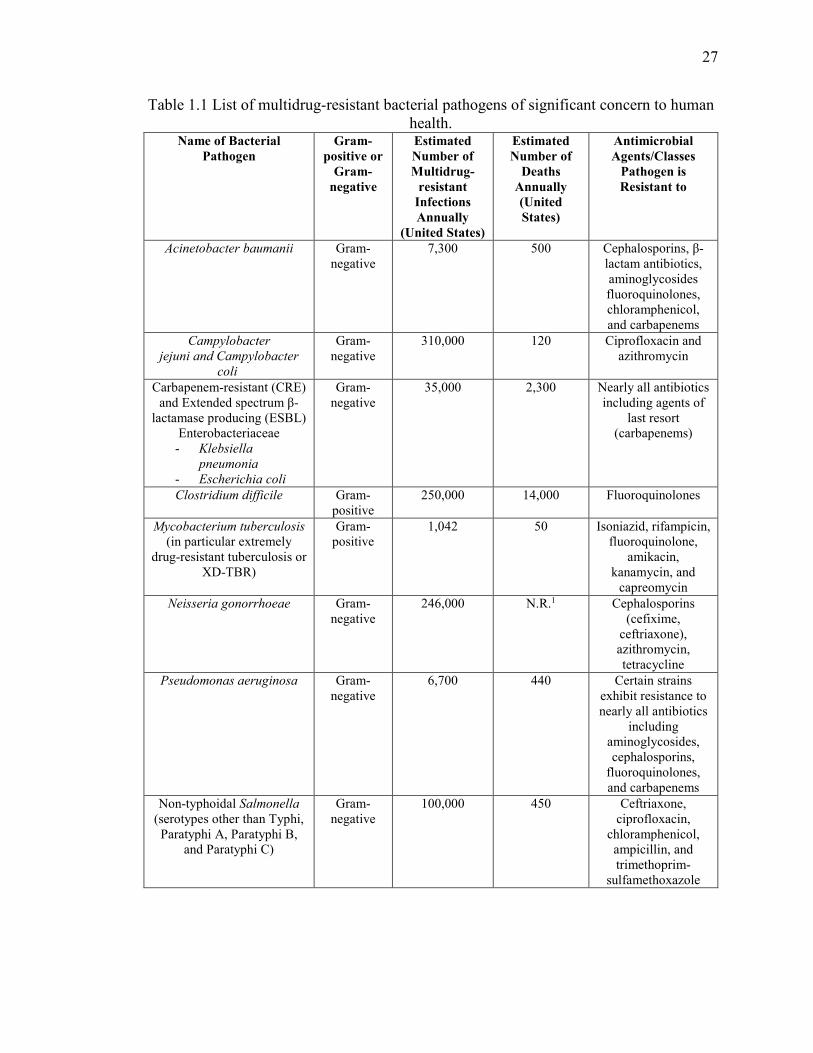
27
Table 1.1 List of multidrug-resistant bacterial pathogens of significant concern to human health.
Name of Bacterial Pathogen
Gram-positive or
Gram-negative
Estimated Number of Multidrug-
resistant Infections Annually
(United States)
Estimated Number of
Deaths Annually (United States)
Antimicrobial Agents/Classes
Pathogen is Resistant to
Acinetobacter baumanii Gram-negative
7,300 500 Cephalosporins, β-lactam antibiotics, aminoglycosides fluoroquinolones, chloramphenicol, and carbapenems
Campylobacter
jejuni and Campylobacter
coli
Gram-negative
310,000 120 Ciprofloxacin and azithromycin
Carbapenem-resistant (CRE) and Extended spectrum β-
lactamase producing (ESBL) Enterobacteriaceae
- Klebsiella
pneumonia
- Escherichia coli
Gram-negative
35,000 2,300 Nearly all antibiotics including agents of
last resort (carbapenems)
Clostridium difficile Gram-positive
250,000 14,000 Fluoroquinolones
Mycobacterium tuberculosis (in particular extremely
drug-resistant tuberculosis or XD-TBR)
Gram-positive
1,042 50 Isoniazid, rifampicin, fluoroquinolone,
amikacin, kanamycin, and
capreomycin
Neisseria gonorrhoeae Gram-negative
246,000 N.R.1 Cephalosporins (cefixime,
ceftriaxone), azithromycin, tetracycline
Pseudomonas aeruginosa Gram-negative
6,700 440 Certain strains exhibit resistance to nearly all antibiotics
including aminoglycosides, cephalosporins,
fluoroquinolones, and carbapenems
Non-typhoidal Salmonella (serotypes other than Typhi, Paratyphi A, Paratyphi B,
and Paratyphi C)
Gram-negative
100,000 450 Ceftriaxone, ciprofloxacin,
chloramphenicol, ampicillin, and trimethoprim-
sulfamethoxazole
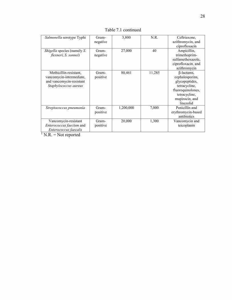
28
Table 7.1 continued
Salmonella serotype Typhi Gram-negative
3,800 N.R. Ceftriaxone, azithromycin, and
ciprofloxacin
Shigella species (namely S.
flexneri, S. sonnei) Gram-
negative 27,000 40 Ampicillin,
trimethoprim-sulfamethoxazole, ciprofloxacin, and
azithromycin
Methicillin-resistant, vancomycin-intermediate, and vancomycin-resistant
Staphylococcus aureus
Gram-positive
80,461 11,285 β-lactams, cephalosporins, glycopeptides, tetracycline,
fluoroquinolones, tetracycline,
mupirocin, and linezolid
Streptococcus pneumonia Gram-positive
1,200,000 7,000 Penicillin and erythromycin-based
antibiotics
Vancomycin-resistant Enterococcus faecium and
Enterococcus faecalis
Gram-positive
20,000 1,300 Vancomycin and teicoplanin
1 N.R. = Not reported
29
CHAPTER 2. DISCOVERY AND CHARACTERIZATION OF POTENT THIAZOLES VERSUS METHICILLIN- AND
VANCOMYCIN-RESISTANT STAPHYLOCOCCUS AUREUS
THIS IS A PUBLISHED JOURNAL ARTICLE. Reprinted with permission from
Discovery and Characterization of Potent Thiazoles versus Methicillin- and
Vancomycin-Resistant Staphylococcus aureus. Haroon Mohammad, Abdelrahman S.
Mayhoub, Adil Ghafoor, Muhammad Soofi, Ruba A. Alajlouni, Mark Cushman, and
Mohamed N. Seleem. Journal of Medicinal Chemistry 2014 57 (4), 1609-1615 DOI:
10.1021/jm401905m. Copyright 2014 American Chemical Society
2.1 Introduction
Methicillin-resistant Staphylococcus aureus (MRSA) is a rapidly expanding global health
concern. It is currently the most common pathogen linked to patients with skin and soft-
tissue infections.(1) Apart from the high mortality and rapid transmission rates, MRSA
infections result in an estimated $3 billion to $4 billion of additional health care costs per
year.(2) Resistance has also emerged to therapeutic agents once deemed to be the drugs of
choice in treating MRSA infections, such as vancomycin (3) and linezolid.(4) Researchers
and clinical-care providers are thus facing an increasingly difficult challenge trying to
construct novel antimicrobials and new therapeutic options to treat MRSA-related
infections.
The thiazole ring is a key structural component for a wide spectrum of therapeutic
agents including anticonvulsants,(5) anticancer,(6, 7) and antiviral agents.(8) In this study,
whole-cell screening assays of libraries of substituted thiazoles and thiadiazoles identified
a novel lead compound that displayed notable antibacterial activity against MRSA. The
lead compound 1a (Figure 2.1) consists of a thiazole central ring connected to two unique
structural features – a cationic element at the C5-position and a lipophilic moiety at the C2-
position. These two structural components have been hypothesized to contribute to the
antibacterial activity of the lead compound. Structural optimizations were focused on the
lipophilic side chain at thiazole-C2 of the lead compound in an attempt to enhance the
antimicrobial activity of the lead compound against MRSA and VRSA. Chemical
30
modifications reported here involved building a focused library of phenylthiazoles with
different lipophilic moieties at the phenyl para position to define the structure-activity-
relationships (SARs) at the thiazole-C2 position in a rigorous way. Our objectives were to
investigate the antimicrobial activities of the thiazole derivatives against MRSA and
VRSA, ascertain the killing kinetics of MRSA in vitro by the lead compound and two
derivatives, determine the cytotoxic impact of the derivatives on mammalian cells in vitro,
and to investigate the physicochemical properties (namely solubility and permeability) of
the thiazole compounds.
2.2 Materials and Methods
2.2.1 Chemistry
General. All biologically tested compounds produced HPLC traces in which the major
peak accounted for ≥ 95% of the combined total peak area when monitored by a UV
detector at 254 nm. 1H NMR spectra were run at 300 MHz and 13C spectra were determined
at 75.46 MHz in CDCl3, DMSO-d6, or CD3OD. Chemical shifts are given in parts per
million (ppm) on the delta (δ) scale. Chemical shifts were calibrated relative to those of the
solvents. Flash chromatography was performed on 230-400 mesh silica and preparative
TLC separations utilized Analtech Uniplates with glass-supported silica (20 × 20 cm, 2000
micron thickness) and UV indicator (254 nM). The progress of reactions was monitored
with Baker-flex silica gel IB2-F plates (0.25 mm thickness). Mass spectra were recorded
at 70 eV. High resolution mass spectra for all ionization techniques were obtained from a
FinniganMAT XL95. Melting points were determined using capillary tubes with a Mel-
Temp apparatus and are uncorrected. HPLC analyses were performed on a Waters binary
HPLC system (Model 1525, 20 μL injection loop) equipped with a Waters dual wavelength
absorbance UV detector (Model 2487) set for 254 nm, and using a 5 μM C-18 reverse
phase column. All reactions were conducted under argon or nitrogen atmosphere, unless
otherwise specified. All yields reported refer to isolated yields. Thiazole ethylketone derivatives 4a-g were prepared in moderate yields by heating
thioamides 3a-g, obtained by treatment of the corresponding amides with Lawesson’s
reagent in dry THF, with 3-chloropentane-2,4-dione in absolute ethanol (Figure 2,2). The
31
methyl ketones 4a-g were gently heated with aminoguanidine hydrochloride in the
presence of lithium chloride as a catalyst to afford hydrazinecarboximidamide derivatives
1a-g (Figure 2.2). Similarly, the final products 1h, 7, 8 and 12 were obtained using a similar
synthetic protocol (Figures 2.3-2.5). A more detailed explanation of the synthetic scheme
for each compound is described below.
Preparation of Hydrazinecarboximidamides 1a-h, 7, 8, and 12. General Procedure.
The ketone derivatives 3a-h, 5, 6, 11 or 16 (1-10 mmol) were dissolved in absolute ethanol
(10-50 mL). Aminoguanidine hydrochloride (1 equivalent) and a catalytic amount of LiCl
(5-20 mg) were added. The reaction mixtures were heated at reflux for 24 hours. The
solvent was evaporated under reduced pressure. The crude product was purified by
crystallization from 70% methanol, and then recrystallized from absolute methanol to
afford the desired compounds as solids. Compound 1a (9) is previously reported.
Preparation of Thioamides 3a-g. Thioamides 3a-g (1-5 mmol), which were obtained
by treatment of their corresponding carboxylic acids 2a-g with thionyl chloride followed
by gradual addition to ammonia solution, and Lawesson’s reagent (1.2 equiv.) were added
to dry THF (15-40 mL). The reaction mixtures were stirred at room temperature for 5-12
hours. The solvent was evaporated under reduced pressure and the residues were
partitioned between aqueous NaHCO3 (2 M, 25-50 mL) and ethyl acetate (25-75 mL). The
organic solvent was separated and dried over anhydrous MgSO4. After solvent evaporation,
the crude products were further purified by silica gel flash chromatography, using hexane-
ethyl acetate (4:1), to yield the corresponding thioamides as yellow solids (55-57%) in the
desired purity degree. Compounds 3a-g were characterized previously as follows. 4-n-
Butylbenzamide,(9) butylthiobenzamide (3a),(9) thiobenzamide (3b),(10) 4-n-
propylbenzamide,(9) propylthiobenzamide (3c),(9) 4-n-pentylbenzamide,(9)
pentylthiobenzamide (3d),(9) 4-n-heptylbenzamide,(11) 4-n-heptylthiobenzamide (3e)(12,
13) 4-nonylbenzamide,(14) 4-t-butylbenzamide,(15) 4-t-butylthiobenzamide (3g)(16) are
reported.
Preparation of Methyl Ketones 4a-i. General Procedure. Thiobenzamides 3a-i (2-
10 mmol) and 3-chloropentane-2,4-dione (1.4 equivalent) were added to absolute ethanol
(10-30 mL). The reaction mixtures were heated at reflux for 12-24 hours. After evaporation
of solvent under reduced pressure, the brown residues were collected and purified by silica
32
gel flash chromatography, using hexane-ethyl acetate (9:1), to yield compounds 4a-i in the
desired purity. Compounds 4a(9) and 4b(17) are previously reported. Characterizations of
compounds 4c-i are listed below.
4-Nonylthiobenzamide (3f). Yellow solid (550 mg, 76%): mp 57 °C. 1H NMR
(DMSO-d6) δ 9.76 (brs, 1 H), 9.39 (brs, 1 H), 7.82 (d, J = 8.1 Hz, 2 H), 7.21 (d, J = 8.1 Hz,
2 H), 2.58 (t, J = 7.5 Hz, 2 H), 1.55 (m, 2 H), 1.25 (m, 12 H), 0.83 (t, J = 6.9 Hz, 3 H); 13C
NMR (DMSO-d6) δ 200.65, 146.95, 137.71, 128.66, 128.32, 35.78, 32.20, 31.61, 29.88,
29.78, 29.62, 29.56, 23.02, 14.88; ESIMS m/z (rel intensity) 264 (MH+, 100); HREISMS,
m/z 264.1784 MH+, calcd for C16H26NS 264.1786.
1-[4-Methyl-2-(4-propylphenyl)thiazol-5-yl]ethanone (4c). White solid (135 mg,
61%): mp 57 ˚C. 1H NMR (CDCl3) δ 8.10 (d, J = 9.0 Hz, 2 H), 7.32 (d, J = 9.0 Hz, 2 H),
2.93 (s, 3 H), 2.6 (m, 5 H), 1.67 (m, 2 H), 0.96 (t, J = 6.3 Hz, 3 H); 13C NMR (CDCl3) δ
189.43, 171.04, 155.41, 148.87, 131.59, 129.63, 127.87, 126.96, 38.00, 30.36, 24.06, 16.88,
13.67; CIMS m/z (rel intensity) 260 (MH+, 100); HRMS (EI), m/z 259.1033 M+, calcd for
C15H17NOS 259.1031.
1-[4-Methyl-2-(4-pentylphenyl)thiazol-5-yl]ethanone (4d). Colorless oil (159 mg,
76%). 1H NMR (CDCl3) δ 7.85 (d, J = 8.7 Hz, 2 H), 7.22 (d, J = 8.7 Hz, 2 H), 2.73 (s, 3
H), 2.60 (t, J = 6.0 Hz, 2 H), 2.50 (s, 3 H), 1.63 (m, 2 H), 1.33 (m, 4 H), 0.87 (t, J = 6.0 Hz,
3 H); 13C NMR (CDCl3) δ 190.33, 169.56, 159.39, 146.64, 130.71, 130.25, 129.04, 126.77,
35.79, 31.37, 30.79, 30.67, 22.45, 18.41, 13.97; CIMS m/z (rel intensity) 288 (MH+, 100);
HRMS (EI), m/z 287.1347 M+, calcd for C17H21NOS 287.1344.
1-[4-Methyl-2-(4-hepylphenyl)thiazol-5-yl]ethanone (4e). Colorless oil (360 mg,
44%). 1H NMR (CDCl3) δ 7.86 (d, J = 8.1 Hz, 2 H), 7.25 (d, J = 8.1 Hz, 2 H), 2.76 (s, 3
H), 2.63 (t, J = 6.0 Hz, 2 H), 2.55 (s, 3 H), 1.62 (m, 2 H), 1.28 (m, 8 H), 0.87 (t, J = 6.0 Hz,
3 H); 13C NMR (CDCl3) δ 190.35, 169.64, 159.42, 146.70, 131.36, 130.73, 130.26, 129.06,
126.80, 35.84, 31.74, 31.13, 30.70, 29.16, 29.09, 22.60, 18.42, 14.05; CIMS m/z (rel
intensity) 316 (MH+, 100); HRMS (EI), m/z 315.1655 M+, calcd for C19H25NOS 315.1657.
1-[4-Methyl-2-(4-nonylphenyl)thiazol-5-yl]ethanone (4f). Yellow oil (450 mg, 60%).
1H NMR (CDCl3) δ 7.87 (d, J = 8.1 Hz, 2 H), 7.24 (d, J = 8.1 Hz, 2 H), 2.77 (s, 3 H), 2.63
(t, J = 7.0 Hz, 2 H), 2.55 (s, 3 H), 1.62 (m, 2 H), 1.29 (m, 12 H), 0.87 (t, J = 6.9 Hz, 3 H);
33
13C NMR (CDCl3) δ 190.42, 169.65, 159.44, 146.71, 130.75, 130.29, 129.07, 126.81, 35.86,
31.83, 31.14, 30.72, 29.48, 29.43, 29.25, 22.63, 18.45, 14.07; ESIMS m/z (rel intensity)
344 (MH+, 100); HRESIMS, m/z 344.2052 M+, calcd for C21H30NOS 344.2048.
1-{2-[4-(tert-Butyl)phenyl]-4-methylthiazol-5-yl}ethanone (4g). White solid (557
mg, 62%): mp 53 ˚C. 1H NMR (CDCl3) δ 7.84 (d, J = 8.4 Hz, 2 H), 7.43 (d, J = 8.4 Hz, 2
H), 2.72 (s, 3 H), 2.49 (s, 3 H), 1.31 (s, 9 H); 13C NMR (CDCl3) δ 190.27, 169.35, 159.36,
154.65, 130.70, 129.97, 126.57, 125.90, 34.85, 31.00, 30.62, 18.37; CIMS m/z (rel intensity)
274 (MH+, 100); HRMS (EI), m/z 273.1182 M+, calcd for C16H19NOS 273.1187.
1-[4-Methyl-2-(naphthalen-2-yl)thiazol-5-yl]ethanone (4h). White solid (110 mg,
67%): mp 109 °C. 1H NMR (CDCl3) δ 8.41 (d, J = 0.9 Hz, 1 H), 7.94 (d, J = 1.8 Hz, 1 H),
7.84 (m, 3 H), 7.50 (d, J = 9.0 Hz, 2 H), 2.76 (s, 3 H), 2.51 (s, 3 H); 13C NMR (CDCl3) δ
190.31, 169.28, 159.49, 134.50, 132.98, 131.21, 129.94, 128.79, 127.80, 127.54, 126.93,
126.67, 123.71, 30.69, 18.45; ESIMS m/z (rel intensity) 268 (MH+, 100); HRESIMS, m/z
268.0793 MH+, calcd for C16H14NOS 264.0796.
1-[2-(4-Iodophenyl)-4-methylthiazol-5-yl]ethanone (4i). Brown solid (1050 mg,
58%): mp 123 ˚C. 1H NMR (CDCl3) δ 7.80 (d, J = 8.7 Hz, 2 H), 7.68 (d, J = 8.7 Hz, 2 H),
2.76 (s, 3 H), 2.56 (s, 3 H); 13C NMR (CDCl3) δ 190.29, 168.05, 159.44, 138.14, 132.11,
131.43, 128.13, 97.66, 30.70, 18.36; CIMS m/z (rel intensity) 344 (MH+, 100); HRMS (EI),
m/z 342.9535 M+, calcd for C12H10INOS 342.9528.
1-{2-[4-(1-Cyclohexenyl)phenyl]-4-methylthiazol-5-yl}ethanone (5). A solution of
4-iodophenylthiazole 4i (100 mg, 0.3 mmol) in dry DMF (5 mL) was charged with
Pd(OAc)2 (5 mg), cyclohexene (1 mL) and triethylamine (0.5 mL). The reaction mixture
was heated at 80 ˚C for 5 h. The reaction mixture was quenched with distilled water (10
mL) and extracted with ethyl acetate (30 mL). The organic layer was separated and dried
over anhydrous Na2SO4. After evaporation of solvent under reduced pressure, the oily
residue was collected and purified by silica gel flash chromatography, using hexane-ethyl
acetate (9:1), to yield faint yellow oil (35 mg, 39%). 1H NMR (CDCl3) δ 7.90 (d, J = 8.1
Hz, 2 H), 7.29 (d, J = 8.1 Hz, 2 H), 5.76 (m, 2 H), 2.80 (m, 1 H), 2.77 (s, 3 H), 2.55 (s, 3
H), 2.27-1.24 (m, 6 H); 13C NMR (CDCl3) δ 190.45, 169.54, 159.46, 150.94, 130.84,
130.63, 128.42, 127.60, 126.97, 126.33, 40.06, 32.98, 30.73, 29.40, 25.59, 18.45; CIMS
34
m/z (rel intensity) 298 (MH+, 100); HRMS (EI), m/z 297.1189 M+, calcd for C18H19NOS
297.1187.
1-[2-(4-Cyclohexylphenyl)-4-methylthiazol-5-yl]ethanone (6). Compound 5 (100
mg, 0.3 mmol) and Pd (50 mg, 10% on activated charcoal) were added to deoxygenated
absolute methanol (10 mL). Hydrogen was applied via a balloon. The reaction mixture was
stirred at room temperature for 24 h, and then filtered through celite. The filtrate was
collected and the solvent was evaporated under reduced pressure to yield compound 7 as a
colorless oil (100 mg, 100%). 1H NMR (CDCl3) δ 7.87 (d, J = 8.4 Hz, 2 H), 7.27 (d, J =
8.4 Hz, 2 H), 2.75 (s, 3 H), 2.53 (s, 3 H), 2.51 (m, 1 H), 1.85-1.73 (m, 5 H), 1.43-1.24 (m,
5 H); 13C NMR (CDCl3) δ 190.45, 169.65, 159.44, 151.70, 130.74, 130.40, 127.51, 126.88,
44.50, 34.10, 30.70, 26.69, 25.99, 18.44; CIMS m/z (rel intensity) 300 (MH+, 100); HRMS
(EI), m/z 299.1350 M+, calcd for C18H21NOS 299.1344.
1-(2-([1,1'-Biphenyl]-4-yl)-4-methylthiazol-5-yl)ethanone (11). The aldehyde 9 (800
mg, 5.2 mmol) was added to a solution of hydroxylamine hydrochloride (725, 10.5 mmol)
in DMSO (10 mL), and the reaction mixture was stirred at 100 ˚C for 20 min. The heater
was turned off and aqueous NaOH solution (600 mg dissolved in 5 mL distilled water) was
slowly added to the reaction mixture over a 2 min period with stirring, and then hydrogen
peroxide 50% (5 mL) was slowly and carefully added over a 10 min period. The reaction
mixture was further stirred for 12 h and extracted with ethyl acetate (3 x 10 mL), dried over
anhydrous Na2SO4, and evaporated under reduced pressure to afford the corresponding
amide as a white solid. The crude amide (2 mmol) and Lawesson’s reagent (980 mg, 2.4
mmol) were added to dry THF (15 mL). The reaction mixture was stirred at room
temperature for 12 h. The solvent was evaporated under reduced pressure and the residue
was partitioned between aq NaHCO3 (25 mL) and ethyl acetate (25 mL). The organic
solvent was separated and dried over anhydrous Na2SO4.
The crude product was further purified by silica gel flash chromatography, using hexane-
ethyl acetate (4:1), to yield the corresponding thioamide 10 as a yellow solid. The obtained
thioamide 10 (215 mg, 1 mmol) and 3-chloro-2,4-pentanedione (0.3 mL, 2.5 mmol) were
added to absolute ethanol (10 mL). The reaction mixture was heated at reflux for 24 h.
After evaporation of solvent under reduced pressure, the oily residue was collected and
35
purified by silica gel flash chromatography, using hexane-ethyl acetate (9:1), to yield
compound 11 as an off-white solid (290 mg, 49%): mp 124-125 ˚C. 1H NMR (CDCl3) δ
8.36 (d, J = 8.4 Hz, 2 H), 7.77 (d, J = 8.4 Hz, 2 H), 7.64 (d, J = 8.7 Hz, 2 H), 7.46 (m, 3
H), 3.03, 2.64; 13C NMR (CD3OD) δ 190.45, 153.24, 148.20, 143.78, 140.00, 130.51,
130.28, 128.49, 127.94, 127.14, 126.14, 126.01, 23.90, 17.99; ESIMS m/z (rel intensity)
290 (MH+, 100); HRESIMS, m/z 290.1039 MH+, calcd for C18H27NS 290.1939.
2.2.2 Bacterial Strains, Reagents, and Antibiotics
The complete list and description of bacterial strains presented in this study is located in
Table 2.1. MRSA clinical isolates, vancomycin-intermediate Staphylococcus aureus
(VISA), vancomycin-resistant Staphylococcus aureus (VRSA) and linezolid-resistant
strains were obtained through the Network of Antimicrobial Resistance in Staphylococcus
aureus (NARSA) program. In addition, MRSA ATCC 43300 was obtained from the
American Type Cultural Collection (Manassas, VA, USA). Lysostaphin (Sigma-Aldrich,
St. Louis, MO, USA) at 20 µg/mL was prepared in 50 mM Tris-HCl (pH 8.00) (Sigma-
Aldrich, St. Louis, MO, USA). Vancomycin hydrochloride powder was purchased
commercially (Gold Biotechnology Inc., St. Louis, MO, USA) and dissolved in dimethyl
sulfoxide (DMSO) (Sigma-Aldrich, St. Louis, MO, USA).
2.2.3 Determination of Minimum Inhibitory Concentration (MIC)
The MICs of the lead thiazole compound (compound 1a) and nine derivatives tested against
all 18 MRSA strains were determined, in triplicate samples, using the broth microdilution
method in accordance with the recommendations contained in the Clinical and Laboratory
Standards Institute guidelines. The MIC was categorized as the concentration at which no
visible growth of bacteria was observed in a particular well. The average of triplicate MIC
determinations is reported along with standard deviation values.
2.2.4 Time-kill Assay
MRSA (USA300) cells, in the logarithmic growth phase, were diluted to 1.0 × 10⁶ colony-
forming units (CFU/mL) and exposed to concentrations equivalent to 3.0 × MIC (in
triplicate) of compounds 1a, 1d, 8, and vancomycin in tryptic soy broth (TSB) (Becton,
Dickinson and Company, Sparks, MD, USA). Viable CFU/mL was determined by serial
36
dilution and plating on tryptic soy agar (TSA) (Becton, Dickinson and Company, Sparks,
MD, USA) plates after 0, 2, 4, 6, 8, 10, and 12 hours of incubation at 37 °C to identify the
time required to reduce the bacterial cell count by 3-log10.
2.2.5 In Vitro Cytotoxicity Analysis
Compounds were assayed at a concentration of 11 µg/mL in human cervical
adenocarcinoma cells (HeLa) to determine the potential toxic effect in vitro. Cells were
cultured in Dulbeco’s modified Eagle’s medium (Sigma-Aldrich, St. Louis, MO, USA)
with 10% fetal bovine serum (USA Scientific, Inc.) at 37 °C with 5% CO2. Controls
received DMSO alone at a concentration equal to that in drug-treated cell samples. The
cells were incubated with the compounds in a 96-well plate at 37 ºC and 5.0% CO2 for two
hours prior to addition of the assay reagent MTS 3-(4,5-dimethylthiazol-2-yl)-5-(3-
carboxymethoxyphenyl)-2-(4-sulfophenyl)-2H-tetrazolium) (Promega, Madison, WI,
USA). Corrected absorbance readings (actual absorbance readings for each treatment
subtracted from background absorbance) were taken using a kinetic ELISA microplate
reader (Molecular Devices, Sunnyvale, CA, USA). The quantity of viable cells after
treatment with each compound was expressed as a percentage of the control, DMSO.
2.2.6 Calculation of Partition Coefficient (log P) and Topological Polar Surface Area (TPSA)
Calculated log P and topological polar surface area (TPSA) values for the thiazole
compounds were obtained using Molinspiration Cheminformatics software available on
the internet (http://www.molinspiration.com/).
2.2.7 Caco-2 Permeability Assay
Caco-2 cells grown in tissue culture flasks were trypsinized, suspended in medium, and the
suspensions were applied to wells of a Millipore 96 well Caco-2 plate. The cells were
allowed to grow and differentiate for three weeks, feeding at 2-day intervals. For Apical to
Basolateral (A→B) permeability, compound 1a was added to the apical (A) side and
amount of permeation was determined on the basolateral (B) side; for Basolateral to Apical
(B→A) permeability, compound 1a was added to the B side and the amount of permeation
was determined on the A side. The A-side buffer contained 100 μM Lucifer yellow dye, in
37
Transport Buffer (1.98 g/L glucose in 10 mM HEPES, 1.0 × Hank’s Balanced Salt Solution)
pH 6.5, and the B-side buffer contained Transport Buffer at pH 7.4. Caco-2 cells were
incubated with these buffers for two hours, and the receiver side buffer was removed for
analysis by LC/MS/MS. To verify the Caco-2 cell monolayers were properly formed,
aliquots of the cell buffers were analyzed by fluorescence to determine the transport of the
impermeable dye Lucifer Yellow. Any deviations from control values are reported. Data
are expressed as permeability (Papp) = (dQ/dt)/C0A where dQ/dt is the rate of permeation,
C0 is the initial concentration of test agent, and A is the area of the monolayer. In
bidirectional permeability studies, the efflux ratio (RE) is also calculated: RE =
(Papp(B→A))/(Papp(A→B)). An RE > 2 indicates a potential substrate for P-glycoprotein
or other active efflux transporters.
2.2.8 MDCK-MDR1 Permeability Assay
MDCK-MDR1 cells were grown in tissue culture flasks, trypsinized, suspended in medium,
and the suspensions were applied to membranes plate wells (96-well format). The cells
were allowed to grow and differentiate for five days. For Apical to Basolateral (A→B)
permeability, compound 1a was added to the apical (A) side and the amount of permeation
was determined on the basolateral (B) side; for Basolateral to Apical (B→A) permeability,
compound 1a was added to the B side and the amount of permeation was determined on
the A side. The A-side buffer contained 100 μM Lucifer Yellow dye, in Transport Buffer
(1.98 g/L glucose, 10 mM HEPES, in 1.0 × Hank’s Balanced Salt Solution) pH 7.4. The
B-side buffer was Transport Buffer, pH 7.4. MDCK-MDR1 cells were incubated with these
buffers for two hours, and the receiver side buffer was removed for analysis by LC/MS-
MS. To verify the MDCK-MDR1 cell monolayers were properly formed, aliquots of the
cell buffers were analyzed by fluorescence to determine the transport of the impermeable
dye Lucifer Yellow. Data are expressed as permeability (Papp) = (dQ/dt)/C0A where dQ/dt
is the rate of permeation, C0 is the initial concentration of compound 1a, and A is the area
of the monolayer. In bidirectional permeability studies, the efflux ratio (RE) is also
calculated: RE = (Papp(B→A))/(Papp(A→B)). An RE > 2 indicates a potential substrate
for P-glycoprotein or other active efflux transporters.
38
2.2.9 PBS Solubility Screen
Serial dilutions of lead compound 1a, reserpine, tamoxifen, and verapamil were prepared
in phosphate buffered saline (PBS) at 100 × the final concentration. The solutions were
diluted 100-fold into PBS in a 96-well plate and mixed. The absorbance of the PBS-
containing plate was measured prior to addition of the test agents to determine the
background absorbance. After two hours, the presence of precipitate was detected by
turbidity (absorbance at 540 nm). An absorbance value of greater than (mean + 3 × standard
deviation of the blank), after subtracting the pre-experiment background, is indicative of
turbidity. The solubility limit is reported as the highest experimental concentration with no
evidence of turbidity.
2.2.10 Microsomal Stability Analysis
Compound 1a was incubated in duplicate with microsomes at 37 ºC. The reaction contained
microsomal protein in 100 mM potassium phosphate, 2 mM NADPH, 3 mM MgCl2, pH
7.4. A control was run for each test agent omitting NADPH to detect NADPH-free
degradation. At 0, 10, 20, 40, and 60 minutes, an aliquot was removed from each
experimental and control reaction and mixed with an equal volume of ice-cold Stop
Solution (methanol containing haloperidol, diclofenac, or other internal standard). Stopped
reactions are incubated at least ten minutes at -20 ºC, and an additional volume of water
was added. The samples were centrifuged to remove precipitated protein, and the
supernatants were analyzed by LC/MS/MS to quantitate the remaining parent. Data are
converted to % remaining by dividing by the time zero concentration value. Data are fit to
a first-order decay model to determine half-life. Intrinsic clearance is calculated from the
half-life and the protein concentrations: CLint = ln(2) /(T1/2 [microsomal protein]).
2.2.11 Statistical Analysis
All statistical analysis was conducted using Kaleida Graph, version 4.03 (Synergy software,
Reading, PA). Statistical significance was determined using ANOVA and the Fisher’s
Least Significant Difference (LSD) test with α = 0.05.
39
2.3 Results and Discussion
2.3.1 Antibacterial activity of thiazole compounds against MRSA and VRSA
The 10 substituted thiazole compounds we synthesized inhibited growth of 18 different
strains of MRSA and VRSA at a concentration ranging from 0.4-5.5 µg/mL (Table 2.2).
The lead compound 1a inhibited the growth of MRSA strains at concentrations ranging
from 1.4 – 5.5 µg/mL. Subsequently synthesized derivatives demonstrated a two- to five-
fold improvement in the MIC values. Initially, the effect of increasing the length of the
alkyl side chain, through insertion of methylene units, was explored. As the length of the
alkyl side chain increased from two (compound 1c) to three (compound 1a) to four
(compound 1d) methylene units, there was a consistent improvement in the MIC values
observed against all MRSA strains tested. However, additional lengthening of the alkyl
side chain appeared to nullify the improvement observed in the antimicrobial activity, as
the MIC for compound 1e (containing six methylene units) nearly matched or exceeded the
values obtained for compound 1d. This result held true as an increase to eight methylene
units (compound 1f) resulted in an MIC value that nearly matched or exceeded the MIC
value attained for compound 1a. Altogether this indicates that an alkyl side chain with four
methylene units exhibits the optimum potency against MRSA and addition of methylene
units to the alkyl side beyond four units will not significantly enhance the antimicrobial
activity of the lead compound.
Replacement of the linear alkyl side chain with a branched alkane (compound 1g)
produced mixed results. There was a modest improvement in the MIC values against six
MRSA strains (1.0 µg/mL for 1g compared to 1.4 µg/mL for 1a) and a nearly two- to five-
fold enhancement in the MIC for five additional strains. Substitution of the alkyl side chain
with a fused ring system (compound 1h) did not significantly enhance the activity of the
derivative against the MRSA strains tested, with the exception of VRSA (a near three-fold
reduction in MIC was observed compared to 1a). However, replacement of the alkyl side
chain with conformationally restricted analogues (compounds 7, 8, and 12) demonstrated
the most consistent, significant improvement in the MIC value obtained relative to the lead
compound (two- to four-fold improvement against 16 MRSA strains tested).
40
The MIC values obtained for compounds 7, 8, and 12 on multiple occasions
matched or were lower than the antibiotic vancomycin against the MRSA strains tested.
Furthermore, all ten thiazole compounds proved to be more potent than vancomycin in
inhibiting growth of VISA and VRSA strains. Compounds 7, 8, and 12 also proved more
effective at eliminating growth of MRSA NRS119, a strain resistant to linezolid (a drug of
last resort in treatment of MRSA infections), and several strains resistant to multiple
antibiotic classes including lincosamides, aminoglycosides, fluoroquinolones, and
macrolides (USA100, USA200, and USA500). In addition to this, all 10 compounds
exhibited excellent activity against MRSA USA300, a strain responsible for most cases of
community-acquired MRSA (CA-MRSA) and MRSA skin and soft tissue infections
(SSTIs) in the United States.(18, 19).
2.3.2 Time-kill assay of thiazole compounds against MRSA
A drawback of several commercial antimicrobials used to treat MRSA infections, including
vancomycin and linezolid, is either they are only capable of inhibiting bacterial growth
(but do not kill the bacteria), or they exhibit a very slow bactericidal effect resulting in
difficulty in clearing the infection. (20, 21) Thus a compound that demonstrates the ability
to rapidly kill MRSA is highly desirable since it limits the possibility of developing
bacterial resistance/tolerance. We studied the rate at which the compounds were able to
eliminate MRSA (USA300) in vitro in a time-kill assay. The results (Figure 2.6) indicate
that at 3.0 × MIC, lead compound 1a, 1d (derivative which contains one more methylene
unit in the alkyl side chain), and 8 (derivative which replaces the alkyl side chain with a
cyclohexane ring) are bactericidal. However, the rate of clearance of MRSA (USA300)
varies among the three compounds. Compound 1d mimics the action of compound 1a,
rapidly eliminating MRSA completely within two hours. This would appear logical as
compounds 1a and 1d are similar in structure, the major difference resulting from the
number of methylene units contained in the alkyl side chain. Compound 8 requires more
than double the time (six hours) to logarithmically reduce MRSA colony forming units
(CFU) to zero. Though compound 8 appears more potent compared to compounds 1a and
1d when comparing MIC values), the latter two appear capable of clearing MRSA colonies
(albeit at a higher concentration) more rapidly. Vancomycin was not able to reduce the
41
number of CFU by 3-log10 within a 12-hour window. Collectively this indicates the thiazole
compounds possess a selective advantage over vancomycin in terms of rate of elimination
of MRSA cells. This information is clinically significant as it would impact the size and
timing of the dose given to patients with an infection caused by MRSA.
In addition to this, combination therapy using multiple antibiotics to treat MRSA
infections is commonly used in clinical practice. Antibiotics that are bacteriostatic or
exhibit a slow bactericidal effect (such as vancomycin) (20) are often paired with
antibiotics exhibiting a rapid bactericidal effect (such as rifampin) in order to limit the
emergence of bacterial strains with reduced susceptibility to vancomycin.(20) As the
thiazole compounds presented here exhibit a rapid bactericidal effect against MRSA,
analysis of synergy between the thiazole compounds and commercial antimicrobials (such
as vancomycin and linezolid) for potential use in combination therapy would be an
interesting avenue to further explore.
2.3.3 Evaluating toxicity of thiazole compounds against a HeLa cell line
The cytotoxicity assay (Figure 2.7) confirmed that all of the compounds are selective for
bacterial cell inhibition over mammalian cells. All compounds tested were not toxic to
human cervical adenocarcinoma (HeLa) cells up to 11 µg/mL; this concentration is more
than 20-fold higher than the MIC value for the most potent thiazole derivatives (8 and 12).
Irrespective of the modification made to the alkane side chain of the lead compound
(addition of methylene units or substitution with a cyclic moiety), the subsequent
derivatives maintained a good toxicity profile when tested against HeLa cells.
2.3.4 Physicochemical properties of the most promising analogues
Physicochemical properties, including solubility and permeability, of potential therapeutic
agents are critical factors that need to be explored early in drug development. Though a
compound proves potent against a target organism during in vitro studies and exhibits
limited toxicity to cultured mammalian cells, the drug-candidate can fail in animal and
human studies if the drug is poorly soluble in aqueous solutions or is incapable of passing
through cellular barriers. Analysis of the hydrogen bonding potential and lipophilicity of a
compound can lend valuable insight into potential solubility and permeability issues. After
42
documenting the strong antimicrobial activity of the thiazole compounds and determining
their limited toxicity against murine macrophage cells, it was critical to establish whether
the compounds possess potential solubility and permeability issues. Using Lipinski’s Rule
of 5 and topological polar surface area (TPSA) as guidelines, the results in Table 2.3
demonstrate all 10 thiazole compounds possess clog P and TPSA values that are associated
with good solubility and permeability qualities. Two derivatives (1e and 1f) violate one
parameter of the Rule of 5 with each derivative possessing a calculated log P value above
5. These derivatives contain the longest linear alkyl chain (six and eight methylene units
for 1e and 1f, respectively) connected to the phenylthiazole nucleus. This result supports
the notion that an ideal thiazole side chain should have four methylene units, as compounds
possessing an alkyl side chain with more than four methylene units exhibit a decrease in
the antimicrobial activity against MRSA and pose potential solubility issues.
2.3.5 Examination of the solubility of lead 1a
To confirm if the thiazole compounds possess good physicochemical properties as
predicted, the lead compound was analyzed using the Caco-2 permeability assay, MDCK-
MDR1 permeability assay, and a solubility screen utilizing phosphate buffered saline
(PBS). The solubility screen was used to determine the highest concentration lead
compound 1a and three control drugs were able to dissolve in PBS before precipitate
formed. The PBS solubility screen indicates the lead thiazole compound possesses modest
solubility compared to the reference drugs tested. When compared to drugs with poor
aqueous solubility, compound 1a was soluble at twice the concentration of the
antihypertensive drug reserpine (31.3 µM) and nearly four times the concentration of the
cancer drug tamoxifen (15.6 µM) as presented in Table 2.4. As good solubility is a critical
property needed for the development of a drug to be used in an oral formulation,
modifications to the lead compound need to be explored to enhance its solubility profile.
The solubility profile of the lead thiazole compound can be further enhanced by
constructing subsequent derivatives containing ionizable groups (such as a basic amine or
carboxylic acid moiety), inclusion of additional hydrogen bond donor groups (OH and NH2
groups), or incorporating one or more polar groups (such as an ester) into the core structure.
43
2.3.6 Caco-2 and MDCK-MDR1 bidirectional permeability analysis of compound 1a
The Caco-2 permeability assay revealed the lead compound (1a) surprisingly exhibited
poor permeability across the membrane from the apical (A) to basolateral (B) direction as
demonstrated in Table 2.5. Compound 1a exhibits a higher apparent permeability
coefficient (Papp) in the basolateral to apical direction (Papp = 2.2 × 10⁻⁶ cm/sec) which
mimics the result obtained with the control drug ranitidine (Papp = 1.2 × 10⁻⁶ cm/sec in the
B to A direction compared to 0.2 × 10⁻⁶ cm/sec in the A to B direction). Transporters in
the membranes can enhance or reduce the permeability of a compound. The presence of
efflux transporters on the apical surface of epithelial cells in the intestine may play a role
in preventing the absorption of the thiazole compounds and passage through Caco-2 cells.
The efflux ratio >2 for the lead thiazole compound supports the notion that the compound
may be a substrate for an efflux transporter (possibly P-glycoprotein which is a potential
reason for the higher rate of transfer of compound from the B to A direction). To confirm
if this was the case, compound 1a was analyzed using Madin-Darby canine kidney (MDCK)
cells transfected with a gene overexpressing multidrug resistance protein 1 (MDR1), also
referred to as P-glycoprotein 1 (Pgp). As presented in Table 2.6, a higher rate of transfer
of compound 1a is observed in the B to A direction (Papp = 3.6 × 10⁻⁶ cm/sec) compared to
the A to B direction (Papp = 0.7 × 10⁻⁶ cm/sec), consistent with what is observed with the
Caco-2 permeability assay. The efflux ratio determined from the MDCK-MDR1
permeability assay for compound 1a is 5.0, indicating the compound may be subject to the
effect of Pgp.
An increasing number of hydrogen bond acceptors (oxygen and nitrogen atoms)
has been shown to increase the likelihood of Pgp efflux of drugs (22). Thus constructing
derivatives of the lead thiazole compound focusing on modifications to the cationic head
(where the hydrogen bond acceptor groups are present) is one mechanism to enhance
permeability. A delicate balance between addition or substitution of functional groups
would need to be achieved to ensure permeability is enhanced without reducing the
solubility profile of the thiazole compounds. Another method to enhance passage of the
thiazole compounds across the intestinal membrane is to use a higher concentration of the
lead compound, especially if the compound is a substrate for efflux transporters. The Caco-
2 assay utilized a low concentration (10 µM) of the lead compound. The concentration of
44
a drug in the gastrointestinal lumen after an oral dose is typically between 50 to 100 µM
(22). Thus testing the lead compound at a higher concentration is necessary to confirm if
the poor permeability observed is potentially due to the low concentration of compound
used in the assay. A higher concentration may help the thiazole compound to effectively
cross the intestinal barrier as efflux transporters will eventually become saturated in a
concentration-dependent manner permitting compound that has passively diffused across
the membrane to remain in the lumen of the intestine. Taken collectively, the permeability
profile of the thiazole compounds can be potentially improved by modifying the structure
of the lead compound or increasing the concentration of the compound used.
2.3.7 Metabolic stability analysis of compound 1a in human microsomes
In addition to testing the solubility and permeability characteristics of the lead thiazole
compound, the metabolic stability of the compound was investigated using human liver
microsomes. As Table 2.7 demonstrates, compound 1a is subject to metabolism by the liver
with a microsomal intrinsic clearance of 80.3 µL/min-mg and a half-life of 28.8 minutes.
These values align with the values obtained with verapamil (the metabolized control drug)
rather than for warfarin (non-metabolized control drug). Removing the cofactor NADPH
significantly reduced the metabolism of compound 1a indicating that these metabolic
processes are NADPH-dependent. The experiment doesn’t eliminate other tissues which
may play a role in metabolism of the thiazole compounds. The half-life and microsomal
intrinsic clearance are important parameters for determining an appropriate dosing regimen
for a drug (as drugs which have a short half-life and are rapidly metabolized in the liver
will require additional doses for patients in order to clear an infection). To improve
metabolic stability of the thiazole compounds, consideration must be given to removing
labile groups or altering atoms in the compound involved in its metabolism.
2.4 Conclusion
In conclusion, the identification of novel antimicrobial agents to treat an array of infections
caused by methicillin-resistant and vancomycin-resistant S. aureus requires a multi-fold
approach from whole-cell screening of chemical libraries to rational drug design. We
present the exciting discovery of a lead antimicrobial compound, identified from whole-
45
cell screening of a library of thiazole and thiadiazole compounds, which is capable of
inhibiting growth of 18 strains of MRSA and VRSA. The lead compound consists of a
thiazole central ring connected to two structural elements critical for activity, namely a
cationic element at the C5-position and a lipophilic moiety at the C2-position. A focused
library of derivatives containing modifications to the lipophilic moiety was constructed to
enhance the antimicrobial activity of the lead compound against MRSA and VRSA. The
lead compound and nine derivatives are capable of inhibiting growth of 18 different clinical
isolates of MRSA and VRSA at a concentration ranging from 0.5 to 3.0 µg/mL.
Furthermore, the lead compound and two derivatives exhibit a rapid bactericidal effect,
eliminating MRSA growth in vitro within six hours. In addition to this, six derivatives,
including the three most potent compounds against MRSA, are not toxic. The ten thiazole
compounds were predicted to have good solubility and permeability characteristics based
upon the criterion set forth by Lipinski’s Rule of 5. However, analysis of permeability of
the lead compound via the Caco-2 and MDCK-MDR1 assays indicated the compound had
poor permeability from the apical to basolateral surface of the membrane (possibly due to
the effect of the Pgp efflux transporter). We confirmed the lead compound does not target
the integrity of the bacterial cell wall or cytoplasmic membrane (data not published);
further explanation of the molecular target of the thiazole compounds will be presented in
a future study. The characterization of the novel thiazole compounds presents an intriguing
step in the development of a novel class of therapeutic agents effective for treating MRSA
and VRSA infections.
46
2.5 References
1. Moran G, Krishnadasan A, Gorwitz R, al. e. 2006. Methicillin-resistant S-aureus
infections among patients in the emergency department. New England Journal of
Medicine 355:666-674.
2. Fischbach MA, Walsh CT. 2009. Antibiotics for Emerging Pathogens. Science
325:1089-1093.
3. Hiramitsu K. 2001. Vancomycin-resistant Staphylococcus aureus: a new model of
antibiotic resistance. The Lancet Infectious Diseases 1:147-155.
4. Peter Wilson, J. A. Andrews, R. Charlesworth, R. Walesby, M. Singer, D. J.
Farrell, Robbins M. 2003. Linezolid resistance in clinical isolates of
Staphylococcus aureus. Journal of Antimicrobial Chemotherapy 51:186-188.
5. Hays SJ, Rice MJ, Ortwine DF, Johnson G, Schwarz RD, Boyd DK, Copeland
LF, Vartanian MG, Boxer PA. 1994. Substituted 2‐benzothiazolamines as
sodium flux inhibitors: Quantitative structure–activity relationships and
anticonvulsant activity. Journal of pharmaceutical sciences 83:1425-1432.
6. Das J MR, Lin J, Liu C, Doweyko AM, DeFex HF, Fang Q, Pang S, Pitt S,
Shen DR, Schieven GL, Barrish JC, Wityak J. 2003. Discovery of 2-amino-
heteroaryl-benzothiazole-6-anilides as potent p56(lck) inhibitors. Bioorganic &
Medicinal Chemistry Letters 13:2587-2590.
7. Hutchinson I, Bradshaw TD, Matthews CS, Stevens MF, Westwell AD. 2003.
Antitumour benzothiazoles. Part 20: 3′-Cyano and 3′-Alkynyl-substituted 2-(4′-
Aminophenyl) benzothiazoles as new potent and selective analogues. Bioorganic
& Medicinal Chemistry Letters 13:471-474.
8. Paget CJ, Kisner K, Stone RL, DeLong DC. 1969. Heterocyclic substituted
ureas. II. Immunosuppressive and antiviral activity of benzothiazolyl-and
benzoxazolylureas. Journal of Medicinal Chemistry 12:1016-1018.
9. Mayhoub AS, Khaliq M, Botting C, Li Z, Kuhn RJ, Cushman M. 2011. An
investigation of phenylthiazole antiflaviviral agents. Bioorganic & Medicinal
Chemistry 19:3845-3854.
47
10. Mahammed KA, Jayashankara VP, Rai NP, Raju KM, Arunachalam PN.
2009. A Mild and Versatile Synthesis of Thioamides. Synlett doi:DOI 10.1055/s-
0029-1217711:2338-2340.
11. Fahim HA, Fleifel AM. 1952. The Preparation of Some 4-Normal-Alkyl-3-
Nitrobenzoic Acids. Journal of the Chemical Society:4519-4521.
12. Dimitrowa K, Hauschild J, Zaschke H, Schubert H. 1980. Liquid-Crystalline
1,3,4-Thiadiazoles .1. Biphenyl and Terphenyl Analogous 1,3,4-Thiadiazoles.
Journal Fur Praktische Chemie 322:933-944.
13. Dolling K, Zaschke H, Schubert H. 1979. Liquid-Crystal Thiazoles. Journal Fur
Praktische Chemie 321:643-654.
14. Miyazawa K, Ushioda M, Inoue H, Saito S, Ohno K. 1989. 2,5-
Diphenylpyrimidine derivatives and liquid-crystal compositions and electrooptical
display devices containing them EP 313284 A2 19890426. Eur. Pat. Appl. (1989).
15. Addison SJ, Cunningham BDM, Gate EN, Shah PZ, Threadgill MD. 1985. The
Formation and Metabolism of N-Hydroxymethyl Compounds .6. The Synthesis of
S-Amidomethyl, S-Ureidomethyl, and S-(1,3,5-Triazin-2-Ylaminomethyl)-
Glutathione Derivatives. Journal of the Chemical Society-Perkin Transactions
1:75-79.
16. Papadopoulos EP. 1976. Friedel-Crafts Thioacylation with Ethoxycarbonyl
Isothiocyanate - One-Step Synthesis of Aromatic Thioamides. Journal of Organic
Chemistry 41:962-965.
17. Ohler E, Elbadawi M, Zbiral E. 1985. Dialkyl (1,2-Epoxy-3-
Oxoalkyl)Phosphonates as Synthons for Heterocyclic Carbonyl-Compounds -
Synthesis of Acyl-Substituted Thiazoles, Indolizines, Imidazo[1,2-Alpha]Pyridines
and Imidazo[1,2-Alpha]Pyrimidines. Chemische Berichte-Recueil 118:4099-4130.
18. King MD, Humphrey BJ, Wang YF, Kourbatova EV, Ray SM, Blumberg HM.
2006. Emergence of community-acquired methicillin-resistant Staphylococcus
aureus USA 300 clone as the predominant cause of skin and soft-tissue infections.
Ann Intern Med 144:309-317.
48
19. Johnson JK, Khoie T, Shurland S, Kreisel K, Stine OC, Roghmann M-C. 2007.
Skin and soft tissue infections caused by methicillin-resistant Staphylococcus
aureus USA300 clone. Emerging Infectious Diseases 13:1195.
20. Deresinski S. 2009. Vancomycin in combination with other antibiotics for the
treatment of serious methicillin-resistant Staphylococcus aureus infections. Clin
Infect Dis 49:1072-1079.
21. Singh SR, Bacon AE, Young DC, Couch KA. 2009. In vitro 24-hour time-kill
studies of vancomycin and linezolid in combination versus methicillin-resistant
Staphylococcus aureus. Antimicrob Agents Chemother 53:4495-4497.
22. Kerns EH, Di, L. 2008. Drug-like Properties: Concepts, Structure Design and
Methods from ADME to Toxicity Optimization. Elsevier Inc., Burlington, MA.
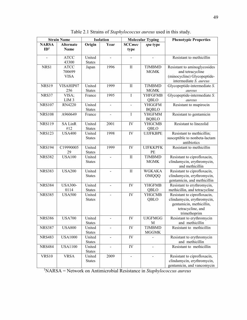
49
Table 2.1 Strains of Staphylococcus aureus used in this study.
Strain Name Isolation Molecular Typing Phenotypic Properties NARSA
ID1 Alternate
Name Origin Year SCCmec
type spa type
- ATCC 43300
United States
- - - Resistant to methicillin
NRS1 ATCC 700699 VISA
Japan 1996 II TJMBMDMGMK
Resistant to aminoglycosides and tetracycline
(minocycline) Glycopeptide-intermediate S. aureus
NRS19 VISAHIP07256
United States
1999 II TJMBMDMGMK
Glycopeptide-intermediate S.
aureus
NRS37 VISA; LIM 3
France 1995 I YHFGFMBQBLO
Glycopeptide-intermediate S.
aureus
NRS107 RN4220 United States
- - YHGGFMBQBLO
Resistant to mupirocin
NRS108 A960649 France - I YHGFMMBQBLO
Resistant to gentamicin
NRS119 SA LinR #12
United States
2001 IV YHGCMBQBLO
Resistant to linezolid
NRS123 USA400 United States
1998 IV UJJFKBPE Resistant to methicillin; susceptible to nonbeta-lactam
antibiotics
NRS194 C1999000529
United States
1999 IV UJFKKPFKPE
Resistant to methicillin
NRS382 USA100 United States
- II TJMBMDMGMK
Resistant to ciprofloxacin, clindamycin, erythromycin,
and methicillin
NRS383 USA200 United States
- II WGKAKAOMQQQ
Resistant to ciprofloxacin, clindamycin, erythromycin, gentamicin, and methicillin
NRS384 USA300-0114
United States
- IV YHGFMBQBLO
Resistant to erythromycin, methicillin, and tetracycline
NRS385 USA500 United States
- IV YHGCMBQBLO
Resistant to ciprofloxacin, clindamycin, erythromycin,
gentamicin, methicillin, tetracycline, and
trimethoprim
NRS386 USA700 United States
- IV UJGFMGGM
Resistant to erythromycin and methicillin
NRS387 USA800 United States
- IV TJMBMDMGGMK
Resistant to methicillin
NRS483 USA1000 United States
- IV - Resistant to erythromycin and methicillin
NRS484 USA1100 United States
- IV - Resistant to methicillin
VRS10
VRSA United States
2009 - - Resistant to ciprofloxacin, clindamycin, erythromycin, gentamicin, and vancomycin
1NARSA = Network on Antimicrobial Resistance in Staphylococcus aureus
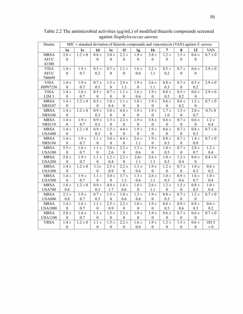
50
Table 2.2 The antimicrobial activities (µg/mL) of modified thiazole compounds screened against Staphylococcus aureus.
Strains MIC ± standard deviation of thiazole compounds and vancomycin (VAN) against S. aureus
1a 1c 1d 1e 1f 1g 1h 7 8 12 VAN
MRSA ATCC 43300
2.8 ± 0
1.2 ± 0 0.8 ± 0
3.0 ± 0
2.2 ± 0
1.9 ± 0
3.8 ± 0
1.2 ± 0
1.5 ± 0
0.6 ± 0
0.7 ± 0
VISA ATCC 700699
1.4 ± 0
1.9 ± 0.7
0.5 ± 0.2
0.7 ± 0
1.1 ± 0
1.6 ± 0.6
3.2 ± 1.1
0.5 ± 0.2
0.7 ± 0
0.6 ± 0
2.9 ± 0
VISA HIP07256
1.4 ± 0
1.9 ± 0.7
0.7 ± 0.3
1.5 ± 0
2.9 ± 1.3
1.9 ± 0
2.6 ± 1.1
0.8 ± 0.3
0.7 ± 0
0.5 ± 0.2
2.9 ± 0
VISA LIM 3
1.4 ± 0
1.6 ± 0.7
0.5 ± 0
0.7 ± 0
1.1 ± 0
1.6 ± 0.6
1.9 ± 0
0.8 ± 0.3
0.5 ± 0.2
0.6 ± 0
2.9 ± 0
MRSA NRS107
1.4 ± 0
1.2 ± 0 0.5 ± 0
1.0 ± 0.4
1.1 ± 0
1.0 ± 0
1.9 ± 0
0.6 ± 0
0.6 ± 0.2
1.2 ± 0
0.7 ± 0
MRSA NRS108
1.4 ± 0
1.2 ± 0 0.9 ± 0.3
3.0 ± 0
4.4 ± 0
1.9 ± 0
1.9 ± 0
1.7 ± 1.0
1.5 ± 0
2.0 ± 0.7
0.7± 0
MRSA NRS119
1.4 ± 0
1.9 ± 0.7
0.9 ± 0.3
1.5 ± 0
2.2 ± 0
1.9 ± 0
3.8 ± 0
0.6 ± 0
0.7 ± 0
0.6 ± 0
1.2 ± 0.4
MRSA USA400
1.4 ± 0
1.2 ± 0 0.9 ± 0.3
1.5 ± 0
4.4 ± 0
1.9 ± 0
1.9 ± 0
0.6 ± 0
0.7 ± 0
0.8 ± 0.3
0.7 ± 0
MRSA NRS194
1.4 ± 0
1.9 ± 0.7
1.1 ± 0
3.0 ± 0
4.4 ± 0
2.6 ± 1.1
1.9 ± 0
0.8 ± 0.3
0.7 ± 0
1.4 ± 0.9
0.7 ± 0
MRSA USA100
5.5 ± 0
1.6 ± 0.7
1.1 ± 0
3.0 ± 2.6
2.2 ± 0
1.3 ± 0.6
1.9 ± 0
1.0 ± 0.3
0.7 ± 0
2.0 ± 0.7
1.2 ± 0.4
MRSA USA200
2.8 ± 0
1.9 ± 0.7
1.1 ± 0
1.2 ± 0.4
2.2 ± 0
2.6± 1.1
2.6 ± 1.1
1.0 ± 0.3
1.2 ± 0.4
0.6 ± 0
0.4 ± 0
MRSA USA300
1.4 ± 0
1.2 ± 0 1.1± 0
2.5 ± 0.9
2.2 ± 0
1.3 ± 0.6
1.9 ± 0
1.2 ± 0
0.7 ± 0
1.0 ± 0.3
0.6 ± 0.2
MRSA USA500
1.4 ± 0
1.9 ± 0.7
1.1 ± 0
3.0 ± 0
3.7 ± 1.3
1.3 ± 0.6
2.6 ± 1.1
1.0 ± 0.3
0.9 ± 0.6
1.6 ± 0.7
1.0 ± 0.4
MRSA USA700
1.8 ± 0.8
1.2 ± 0 0.9 ± 0.3
4.0 ± 1.7
1.8 ± 0.6
1.0 ± 0
2.6 ± 1.1
1.2 ± 0
1.5 ± 0
0.8 ± 0.3
1.0 ± 0.4
MRSA USA800
2.3 ± 0.8
1.9 ± 0.7
0.7 ± 0.3
1.5 ± 0
1.8 ± 0.6
1.3 ± 0.6
1.9 ± 0
0.8 ± 0.3
0.7 ± 0
1.2 ± 0
0.7 ± 0
MRSA USA1000
1.4 ± 0
1.6 ± 0.7
1.1 ± 0
2.5 ± 0.9
2.2 ± 0
1.0 ± 0
1.9 ± 0
0.8 ± 0.3
0.9 ± 0.6
0.8 ± 0.3
0.6 ± 0.2
MRSA USA1100
2.8 ± 0
1.6 ± 0.7
1.1 ± 0
1.5 ± 0
2.2 ± 0
1.9 ± 0
1.9 ± 0
0.6 ± 0
0.7 ± 0
0.6 ± 0
0.7 ± 0
VRSA 1.4 ± 0
1.2 ± 0 1.1 ± 0
1.5 ± 0
2.2 ± 0
1.6 ± 0.6
1.9 ± 0
1.2 ± 0
1.5 ± 0
0.6 ± 0
185.5 ± 0
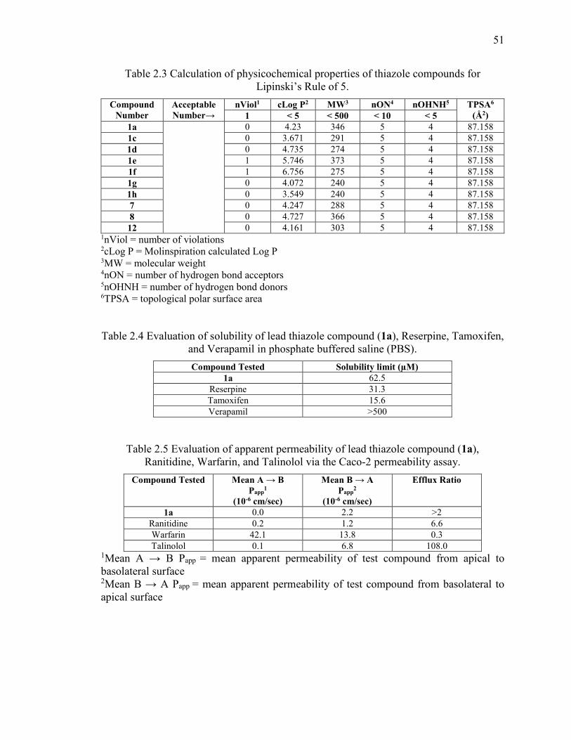
51
Table 2.3 Calculation of physicochemical properties of thiazole compounds for Lipinski’s Rule of 5.
Compound Number
Acceptable Number→
nViol1 cLog P2 MW3 nON4 nOHNH5 TPSA6 (Å2) 1 < 5 < 500 < 10 < 5
1a 0 4.23 346 5 4 87.158
1c 0 3.671 291 5 4 87.158
1d 0 4.735 274 5 4 87.158
1e 1 5.746 373 5 4 87.158
1f 1 6.756 275 5 4 87.158
1g 0 4.072 240 5 4 87.158
1h 0 3.549 240 5 4 87.158
7 0 4.247 288 5 4 87.158
8 0 4.727 366 5 4 87.158
12 0 4.161 303 5 4 87.158 1nViol = number of violations 2cLog P = Molinspiration calculated Log P 3MW = molecular weight 4nON = number of hydrogen bond acceptors 5nOHNH = number of hydrogen bond donors 6TPSA = topological polar surface area
Table 2.4 Evaluation of solubility of lead thiazole compound (1a), Reserpine, Tamoxifen, and Verapamil in phosphate buffered saline (PBS).
Compound Tested Solubility limit (µM) 1a 62.5
Reserpine 31.3
Tamoxifen 15.6
Verapamil >500
Table 2.5 Evaluation of apparent permeability of lead thiazole compound (1a), Ranitidine, Warfarin, and Talinolol via the Caco-2 permeability assay.
Compound Tested Mean A → B Papp
1 (10-6 cm/sec)
Mean B → A Papp
2 (10-6 cm/sec)
Efflux Ratio
1a 0.0 2.2 >2
Ranitidine 0.2 1.2 6.6
Warfarin 42.1 13.8 0.3
Talinolol 0.1 6.8 108.0 1Mean A → B Papp = mean apparent permeability of test compound from apical to basolateral surface 2Mean B → A Papp = mean apparent permeability of test compound from basolateral to apical surface
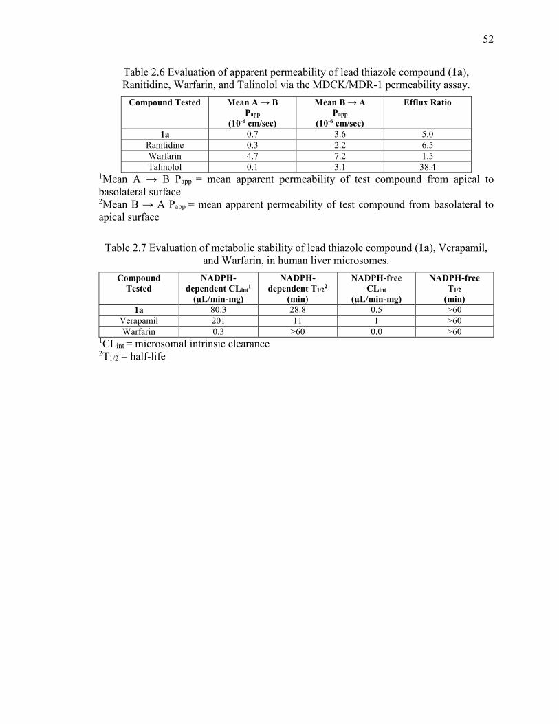
52
Table 2.6 Evaluation of apparent permeability of lead thiazole compound (1a), Ranitidine, Warfarin, and Talinolol via the MDCK/MDR-1 permeability assay.
Compound Tested Mean A → B Papp
(10-6 cm/sec)
Mean B → A Papp
(10-6 cm/sec)
Efflux Ratio
1a 0.7 3.6 5.0
Ranitidine 0.3 2.2 6.5
Warfarin 4.7 7.2 1.5
Talinolol 0.1 3.1 38.4 1Mean A → B Papp = mean apparent permeability of test compound from apical to basolateral surface 2Mean B → A Papp = mean apparent permeability of test compound from basolateral to apical surface
Table 2.7 Evaluation of metabolic stability of lead thiazole compound (1a), Verapamil, and Warfarin, in human liver microsomes.
Compound Tested
NADPH-dependent CLint
1 (µL/min-mg)
NADPH-dependent T1/2
2 (min)
NADPH-free CLint
(µL/min-mg)
NADPH-free T1/2
(min) 1a 80.3 28.8 0.5 >60
Verapamil 201 11 1 >60
Warfarin 0.3 >60 0.0 >60 1CLint = microsomal intrinsic clearance 2T1/2 = half-life
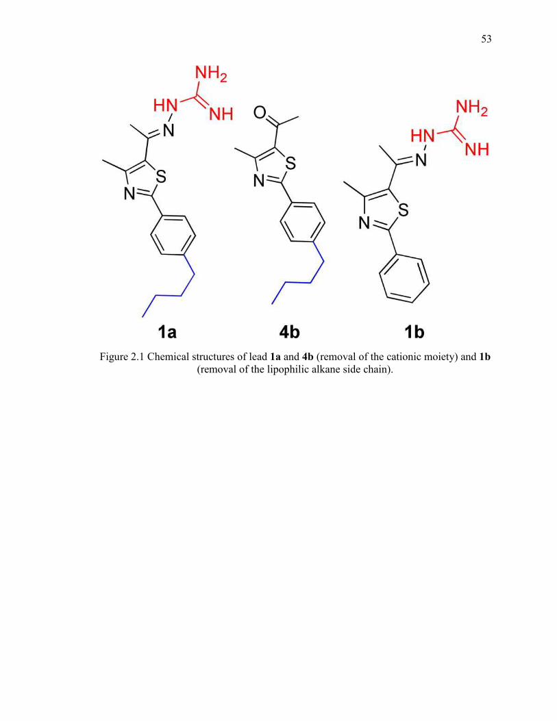
53
Figure 2.1 Chemical structures of lead 1a and 4b (removal of the cationic moiety) and 1b (removal of the lipophilic alkane side chain).
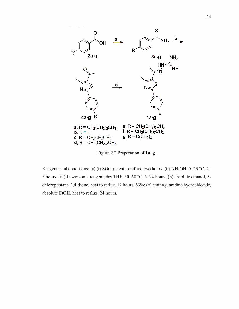
54
Figure 2.2 Preparation of 1a–g.
Reagents and conditions: (a) (i) SOCl2, heat to reflux, two hours, (ii) NH4OH, 0–23 °C, 2–
5 hours, (iii) Lawesson’s reagent, dry THF, 50–60 °C, 5–24 hours; (b) absolute ethanol, 3-
chloropentane-2,4-dione, heat to reflux, 12 hours, 63%; (c) aminoguanidine hydrochloride,
absolute EtOH, heat to reflux, 24 hours.
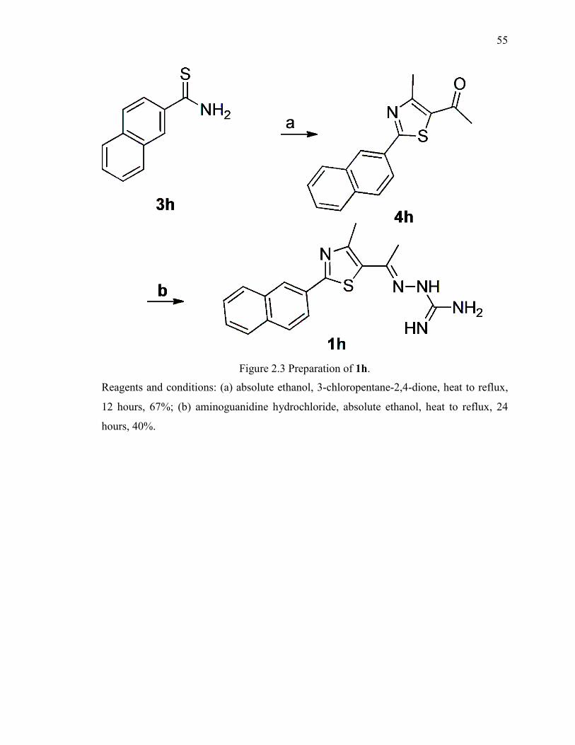
55
Figure 2.3 Preparation of 1h.
Reagents and conditions: (a) absolute ethanol, 3-chloropentane-2,4-dione, heat to reflux,
12 hours, 67%; (b) aminoguanidine hydrochloride, absolute ethanol, heat to reflux, 24
hours, 40%.
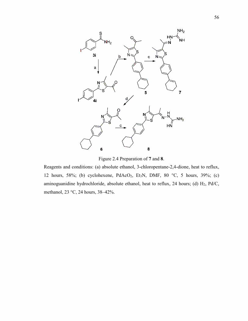
56
Figure 2.4 Preparation of 7 and 8.
Reagents and conditions: (a) absolute ethanol, 3-chloropentane-2,4-dione, heat to reflux,
12 hours, 58%; (b) cyclohexene, PdAcO2, Et3N, DMF, 80 °C, 5 hours, 39%; (c)
aminoguanidine hydrochloride, absolute ethanol, heat to reflux, 24 hours; (d) H2, Pd/C,
methanol, 23 °C, 24 hours, 38–42%.
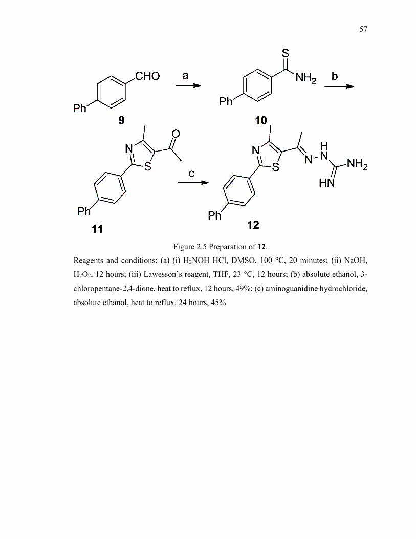
57
Figure 2.5 Preparation of 12.
Reagents and conditions: (a) (i) H2NOH HCl, DMSO, 100 °C, 20 minutes; (ii) NaOH,
H2O2, 12 hours; (iii) Lawesson’s reagent, THF, 23 °C, 12 hours; (b) absolute ethanol, 3-
chloropentane-2,4-dione, heat to reflux, 12 hours, 49%; (c) aminoguanidine hydrochloride,
absolute ethanol, heat to reflux, 24 hours, 45%.
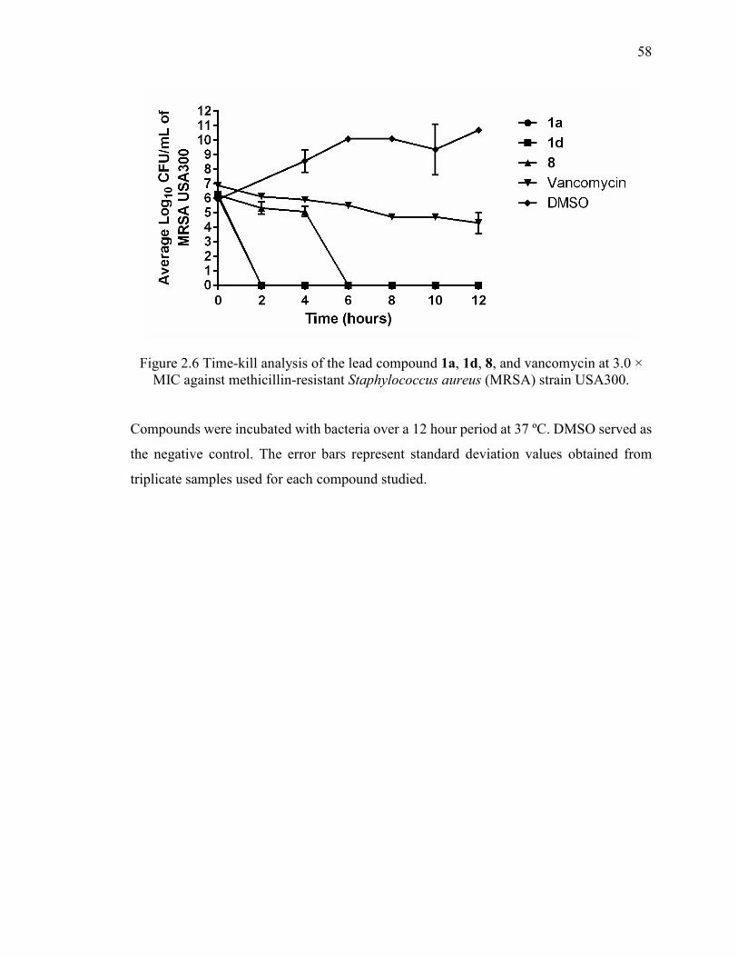
58
Figure 2.6 Time-kill analysis of the lead compound 1a, 1d, 8, and vancomycin at 3.0 × MIC against methicillin-resistant Staphylococcus aureus (MRSA) strain USA300.
Compounds were incubated with bacteria over a 12 hour period at 37 ºC. DMSO served as
the negative control. The error bars represent standard deviation values obtained from
triplicate samples used for each compound studied.
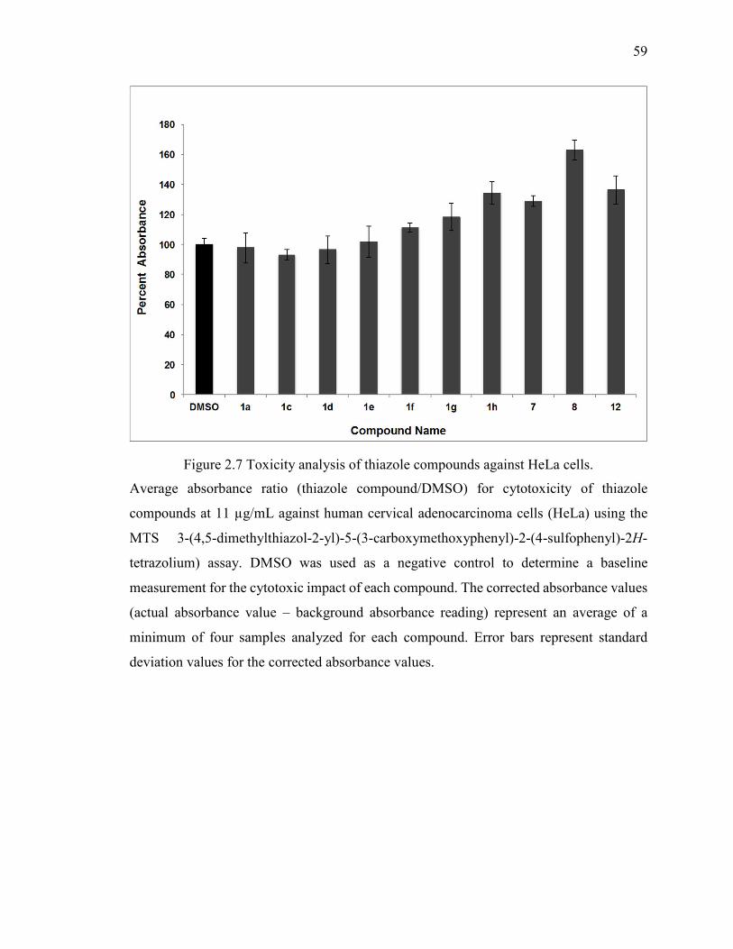
59
Figure 2.7 Toxicity analysis of thiazole compounds against HeLa cells.
Average absorbance ratio (thiazole compound/DMSO) for cytotoxicity of thiazole
compounds at 11 µg/mL against human cervical adenocarcinoma cells (HeLa) using the
MTS 3-(4,5-dimethylthiazol-2-yl)-5-(3-carboxymethoxyphenyl)-2-(4-sulfophenyl)-2H-
tetrazolium) assay. DMSO was used as a negative control to determine a baseline
measurement for the cytotoxic impact of each compound. The corrected absorbance values
(actual absorbance value – background absorbance reading) represent an average of a
minimum of four samples analyzed for each compound. Error bars represent standard
deviation values for the corrected absorbance values.

60
CHAPTER 3. ANTI-BIOFILM ACTIVITY AND SYNERGISM OF NOVEL THIAZOLE COMPOUNDS WITH GLYCOPEPTIDE
ANTIBIOTICS AGAINST MULTIDRUG-RESISTANT STAPHYLOCOCCI
THIS IS A PUBLISHED JOURNAL ARTICLE. Reprinted with permission from Anti-
biofilm activity and synergism of novel thiazole compounds with glycopeptide
antibiotics against multidrug-resistant Staphylococci. Haroon Mohammad,
Abdelrahman S. Mayhoub, Mark Cushman, and Mohamed N. Seleem. The Journal of
Antibiotics (2015) 68, 259–266; doi:10.1038/ja.2014.142 Copyright 2014 Japan
Antibiotics Research Association
3.1 Introduction
Antibiotic-resistant bacteria are a major global health concern resulting in 23,000
deaths each year in the United States alone (1). Two species alone, methicillin-resistant
Staphylococcus aureus (MRSA) and Staphylococcus epidermidis, are responsible annually
for the majority of skin and soft-tissue infections and infections caused by bacterial
biofilms present on indwelling medical devices (2, 3). Biofilms are responsible for 80% of
microbial infections which develop in the human body and bacterial biofilms on implanted
biomedical devices and tissue surfaces (chronic wound) constitute an ever-increasing threat
to human health and place a significant burden on healthcare systems (4). Biofilms consist
of a cluster of bacterial cells enclosed within an extracellular matrix which collectively
attach to an animate or inanimate surface (4). Cells present within a biofilm pose a key
challenge as they demonstrate increased resistance to the effect of antimicrobials (5).
Antibiotics have been key allies in the treatment of bacterial infections for more
than 80 years. While several classes of antibiotics were once capable of treating
staphylococci-induced infections, strains have emerged which are resistant to an array of
antimicrobials once deemed effective including β-lactams (6), macrolides (2), and
fluoroquinolones (6, 7). Further exacerbating the issue is the rise of strains (such as
vancomycin-resistant Staphylococcus aureus (VRSA)), which are resistant to antibiotics
deemed drugs of last resort for treatment of staphylococcal infections, including
61
glycopeptide antibiotics like vancomycin (8). Conventional antibiotics face an added
challenge in the treatment of biofilm infections as bacteria present within a biofilm may be
1000-fold more resistant to antibiotics compared to their planktonic equivalents (5). Thus
there is a critical need for the discovery of novel antimicrobials and treatment strategies to
circumvent this growing public health concern.
Several thiazole compounds have been shown to be effective anticonvulsant(9),
anticancer (10, 11), and antiviral agents (12). However, limited studies have been
performed to characterize their abilities as antimicrobial agents, particularly against
MRSA. Darwish et al, synthesized a series of thiadiazole analogues incorporating a
sulfonamide group and found they possessed activity against Streptococcus pneumoniae
and Bacillus subtilis (13). Additionally, Desai et al, constructed a series of novel hybrid
compounds which combined the thiazole and 1,3,4-oxadiazole pharmacophores but found
they had limited activity against S. aureus (minimum inhibitory concentration (MIC) of six
of 12 analogues constructed was 500 µg/mL or higher) (14). Furthermore, a third study
assessing a series of disubstituted 1,3-thiazole derivatives found the most potent analogue
possessed modest activity against a single strain of S. aureus tested (MIC of 50 µg/mL)
(15). None of these studies assessed broader therapeutic applications of thiazole
compounds beyond use as single agents to inhibit bacterial growth in vitro.
We recently discovered a novel lead thiazole compound 1 which exhibited potent
antimicrobial activity against MRSA (Figure 3.1) (16). The lead compound is composed
of a thiazole nucleus connected to a cationic amino head at the C5-position and a lipophilic
phenylalkyl tail at the C2-position. The aims of the present study are to identify if the lead
compound 1 and the most potent synthesized derivative 2 have potential to be used in
combination with glycopeptide antibiotics commonly used to treat MRSA infections, to
analyze the ability of 1 and 2 to re-sensitize VRSA strains to glycopeptide antibiotics, and
to assess if these compounds are capable of disrupting staphylococcal biofilms using an in
vitro model of S. epidermidis. Results garnered from this study will provide valuable
insight into potential therapeutic applications of thiazole compounds for use as antibacterial
agents.
62
3.2 Materials and Methods
3.2.1 Bacterial Strains and Reagents
The bacterial strains of methicillin-resistant and vancomycin-intermediate Staphylococcus
aureus utilized were obtained from the Network on Antimicrobial Resistance in
Staphylococcus aureus (NARSA). Staphylococcus epidermidis ATCC 35984 was obtained
from the American Tissue Culture Collection. Antibiotics were purchased commercially
from Gold Biotechnology (St. Louis, MO, USA) (vancomycin hydrochloride) and Biotang
Inc. (Waltham, MA, USA) (teicoplanin). Both antibiotics were dissolved in dimethyl
sulfoxide to obtain a stock 10 mM solution.
3.2.2 Synthesis of Thiazole Compounds 1 and 2
The detailed synthetic protocols and spectral data of final products 1 and 2 as well as all
intermediates have been previously reported (16),(17).
3.2.3 Determination of Minimum Inhibitory Concentration (MIC) and Minimum Bactericidal Concentration (MBC) Against MRSA, VISA, VRSA, and S.
epidermidis
The MICs of the thiazole compounds, vancomycin, and teicoplanin against MRSA, VISA,
VRSA, and S. epidermidis were determined using the broth microdilution method, in
accordance with the recommendations contained in the CLSI guidelines (with the
exception that Mueller-Hinton broth (MHB) was used instead of cation-adjusted MHB)
(18). Bacteria were prepared in phosphate buffered saline (PBS) until a McFarland standard
of 0.5 was achieved. The solution was diluted 1:300 in MHB to reach a starting inoculum
of 1 × 10⁵ colony-forming units (CFU/mL). Bacteria were transferred to a 96-well
microtiter plate. Thiazole compounds and antibiotics were added (in triplicate) to wells in
the first row of the microtitier plate and then serially diluted along the ordinate. The plate
was incubated at 37 °C for 20-24 hours before the MIC was determined. The MIC was
categorized as the concentration at which no visible growth of bacteria was observed in a
particular well.
The MBC was determined by plating 5 µL from wells on the 96-well microtiter plate
(where the MIC was determined) were no growth was observed onto tryptic soy agar (TSA)
plates. The TSA plates were then incubated at 37 °C for 20-24 hours before the MBC was
63
determined. The MBC was categorized as the concentration where ≥99% reduction in
bacterial cell count was observed.
3.2.4 Time-kill Analysis of Thiazole Compounds and Glycopeptide Antibiotics Against MRSA
MRSA NRS123 (USA400) cells in the logarithmic growth phase were diluted to ~1 × 10⁸
colony-forming units (CFU/mL) and exposed to concentrations equivalent to 2, 4, and 8 ×
MIC (in triplicate) of thiazole compounds 1 and 2, teicoplanin, and vancomycin in Mueller-
Hinton broth (MHB). 20 µL samples were collected after 0, 2, 4, 6, 8, 10, 12, and 24 hours
of incubation at 37 °C and subsequently serially diluted in PBS. Bacteria were then
transferred to TSA plates and incubated at 37 °C for 18-20 hours before viable CFU/mL
was determined. The test agent was deemed bactericidal if it successfully produced a 3-
log10 reduction in the bacterial count within 24 hours, as reported elsewhere (19).
3.2.5 Single-step Resistance Selection
The frequency of spontaneous single-step resistance of the thiazole compounds and
glycopeptide antibiotics to five MRSA strains was determined as reported elsewhere (20,
21). Briefly, bacterial cultures (>1 × 10⁹ CFU/mL) were spread onto Mueller-Hinton agar
plates (10-mm diameter) containing each compound/antibiotic at 4 × MIC. Plates were
incubated aerobically at 37 °C for 48 hours. The frequency of resistance was calculated as
the number of resistant colonies per inoculum (21).
3.2.6 Combination Therapy Analysis of Thiazole Compounds with Glycopeptide Antibiotics
The relationship between the thiazole compounds and glycopeptide antibiotics
(vancomycin and teicoplanin) was assessed via a standard checkerboard assay (22).
Bacteria equivalent to a McFarland standard of 0.5 were prepared in PBS. The bacteria
were then diluted in MHB to achieve a starting cell density of 1 × 10⁵ CFU/mL. MHB was
transferred to all wells of a 96-well microtiter plate. The thiazole compounds and
glycopeptide antibiotics were diluted in MHB to achieve a starting concentration
equivalent to 2 × or 4 × MIC, respectively. The glycopeptide antibiotic was serially diluted
along the abscissa of the microtiter plate while the thiazole compound was serially diluted
64
along the ordinate. The plate was incubated for 20-24 hours at 37 °C. The MIC of the test
compound in combination with each glycopeptide antibiotic studied was determined as the
lowest concentration of each compound/antibiotic where no visible growth of bacteria was
observed. The fractional inhibitory concentration index (ƩFIC) was calculated for each
combination as follows:
ƩFIC =(�������� �������� �� ���������� ���� ������ ���� ���������
�������� �������� ��� ) +
(��������� ���� ��������� �� ���������� ���� ����� ��������
��������� ���� ���������)
A synergistic relationship was classified as an FIC index less than or equal to 0.5. FIC
values above 0.5 but less than 4.0 were characterized as indifference while FIC values
above 4.0 were classified as antagonistic.
3.2.7 Re-sensitization of VRSA Strains to Vancomycin Using Broth Microdilution Method
MHB was inoculated with VRSA (5×10⁵ CFU/mL), as described elsewhere (23). 5-ml
aliquots of the bacterial suspension were divided into microcentrifuge tubes. Compound 1
or 2 (at ½ × MIC) was introduced into each tube. After sitting at room temperature for 30
minutes, 1 ml samples from each tube were transferred to a new centrifuge tube prior to
addition of the antibiotic (either vancomycin or teicoplanin at a concentration equivalent
to their MIC). Using a 96-well microtiter plate, rows 2-12 were filled with the remaining 4
ml bacterial suspension (containing either compound 1 or 2). 200-µl aliquots from tubes
containing both the thiazole compound and glycopeptide antibiotic were transferred to row
1 of the 96-well plate. After aspirating contents in the first row 4-6 times, 100 µL was
transferred from wells in row 1 to row 2. This process was repeated to dilute the remaining
wells containing no antibiotic. Untreated bacteria served as a control. The plate was
incubated at 37 °C for 20 hours before the MIC was recorded. The MIC was categorized
as the concentration at which no visible growth of bacteria was observed in a particular
well. A fold reduction was calculated by comparing the MIC of the antibiotic alone
compared to the MIC of the antibiotic given in combination with 1 or 2.
65
3.2.8 Staphylococcus Biofilm Mass Reduction Determination
The microtiter dish biofilm formation assay (24) was utilized to assess the ability of the
thiazole compounds to disrupt an adherent staphylococcal biofilm, similar to what has been
described elsewhere (25). S. epidermidis ATCC 35984 was transferred to tryptic soy broth
and incubated at 37 °C for 24 hours before being diluted 1:200 in tryptic soy broth + 1%
glucose. This solution was transferred to each well of a 96-well microtiter plate and
incubated at 37 °C for 24 hours to permit biofilm formation on the well surface. Bacteria
were removed and wells were washed twice with PBS. Compounds 1, 2, or vancomycin
were added (in triplicate) to wells and serially diluted. The microtiter plate was then
incubated at 37 °C for 24 hours. The plate was washed twice by submerging in deionized
water. 0.1% (w/v) crystal violet was added to each well and allowed to stain the biofilm
for 20 minutes before addition of 95% ethanol to decolorize. Using a kinetic microplate
reader (Molecular Devices, Sunnyvale, CA, USA), the optical density of each well at 595
nm was measured. Percent biofilm mass reduction was calculated for each treatment
regimen as compared to the control (wells receiving no treatment).
3.2.9 Kinetic Solubility Determination of Compound 2
Serial dilutions of compound 2 were prepared in DMSO at 100× the final concentration.
Compound 2 was then diluted 100-fold into PBS in a 96-well plate and mixed. The
absorbance of the PBS-containing plate was measured prior to addition of the test agents
to determine the background absorbance. After two hours, the presence of precipitate was
detected by turbidity (absorbance at 540 nm). An absorbance value of greater than (mean
+ 3× standard deviation of the blank), after subtracting the pre-experiment background,
was indicative of turbidity. The solubility limit is reported as the highest experimental
concentration for compound 2 with no evidence of turbidity.
3.2.10 Caco-2 Bidirectional Permeability Assessment of Compound 2
To assess the ability of compound 2 to passively permeate through epithelial tissue, a Caco-
2 permeability assay was performed as described previously (16). Caco-2 cells grown in
tissue culture flasks were trypsinized, suspended in medium, and the suspensions were
applied to wells of a Millipore 96 well Caco-2 plate. The cells were allowed to grow and
66
differentiate for three weeks, feeding at 2-day intervals. For Apical to Basolateral (A→B)
permeability, compound 2 was added to the apical (A) side and amount of permeation was
determined on the basolateral (B) side; for Basolateral to Apical (B→A) permeability,
compound 2 was added to the B side and the amount of permeation was determined on the
A side. The A-side buffer contained 100 μM Lucifer yellow dye, in Transport Buffer (1.98
g/L glucose in 10 mM HEPES, 1.0 × Hank’s Balanced Salt Solution) at pH 6.5, and the B-
side buffer contained Transport Buffer at pH 7.4. Caco-2 cells were incubated with these
buffers for two hours, and the receiver side buffer was removed for analysis by LC/MS/MS.
To verify the Caco-2 cell monolayers were properly formed, aliquots of the cell buffers
were analyzed by fluorescence to determine the transport of the impermeable dye Lucifer
Yellow. Any deviations from control values are reported. Data are expressed as
permeability ���� =�(
��
� )
"#$ where
%&
%' is the rate of permeation, C0 is the initial
concentration of test agent, and A is the area of the monolayer. In bidirectional permeability
studies, the efflux ratio (RE) is also calculated: RE = (��()→+)
(��(+→)). An RE > 2 indicates a
potential substrate for P-glycoprotein or other active efflux transporters.
3.2.11 Statistical Analysis
All statistical analysis was performed using the two-tailed Student’s t-test (P < 0.05)
utilizing Microsoft Excel software.
3.3 Results and Discussion
3.3.1 Determination of the Antimicrobial Activity of the Thiazole Compounds and Glycopeptide Antibiotics
We have designed and synthesized a series of thiazole derivatives containing modifications
to the lipophilic alkyl side chain of 1 (16). Antimicrobial susceptibility analysis of these
derivatives, using the standard broth microdilution assay (26), revealed compound 2
exhibited the most potent antibacterial activity against multidrug-resistant staphylococci.
As Table 3.1 demonstrates, the minimum inhibitory concentration (MIC) for 1 was 1.38
µg/mL; compound 2 showed similar activity inhibiting growth of the same strains at a
67
concentration of 1.40 µg/mL. At these concentrations, the compounds are not toxic to
mammalian cells as confirmed in a previous study (16).
The thiazole compounds exhibited activity against MRSA strains resistant to several
different classes of antibiotics including macrolides (NRS384), fluoroquinolones
(NRS385), aminoglycosides (NRS385), tetracyclines (NRS384), and oxazolidinones
(NRS119). Additionally, both 1 (MIC from 1.38-2.77 µg/mL) and 2 (MIC from 0.70-1.40
µg/mL), unlike vancomycin (MIC from 2.97-760.68 µg/mL), retained their antimicrobial
activity against strains of vancomycin-intermediate S. aureus (VISA) and VRSA strains.
Furthermore, both thiazole compounds were more potent than teicoplanin against two
VISA strains (MICTeicoplanin from 0.94-7.52 µg/mL) and all three VRSA strains tested
(MICTeicoplanin from 60.51-120.30 µg/mL1). Thus, 1 and 2 exhibit a selective advantage over
vancomycin and teicoplanin in their antibacterial activity against both VISA and VRSA.
Antimicrobial agents that exhibit bactericidal activity are hypothesized to contribute to
a more rapid recovery from infection and a better clinical outcome, compared to their
bacteriostatic counterparts (27). To ascertain whether the thiazole compounds were
bacteriostatic or bactericidal, the minimum bactericidal concentration (MBC) was
determined. The MBC was calculated as the lowest concentration of compound/drug that
produced a ≥99.9% reduction in the bacterial cell count as compared to the initial inoculum
(28). As Table 3.1 demonstrates, both thiazole compounds are bactericidal. Against five
MRSA strains (NRS107, NRS119, NRS123, NRS385, and ATCC 43300), all VISA strains,
and two VRSA strains (VRS1 and VRS4), 1 and 2 possess MBC values equivalent to their
MICs or one-fold higher than the MICs. This is similar to what is observed with
vancomycin, a known bactericidal antibiotic, with MBC values equal to or one-fold higher
than the MICs for all MRSA and VISA strains tested. Teicoplanin exhibits MBC values
equivalent to its MIC against two MRSA strains, four-fold higher than its MIC against two
additional MRSA strains, and MBC values 16-fold higher than the MIC values against
three strains of MRSA (NRS194, USA300, and USA400).
3.3.2 Time-kill Analysis of Thiazole Compounds and Glycopeptide Antibiotics
In order to confirm that 1 and 2 were bactericidal agents, we next examined how rapidly
the thiazole compounds were able to kill a high inoculum of MRSA. Using a standard time-
68
kill assay, MRSA USA400 (NRS123), a predominant strain linked to many community-
acquired MRSA infections (29), was exposed to 2, 4, and 8 × MIC of 1, 2, teicoplanin, or
vancomycin. Samples were collected at specific time points and transferred to tryptic soy
agar (TSA) plates to determine the number of viable bacteria remaining post-treatment.
As depicted in Figure 3.2, both 1 and 2 exhibit bactericidal activity at all concentrations
tested; however the time to achieve a 3-log10 reduction in CFU/mL differs depending on
the concentration of the test agent. For compound 1, MRSA is completely eliminated after
24 hours at 2 × MIC, after 10 hours at 4 × MIC, and after only two hours at 8 × MIC.
Analogue 2 produces a 3-log10 reduction in CFU/mL after 10 hours at 2 × MIC; however,
it is not able to completely eradicate MRSA similar to the parent compound. At higher
concentrations, 2 successfully eliminates MRSA completely after 24 hours (at 4 × MIC);
at the highest concentration tested (8 × MIC), analogue 2 proves superior to both antibiotics
tested as it rapidly eliminates MRSA within 10 hours. Vancomycin required 24 hours to
completely eradicate MRSA at both 4 and 8 × MIC; at 2 × MIC, vancomycin produced a
3-log10 reduction in CFU/mL within 24 hours but was not able to eliminate all bacteria
completely (similar to analogue 2). These results are similar to what has been previously
published regarding vancomycin’s slow bactericidal activity (30). Teicoplanin required 24
hours to completely eliminate MRSA at all three concentrations tested. Thus, in addition
to retaining antimicrobial activity against VISA and VRSA strains, 1 and 2 possess an
additional advantage over vancomycin and teicoplanin in their ability to rapidly kill MRSA,
particularly at higher concentrations.
Rapid bactericidal activity is an important factor in reducing the emergence of
bacterial resistance to an antimicrobial agent and is important clinically in preventing an
infection from spreading (27). Additionally, bactericidal agents have been shown both
clinically and in in vivo studies to be superior to bacteriostatic agents for the treatment of
certain invasive diseases such as endocarditis (31). Furthermore, rapid bactericidal activity
is an important quality for consideration in using a particular agent in combination with
other antibiotics, such as vancomycin (30). The results from the time-kill assay provided
valuable insight into the possibility that the thiazole compounds could be potentially paired
with other antibiotics against MRSA, given 1 and 2 possess rapid bactericidal activity.

69
3.3.3 Assessment of Single-step Resistance
After confirming compounds 1 and 2 possessed rapid bactericidal activity against MRSA,
we next turned our attention to assessing the likelihood MRSA would develop resistance
quickly to these thiazole compounds. A single-step resistance selection experiment was
performed by subculturing a high inoculum of MRSA (>1 × 10⁹ CFU/mL) onto TSA plates
containing 1, 2, vancomycin, or teicoplanin at a concentration equivalent to 4 × MIC. The
likelihood of bacterial resistance arising (via spontaneous mutations in the bacterial
genome) to these compounds/antibiotics was examined using five MRSA strains. Table 3.2
presents the mutation frequencies generated against each tested agent: for 1, 1.19×10⁻⁸ to
>1.73×10⁻¹⁰; for 2, >1.73×10⁻¹⁰ to >2.33×10⁻¹⁰; for teicoplanin, 2.73×10⁻⁷ to 3.03×10⁻⁹; and
for vancomycin, 3.03×10⁻¹⁰ to >8.47×10⁻¹⁰. The values obtained for teicoplanin and
vancomycin are similar to what has been reported elsewhere (32).
The thiazole compounds produce a similar mutation frequency as both teicoplanin
and vancomycin. Interestingly, 2 demonstrates a mutation frequency similar to or better
than vancomycin against the five MRSA strains tested. Even at lower (2 × MIC)
concentrations, resistant mutants are difficult to isolate against this particular compound
(data not published). It took 30 years to isolate a strain of S. aureus exhibiting resistance
to vancomycin (1). Thus the results presented here support the notion that MRSA is
unlikely to develop rapid resistance to the thiazole compounds, in particular compound 2.
The data obtained from both the time-kill and single-step resistance selection experiments
demonstrate the thiazole compounds possess two important characteristics necessary for
an ideal antibiotic for MRSA, rapid bactericidal activity and low potential for bacterial
resistance development (33).
3.3.4 Combination Testing of Thiazole Compounds with Glycopeptide Antibiotics
Glycopeptide antibiotics, chiefly vancomycin, have been a principal source of treatment of
MRSA infections for many years (33). However, extensive use of these antibiotics opens
the door for the emergence of strains with reduced susceptibility to these antibiotics (30).
Combination therapy, pairing vancomycin with another antimicrobial, has been used in the
healthcare setting both to reduce the likelihood of resistant strains to vancomycin from
rapidly emerging and to improve the morbidity associated with MRSA infections. For

70
example, vancomycin has been combined with subinhibitory concentrations of
clindamycin and linezolid to reduce toxins generated by S. aureus during infection (34,
35). Identifying other antimicrobial partners capable of being paired with vancomycin can
potentially prolong the clinical utility of this antibiotic.
To ascertain whether 1 and 2 have potential to be combined with vancomycin
against MRSA, the checkerboard assay was utilized (22). In this assay, one
antibiotic/compound is serially diluted along the abscissa followed by diluting the second
antibiotic/compound along the ordinate in a 96-well plate. The fractional inhibitory
concentration (FIC) is then calculated as a ratio of the MIC of each antibiotic/compound
when given in combination relative to the MIC of each antibiotic/compound given alone.
The FIC index (ƩFIC) is a summation of the FICs for each antibiotic/compound tested in
combination. ƩFIC ≤ 0.50 is indicative of synergism between the antibiotic and compound.
Results from the checkerboard assay experiment are presented in Table 3.3. Both thiazole
compounds were found to exhibit a synergistic relationship with vancomycin against six
of the seven MRSA strains tested with ƩFIC values ranging from 0.07 to 0.50 for 1 and
ƩFIC values ranging from 0.13 to 0.50 for 2. At ¼ × MIC for 2, a 16-fold reduction in the
MIC for vancomycin (when combined with 2) was observed for all six MRSA strains
where synergy was detected (data not presented). As vancomycin is known to be a
nephrotoxic agent, using a lower concentration of this drug in MRSA infections is highly
desirable as it has the potential benefit of reducing this side effect in patients (33). When
tested against VISA, 1 failed to exhibit synergy with vancomycin while 2 demonstrated a
synergistic relationship with vancomycin against one strain (NRS19).
We were curious to explore if the synergistic relationship observed was limited just
to vancomycin or could be observed with other glycopeptide antibiotics as well.
Teicoplanin was used to further explore the partnership between thiazole compounds and
glycopeptide antibiotics. Interestingly, the checkerboard assay revealed that neither 1 nor
2 exhibited a synergistic relationship with teicoplanin against MRSA. This suggests that
combination therapy involving the thiazole compounds may be limited to only being paired
with vancomycin though further studies with other glycopeptide antibiotics are needed to
confirm this observation. Additionally, as vancomycin targets cell wall biosynthesis in S.
aureus, it would be worthwhile to explore if a synergistic relationship would be observed
71
between these thiazole compounds and other cell wall biosynthesis inhibitors (such as β-
lactam antibiotics). Collectively, the results shed valuable insight into thiazole compounds
serving as potential future partners with vancomycin against MRSA. This discovery can
potentially prolong the usage of vancomycin as a therapeutic agent for MRSA infections
by reducing the likelihood of strains developing resistance to vancomycin used in
monotherapy.
3.3.5 Re-sensitization of VRSA to Glycopeptide Antibiotics
The emergence of S. aureus strains resistant to vancomycin presents an additional
challenge to clinical care providers dealing with the growing epidemic of multidrug-
resistant bacterial infections. Identifying clever strategies to prolong the use of current
antibiotics against multidrug-resistant bacteria is necessary. One strategy that has been
explored recently is suppressing antibiotic resistance by re-sensitizing resistant bacteria
using a secondary compound (23). As the thiazole compounds were found to possess a
synergistic relationship with vancomycin against MRSA, we postulated that the thiazole
compounds may be capable of re-sensitizing VRSA strains to vancomycin. Initially, the
MIC of 1 and 2 was determined using the broth microdilution assay. Next, Mueller-Hinton
broth was inoculated with either compound 1 or 2 (at ½ × MIC). Vancomycin was then
serially diluted in both the inoculated media alone and media supplemented with the
thiazole compounds. The MICs of vancomycin in the presence of the thiazole compounds
was compared to vancomycin used alone. A fold-reduction was calculated by dividing the
MIC of vancomycin alone by the MIC of vancomycin + the thiazole compound.
As Table 3.4 presents, both thiazole compounds were capable of re-sensitizing
VRSA to vancomycin. Compound 1 was able to produce a four-fold reduction in the MIC
of vancomycin when the two agents were combined against VRSA. Amazingly, compound
2 proved to be superior to 1 as it produced a 512-fold reduction in the MIC of vancomycin
against two VRSA strains tested. Furthermore, compound 2 produced a 32-fold reduction
in the MIC of teicoplanin against two VRSA strains (VRS4 and VRS5) and a 64-fold
reduction against strain VRS1. Thus compound 2 was capable of re-sensitizing VRSA to
both vancomycin and teicoplanin. Substitution of the alkane side chain (in 1) with a phenyl
group (in 2) produced a dramatic improvement in the thiazole compounds’ ability to re-
72
sensitize VRSA to the effect of glycopeptide antibiotics. Using the checkerboard assay, we
found that compound 2 exhibited a synergistic relationship with both vancomycin and
teicoplanin against two VRSA strains (VRS4 and VRS5) with ƩFIC = 0.50. Thus,
compound 2 holds promise for future use to suppress vancomycin-resistance in VRSA
strains, prolonging the utility of glycopeptide antibiotics against these strains.
3.3.6 S. epidermidis Biofilm Mass Reduction
Bacterial biofilms which form on the surface of indwelling medical devices, such as
intravascular catheters, are a major problem in hospitals. These biofilms can lead to life-
threatening bloodstream infections associated with high mortality and treatment costs (36).
Staphylococci, primarily S. epidermidis and S. aureus, are responsible for many invasive
infections which develop from bacterial biofilms that form on the surface of medical
devices (3, 37). Further exacerbating this problem, traditional antibiotics are not effective
at disrupting these biofilms as cells present within the biofilm exhibit increased resistance
to antibiotics (5). Identifying antimicrobials capable of disrupting these biofilms is
necessary to combat this growing problem.
Recent studies have demonstrated that thiazole and thiazolidinone compounds
possess the ability to disrupt bacterial biofilms (38, 39). To examine if the potential
therapeutic application of 1 and 2 could be expanded beyond just inhibition of planktonic
bacteria, the ability of both thiazole compounds to disrupt staphylococcal biofilm was
analyzed. First, to confirm the thiazole compounds were capable of inhibiting planktonic
bacteria, the MIC of each compound and vancomycin against a biofilm-forming clinical
isolate of methicillin-resistant S. epidermidis was assessed using the broth microdilution
technique. Compounds 1 and 2 were found to inhibit bacterial growth at 2.77 and 0.61
µg/mL, respectively (Table 3.1). Vancomycin inhibited growth of planktonic S.
epidermidis at a concentration of 0.74 µg/mL. Next, to determine if compounds 1 and 2
had the potential to disrupt staphylococcal biofilm, the crystal violet reporter assay was
used against a mature S. epidermidis biofilm (24). As Figure 3.3 demonstrates, 1 (at 8 ×
MIC) and 2 (at 32 × MIC) significantly disrupted S. epidermidis biofilm, reducing the
biofilm mass by 56.7% and 65.2% respectively. These compounds proved to be far superior
to vancomycin; even at 128 × MIC, vancomycin was only able to reduce S. epidermidis
73
biofilm mass by 21.5%. The thiazole compounds thus possess anti-biofilm activity and are
capable of disrupting adherent staphylococci biofilm much better than a traditional
antibiotic, vancomycin.
3.3.7 In vitro Pharmacokinetic Analysis of Compound 2
Assessment of a compound’s drug-like properties is important early in drug development
to identify and address potential issues, especially those associated with aqueous solubility
and permeability. Previously we reported the lead thiazole compound 1 possessed
moderate aqueous solubility (21.6 µg/mL) but poor permeability across a biological
membrane (Caco-2 apparent permeability, Papp (A → B) = 0.0 × 10⁻⁶ cm/sec) (16). We
were interested to examine if compound 2, containing a phenyl ring substitution in place
of the linear alkane side chain present in 1, would exhibit an improved pharmacokinetic
profile. Initially, a turbidometric solubility screen was used to assess the maximum
concentration compound 2 was able to dissolve in an aqueous buffer (phosphate-buffered
saline). Table 3.5 demonstrates, the phenyl ring substitution resulted in a significant
decrease in the aqueous solubility of compound 2 (2.70 µg/mL) relative to 1. After
determining compound 2 exhibited poor aqueous solubility, this compound’s ability to
passively diffuse across a biological membrane was assessed.
The Caco-2 permeability assay was utilized to determine if compound 2 was more
permeable than compound 1. 10 µM of 2 was added to the apical (A) surface of a Caco-2
bilayer and the rate of transfer of the compound to the basolateral (B) surface was
measured. The process was then repeated in reverse to assess the rate of transfer from the
B to A direction. As Table 3.6 demonstrates, the rate of transfer of compound 2 from the
apical to basolateral surface was not measurable (Papp (A → B) = 0.0 × 10⁻⁶ cm/sec).
However, the rate of transfer from the B to A surface was measured to be 1.2 × 10⁻⁶ cm/sec.
This is similar to what is observed with the poorly permeable drug control ranitidine (Papp
(B → A) = 1.7 × 10⁻⁶ cm/sec). Thus the results indicate compound 2 does not exhibit
improved permeability relative to 1. The discrepancy between the rate of transfer of
compound 2 across the basolateral and apical surfaces results in an efflux ratio >2; this
suggests that 2 may be a substrate for an efflux transporter (such as P-glycoprotein). One
method to overcome the effect of efflux transporters is to saturate the transporters, by using
74
a higher concentration than 10 µM of compound 2 used for the assay, thus permitting
passive transfer of the compound across the apical surface of the membrane.
While limited solubility and permeability characteristics are not encouraging to
consider biologically-active compounds as drug-candidates for subsequent clinical steps,
recent formulation technology has been able to overcome such limitations to propel
valuable compounds with similar kinetic profiles into the market. For instance, the orally
administrated protease inhibitor telaprevir possesses an aqueous solubility profile similar
to compound 2. By using a spray drying dispersion technique, telaprevir’s water solubility,
permeability and the consequent bioavailability were dramatically improved (40).
Moreover, formulators have more techniques to handle poor water solubility such as using
the solvent/antisolvent method (41). By shedding light on the limited pharmacokinetic
profile of compound 2, we are opening a gate for formulators to investigate their time and
effort improving the pharmacokinetic profile of this very promising antimicrobial agent.
3.4 Conclusion
We have successfully developed an approach to synthesize phenylthiazole compounds with
potent antibacterial activity against methicillin-resistant (MRSA), vancomycin-
intermediate (VISA), and vancomycin-resistant Staphylococcus aureus (VRSA). The most
potent derivative 2 exhibited MIC values ranging from 0.70 to 1.40 µg/mL and MBC
values ranging from 1.40 to 11.17 µg/mL against MRSA, VISA, and VRSA. Both
compounds 1 and 2 rapidly eliminated MRSA within 10 hours, at 8 × MIC, while
vancomycin required 24 hours; additionally both thiazole compounds exhibited low
resistance frequencies, similar to vancomycin. Lead 1 behaved synergistically when
combined with vancomycin exhibiting ƩFIC ranging from 0.07 to 0.50 against six MRSA
strains while derivative 2 behaved synergistically with vancomycin exhibiting ƩFIC
ranging from 0.09 to 0.50 against six MRSA strains. Interestingly, compound 2
demonstrated the ability to re-sensitize two VRSA strains to vancomycin and teicoplanin
reducing their MIC by 512-fold and 32-fold, respectively. Additionally, both compounds
1 and 2 exhibited strong anti-biofilm activity reducing adherent S. epidermidis biofilm by
56.7% and 65.2%, respectively. As compound 2 did not demonstrate good solubility or
permeability properties, incorporating advanced formulation techniques are a must to
75
improve its pharmacokinetic profile. In addition, further derivatives will be constructed
with the aim of improving the thiazole compounds’ drug-like properties while maintaining
their strong antibacterial properties. Collectively, the thiazole compounds prepared here
have the versatility to potentially be used for multiple therapeutic applications including
being used alone or in combination with vancomycin against multidrug-resistant
staphylococci, to re-sensitize VRSA to vancomycin, or to disrupt mature staphylococcal
biofilms.
76
3.4.1 References
1. CDC. 2013. Antibiotic Resistance Threats in the United States, 2013.1-114.
2. Moran GJ, Krishnadasan A, Gorwitz RJ, Fosheim GE, McDougal LK, Carey
RB, Talan DA. 2006. Methicillin-resistant S. aureus infections among patients in
the emergency department. N Engl J Med 355:666-674.
3. Wang R, Khan BA, Cheung GY, Bach TH, Jameson-Lee M, Kong KF, Queck
SY, Otto M. 2011. Staphylococcus epidermidis surfactant peptides promote
biofilm maturation and dissemination of biofilm-associated infection in mice. J
Clin Invest 121:238-248.
4. Davies D. 2003. Understanding biofilm resistance to antibacterial agents. Nat Rev
Drug Discov 2:114-122.
5. Mah TF, O'Toole GA. 2001. Mechanisms of biofilm resistance to antimicrobial
agents. Trends Microbiol 9:34-39.
6. Chambers HF. 2005. Community-associated MRSA--resistance and virulence
converge. N Engl J Med 352:1485-1487.
7. Moran GJ, Amii RN, Abrahamian FM, Talan DA. 2005. Methicillin-resistant
Staphylococcus aureus in community-acquired skin infections. Emerg Infect Dis
11:928-930.
8. Hiramatsu K. 2001. Vancomycin-resistant Staphylococcus aureus: a new model
of antibiotic resistance. Lancet Infect Dis 1:147-155.
9. Hays SJ, Rice MJ, Ortwine DF, Johnson G, Schwarz RD, Boyd DK, Copeland
LF, Vartanian MG, Boxer PA. 1994. Substituted 2-Benzothiazolamine as Sodium
Flux Inhibitors - Quantitative Structure-Activity-Relationships and Anticonvulsant
Activity. Journal of Pharmaceutical Sciences 83:1425-1432.
10. Das J, Moquin RV, Lin J, Liu C, Doweyko AM, DeFex HF, Fang Q, Pang S,
Pitt S, Shen DR, Schieven GL, Barrish JC, Wityak J. 2003. Discovery of 2-
amino-heteroaryl-benzothiazole-6-anilides as potent p56(lck) inhibitors. Bioorg
Med Chem Lett 13:2587-2590.
77
11. Hutchinson I, Bradshaw TD, Matthews CS, Stevens MF, Westwell AD. 2003.
Antitumour benzothiazoles. Part 20: 3'-cyano and 3'-alkynyl-substituted 2-(4'-
aminophenyl)benzothiazoles as new potent and selective analogues. Bioorg Med
Chem Lett 13:471-474.
12. Paget CJ, Kisner K, Stone RL, DeLong DC. 1969. Heterocyclic substituted
ureas. II. Immunosuppressive and antiviral activity of benzothiazole- and
benzoxazoleureas. J Med Chem 12:1016-1018.
13. Darwish ES, Fattah AMA, Attaby FA, Al-Shayea ON. 2014. Synthesis and
Antimicrobial Evaluation of Some Novel Thiazole, Pyridone, Pyrazole, Chromene,
Hydrazone Derivatives Bearing a Biologically Active Sulfonamide Moiety.
International Journal of Molecular Sciences 15:1237-1254.
14. Desai NC, Bhatt N, Somani H, Trivedi A. 2013. Synthesis, antimicrobial and
cytotoxic activities of some novel thiazole clubbed 1,3,4-oxadiazoles. European
Journal of Medicinal Chemistry 67:54-59.
15. Sadek B, Al-Tabakha MM, Fahelelbom KMS. 2011. Antimicrobial Prospect of
Newly Synthesized 1,3-Thiazole Derivatives. Molecules 16:9386-9396.
16. Mohammad H, Mayhoub AS, Ghafoor A, Soofi M, Alajlouni RA, Cushman
M, Seleem MN. 2014. Discovery and Characterization of Potent Thiazoles versus
Methicillin- and Vancomycin-Resistant Staphylococcus aureus. J Med Chem
doi:10.1021/jm401905m.
17. Mayhoub AS, Khaliq M, Botting C, Li Z, Kuhn RJ, Cushman M. 2011. An
investigation of phenylthiazole antiflaviviral agents. Bioorg Med Chem 19:3845-
3854.
18. Standards NCfCL. 2011. Methods for Dilution Antimicrobial Susceptibility Tests
for Bacteria That Grow Aerobically - Seventh Edition: Approved Standard M7-A7,
Wayne, PA, USA.
19. Messick CR, Rodvold KA, Pendland SL. 1999. Modified time-kill assay against
multidrug-resistant Enterococcus faecium with novel antimicrobial combinations.
Journal of Antimicrobial Chemotherapy 44:831-834.
78
20. Baldoni D, Haschke M, Rajacic Z, Zimmerli W, Trampuz A. 2009. Linezolid
alone or combined with rifampin against methicillin-resistant Staphylococcus
aureus in experimental foreign-body infection. Antimicrob Agents Chemother
53:1142-1148.
21. Nagai K, Davies TA, Pankuch GA, Dewasse BE, Jacobs MR, Appelbaum PC.
2000. In vitro selection of resistance to clinafloxacin, ciprofloxacin, and
trovafloxacin in Streptococcus pneumoniae. Antimicrob Agents Chemother
44:2740-2746.
22. Orhan G, Bayram A, Zer Y, Balci I. 2005. Synergy tests by E test and
checkerboard methods of antimicrobial combinations against Brucella melitensis.
J Clin Microbiol 43:140-143.
23. Furlani RE, Yeagley, A.A., Melander, C. 2013. A flexible approach to 1,4-
disubstituted 2-aminoimidazoles that inhibit and disperse biofilms and potentiate
the effects of B-lactams against multi-drug resistant bacteria. European Journal of
Medicinal Chemistry 62:59-70.
24. O'Toole GA. 2011. Microtiter dish biofilm formation assay. J Vis Exp
doi:10.3791/2437.
25. Mohamed MF, Hamed MI, Panitch A, Seleem MN. 2014. Targeting Methicillin-
Resistant Staphylococcus aureus with Short Salt-Resistant Synthetic Peptides.
Antimicrob Agents Chemother 58:4113-4122.
26. Clinical and Laboratory Standards Institute. 2012. Methods for dilution
antimicrobial susceptibility tests for bacteria that grow aerobically. NCCLS
approved standard M7-A9. NCCLS, Wayne, Pa., 9th edition ed.
27. Alder J, Eisenstein B. 2004. The Advantage of Bactericidal Drugs in the
Treatment of Infection. Curr Infect Dis Rep 6:251-253.
28. Pankey GA, Sabath LD. 2004. Clinical relevance of bacteriostatic versus
bactericidal mechanisms of action in the treatment of Gram-positive bacterial
infections. Clin Infect Dis 38:864-870.
79
29. McDougal LK, Steward CD, Killgore GE, Chaitram JM, McAllister SK,
Tenover FC. 2003. Pulsed-field gel electrophoresis typing of oxacillin-resistant
Staphylococcus aureus isolates from the United States: establishing a national
database. J Clin Microbiol 41:5113-5120.
30. Deresinski S. 2009. Vancomycin in combination with other antibiotics for the
treatment of serious methicillin-resistant Staphylococcus aureus infections. Clin
Infect Dis 49:1072-1079.
31. Finberg RW, Moellering RC, Tally FP, Craig WA, Pankey GA, Dellinger EP,
West MA, Joshi M, Linden PK, Rolston KV, Rotschafer JC, Rybak MJ. 2004.
The importance of bactericidal drugs: future directions in infectious disease. Clin
Infect Dis 39:1314-1320.
32. Kosowska-Shick K, Clark C, Pankuch GA, McGhee P, Dewasse B, Beachel L,
Appelbaum PC. 2009. Activity of telavancin against staphylococci and
enterococci determined by MIC and resistance selection studies. Antimicrob
Agents Chemother 53:4217-4224.
33. Nguyen HM, Graber CJ. 2010. Limitations of antibiotic options for invasive
infections caused by methicillin-resistant Staphylococcus aureus: is combination
therapy the answer? Journal of Antimicrobial Chemotherapy 65:24-36.
34. Dumitrescu O, Badiou C, Bes M, Reverdy ME, Vandenesch F, Etienne J, Lina
G. 2008. Effect of antibiotics, alone and in combination, on Panton-Valentine
leukocidin production by a Staphylococcus aureus reference strain. Clin Microbiol
Infect 14:384-388.
35. Stevens DL, Ma Y, Salmi DB, McIndoo E, Wallace RJ, Bryant AE. 2007.
Impact of antibiotics on expression of virulence-associated exotoxin genes in
methicillin-sensitive and methicillin-resistant Staphylococcus aureus. J Infect Dis
195:202-211.
36. Leonidou L, Gogos CA. 2010. Catheter-related bloodstream infections: catheter
management according to pathogen. Int J Antimicrob Agents 36:S26-S32.
37. Vuong C, Otto M. 2002. Staphylococcus epidermidis infections. Microbes Infect
4:481-489.
80
38. More PG, Karale NN, Lawand AS, Narang N, Patil RH. 2014. Synthesis and
anti-biofilm activity of thiazole Schiff bases. Medicinal Chemistry Research
23:790-799.
39. Rane RA, Sahu NU, Shah CP. 2012. Synthesis and antibiofilm activity of marine
natural product-based 4-thiazolidinones derivatives. Bioorg Med Chem Lett
22:7131-7134.
40. Kwong AD, Kauffman RS, Hurter P, Mueller P. 2011. Discovery and
development of telaprevir: an NS3-4A protease inhibitor for treating genotype 1
chronic hepatitis C virus. Nat Biotechnol 29:993-1003.
41. Allegrini PB, Enrico;. 2013. Process for the preparation of a viral protease
inhibitor in amorphous form patent WO2013153055 A1.
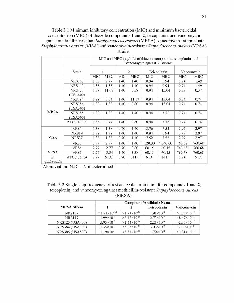
81
Table 3.1 Minimum inhibitory concentration (MIC) and minimum bactericidal concentration (MBC) of thiazole compounds 1 and 2, teicoplanin, and vancomycin
against methicillin-resistant Staphylococcus aureus (MRSA), vancomycin-intermediate Staphylococcus aureus (VISA) and vancomycin-resistant Staphylococcus aureus (VRSA)
strains.
Strain
MIC and MBC (µg/mL) of thiazole compounds, teicoplanin, and vancomycin against S. aureus
1
2
Teicoplanin
Vancomycin
MIC MBC MIC MBC MIC MBC MIC MBC
MRSA
NRS107 1.38 2.77 1.40 1.40 0.94 0.94 0.74 1.49
NRS119 1.38 1.38 1.40 1.40 0.94 0.94 0.74 1.49
NRS123 (USA400)
1.38 11.07 1.40 5.58 0.94 15.04 0.37 0.37
NRS194 1.38 5.54 1.40 11.17 0.94 15.04 0.74 0.74
NRS384 (USA300)
1.38 1.38 1.40 2.80 0.94 15.04 0.74 0.74
NRS385 (USA500)
1.38 1.38 1.40 1.40 0.94 3.76 0.74 0.74
ATCC 43300 1.38 2.77 1.40 2.80 0.94 3.76 0.74 0.74
VISA
NRS1 1.38 1.38 0.70 1.40 3.76 7.52 2.97 2.97
NRS19 1.38 1.38 1.40 1.40 0.94 0.94 2.97 2.97
NRS37 1.38 1.38 0.70 1.40 7.52 7.52 2.97 2.97
VRSA
VRS1 2.77 2.77 1.40 1.40 120.30 >240.60 760.68 760.68
VRS4 2.77 2.77 0.70 2.80 60.15 60.15 760.68 760.68
VRS5 2.77 5.54 1.40 5.58 60.15 60.15 760.68 760.68
S.
epidermidis
ATCC 35984 2.77 N.D.1 0.70 N.D. N.D. N.D. 0.74 N.D.
1Abbreviation: N.D. = Not Determined
Table 3.2 Single-step frequency of resistance determination for compounds 1 and 2, teicoplanin, and vancomycin against methicillin-resistant Staphylococcus aureus
(MRSA).
MRSA Strain
Compound/Antibiotic Name 1 2 Teicoplanin Vancomycin
NRS107 >1.73×10⁻¹⁰ >1.73×10⁻¹⁰ 1.91×10⁻⁸ >1.73×10⁻¹⁰
NRS119 1.99×10⁻⁸ >8.47×10⁻¹⁰ 2.73×10⁻⁷ >8.47×10⁻¹⁰
NRS123 (USA400) 5.93×10⁻⁹ >2.33×10⁻¹⁰ 2.21×10⁻⁹ >2.33×10⁻¹⁰
NRS384 (USA300) 1.35×10⁻⁸ >3.03×10⁻¹⁰ 3.03×10⁻⁹ 3.03×10⁻¹⁰
NRS385 (USA500) 1.19×10⁻⁸ >3.31×10⁻¹⁰ 1.79×10⁻⁸ >3.31×10⁻¹⁰
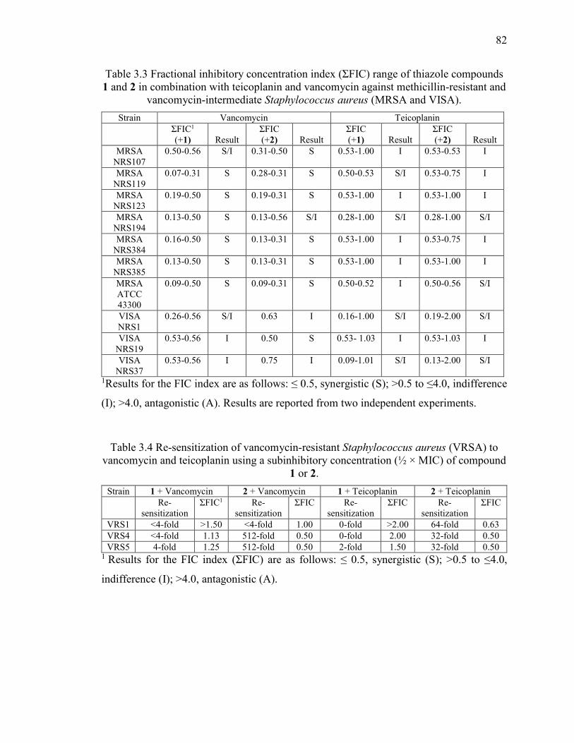
82
Table 3.3 Fractional inhibitory concentration index (ƩFIC) range of thiazole compounds 1 and 2 in combination with teicoplanin and vancomycin against methicillin-resistant and
vancomycin-intermediate Staphylococcus aureus (MRSA and VISA).
Strain Vancomycin Teicoplanin
ƩFIC1 (+1)
Result
ƩFIC (+2)
Result
ƩFIC (+1)
Result
ƩFIC (+2)
Result
MRSA NRS107
0.50-0.56 S/I 0.31-0.50 S 0.53-1.00 I 0.53-0.53 I
MRSA NRS119
0.07-0.31 S 0.28-0.31 S 0.50-0.53 S/I 0.53-0.75 I
MRSA NRS123
0.19-0.50 S 0.19-0.31 S 0.53-1.00 I 0.53-1.00 I
MRSA NRS194
0.13-0.50 S 0.13-0.56 S/I 0.28-1.00 S/I 0.28-1.00 S/I
MRSA NRS384
0.16-0.50 S 0.13-0.31 S 0.53-1.00 I 0.53-0.75 I
MRSA NRS385
0.13-0.50 S 0.13-0.31 S 0.53-1.00 I 0.53-1.00 I
MRSA ATCC 43300
0.09-0.50 S 0.09-0.31 S 0.50-0.52 I 0.50-0.56 S/I
VISA NRS1
0.26-0.56 S/I 0.63 I 0.16-1.00 S/I 0.19-2.00 S/I
VISA NRS19
0.53-0.56 I 0.50 S 0.53- 1.03 I 0.53-1.03 I
VISA NRS37
0.53-0.56 I 0.75 I 0.09-1.01 S/I 0.13-2.00 S/I
1Results for the FIC index are as follows: ≤ 0.5, synergistic (S); >0.5 to ≤4.0, indifference
(I); >4.0, antagonistic (A). Results are reported from two independent experiments.
Table 3.4 Re-sensitization of vancomycin-resistant Staphylococcus aureus (VRSA) to vancomycin and teicoplanin using a subinhibitory concentration (½ × MIC) of compound
1 or 2.
Strain 1 + Vancomycin 2 + Vancomycin 1 + Teicoplanin 2 + Teicoplanin
Re-sensitization
ƩFIC1 Re-sensitization
ƩFIC Re-sensitization
ƩFIC Re-sensitization
ƩFIC
VRS1 <4-fold >1.50 <4-fold 1.00 0-fold >2.00 64-fold 0.63
VRS4 <4-fold 1.13 512-fold 0.50 0-fold 2.00 32-fold 0.50
VRS5 4-fold 1.25 512-fold 0.50 2-fold 1.50 32-fold 0.50 1 Results for the FIC index (ƩFIC) are as follows: ≤ 0.5, synergistic (S); >0.5 to ≤4.0,
indifference (I); >4.0, antagonistic (A).
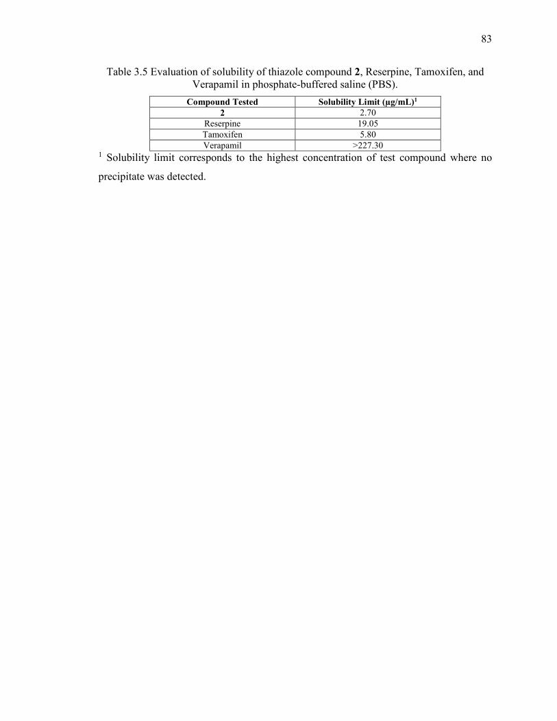
83
Table 3.5 Evaluation of solubility of thiazole compound 2, Reserpine, Tamoxifen, and Verapamil in phosphate-buffered saline (PBS).
Compound Tested Solubility Limit (µg/mL)1 2 2.70
Reserpine 19.05
Tamoxifen 5.80
Verapamil >227.30 1 Solubility limit corresponds to the highest concentration of test compound where no
precipitate was detected.
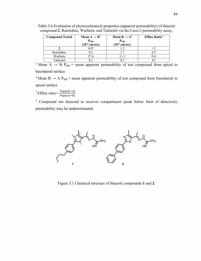
84
Table 3.6 Evaluation of physicochemical properties (apparent permeability) of thiazole compound 2, Ranitidine, Warfarin, and Talinolol via the Caco-2 permeability assay.
Compound Tested Mean A → B1 Papp
(10-6 cm/sec)
Mean B → A2 Papp
(10-6 cm/sec)
Efflux Ratio3
2 0.04 1.2 >2
Ranitidine 0.2 1.7 8.5
Warfarin 27.6 11.1 0.4
Talinolol 0.1 8.3 83 1 Mean A → B Papp = mean apparent permeability of test compound from apical to
basolateral surface
2 Mean B → A Papp = mean apparent permeability of test compound from basolateral to
apical surface
3 Efflux ratio = (��()→+)
(��(+→))
4 Compound not detected in receiver compartment (peak below limit of detection);
permeability may be underestimated
Figure 3.1 Chemical structure of thiazole compounds 1 and 2.
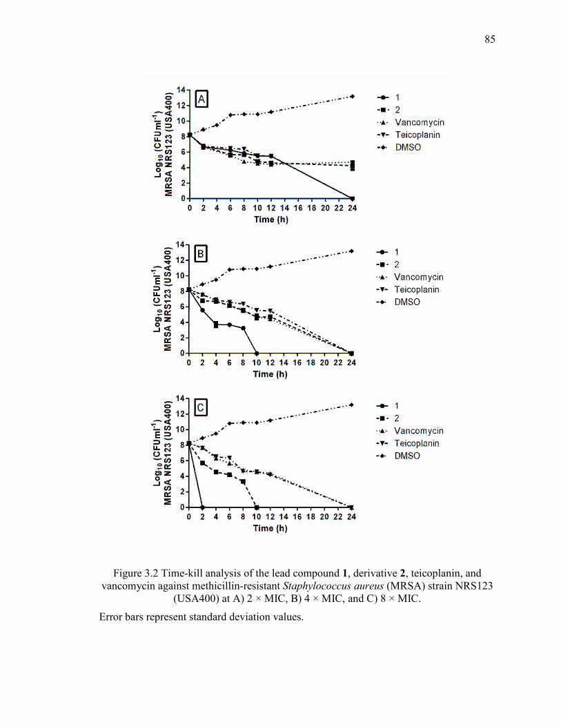
85
Figure 3.2 Time-kill analysis of the lead compound 1, derivative 2, teicoplanin, and vancomycin against methicillin-resistant Staphylococcus aureus (MRSA) strain NRS123
(USA400) at A) 2 × MIC, B) 4 × MIC, and C) 8 × MIC.
Error bars represent standard deviation values.
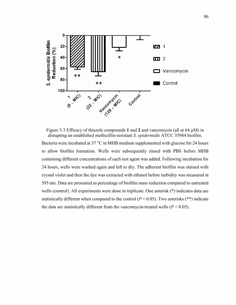
86
Figure 3.3 Efficacy of thiazole compounds 1 and 2 and vancomycin (all at 64 µM) in disrupting an established methicillin-resistant S. epidermidis ATCC 35984 biofilm.
Bacteria were incubated at 37 °C in MHB medium supplemented with glucose for 24 hours
to allow biofilm formation. Wells were subsequently rinsed with PBS before MHB
containing different concentrations of each test agent was added. Following incubation for
24 hours, wells were washed again and left to dry. The adherent biofilm was stained with
crystal violet and then the dye was extracted with ethanol before turbidity was measured at
595 nm. Data are presented as percentage of biofilm mass reduction compared to untreated
wells (control). All experiments were done in triplicate. One asterisk (*) indicates data are
statistically different when compared to the control (P < 0.05). Two asterisks (**) indicate
the data are statistically different from the vancomycin-treated wells (P < 0.05).
87
CHAPTER 4. SYNTHESIS AND ANTIBACTERIAL EVALUATION OF A NOVEL SERIES OF SYNTHETIC
PHENYLTHIAZOLE COMPOUNDS AGAINST METHICILLIN-RESISTANT STAPHYLOCOCCUS AUREUS (MRSA)
THIS IS A PUBLISHED JOURNAL ARTICLE. Reproduced with permission from
Synthesis and antibacterial evaluation of a novel series of synthetic phenylthiazole
compounds against methicillin-resistant Staphylococcus aureus (MRSA). Haroon
Mohammad, P.V. Narasimha Reddy, Dennis Monteleone, Abdelrahman S. Mayhoub,
Mark Cushman, and Mohamed N. Seleem. European Journal of Medicinal
Chemistry (2015) 10, 306–316; doi: 10.1016/j.ejmech.2015.03.015 Copyright 2015
Elsevier Masson SAS. All rights reserved.
4.1 Introduction
Methicillin-resistant Staphylococcus aureus (MRSA) infections remain a
significant public health challenge globally. Though reports have indicated the incidence
of healthcare-associated MRSA (HA-MRSA) infections have diminished (1, 2),
transmission of community-associated MRSA (CA-MRSA) infections, primarily strains
USA300 and USA400 (3), has continued to present major problems amongst a diverse
population including healthcare workers (4), prison inmates (5, 6), military service
personnel (6), contact sport athletes (7, 8), homeless individuals (9), intravenous drug users
(9, 10), tattoo recipients (11), neonates (12), and young children (13, 14). Moreover, CA-
MRSA infections are typically associated with more severe morbidity and mortality than
their HA-MRSA counterparts (15). While CA-MRSA is a leading cause of skin and soft-
tissue infections (16, 17), MRSA has also been associated with more complicated medical
diseases including necrotizing pneumonia (18), osteomyelitis (19), and sepsis (20), leading
to over 11,000 deaths annually (21).
A recent study has estimated the total annual burden upon society for treatment of
CA-MRSA infections alone may exceed US$13 billion (22). Part of the associated cost is
due to failure of current antimicrobials to treat certain clinical isolates of MRSA that have
developed resistance to these therapeutic agents. Indeed, clinical isolates of both CA-
88
MRSA and HA-MRSA have been documented that exhibit resistance to an array of
different antibiotic classes including the β-lactams (23), macrolides (24), quinolones (25,
26), tetracyclines (27), and lincosamides (27). Further exacerbating the problem, strains
have emerged which exhibit resistance to first-line antibiotics (such as mupirocin (27, 28)
for the treatment of MRSA skin infections) and drugs deemed agents of last resort (such as
linezolid (29, 30) and vancomycin (31)). Prudent use and development of effective
antimicrobials is a critical step to alleviate complications and costs associated with MRSA
infections. Therefore there is an urgent need for the development of novel therapeutic
agents and treatment strategies to circumvent this significant global health issue.
Utilizing whole-cell screening of a library of substituted thiazoles, our research group
identified a novel lead thiazole compound that possesses potent antimicrobial activity
against clinically relevant isolates of MRSA, vancomycin-intermediate S. aureus (VISA),
and vancomycin-resistant S. aureus (VRSA) (32). The basic structure of the lead 1 consists
of a central thiazole ring connected to two distinct moieties – a lipophilic side chain at C2
and a cationic amino group at C5. The objectives of the present study were to construct a
series of analogues to the lead 1 (Table 4.1) with modifications to the functional groups at
both the thiazole-C2 and C5 positions to more rigorously ascertain the structure-activity
relationship of these compounds against a diverse array of HA-MRSA and CA-MRSA
isolates, identify new derivatives exhibiting an improved toxicity profile against
mammalian cells, and to enhance the metabolic stability profile of the lead 1.
4.2 Materials and Methods
4.2.1 Chemistry
The detailed synthetic protocols and spectral data of the lead 1 (Figure 4.1) in
addition to all intermediates have been reported elsewhere (32, 33). All thiazole
compounds were dissolved in dimethyl sulfoxide (DMSO) (Sigma-Aldrich, St. Louis, MO,
USA) to achieve a stock 10 mM solution.
The (4-iodophenyl)thiazole derivative 3 was prepared by heating a mixture of the
commercially available 4-iodothiobenzamide 2 and 3-chloro-2,4-pentanedione in absolute
ethanol, as illustrated in Figure 4.2. The phenylthiazolyl methyl ketone derivatives 4 and 6
89
were prepared via the Sonogashira cross coupling of the (4-iodophenyl)thiazole derivative
3 with commercially available 1-hexyne and 1-nonyne, respectively, in DMF using a
bis(triphenylphosphine)palladium(II) dichloride catalyst, copper(I) iodide co-catalyst, and
cesium carbonate base (Figure 4.2). The hydrazinecarboximidamide derivatives 5 and 7
were synthesized by treatment of the phenylthiazolyl methyl ketone derivatives 4 and 6,
respectively, with aminoguanidine hydrochloride in the presence of a catalytic amount of
lithium chloride in absolute ethanol (Figure 4.2).
The amide derivatives 10-13 were prepared in quantitative yields by reacting the 4-
butylphenylthiazole acid chloride intermediate 8 (34) with the appropriate amines in THF,
as illustrated in Figure 4.3. Compound 16 was synthesized in three steps, starting with the
formation of the amide derivative 9 by way of reacting the acid chloride intermediate 8
with ammonium hydroxide in THF at room temperature. The amide intermediate 9 was
then heated in thionyl chloride to give the nitrile intermediate 14, which upon subsequent
treatment with NaN3 in the presence of iodine gave the tetrazole-containing thiazole
derivative 16 as shown in Figure 4.3. The nitrile intermediate 14 was also treated with
hydroxylamine hydrochloride in absolute ethanol with a catalytic amount of potassium
carbonate to afford the thiazole derivative 15. The phenylthiazolyl methyl ketone
derivative 18 was prepared by treatment of the commercially available 4-
aminothiobenzamide 17 with 3-chloro-2,4-pentanedione in absolute ethanol.
Synthesis of the hydrazinecarboximidamide derivative 19 was achieved by
treatment of the phenylthiazolyl methyl ketone derivative 18 with aminoguanidine
hydrochloride in the presence of a catalytic amount of lithium chloride (Figure 4.4).
Phenylthiazole methylketone derivatives 21a-d and 24 were prepared via the
Suzuki-Miyaura cross coupling of the (4-iodophenyl)thiazole derivative 3 with the
commercially available phenylboronic acid derivatives 20a-d and 23, respectively, in the
presence of a catalytic quantity of palladium(II) acetate and (2-
biphenyl)dicyclohexylphosphine ligand, as shown in Figure 4.5. Synthesis of the
hydrazinecarboximidamide derivatives 22a-d and 25 was achieved by treatment of
phenylthiazole methylketone derivatives 21a-d and 24, respectively, with aminoguanidine
hydrochloride in the presence of lithium chloride as catalyst (Figure 4.5).
90
1H NMR spectra were recorded in CDCl3 or DMSO-d6 using a 300 MHz spectrometer.
Chemical shifts are reported in units of ppm on the delta (δ) scale and coupling constants
(J) are reported in units of Hz. The following splitting abbreviations are used: s = singlet,
d = doublet, t = triplet and m = multiplet. All melting points were recorded using capillary
tubes on a Mel-Temp apparatus and are not corrected. Mass spectral analyses were
performed at the Purdue University Campus-Wide Mass Spectrometry Center. Reagents
and solvents were purchased from commercial vendors and were used as received without
further purification, unless otherwise stated.
1-(2-(4-Iodophenyl)-4-methylthiazol-5-yl)ethanone (3).
4-Iodothiobenzamide (2, 3.80 mmol) and α-chloropentanedione (0.611 mg, 4.56 mmol)
were added to absolute ethanol (50 mL). The reaction mixture was heated at reflux for 24
hours. After removal of solvent under reduced pressure, the residue was purified by silica
gel chromatography using hexanes–ethyl acetate (7:3) to provide the desired compound as
light orange solid (0.800 g, 62%): mp 105-106 oC. 1H NMR (300 MHz, CDCl3) δ 7.81 (d,
J = 6.6 Hz, 2 H), 7.70 (d, J = 6.6 Hz, 2 H), 2.77 (s, 3 H), 2.56 (s, 3 H).
1-(2-(4-(Hex-1-yn-1-yl)phenyl)-4-methylthiazol-5-yl)ethanone (4).
1-(2-(4-Iodophenyl)-4-methylthiazol-5-yl)ethanone (3, 0.5 g, 1.45 mmol), 1-hexyne (0.373
g, 7.73 mmol), cesium carbonate (0.947 g, 2.91 mmol), dichloro-
bis(triphenylphosphine)palladium(II) (0.051 g, 0.072 mmol) and CuI (0.027 g, 0.145
mmol) were dissolved in DMF (6 mL). The reaction mixture was purged with argon for 20
minutes. The sealed tube was closed, placed in an oil bath and stirred at 65 oC for 15 hours.
The reaction mixture was filtered through celite, and the celite was washed with chloroform
(50 mL). The organic phase was washed with 1% hydrochloric acid (30 mL), water (3 x 40
mL) and brine (30 mL). The organic layer was dried over Na2SO4, concentrated and
purified by silica gel flash column chromatography using hexanes–ethyl acetate (8:2) to
provide the desired compound as yellow syrup (0.400 g, 92.5%): IR (film) 1945, 1675,
1111, 819, 666 cm-1; 1H NMR (300 MHz, CDCl3) δ 7.90 (d, J = 8.5 Hz, 2 H), 7.46 (d, J =
8.3 Hz, 2 H), 2.76 (s, 3 H), 2.56 (s, 3 H), 2.45 (t, J = 6.9 Hz, 2 H), 1.59 (m, 4 H), 0.97 (t, J
= 7.3 Hz, 3 H); ESIMS m/z (rel intensity) 298 (MH+, 100).
91
(Z)-2-(1-(2-(4-(Hex-1-yn-1-yl)phenyl)-4-methylthiazol-5-yl)ethylidene)
hydrazinecarboximidamide (5).
The thiazole derivative 4 (200 mg, 0.673 mmol) was dissolved in absolute ethanol (10 mL),
and aminoguanidine hydrochloride (0.088 mg, 0.808 mmol) and a catalytic amount of LiCl
(5 mg) were added. The reaction mixture was heated at reflux for 24 hours. The solvent
was evaporated under reduced pressure. The crude product was purified by crystallization
from 70% methanol and then recrystallized from methanol to afford the desired compound
as a yellow solid (80 mg, 46%): mp 253-254 oC. IR (KBr) 3329, 2227, 1678, 1143, 836,
657 cm-1; 1H NMR (DMSO-d6) δ 11.35 (br s, 1 H), 7.88 (d, J = 8.1 Hz, 2 H), 7.69 (br s, 3
H), 7. 49 (d, J = 8.1 Hz, 2 H), 2.60 (s, 3 H), 2.44 (s, 3 H), 2.41 (t, J = 7.1 Hz, 2 H), 1.48
(m, 4 H), 0.93 (t, J = 7.1 Hz, 3 H); ESIMS m/z (rel intensity) 354 (MH+, 100); HRESIMS
calcd for C19H24N5S 354.1509 (MH+), found 354.1514; HPLC purity 98.07% (1% TFA in
MeOH:H2O – 85:15).
1-(4-Methyl-2-(4-(non-1-yn-1-yl)phenyl)thiazol-5-yl)ethan-1-one (6).
The thiazole derivative 3 (750 mg, 2.19 mmol), 1-nonyne (1.44 mL, 8.76 mmol),
PdCl2(PPh3)2 (76.6 mg, 0.11mmol), copper(I) iodide (41.6 mg, 0.22 mmol) and Cs2CO3
(1.42 g, 4.38 mmol) were added to a sealed tube under argon for 10 minutes, and then DMF
(7.5 mL) was added. The tube was once again evacuated and purged with argon for 5
minutes and heated to 70 °C for 15 hours. The tube was allowed to cool to room
temperature, and the solids were removed by filtration and the filter cake was extracted
with additional CHCl3 (50 mL). The combined filtrate and extracts were concentrated under
vacuum and extracted with EtOAc (2 × 50 mL) and washed with water (2 × 50 mL). After
evaporation of the solvent under reduced pressure, the residue was collected and purified
by flash chromatography (SiO2, hexanes-EtOAc, 8.8:1.2) to yield the desired compound 6
as a dark green oil (772 mg, 100%); 1H NMR (300 MHz, CDCl3) δ 7.9 (t, J = 6.8 Hz, 2 H),
7.46 (d, J = 8.4 Hz, 2 H), 2.76 (s, 3 H), 2.55 (s, 3 H), 2.44 (t, J = 7.1 Hz, 3 H), 1.63 (t, J =
7.7 Hz, 2 H), 1.30 (m, J = 3.3 Hz, 9 H), 0.9 (q, J = 6.2 Hz, 4 H).
(E)-2-(1-(4-Methyl-2-[4-(non-1-yn-1-yl)phenyl]thiazol-5-
yl)ethylidene)hydrazinecarboximidamide (7).
The thiazole derivative 6 (140 mg, 0.41 mmol), aminoguanidine hydrochloride (90.85 mg,
0.83 mmol), and a catalytic amount of LiCl (5 mg) were added to absolute ethanol (10 mL).
92
The reaction mixture was heated at reflux for 24 hours. After evaporation of the solvent
under reduced pressure, the crude residue was extracted with CHCl3/MeOH (90:10, 2 × 40
mL) and washed with water (2 × 50 mL) and brine (40 mL). The extracts were pooled
together, dried over Na2SO4 and stripped of solvents under reduced pressure. The residue
was suspended in CHCl3/hexanes (50:50, 50 mL) and filtered through
Whatman filter paper to afford the desired product (7) as a yellow-white solid (200 mg,
100%): mp 255-260 °C dec, 1H NMR (300 MHz, DMSO-d6) δ 7.88 (d, J = 8.3 Hz, 3 H),
7.49 (d, J = 8.4 Hz, 3 H), 2.60 (s, 3 H), 2.49 (q, J = 1.7 Hz, 5 H), 1.26 (s, 12 H), 0.85 (s, 4
H); ESIMS m/z (rel intensity) 395 (M+, 57).
2-(4-Butylphenyl)-4-methylthiazole-5-carboxamide (9).
Acid chloride 8 (34) (0.200 g, 0.682 mmol) was dissolved in THF (20 mL) and then 30%
aq NH4OH (10 mL) was added. The reaction mixture was stirred at room temperature for
24 hours. The THF was removed on a rotary evaporator and the crude product was
extracted with EtOAc (2 × 25 mL) and washed with water (2 × 20 mL) and brine (20 mL).
The combined organic layer was dried over Na2SO4 and concentrated to afford the amide
9 (0.185 g) in quantitative yield: mp 160-161 oC. IR (KBr) 3245, 1691, 1611, 1121, 846,
665 cm-1; 1H NMR (300 MHz, CDCl3) δ 7.84 (d, J = 8.2 Hz, 2 H), 7.26 (d, J = 8.2 Hz, 2
H), 5.79 (br s, 2 H), 2.74 (s, 3 H), 2.66 (t, J = 7.5 Hz, 2 H), 1.63 (m, 2 H), 1.39 (m, 2 H),
0.94 (t, J = 7.3 Hz, 3 H); APCIMS m/z (rel intensity) 275 (MH+, 100); HPLC purity 97.89%
(1% TFA in MeOH:H2O – 90:10).
2-(4-Butylphenyl)-N-(N-carbamimidoylcarbamimidoyl)-4-methylthiazole-5-
carboxamide (10).
Acid chloride 8 (0.200 g, 0.682 mmol) was dissolved in THF (20 mL) and then biguanidine
hydrochloride (0.467 g, 3.41 mmol) followed by triethylamine (0.344 g, 3.41 mmol) were
added. The reaction mixture was stirred at room temperature for 24 hours. The THF was
removed on a rotary evaporator and the crude product was extracted with EtOAc (2 × 30
mL) and washed with water (2 × 20 mL) and brine (20 mL). The combined organic layer
was dried over Na2SO4, concentrated and purified by silica gel flash column
chromatography using chloroform-methanol (9.5:0.5) to provide the desired compound as
a yellow solid (0.070 g, 30%): mp 150-151 oC. IR (KBr) 3312, 1694, 1655, 1148, 823, 666
cm-1; 1H NMR (300 MHz, CDCl3) δ 7.89 (br s, 2 H), 7.25 (br s, 2 H), 2.80 (s, 3 H), 2.67 (t,
93
J = 7.2 Hz, 2 H), 1.60 (m, 2 H), 1.38 (m, 2 H), 0.94 (t, J = 7.1 Hz, 3 H); ESIMS m/z (rel
intensity) 359 (MH+, 65), 341 (MH+-NH3, 68); HRESIMS calcd for C17H23N6OS m/z
359.1248 (MH+), found 359.1251; HPLC purity 95.16% (1% TFA in MeOH:H2O – 90:10).
2-(2-(4-Butylphenyl)-4-methylthiazole-5-
carbonyl)hydrazinecarboximidamide (11).
Acid chloride 8 (0.200 g, 0.682 mmol) was dissolved in THF (20 mL) and then amino
guanidine hydrochloride (0.377 g, 3.41 mmol) followed by triethylamine (0.344 g, 3.41
mmol) were added. The reaction mixture was stirred at room temperature for 24 hours. The
THF was removed on a rotary evaporator and the crude product was extracted with EtOAc
(2 × 30 mL) and washed with water (2 × 20 mL) and brine (20 mL). The combined organic
layer was dried over Na2SO4, concentrated and purified by silica gel flash column
chromatography using hexanes–ethyl acetate (4:6) to provide the desired compound as a
yellow solid. (0.090 g, 40%): mp 174-175 oC. IR (KBr) 3322, 1698, 1658, 1462, 1155, 856,
665 cm-1; 1H NMR (300 MHz, CDCl3) δ 7.87 (d, J = 8.0 Hz, 2 H), 7.24 (d, J = 8.0 Hz, 2
H), 2.78 (s, 3 H), 2.67 (t, J = 7.6 Hz, 2 H), 1.63 (m, 2 H), 1.39 (m, 2 H), 0.94 (t, J = 7.3 Hz,
3 H); ESIMS m/z (rel intensity) 332 (MH+, 100); HRESIMS calcd for C16H22N5OS m/z
332.1112 (MH+), found 332.1210; HPLC purity 96.57% (1% TFA in MeOH:H2O – 90:10).
(R)-2-(4-Butylphenyl)-N-(2,3-dihydroxypropyl)-4methylthiazole-5-
carboxamide (12).
A mixture of the acid chloride derivative 8 (0.25 g, 0.9 mmol) and (R)-(-)-3-amino-1,2-
propanediol (0.154 g, 1.7 mmol) in THF (15 mL) were stirred at room temperature for 48
hours. The THF was removed under reduced pressure and the resulting crude oil was
extracted with chloroform (2 × 50 mL) and the extract was washed with water (2 × 50 mL)
and brine (50 mL). The combined extracts were dried over Na2SO4 (10 g), filtered and
concentrated under vacuum. The residue was purified by flash column chromatography
(SiO2, CHCl3-MeOH, 9.4:0.6) to afford the product 12 as dark red crystals (0.17 g, 57%):
mp 93-95 °C. 1H NMR (300 MHz, CDCl3) δ 7.82 (d, J = 8.2 Hz, 2 H), 7.24 (d, J = 7.2 Hz,
4 H), 6.32 (s, 1 H), 3.89 (d, J = 5.0 Hz, 1 H), 3.65 (m, J = 5.2 Hz, 4 H), 2.73 (s, 3 H), 2.63
(t, J = 7.6 Hz, 2 H), 1.61 (t, J = 7.9 Hz, 2 H), 1.35 (q, J = 7.5 Hz, 2 H), 0.92 (t, J = 6.1 Hz,
3 H); ESIMS m/z (rel intensity) 349 (MH+, 100).
94
2-(4-Butylphenyl)-N-[(dimethylamino)methyl]-4-methylthiazole-5-
carboxamide (13).
A mixture of acid chloride derivative 8 (0.4 g, 1.44 mmol) and N,N-
dimethylethylenediamine (0.63 mL, 5.7 mmol) in THF (15 mL) were stirred at room
temperature for 48 hours. The THF was removed under reduced pressure and the resulting
residue was purified by flash chromatography (SiO2, CHCl3-MeOH, 9.3:0.7) to afford the
product 13 as a pink solid (0.123 g, 24%): mp 79-81 °C. 1H NMR (300 MHz, CDCl3) δ
7.83 (d, J = 8.2 Hz, 2 H), 7.25 (d, J = 9.7 Hz, 4 H), 3.49 (t, J = 5.8 Hz, 2 H), 2.72 (s, 3 H),
2.63 (t, J = 7.6 Hz, 2 H), 2.55 (t, J = 5.9 Hz, 2 H), 2.3 (s, 6 H), 1.61 (t, J = 7.8, 2 H), 1.36
(t, J = 7.4, 2 H), 0.92 (t, J = 7.3 Hz, 3 H); ESIMS m/z (rel intensity) 346 (MH+, 100).
2-(4-Butylphenyl)-4-methylthiazole-5-carbonitrile (14).
Amide 9 (0.400 g, 1.45 mmol) was dissolved in thionyl chloride (20 mL) and the solution
was heated to reflux for seven hours. Thionyl chloride was removed under reduced
pressure, EtOAc (30 mL) was added and the mixture was washed with saturated aqueous
NaHCO3 (2 × 15 mL) and water (2 × 15 mL). The organic layer was dried over Na2SO4,
concentrated and purified by silica gel flash column chromatography using hexanes–ethyl
acetate (9:1) to provide the desired compound as yellow syrup (0.300 g, 81%): IR (KBr)
2246, 1456, 1122, 841, 665 cm-1; 1H NMR (300 MHz, CDCl3) δ 7.83 (d, J = 8.1 Hz, 2 H),
7.27 (d, J = 8.1 Hz, 2 H), 2.67 (s, 3 H), 2.64 (m, 2 H), 1.63 (m, 2 H), 1.38 (m, 2 H), 0.94
(t, J = 7.3 Hz, 3 H); ESIMS m/z (rel intensity) 256 (M+, 33), 213 (M+-C3H7, 100).
(Z)-2-(4-Butylphenyl)-N’-hydroxy-4-methylthiazole-5-carboximidamide (15).
A mixture of the thiazole derivative 14 (0.19 g, 0.74 mmol), hydroxylamine hydrochloride
(0.07 g, 1 mmol) and K2CO3 (0.102 g, 0.74 mmol) in absolute EtOH (15 mL) was stirred
at room temperature for one hour and then heated at reflux overnight. The EtOH was
removed under reduced pressure and the resulting crude residue was purified by flash
column chromatography (SiO2, hexanes-EtOAc, 9:1) to afford the product 15 as an off
white to light yellow solid (35.5 mg, 17%): mp 135-137 °C. 1H NMR (300 MHz, CDCl3)
δ 7.81 (d, J = 8.05 Hz, 2 H), 7.25 (t, J = 5.7 Hz, 2 H), 2.63 (t, J = 7.6 Hz, 5 H), 1.59 (q, J
= 7.4 Hz, 2 H), 1.34 (p, J = 11.2 Hz, 2 H), 0.92 (t, J = 7.28 Hz, 3 H); ESIMS m/z (rel
intensity ) 290 (M+, 100).
2-(4-Butylphenyl)-4-methyl-5-(1H-tetrazol-5-yl)thiazole (16).
95
I2 (20 mg) was added to a mixture of nitrile (14, 0.2 g, 0.781 mmol) and NaN3 (0.076 g,
1.17 mmol) and the mixture was stirred at 120 °C for 15 hours. After completion of the
reaction, EtOAc (15 mL) and 4 M HCl (10 mL) were added and the mixture was stirred
vigorously for 10 minutes. The organic layer was separated and the aqueous layer was
extracted with EtOAc (2 × 10 mL). The combined organic layer was washed with brine (4
× 15 mL), dried over Na2SO4, concentrated and purified by silica gel flash column
chromatography using hexane-ethyl acetate (5:5) to provide the desired compound as a
light yellow solid (0.085 g, 37%): mp 170-171 oC. IR (KBr) 1825, 1415, 1098, 844, 664
cm-1; 1H NMR (300 MHz, CDCl3) δ 7.84 (d, J = 8.1 Hz, 2 H), 7.22 (d, J = 8.1 Hz, 2 H),
2.85 (s, 3 H), 2.64 (t, J = 7.5 Hz, 2 H), 1.63 (m, 2 H), 1.35 (m, 2 H), 0.93 (t, J = 7.3 Hz, 3
H); ESIMS m/z (rel intensity) 300 (MH+, 100); HRESIMS calcd for C15H18N5S m/z
300.1267 (MH+), found 300.1270; HPLC purity 98.25% (1% TFA in MeOH:H2O – 90:10).
1-(4-Methyl-2-(4-aminophenyl)thiazol-5-yl)ethanone (18).
4-Aminothiobenzamide (0.6 g, 3.80 mmol) and α-chloropentanedione (0.611 mg, 4.56
mmol) were added to absolute ethanol (50 mL). The reaction mixture was heated at reflux
for 24 hours. After removal of solvent under reduced pressure, the residue was purified by
silica gel chromatography using hexanes–ethyl acetate (6:4) to provide the desired
compound as light brown solid (0.920 g, 97%): mp 204-205 oC. IR (KBr) 3334, 1745,
1637, 1145, 865, 666 cm-1; 1H NMR (300 MHz, CDCl3) δ 7.89 (d, J = 8.5 Hz, 2 H), 7.07
(d, J = 8.5 Hz, 2 H), 2.66 (s, 3 H), 2.53 (s, 3 H).
(E)-2-(1-(4-Methyl-2-(4-aminophenyl)thiazol-5-yl)ethylidene)
hydrazinecarboximidamide (19).
The thiazole derivative 18 (0.250 g, 0.954 mmol) was dissolved in absolute ethanol (50
mL), and aminoguanidine hydrochloride (0.125 g, 1.14 mmol) and a catalytic amount of
LiCl (15 mg) were added. The reaction mixture was heated at reflux for 24 hours. The
solvent was evaporated under reduced pressure. The crude product was purified by
crystallization from 70% methanol and then recrystallized from methanol to afford the
desired compound as a yellow solid (0.175 g, 58%): mp ˃ 280 oC. IR (KBr) 3402, 1705,
1665, 1156, 826, 665 cm-1; 1H NMR (300 MHz, DMSO-d6) δ 11.44 (br s, 1 H), 7.81 (d, J
= 8.4 Hz, 2 H), 7.75 (br s, 3 H), 7.00 (d, J = 8.4 Hz, 2 H), 2.58 (s, 3 H), 2.41 (s, 3 H);
96
ESIMS m/z (rel intensity) 289 (MH+, 100); HRESIMS calcd for C13H17N6S m/z 289.1123
(MH+), found 289.1120; HPLC purity 96.58% (1% TFA in MeOH:H2O – 90:10).
1-(2-(4'-Hydroxy-[1,1'-biphenyl]-4-yl)-4-methylthiazol-5-yl)ethanone (21a).
Iodide 3 (0.172 g, 0.5 mmol), the 4-hydroxyphenyl boronic acid (20a, 0.205 g, 1.5 mmol),
tripotassium monophosphate (0.424 g, 2 mmol) and (2-biphenyl)dicylohexylphosphine (18
mg) were dissolved in dry toluene (15 mL) and the solution was purged with argon for 20
minutes. Pd(OAc)2 (25 mg, 0.24 mmol) was added to the reaction mixture and the reaction
mixture was heated at 90 ºC under argon for 24 hours. Ethyl acetate (40 mL) was added to
reaction mixture, which was then washed with water (2 × 25 mL). The combined organic
layer was dried over Na2SO4, concentrated and purified by flash column chromatography
(EtOAc:hexanes 3:7 to 0.9:9.1) to afford the desired compound as yellow solid (0.150 g,
93%): mp 212-214 oC. IR (KBr) 3356, 1745, 1123, 856, 665 cm-1; 1H NMR (300 MHz ,
CDCl3) δ 7.86 (d, J = 8.3 Hz, 2 H), 7.51 (d, J = 8.3 Hz, 2 H), 7.38 (d, J = 8.5 Hz, 2 H), 6.79
(d, J = 8.6 Hz, 2 H), 2.64 (s, 3 H), 2.45 (s, 3 H); ESIMS m/z (rel intensity) 309 (M+, 100).
1-(2-(4'-Fluoro-[1,1'-biphenyl]-4-yl)-4-methylthiazol-5-yl)ethanone (21b).
Iodide 3 (0.172 g, 0.5 mmol), 4-fluorophenyl boronic acid (20b, 0.209 1.5 mmol),
tripotassium monophosphate (0.424 g, 2 mmol) and (2-biphenyl)dicylohexylphosphine (18
mg) were dissolved in dry toluene (15 mL) and the solution was purged with argon for 20
minutes. Pd(OAc)2 (25 mg, 0.24 mmol) was added to the reaction mixture and the reaction
mixture was heated at 90 ºC under argon for 24 hours. Ethyl acetate (40 mL) was added to
reaction mixture, which was then washed with water (2 × 25 mL). The combined organic
layer was dried over Na2SO4, concentrated and purified by flash column chromatography
(EtOAc:hexanes 3:7 to 0.9:9.1) to afford the desired compound as an off-white solid (0.140
g, 90%): mp 127-128 oC. IR (KBr) 2956, 1689, 1123, 819, 659 cm-1; 1H NMR (300 MHz,
CDCl3) δ 8.05 (d, J = 7.6 Hz, 2 H), 7.64 (m, 4 H), 7.23 (d, J = 7.5 Hz, 2 H), 2.79 (s, 3 H),
2.58 (s, 3 H); ESIMS m/z (rel intensity) 312 (M+, 100); HPLC purity 98.50% (1% TFA in
MeOH:H2O – 90:10).
97
1-(4-Methyl-2-(4'-(trifluoromethyl)-[1,1'-biphenyl]-4-yl)thiazol-5-yl)ethanone
(21c).
Iodide 3 (0.172 g, 0.5 mmol), 4-trifluromethylphenyl boronic acid (20c, 0.228 g, 1.5
mmol), tripotassium monophosphate (0.424 g, 2 mmol) and (2-
biphenyl)dicylohexylphosphine (18 mg) were dissolved in dry toluene (15 mL) and the
solution was purged with argon for 20 minutes. Pd(OAc)2 (25 mg, 0.24 mmol) was added
to the reaction mixture and the reaction mixture was heated at 90 ºC under argon for 24
hours. Ethyl acetate (40 mL) was added to reaction mixture, which was then washed with
water (2 × 25 mL). The combined organic layer was dried over Na2SO4, concentrated and
purified by flash column chromatography (EtOAc:hexanes 3:7 to 0.9:9.1) to afford the
desired compound as an off-white solid (0.150 g, 80%): mp 116-117 oC. IR (KBr) 2959,
1938, 1650, 1111, 819, 659 cm-1; 1H NMR (300 MHz , CDCl3) δ 8.09 (d, J = 8.3 Hz, 2 H),
7.72 (m, 6 H), 2.80 (s, 3 H), 2.58 (s, 3 H); ESIMS m/z (rel intensity) 361 (M+, 100); HPLC
purity 98.75% (1% TFA in MeOH:H2O – 90:10).
(E)-2-(1-(2-(4'-Hydroxy-[1,1'-biphenyl]-4-yl)-4-methylthiazol-5-
yl)ethylidene)hydrazinecarboximidamide (22a).
Compound 21a (0.150 g, 0.485 mmol) was dissolved in absolute ethanol (50 mL) and
aminoguanidine hydrochloride (0.064 g, 0.588 mmol) and a catalytic amount of LiCl (10
mg) were added. The reaction mixture was heated at reflux for 24 hours. The solvent was
evaporated under reduced pressure. The crude product was purified by crystallization from
70% methanol, and then recrystallized from methanol to afford the desired compound as a
light yellow solid (0.115 g, 66%): mp 252-254 oC. IR (KBr) 3398, 1695, 1655, 1142, 855,
665 cm-1; 1H NMR (300 MHz, DMSO-d6) δ 9.73 (s, 1 H), 8.02 (d, J = 8.3 Hz, 2 H), 7.74
(d, J = 8.4 Hz, 2 H), 7.59 (d, J = 8.6 Hz, 2 H), 6.88 (d, J = 8.6 Hz, 2 H), 2.70 (s, 3 H), 2.56
(s, 3 H); ESIMS m/z (rel intensity) 366 (MH+, 100); HRESIMS calcd for C19H20N5OS
366.1045 (MH+), found 366.1048; HPLC purity 96.11% (1% TFA in MeOH:H2O – 90:10).
(E)-2-(1-(2-(4'-Fluoro-[1,1'-biphenyl]-4-yl)-4-methylthiazol-5-
yl)ethylidene)hydrazinecarboximidamide (22b).
Compound 21b (0.1 g, 0.321 mmol) was dissolved in absolute ethanol (50 mL) and
aminoguanidine hydrochloride (0.064 g, 0.588 mmol) and a catalytic amount of LiCl (10
mg) were added. The reaction mixture was heated at reflux for 24 hours.
98
The solvent was evaporated under reduced pressure. The crude product was purified by
crystallization from 70% methanol, and then recrystallized from methanol to afford the
desired compound as a light yellow solid (0.077 g, 65%): mp 273-274 ºC. IR (KBr) 3308,
1687, 1645, 1146, 823, 666 cm-1; 1H NMR (300 MHz, DMSO-d6) δ 11.73 (br s, 1 H), 8.00
(d, J = 8.3 Hz, 2 H) 7.81 (m, 8 H), 7.32 (m, 2 H), 2.62 (s, 3 H), 2.44 (s, 3 H); ESIMS m/z
(rel intensity) 368 (MH+, 100); HRESIMS calcd for C19H19FN5S m/z 368.1245 (MH+),
found 368.1251; HPLC purity 95.78% (1% TFA in MeOH:H2O – 90:10).
(E)-2-(1-(4-Methyl-2-(4'-(trifluoromethyl)-[1,1'-biphenyl]-4-yl)thiazol-5-
yl)ethylidene)hydrazinecarboximidamide (22c).
Compound 21c (0.1 g, 0.277 mmol) was dissolved in absolute ethanol (50 mL) and
aminoguanidine hydrochloride (0.064 g, 0.588 mmol) and a catalytic amount of LiCl (10
mg) were added. The reaction mixture was heated at reflux for 24 hours. The solvent was
evaporated under reduced pressure. The crude product was purified by crystallization from
70% methanol, and then recrystallized from methanol to afford the desired compounds as
yellow solid (0.088 g, 76%): mp 269-270 ºC. IR (KBr) 3583, 3307, 1678, 1145, 821, 665
cm-1; 1H NMR (300 MHz, DMSO-d6) δ 11.78 (br s, 1 H), 8.05 (d, J = 8.4 Hz, 2 H), 7.98
(d, J = 8.2 Hz, 2 H), 7.90 (m, 8 H), 2.62 (s, 3 H), 2.45 (s, 3 H); ESIMS m/z (rel intensity)
418 (MH+, 100); HRESIMS calcd for C20H19F3N5S m/z 418.1423 (MH+), found 418.1420;
HPLC purity 96.10% (1% TFA in MeOH:H2O – 90:10).
(E)-2-(1-(2-(4'-Acetyl-[1,1'-biphenyl]-4-yl)-4-methylthiazol-5-
yl)ethylidene)hydrazinecarboximidamide (22d).
Compound 11d (0.150 g, 0.472 mmol) was dissolved in absolute ethanol (50 mL) and
aminoguanidine hydrochloride (0.064 g, 0.588 mmol) and a catalytic amount of LiCl (10
mg) were added. The reaction mixture was heated at reflux for 24 hours. The solvent was
evaporated under reduced pressure. The crude product was purified by crystallization from
70% methanol, and then recrystallized from methanol to afford the desired compound as a
light yellow solid (0.062, 35%): mp >280 ºC. IR (KBr) 3402, 1715, 1655, 1446 1125, 856,
665 cm-1; 1H NMR (300 MHz, DMSO-d6) δ 11.68 (br s, 1 H), 8.10 (d, J = 8.2 Hz, 4 H),
7.91 (m, 6 H), 7.80 (d, J = 8.2 Hz, 2 H), 2.72 (s, 3 H), 2.58 (s, 3 H), 2.40 (s, 3 H); ESIMS
m/z (rel intensity) 392 (MH+, 100); HRESIMS calcd for C21H22N5OS m/z 392.1165 (MH+),
found 392.1169.
99
1-(4-Methyl-2-(4-(naphthalen-1-yl)phenyl)thiazol-5-yl)ethanone (24)
Iodide 3 (0.172 g, 0.5 mmol), 1-naphalene boronic acid (23, 258 g, 1.5 mmol), tripotassium
monophosphate (0.424 g, 2 mmol) and (2-biphenyl)dicylohexylphosphine (18 mg) were
dissolved in dry toluene (15 mL) and the solution was purged with argon for 20 minutes.
Pd(OAc)2 (25 mg, 0.24 mmol) was added to the reaction mixture and the reaction mixture
was heated at 90 ºC under argon for 24 hours. Ethyl acetate (40 mL) was added to reaction
mixture, which was then washed with water (2 × 25 mL). The combined organic layer was
dried over Na2SO4, concentrated and purified by flash column chromatography (EtOAc:
hexane 3:7 to 0.9:9.1) to afford the desired compound as a light brown solid (0.170 g,
99%): mp 141-142 ºC. IR (KBr) 2287, 1670, 1006, 801, 663 cm-1; 1H NMR (300 MHz,
CDCl3) δ 8.11 (d, J = 6.5 Hz, 2 H), 7.90 (m, 3 H), 7.61 (m, 7 H), 2.82 (s, 3 H), 2.60 (s, 3
H); ESIMS m/z (rel intensity) 344 (MH+, 100); HRESIMS calcd for C22H18NOS m/z
344.1123 (MH+), found 344.1125.
(E)-2-(1-(4-Methyl-2-(4-(naphthalen-1-yl)phenyl)thiazol-5-
yl)ethylidene)hydrazinecarboximidamide (25).
Compound 24 (0.1 g, 0.291 mmol) was dissolved in absolute ethanol (50 mL) and
aminoguanidine hydrochloride (0.064 g, 0.588 mmol) and a catalytic amount of LiCl (10
mg) were added. The reaction mixture was heated at reflux for 24 hours. The solvent was
evaporated under reduced pressure. The crude product was purified by crystallization from
70% methanol, and then recrystallized from methanol to afford the desired compound as a
yellow solid (0.057, 49 %): mp 241-242 ºC. IR (KBr) 3299, 2300, 1675, 1618, 1142, 800,
664 cm-1; 1H NMR (300 MHz, DMSO-d6) δ 11.63 (br s, 1 H), 8.08 (m, 5 H), 7.65 (d, J =
5.1 Hz, 1 H); 7.61 (m, 9 H), 2.64 (s, 3 H), 2.44 (s, 3 H); ESIMS m/z (rel intensity) 400
(MH+, 100); HRESIMS calcd for C23H22N5S m/z 400.1323 (MH+), found 400.1327; HPLC
purity 95.55% (1% TFA in MeOH:H2O – 85:15).
4.2.2 Bacterial strains and reagents
Clinical isolates of MRSA, VISA, and VRSA were obtained through the Network of
Antimicrobial Resistance in Staphylococcus aureus (NARSA) program. In addition,
MRSA ATCC 43300 was obtained from the American Type Cultural Collection (Manassas,
VA, USA). Vancomycin hydrochloride powder was purchased commercially (Gold
100
Biotechnology Inc., St. Louis, MO, USA) and dissolved in DMSO to prepare a 10 mM
stock solution.
4.2.3 Determination of minimum inhibitory concentration (MIC) and minimum bactericidal concentration (MBC) against MRSA, VISA, and VRSA strains
The MICs of the thiazole compounds and vancomycin against seven clinical isolates of
MRSA, three clinical isolates of VISA, and three clinical isolates of VRSA were
determined using the broth microdilution method in accordance with the recommendations
contained in the CLSI guidelines (35). Bacteria were prepared in phosphate-buffered saline
(PBS) to achieve a McFarland standard of 0.5. The solution was subsequently diluted 1:300
in Mueller-Hinton broth (MHB) to reach a starting inoculum of 1 × 10⁵ colony-forming
units (CFU/mL). Bacteria were then transferred to a 96-well microtiter plate. Thiazole
compounds and vancomycin were added (in triplicate) to wells in the first row of the
microtiter plate and then serially diluted along the vertical axis. The plate was incubated at
37 °C for 18-20 hours before the MIC was determined as the lowest concentration where
visible growth of bacteria was not observed.
The MBC was determined by plating 5 µL from wells on the 96-well microtiter
plate (where the MIC was determined), where no growth was observed, onto tryptic soy
agar (TSA) plates. The TSA plates were then incubated at 37 °C for 18-20 hours before the
MBC was determined. The MBC was classified as the concentration where ≥99% reduction
in bacterial cell count was observed.
4.2.4 Time-kill analysis of thiazole compounds 1, 5, and 25 and vancomycin against MRSA
MRSA USA300 cells in late logarithmic growth phase were diluted to ~1 × 10⁸ colony-
forming units (CFU/mL) and exposed to concentrations equivalent to 3 × MIC (in triplicate)
of thiazole compounds 1, 5, and 25 and vancomycin in MHB. 20 µL samples were collected
after 0, 2, 4, 6, 8, 10, 12, and 24 hours of incubation at 37 °C and subsequently serially
diluted in PBS. Bacteria were then transferred to TSA plates and incubated at 37 °C for 18-
20 hours before viable CFU/mL was determined.
101
4.2.5 In vitro cytotoxicity analysis
Compounds 1, 5, 22b-d and 25 were assayed at concentrations of 5 µg/mL, 10 µg/mL, 20
µg/mL, and 40 µg/mL against a human embryonic kidney (HEK293) cell line to determine
the potential toxic effect to mammalian cells in vitro. Cells were cultured in Dulbeco’s
modified Eagle’s medium (Sigma-Aldrich, St. Louis, MO, USA) with 10% fetal bovine
serum (USA Scientific, Inc.) at 37 °C with 5% CO2. Controls received DMSO alone at a
concentration equal to that in drug-treated cell samples. The cells were incubated with the
compounds in a 96-well plate at 37 ºC and 5% CO2 for two hours prior to addition of the
assay reagent MTS 3-(4,5-dimethylthiazol-2-yl)-5-(3-carboxymethoxyphenyl)-2-(4-
sulfophenyl)-2H-tetrazolium) (Promega, Madison, WI, USA). Absorbance readings (at
OD490) were taken using a kinetic microplate reader (Molecular Devices, Sunnyvale, CA,
USA). The quantity of viable cells after treatment with each compound was expressed as a
percentage of the viability of DMSO-treated control cells.
4.2.6 Microsomal stability analysis
The metabolic stability analysis of analogue 5 was performed as described previously (32).
Compound 5 was incubated in duplicate with human liver microsomes at 37 ºC. The
reaction contained microsomal protein in 100 mM potassium phosphate, 2 mM NADPH,
3 mM MgCl2, pH 7.4. A control was run for each test agent omitting NADPH to detect
NADPH-free degradation. At 0, 10, 20, 40, and 60 minutes, an aliquot was removed from
each experimental and control reaction and mixed with an equal volume of ice-cold Stop
Solution (methanol containing haloperidol, diclofenac, or other internal standard). Stopped
reactions were incubated at least ten minutes at -20 ºC, and an additional volume of water
was added. The samples were centrifuged to remove precipitated protein, and the
supernatants were analyzed by LC/MS/MS to quantitate the remaining parent. Data were
converted to % remaining by dividing by the time zero concentration value. Data were fit
to a first-order decay model to determine half-life. Intrinsic clearance was calculated from
the half-life and the protein concentrations as follows:
CLint = ln(2) /(T1/2 [microsomal protein])
102
4.2.7 Statistical analysis
All statistical analysis was performed using the unpaired t-test (P < 0.05) utilizing
GraphPad Prism 6 software. Data for both the time-kill assay and toxicity analysis of the
tested compounds are presented as mean ± standard deviation (as depicted by the error
bars).
4.3 Results and Discussion
4.3.1 Antibacterial activity of thiazole compounds and vancomycin against MRSA, VISA, and VRSA
To ascertain the structure-activity relationships of the lead thiazole compound more
thoroughly, derivatives were initially constructed with modifications to the thiazole-C5
cationic moiety (keeping the lipophilic alkane side chain at thiazole-C2 intact). Substitution
of the ethylidenehydrazine-1-carboximidamide of the lead 1 with moieties such as a
tetrazole (16), an amide derivative (9-13), or a hydroxamidine (15) results in complete
abolishment of antimicrobial activity against MRSA (minimum inhibitory concentration
(MIC) > 35.1 μg/mL (Table 4.1)). This trend continues when the amino moiety is replaced
with a ketone in derivatives 21b-c and 24. Interestingly, derivatives 22b-c and 25
(consisting of the same cationic head group as the lead 1 but with substitutions to the linear
alkane side chain at thiazole-C2 identical to those in compounds 21b-c and 24) retain
antimicrobial activity; among the groups studied thus far, the ethylidenehydrazine-1-
carboximidamide is the only one to retain potency at this position of the structural series
and we therefore retained it in all future analogs.
Modifications made to the linear alkane side chain at thiazole-C2 revealed
hydrophobic, nonpolar moieties at this position are preferred for the compound to retain
potent antimicrobial activity. The presence of a hydrophilic, polar group, such as an amine
(19) or alcohol (22a) at this position, results in complete loss of antimicrobial activity, with
both compounds possessing a MIC > 36.9 μg/mL (Table 4.1). Replacement of the alkane
side chain with hydrophobic, polar substituents such as an acetyl group (22d, MIC = 6.3
μg/mL), a fluoride (22b), or a trifluoromethyl group (22c) results in the compounds
possessing antimicrobial activity, but with a MIC higher than the parent compound. On the
other hand, substitution of the alkane side chain with a nonpolar, hydrophobic moiety, such
103
as an alkyne (5 with MIC of 1.4 μg/mL) or naphthalene (25 with MIC of 1.6 μg/mL)
functional group, results in derivatives with potent antimicrobial activity (nearly identical
MIC to the lead 1). Once again this confirms that a more nonpolar, hydrophobic functional
group is needed at the C5 position for the thiazole compounds to possess potent
antibacterial activity. This is in agreement with previously reported findings where alkane,
cycloalkane, cycloalkene, and arene substitutions at thiazole-C2 resulted in compounds
with stronger activity against MRSA (32). Interestingly, extending the alkyne length from
a hexyne (5) to a nonyne (7) group results in diminished anti-MRSA activity with the MIC
increasing nine-fold from 1.4 μg/mL to 12.6 μg/mL. This is similar to what was previously
found with lengthening of the alkane side chain at thiazole-C2; increasing the alkane side
chain beyond four methylene units resulted in a drastic reduction in antimicrobial activity
of the compounds. Future studies examining decreasing the alkyne side chain length at
thiazole-C5 and its effect on anti-MRSA activity warrant further exploration. Additionally,
repositioning the nonpolar moiety (at the ortho and meta positions of the phenyl substituent
connected to C2 on the thiazole ring) would be of interest to assess if the para position
plays a crucial role in the antimicrobial activity of the compounds.
After confirming that five derivatives (5, 22b-d and 25) possessed strong
antimicrobial activity against a single strain of MRSA, we next assessed their activity
against an array of clinically relevant multidrug-resistant HA-MRSA and CA-MRSA
strains as well as vancomycin-intermediate (VISA) and vancomycin-resistant (VRSA) S.
aureus isolates. All five compounds maintained their activity (with MICs identical or two-
fold higher than those reported against MRSA ATCC43300) against MRSA isolates
exhibiting resistance to mupirocin (NRS107), linezolid (NRS119), erythromycin
(USA300), tetracycline (USA300), ciprofloxacin (USA500), clindamycin (USA500), and
gentamicin (USA500) (Table 4.2); this indicates cross-resistance between these antibiotics
and the thiazole compounds is unlikely to occur. The thiazole derivatives also exhibited
potent activity against strains of MRSA (USA300 and USA400) responsible for the
majority of MRSA-related skin and soft tissue infections in North America (3, 36).
Additionally, analogues 5 (MIC between 1.3-2.6 μg/mL), 22b (MIC between 2.9-5.9
μg/mL), and 25 (MIC of 1.6 μg/mL) proved to be similar in activity or better than
vancomycin (MIC of 3.0 μg/mL) against two VISA isolates tested. Furthermore, while all
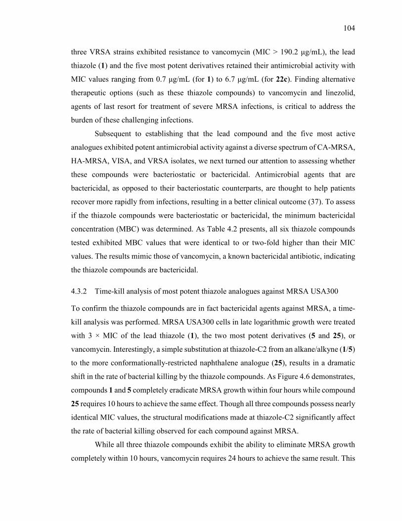
104
three VRSA strains exhibited resistance to vancomycin (MIC > 190.2 μg/mL), the lead
thiazole (1) and the five most potent derivatives retained their antimicrobial activity with
MIC values ranging from 0.7 μg/mL (for 1) to 6.7 μg/mL (for 22c). Finding alternative
therapeutic options (such as these thiazole compounds) to vancomycin and linezolid,
agents of last resort for treatment of severe MRSA infections, is critical to address the
burden of these challenging infections.
Subsequent to establishing that the lead compound and the five most active
analogues exhibited potent antimicrobial activity against a diverse spectrum of CA-MRSA,
HA-MRSA, VISA, and VRSA isolates, we next turned our attention to assessing whether
these compounds were bacteriostatic or bactericidal. Antimicrobial agents that are
bactericidal, as opposed to their bacteriostatic counterparts, are thought to help patients
recover more rapidly from infections, resulting in a better clinical outcome (37). To assess
if the thiazole compounds were bacteriostatic or bactericidal, the minimum bactericidal
concentration (MBC) was determined. As Table 4.2 presents, all six thiazole compounds
tested exhibited MBC values that were identical to or two-fold higher than their MIC
values. The results mimic those of vancomycin, a known bactericidal antibiotic, indicating
the thiazole compounds are bactericidal.
4.3.2 Time-kill analysis of most potent thiazole analogues against MRSA USA300
To confirm the thiazole compounds are in fact bactericidal agents against MRSA, a time-
kill analysis was performed. MRSA USA300 cells in late logarithmic growth were treated
with 3 × MIC of the lead thiazole (1), the two most potent derivatives (5 and 25), or
vancomycin. Interestingly, a simple substitution at thiazole-C2 from an alkane/alkyne (1/5)
to the more conformationally-restricted naphthalene analogue (25), results in a dramatic
shift in the rate of bacterial killing by the thiazole compounds. As Figure 4.6 demonstrates,
compounds 1 and 5 completely eradicate MRSA growth within four hours while compound
25 requires 10 hours to achieve the same effect. Though all three compounds possess nearly
identical MIC values, the structural modifications made at thiazole-C2 significantly affect
the rate of bacterial killing observed for each compound against MRSA.
While all three thiazole compounds exhibit the ability to eliminate MRSA growth
completely within 10 hours, vancomycin requires 24 hours to achieve the same result. This
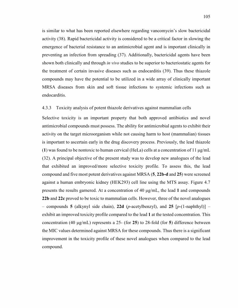
105
is similar to what has been reported elsewhere regarding vancomycin’s slow bactericidal
activity (38). Rapid bactericidal activity is considered to be a critical factor in slowing the
emergence of bacterial resistance to an antimicrobial agent and is important clinically in
preventing an infection from spreading (37). Additionally, bactericidal agents have been
shown both clinically and through in vivo studies to be superior to bacteriostatic agents for
the treatment of certain invasive diseases such as endocarditis (39). Thus these thiazole
compounds may have the potential to be utilized in a wide array of clinically important
MRSA diseases from skin and soft tissue infections to systemic infections such as
endocarditis.
4.3.3 Toxicity analysis of potent thiazole derivatives against mammalian cells
Selective toxicity is an important property that both approved antibiotics and novel
antimicrobial compounds must possess. The ability for antimicrobial agents to exhibit their
activity on the target microorganism while not causing harm to host (mammalian) tissues
is important to ascertain early in the drug discovery process. Previously, the lead thiazole
(1) was found to be nontoxic to human cervical (HeLa) cells at a concentration of 11 μg/mL
(32). A principal objective of the present study was to develop new analogues of the lead
that exhibited an improved/more selective toxicity profile. To assess this, the lead
compound and five most potent derivatives against MRSA (5, 22b-d and 25) were screened
against a human embryonic kidney (HEK293) cell line using the MTS assay. Figure 4.7
presents the results garnered. At a concentration of 40 μg/mL, the lead 1 and compounds
22b and 22c proved to be toxic to mammalian cells. However, three of the novel analogues
– compounds 5 (alkynyl side chain), 22d (p-acetylbenzyl), and 25 [p-(1-naphthyl)] –
exhibit an improved toxicity profile compared to the lead 1 at the tested concentration. This
concentration (40 μg/mL) represents a 25- (for 25) to 28-fold (for 5) difference between
the MIC values determined against MRSA for these compounds. Thus there is a significant
improvement in the toxicity profile of these novel analogues when compared to the lead
compound.
106
4.3.4 Metabolic stability analysis of compound 5
Previously, microsomal stability analysis of the lead 1 revealed this compound was
metabolized fairly rapidly (intrinsic clearance rate of 80.3 µL/min-mg and half-life of 28.8
minutes) via a NADPH-mediated process (such as via the cytochrome P450 system) (32).
As compound 5 demonstrated nearly identical antimicrobial activity to the lead compound,
we were curious to assess if the substitution of an alkane side chain with an alkyne at
thiazole-C2 would enhance the metabolic stability of the compound, preventing its
conversion to potentially inactive metabolites. Using pooled human liver microsomes, 5,
similar to the parent compound, was found to be metabolized via a NADPH-mediated
process (intrinsic clearance rate of 3.7 µL/min-mg as compared to 0.0 µL/min-mg in the
absence of the cofactor, NADPH) (Table 4.3). Interestingly, the slower clearance rate
correlates with an improved half-life for compound 5 (as compared to the lead compound)
that exceeds four hours. This marked improvement in the metabolic stability of the thiazole
compound is important as it has the potential to positively impact the pharmacokinetic
profile of this compound, reduce the frequency of doses needed to be administered for
treatment (fewer doses leads to improved patient compliance), while also ensuring the
active drug circulates within the patient’s system to assist with treating and clearing an
infection. Additionally, compounds that are metabolically stable are less susceptible to
experiencing issues pertaining to toxicity and drug-drug interactions caused by metabolites
(40). The metabolic stability analysis combined with the enhanced toxicity profile of
compound 5 (as compared to the lead) warrants further analysis of this compound as a
potential novel antibiotic for the treatment of MRSA infections.
4.4 Conclusion
We present herein a novel series of 2,5-disubstituted thiazole compounds exhibiting potent
activity against clinically relevant isolates of MRSA, VISA, and VRSA. A rigorous
analysis of the structure-activity relationship of these analogues reveals the
ethylidenehydrazine-1-carboximidamide head group (at thiazole-C5) and a nonpolar,
hydrophobic moiety (at thiazole-C2) are critical for the thiazole compound’s antibacterial
action. Three derivatives with substitutions at thiazole-C2 (an alkyne, p-acetylbenzene, and
107
p-naphthalene) demonstrate an improved toxicity profile against mammalian cells
compared to the lead compound. Furthermore, the alkyne substitution results in a
compound that is more stable to metabolism as assessed via human liver microsomes.
Collectively, the results present critical information necessary for further analysis and
development of these thiazole compounds as novel antimicrobial agents for use in
treatment of infections caused by multidrug-resistant S. aureus.
108
4.5 References
1. Hadler JL, Petit S, Mandour M, Cartter ML. 2012. Trends in invasive infection
with methicillin-resistant Staphylococcus aureus, Connecticut, USA, 2001-2010.
Emerg Infect Dis 18:917-924.
2. Wyllie D, Paul J, Crook D. 2011. Waves of trouble: MRSA strain dynamics and
assessment of the impact of infection control. J Antimicrob Chemother 66:2685-
2688.
3. McDougal LK, Steward CD, Killgore GE, Chaitram JM, McAllister SK,
Tenover FC. 2003. Pulsed-field gel electrophoresis typing of oxacillin-resistant
Staphylococcus aureus isolates from the United States: Establishing a national
database. Journal of Clinical Microbiology 41:5113-5120.
4. Kassis C, Hachem R, Raad II, Perego CA, Dvorak T, Hulten KG, Frenzel E,
Thomas G, Chemaly RF. 2011. Outbreak of community-acquired methicillin-
resistant Staphylococcus aureus skin infections among health care workers in a
cancer center. American Journal of Infection Control 39:112-117.
5. Malcolm B. 2011. The rise of methicillin-resistant staphylococcus aureus in U.S.
correctional populations. J Correct Health Care 17:254-265.
6. Aiello AE, D Lowy F, Wright LN, Larson EL. 2006. Meticillin-resistant
Staphylococcus aureus among US prisoners and military personnel: review and
recommendations for future studies. Lancet Infectious Diseases 6:335-341.
7. Cohen PR. 2005. Cutaneous community-acquired methicillin-resistant
Staphylococcus aureus infection in participants of athletic activities. Southern
Medical Journal 98:596-602.
8. Kazakova SV, Hageman JC, Matava M, Srinivasan A, Phelan L, Garfinkel B,
Boo T, McAllister S, Anderson J, Jensen B, Dodson D, Lonsway D, McDougal
LK, Arduino M, Fraser VJ, Killgore G, Tenover FC, Cody S, Jernigan DB.
2005. A clone of methicillin-resistant Staphylococcus aureus among professional
football players. New England Journal of Medicine 352:468-475.
109
9. Gilbert M, MacDonald J, Gregson D, Siushansian J, Zhang K, Elsayed S,
Laupland K, Louie T, Hope K, Mulvey M, Gillespie J, Nielsen D, Wheeler V,
Louie M, Honish A, Keays G, Conly J. 2006. Outbreak in Alberta of community-
acquired (USA300) methicillin-resistant Staphylococcus aureus in people with a
history of drug use, homelessness or incarceration. Canadian Medical Association
Journal 175:149-154.
10. Craven DE, Rixinger AI, Goularte TA, Mccabe WR. 1986. Methicillin-
Resistant Staphylococcus-Aureus Bacteremia Linked to Intravenous Drug-Abusers
Using a Shooting Gallery. American Journal of Medicine 80:770-776.
11. Anonymous. 2006. Methicillin-resistant Staphylococcus aureus skin infections
among tattoo recipients--Ohio, Kentucky, and Vermont, 2004-2005. MMWR Morb
Mortal Wkly Rep 55:677-679.
12. Regev-Yochay G, Rubinstein E, Barzilai A, Carmeli Y, Kuint J, Etienne J,
Blech M, Smollen G, Maayan-Metzger A, Leavitt A, Rahav G, Keller N. 2005.
Methicillin-resistant Staphylococcus aureus in neonatal intensive care unit. Emerg
Infect Dis 11:453-456.
13. Herold BC, Immergluck LC, Maranan MC, Lauderdale DS, Gaskin RE,
Boyle-Vavra S, Leitch CD, Daum RS. 1998. Community-acquired methicillin-
resistant Staphylococcus aureus in children with no identified predisposing risk.
JAMA 279:593-598.
14. Karamatsu ML, Thorp AW, Brown L. 2012. Changes in community-associated
methicillin-resistant Staphylococcus aureus skin and soft tissue infections
presenting to the pediatric emergency department: comparing 2003 to 2008. Pediatr
Emerg Care 28:131-135.
15. Tristan A, Ferry T, Durand G, Dauwalder O, Bes M, Lina G, Vandenesch F,
Etienne J. 2007. Virulence determinants in community and hospital meticillin-
resistant Staphylococcus aureus. Journal of Hospital Infection 65:105-109.
16. Odell CA. 2010. Community-associated methicillin-resistant Staphylococcus
aureus (CA-MRSA) skin infections. Curr Opin Pediatr 22:273-277.
110
17. Stryjewski ME, Chambers HF. 2008. Skin and soft-tissue infections caused by
community-acquired methicillin-resistant Staphylococcus aureus. Clinical
Infectious Diseases 46 Suppl 5:S368-377.
18. Gillet Y, Issartel B, Vanhems P, Fournet JC, Lina G, Bes M, Vandenesch F,
Piemont Y, Brousse N, Floret D, Etienne J. 2002. Association between
Staphylococcus aureus strains carrying gene for Panton-Valentine leukocidin and
highly lethal necrotising pneumonia in young immunocompetent patients. Lancet
359:753-759.
19. Bocchini CE, Hulten KG, Mason EO, Gonzalez BE, Hammerman WA, Kaplan
SL. 2006. Panton-Valentine leukocidin genes are associated with enhanced
inflammatory response and local disease in acute hematogenous Staphylococcus
aureus osteomyelitis in children. Pediatrics 117:433-440.
20. David MZ, Daum RS. 2010. Community-associated methicillin-resistant
Staphylococcus aureus: epidemiology and clinical consequences of an emerging
epidemic. Clin Microbiol Rev 23:616-687.
21. King MD, Waldron DR, Barber DL, Larson MM, Saunders GK, Troy GC,
Zimmerman-Pope N, Ward DL. 2006. Effect of nephrotomy on renal function
and morphology in normal cats. Vet Surg 35:749-758.
22. Lee BY, Singh A, David MZ, Bartsch SM, Slayton RB, Huang SS, Zimmer
SM, Potter MA, Macal CM, Lauderdale DS, Miller LG, Daum RS. 2013. The
economic burden of community-associated methicillin-resistant Staphylococcus
aureus (CA-MRSA). Clin Microbiol Infect 19:528-536.
23. Chambers HF. 2005. Community-associated MRSA--resistance and virulence
converge. N Engl J Med 352:1485-1487.
24. Moran GJ, Krishnadasan A, Gorwitz RJ, Fosheim GE, McDougal LK, Carey
RB, Talan DA. 2006. Methicillin-resistant S-aureus infections among patients in
the emergency department. New England Journal of Medicine 355:666-674.
25. Frazee BW, Lynn J, Charlebois ED, Lambert L, Lowery D, Perdreau-
Remington F. 2005. High prevalence of methicillin-resistant Staphylococcus
aureus in emergency department skin and soft tissue infections. Ann Emerg Med
45:311-320.
111
26. Fridkin SK, Hageman JC, Morrison M, Sanza LT, Como-Sabetti K, Jernigan
JA, Harriman K, Harrison LH, Lynfield R, Farley MM. 2005. Methicillin-
resistant Staphylococcus aureus disease in three communities. N Engl J Med
352:1436-1444.
27. Han LL, McDougal LK, Gorwitz RJ, Mayer KH, Patel JB, Sennott JM,
Fontana JL. 2007. High frequencies of clindamycin and tetracycline resistance in
methicillin-resistant Staphylococcus aureus pulsed-field type USA300 isolates
collected at a Boston ambulatory health center. Journal of Clinical Microbiology
45:1350-1352.
28. Patel JB, Gorwitz RJ, Jernigan JA. 2009. Mupirocin Resistance. Clinical
Infectious Diseases 49:935-941.
29. Locke JB, Morales G, Hilgers M, G CK, Rahawi S, Jose Picazo J, Shaw KJ,
Stein JL. 2010. Elevated linezolid resistance in clinical cfr-positive
Staphylococcus aureus isolates is associated with co-occurring mutations in
ribosomal protein L3. Antimicrob Agents Chemother 54:5352-5355.
30. Wilson P, Andrews JA, Charlesworth R, Walesby R, Singer M, Farrell DJ,
Robbins M. 2003. Linezolid resistance in clinical isolates of Staphylococcus
aureus. J Antimicrob Chemother 51:186-188.
31. Hiramatsu K. 2001. Vancomycin-resistant Staphylococcus aureus: a new model
of antibiotic resistance. Lancet Infectious Diseases 1:147-155.
32. Mohammad H, Mayhoub AS, Ghafoor A, Soofi M, Alajlouni RA, Cushman
M, Seleem MN. 2014. Discovery and Characterization of Potent Thiazoles versus
Methicillin- and Vancomycin-Resistant Staphylococcus aureus. Journal of
Medicinal Chemistry 57:1609-1615.
33. Mayhoub AS, Khaliq M, Botting C, Li Z, Kuhn RJ, Cushman M. 2011. An
investigation of phenylthiazole antiflaviviral agents. Bioorg Med Chem 19:3845-
3854.
34. Mayhoub AS, Khaliq M, Kuhn RJ, Cushman M. 2011. Design, synthesis, and
biological evaluation of thiazoles targeting flavivirus envelope proteins. J Med
Chem 54:1704-1714.
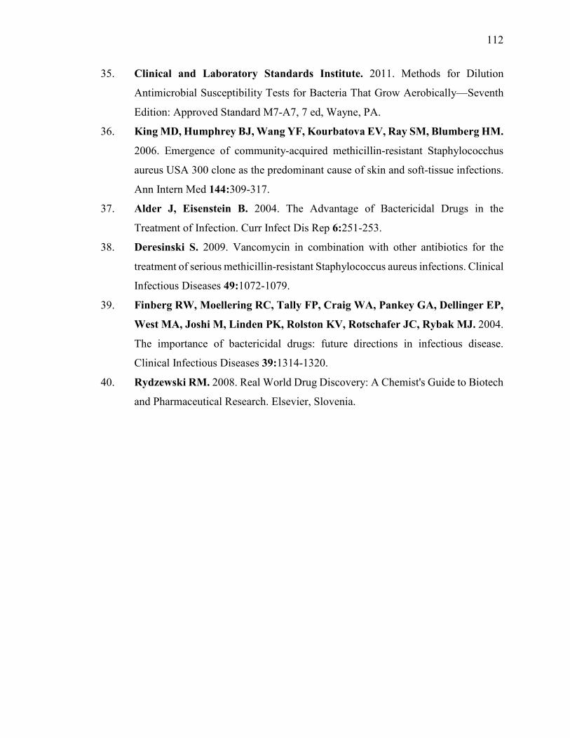
112
35. Clinical and Laboratory Standards Institute. 2011. Methods for Dilution
Antimicrobial Susceptibility Tests for Bacteria That Grow Aerobically—Seventh
Edition: Approved Standard M7-A7, 7 ed, Wayne, PA.
36. King MD, Humphrey BJ, Wang YF, Kourbatova EV, Ray SM, Blumberg HM.
2006. Emergence of community-acquired methicillin-resistant Staphylococchus
aureus USA 300 clone as the predominant cause of skin and soft-tissue infections.
Ann Intern Med 144:309-317.
37. Alder J, Eisenstein B. 2004. The Advantage of Bactericidal Drugs in the
Treatment of Infection. Curr Infect Dis Rep 6:251-253.
38. Deresinski S. 2009. Vancomycin in combination with other antibiotics for the
treatment of serious methicillin-resistant Staphylococcus aureus infections. Clinical
Infectious Diseases 49:1072-1079.
39. Finberg RW, Moellering RC, Tally FP, Craig WA, Pankey GA, Dellinger EP,
West MA, Joshi M, Linden PK, Rolston KV, Rotschafer JC, Rybak MJ. 2004.
The importance of bactericidal drugs: future directions in infectious disease.
Clinical Infectious Diseases 39:1314-1320.
40. Rydzewski RM. 2008. Real World Drug Discovery: A Chemist's Guide to Biotech
and Pharmaceutical Research. Elsevier, Slovenia.
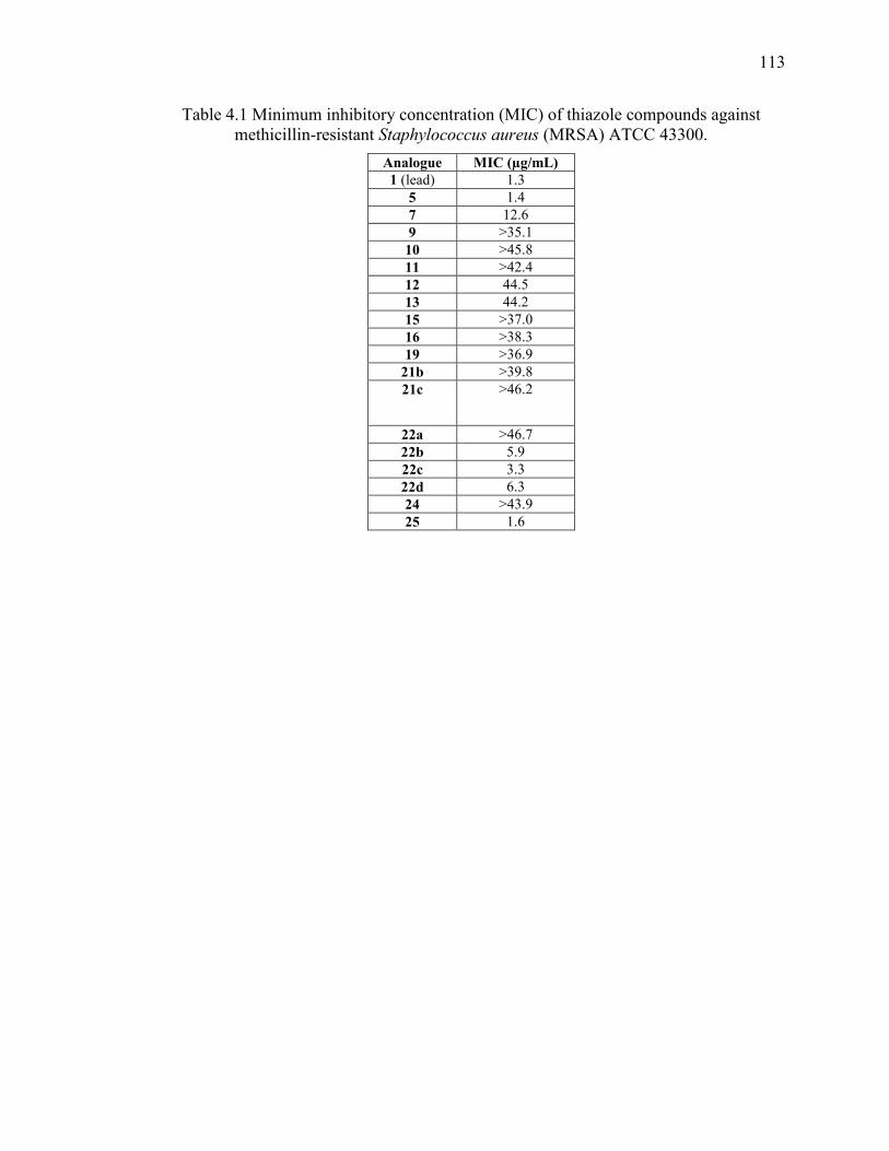
113
Table 4.1 Minimum inhibitory concentration (MIC) of thiazole compounds against methicillin-resistant Staphylococcus aureus (MRSA) ATCC 43300.
Analogue MIC (µg/mL) 1 (lead) 1.3
5 1.4
7 12.6
9 >35.1
10 >45.8
11 >42.4
12 44.5
13 44.2
15 >37.0
16 >38.3
19 >36.9
21b >39.8
21c >46.2
22a >46.7
22b 5.9
22c 3.3
22d 6.3
24 >43.9
25 1.6
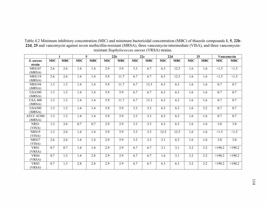
Pa
ge9
2
114
Table 4.2 Minimum inhibitory concentration (MIC) and minimum bactericidal concentration (MBC) of thiazole compounds 1, 5, 22b-22d, 25 and vancomycin against seven methicillin-resistant (MRSA), three vancomycin-intermediate (VISA), and three vancomycin-
resistant Staphylococcus aureus (VRSA) strains.
1 5 22b 22c 22d 25 Vancomycin S. aureus
strain MIC MBC MIC MBC MIC MBC MIC MBC MIC MBC MIC MBC MIC MBC
NRS107 (MRSA)
2.6 2.6 1.4 1.4 2.9 5.9 3.3 6.7 6.3 12.5 1.6 1.6 <1.5 <1.5
NRS119 (MRSA)
2.6 2.6 1.4 1.4 5.9 11.7 6.7 6.7 6.3 12.5 1.6 1.6 <1.5 <1.5
NRS194 (MRSA)
1.3 1.3 1.4 1.4 5.9 11.7 6.7 13.3 6.3 6.3 1.6 1.6 0.7 0.7
USA300 (MRSA)
1.3 1.3 1.4 1.4 5.9 5.9 6.7 6.7 6.3 6.3 1.6 1.6 0.7 0.7
USA 400 (MRSA)
1.3 1.3 1.4 1.4 5.9 11.7 6.7 13.3 6.3 6.3 1.6 1.6 0.7 0.7
USA500 (MRSA)
1.3 1.3 1.4 1.4 5.9 5.9 3.3 3.3 6.3 6.3 1.6 3.2 0.7 0.7
ATCC 43300 (MRSA)
1.3 1.3 1.4 1.4 5.9 5.9 3.3 3.3 6.3 6.3 1.6 1.6 0.7 0.7
NRS1 (VISA)
1.3 2.6 0.7 0.7 2.9 2.9 3.3 3.3 6.3 6.3 1.6 1.6 3.0 3.0
NRS19 (VISA)
1.3 2.6 1.4 1.4 5.9 5.9 3.3 3.3 12.5 12.5 1.6 1.6 <1.5 <1.5
NRS37 (VISA)
2.6 2.6 1.4 1.4 2.9 5.9 3.3 3.3 3.1 6.3 1.6 1.6 3.0 3.0
VRS1 (VRSA)
0.7 0.7 1.4 1.4 2.9 2.9 6.7 6.7 3.1 3.1 3.2 3.2 >190.2 >190.2
VRS4 (VRSA)
0.7 1.3 1.4 2.8 2.9 2.9 6.7 6.7 1.6 3.1 3.2 3.2 >190.2 >190.2
VRS5 (VRSA)
0.7 1.3 2.8 2.8 2.9 2.9 6.7 6.7 6.3 6.3 3.2 3.2 >190.2 >190.2
115
Table 4.3 Evaluation of metabolic stability of thiazole compound 5, Verapamil, and Warfarin in human liver microsomes.
Compound/Drug Tested
NADPH-dependent CLint
1
(µL/min/mg)
NADPH-dependent T1/2
2 (min)
NADPH-free CLint
(µL/min/mg)
NADPH-free T1/2
(min) 5 3.7 >240 0.0 >240
Verapamil 213 10.8 0.0 >240
Warfarin 0.0 >240 0.0 >240 1 CLint= microsomal intrinsic clearance 2 T1/2 = half-life
Figure 4.1 Chemical structure of the lead compound 1.
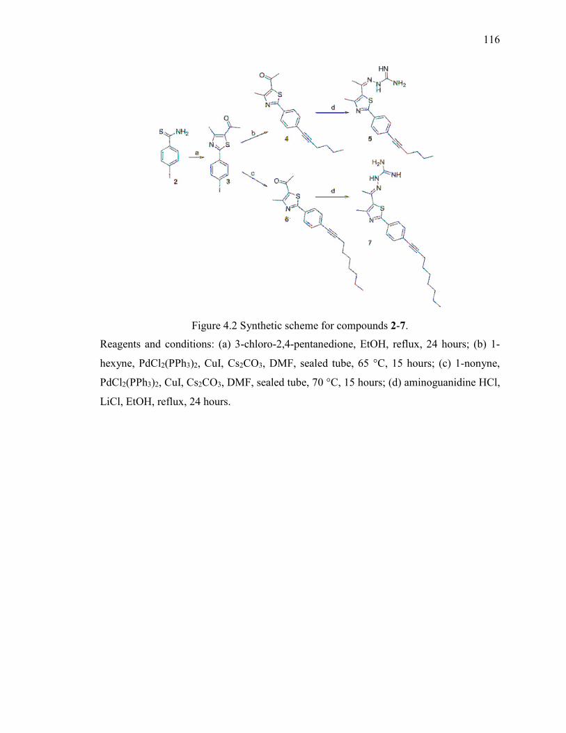
116
Figure 4.2 Synthetic scheme for compounds 2-7.
Reagents and conditions: (a) 3-chloro-2,4-pentanedione, EtOH, reflux, 24 hours; (b) 1-
hexyne, PdCl2(PPh3)2, CuI, Cs2CO3, DMF, sealed tube, 65 °C, 15 hours; (c) 1-nonyne,
PdCl2(PPh3)2, CuI, Cs2CO3, DMF, sealed tube, 70 °C, 15 hours; (d) aminoguanidine HCl,
LiCl, EtOH, reflux, 24 hours.
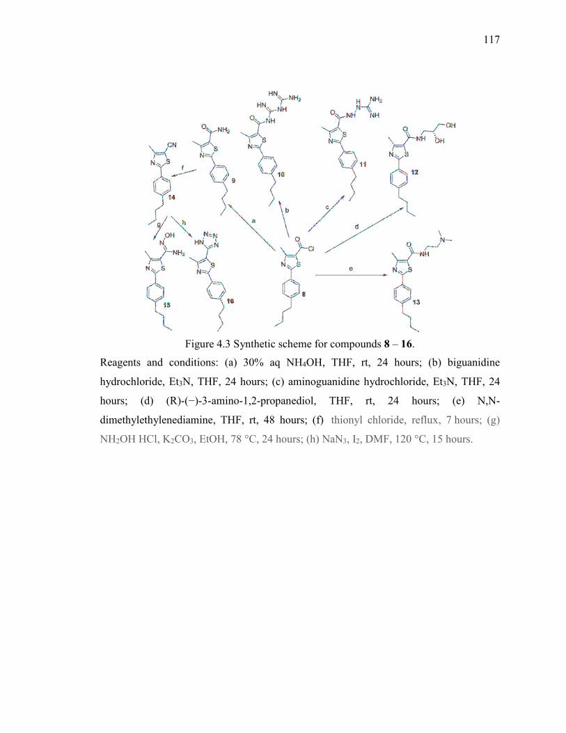
117
Figure 4.3 Synthetic scheme for compounds 8 – 16.
Reagents and conditions: (a) 30% aq NH4OH, THF, rt, 24 hours; (b) biguanidine
hydrochloride, Et3N, THF, 24 hours; (c) aminoguanidine hydrochloride, Et3N, THF, 24
hours; (d) (R)-(−)-3-amino-1,2-propanediol, THF, rt, 24 hours; (e) N,N-
dimethylethylenediamine, THF, rt, 48 hours; (f) thionyl chloride, reflux, 7 hours; (g)
NH2OH HCl, K2CO3, EtOH, 78 °C, 24 hours; (h) NaN3, I2, DMF, 120 °C, 15 hours.
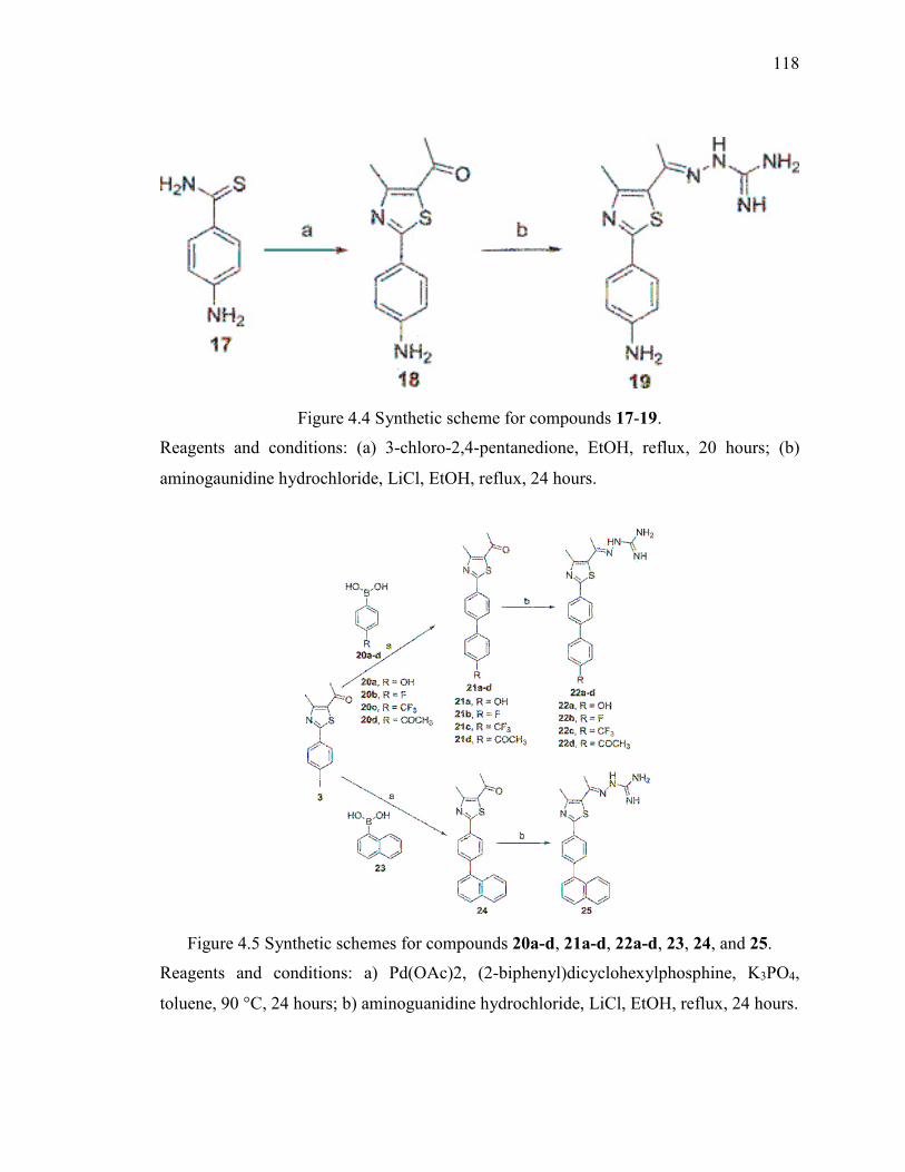
118
Figure 4.4 Synthetic scheme for compounds 17-19.
Reagents and conditions: (a) 3-chloro-2,4-pentanedione, EtOH, reflux, 20 hours; (b)
aminogaunidine hydrochloride, LiCl, EtOH, reflux, 24 hours.
Figure 4.5 Synthetic schemes for compounds 20a-d, 21a-d, 22a-d, 23, 24, and 25.
Reagents and conditions: a) Pd(OAc)2, (2-biphenyl)dicyclohexylphosphine, K3PO4,
toluene, 90 °C, 24 hours; b) aminoguanidine hydrochloride, LiCl, EtOH, reflux, 24 hours.
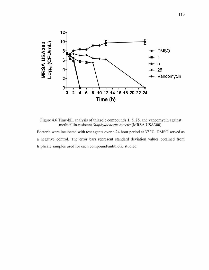
119
Figure 4.6 Time-kill analysis of thiazole compounds 1, 5, 25, and vancomycin against methicillin-resistant Staphylococcus aureus (MRSA USA300).
Bacteria were incubated with test agents over a 24 hour period at 37 °C. DMSO served as
a negative control. The error bars represent standard deviation values obtained from
triplicate samples used for each compound/antibiotic studied.
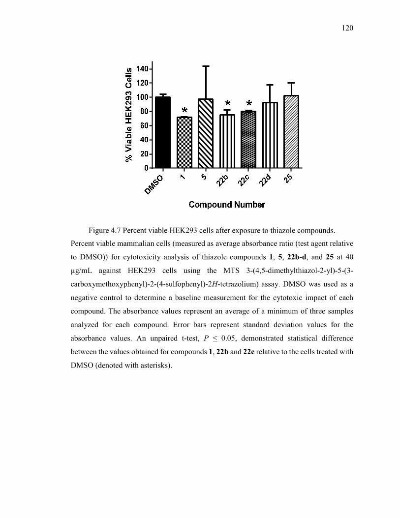
120
Figure 4.7 Percent viable HEK293 cells after exposure to thiazole compounds.
Percent viable mammalian cells (measured as average absorbance ratio (test agent relative
to DMSO)) for cytotoxicity analysis of thiazole compounds 1, 5, 22b-d, and 25 at 40
µg/mL against HEK293 cells using the MTS 3-(4,5-dimethylthiazol-2-yl)-5-(3-
carboxymethoxyphenyl)-2-(4-sulfophenyl)-2H-tetrazolium) assay. DMSO was used as a
negative control to determine a baseline measurement for the cytotoxic impact of each
compound. The absorbance values represent an average of a minimum of three samples
analyzed for each compound. Error bars represent standard deviation values for the
absorbance values. An unpaired t-test, P ≤ 0.05, demonstrated statistical difference
between the values obtained for compounds 1, 22b and 22c relative to the cells treated with
DMSO (denoted with asterisks).
121
CHAPTER 5. ANTIBACTERIAL EVALUATION OF SYNTHETIC THIAZOLE COMPOUNDS IN VITRO AND IN
VIVO IN A METHICILLIN-RESISTANT STAPHYLOCOCCUS
AUREUS (MRSA) SKIN INFECTION MOUSE MODEL
THIS IS A PUBLISHED JOURNAL ARTICLE. Reprinted with permission from
Antibacterial Evaluation of Synthetic Thiazole Compounds in vitro and in vivo in a
Methicillin-resistant Staphylococcus aureus (MRSA) Skin Infection Mouse Model.
Haroon Mohammad, Mark Cushman, and Mohamed N. Seleem. PLoS
ONE (2015) 10, e0142321; doi: 10.1371/journal.pone.0142321 Copyright 2015 PLoS
ONE
5.1 Introduction
Ten percent of all hospital admissions in the United States each year are due to patients
suffering from skin and soft tissue infections (SSTIs) (1). SSTIs can range from simple
abscesses, cellulitis, and traumatic wound infections to complicated infections (infected
burns, diabetic foot ulcers, and major abscesses) (1). SSTIs are often caused by the bacterial
pathogen methicillin-resistant Staphylococcus aureus (MRSA) (2, 3). Indeed, 58% of all
SSTIs treated in the United States alone were caused by MRSA, according to an
epidemiological study of one national health care system (4). This agrees with a 2004 study
conducted in emergency room departments in 11 cities where MRSA was responsible for
59% of patients presenting with a SSTI (5). The large number of S. aureus-based SSTIs
has placed a significant economic burden on the healthcare system. A recent report
examining the increase in S. aureus-SSTI hospitalizations in the United States documented
a dramatic rise in the annual cost of treating infected patients from $3.36 billion to $4.50
billion (from the years 2001 through 2009) (6). A recent increase in skin abscesses has
been observed and has been associated with a rise in strains of community-associated
MRSA (CA-MRSA) (7). Of these strains, MRSA USA300 has been linked most frequently
to skin infections in the United States (5, 8).
According to guidelines provided by the Infectious Diseases Society of America,
treatment of moderate to severe skin infections caused by MRSA involves incision and
122
drainage of the affected region combined with administration of empirical antibiotics (such
as clindamycin, vancomycin, linezolid, and mupirocin) (9, 10). However, strains of MRSA
exhibiting resistance to several of these antibiotics including vancomycin (11, 12),
clindamycin (13, 14), and topical ointments like mupirocin (14-16), indicate that such
therapies may be rendered ineffective in the future. Therefore, development of novel
antimicrobials capable of treating MRSA-induced SSTIs is an important step necessary to
circumvent the burden of this public health issue.
Previous research by our group has identified a lead disubstituted phenylthiazole
compound (compound 1, Figure 5.1) that exhibited potent antimicrobial activity in vitro
against a diverse array of clinically-significant strains of MRSA (17). Derivatives of the
lead 1 were synthesized to elucidate the structure-activity relationships of this compound.
These derivatives revealed that the aminoguanidine moiety at thiazole-C5 is critical for
antibacterial activity (18). Furthermore, a nonpolar, hydrophobic group is favored at
thiazole-C2. Analogues to the lipophilic alkyl tail of the lead 1 were subsequently
constructed in order to enhance the antimicrobial activity of these thiazole compounds, to
improve their toxicity profile, and to refine their physicochemical properties (18). These
particular phenylthiazole compounds possess several excellent characteristics in vitro
including rapid bactericidal activity against MRSA, a low frequency of bacterial resistance
developing, and they have been shown to possess the ability to be used in combination with
currently approved antibiotics, such as vancomycin, against MRSA (19). Studies
performed to date indicate these compounds have potential to be used as topical
antimicrobials for treatment of MRSA skin infections. The objectives of the current study
were to assess the antibacterial activity of the lead thiazole compound and four analogues
(Fig. 1) in vitro against antibiotic-resistant S. aureus strains isolated from/responsible for
skin infections, to assess the ability of these compounds to be paired with mupirocin as a
treatment option against MRSA, to confirm the compounds have limited toxicity to human
keratinocytes, and to verify the thiazole compounds can retain their antimicrobial activity
in vivo in an established murine MRSA skin infection model. Confirmation of the ability
of these compounds to successfully treat mice infected with a MRSA skin infection will
lay the foundation for further assessment of these compounds as novel antimicrobials for
treatment of MRSA skin infections.
123
5.2 Materials and Methods
5.2.1 Synthesis of thiazole compounds 1-5
Synthetic schemes, spectral data, and purity (>95%, determined by HPLC) of thiazole
compounds 1-5 (Fig. 1), in addition to all intermediates, have been reported elsewhere (17,
18, 20).
5.2.2 Bacterial strains and reagents used in this study
Clinical isolates of S. aureus were obtained through the Network of Antimicrobial
Resistance in Staphylococcus aureus (NARSA) program (Table 5.1). Clindamycin
hydrochloride monohydrate (TCI America, Portland, OR, USA, >98.0% purity) and
mupirocin (pure USP) (AppliChem, St. Louis, MO, USA) powders were purchased
commercially and dissolved in dimethyl sulfoxide (DMSO) (for clindamycin) or ethanol
(for mupirocin) to prepare a stock solution (10 μg/mL). Lipoderm was purchased from the
Professional Compounding Centers of America (Houston, TX, USA). Cation-adjusted
Mueller-Hinton broth (CAMHB) (Sigma-Aldrich, St. Louis, MO, USA), tryptic soy broth
(TSB) (Becton, Dickinson and Company, Franklin Lakes, NJ, USA), mannitol salt agar
(Hardy Diagnostics, Santa Maria, CA, USA), phosphate-buffered saline (PBS) (Sigma-
Aldrich, St. Louis, MO, USA), Dulbeco’s modified Eagle’s medium (DMEM) (Sigma-
Aldrich, St. Louis, MO, USA), fetal bovine serum (FBS) (USA Scientific, Inc., Orlando,
FL, USA), petroleum jelly (Equate [Walmart, Inc.], Bentonville, AR, USA), and 96-well
plates (CellTreat Scientific Products, Shirley, MA, USA) were all purchased from
commercial vendors.
5.2.3 Determination of minimum inhibitory concentration (MIC) against drug-resistant S. aureus strains
The MIC of thiazole compounds 1-5, clindamycin, and mupirocin was determined against
five different MRSA strains and one highly mupirocin-resistant S. aureus strain isolated
from skin wounds, using the broth microdilution method, following the guidelines outlined
by the Clinical and Laboratory Standards Institute (21). A bacterial suspension equivalent
to a McFarland standard of 0.5 was prepared and subsequently diluted 1:300 in CAMHB.
This bacterial suspension (~1 × 10⁵ colony forming unit (CFU/mL)) was then added to
124
each well of a 96-well microtiter plate. Compounds 1-5, clindamycin, or mupirocin were
added (in triplicate) to the first row of the plate and then serially diluted down the ordinate.
Plates were incubated at 37 °C for 18-20 hours and then the MIC was ascertained. The
MIC was classified as the lowest concentration of each test agent where bacterial growth
could not be visualized.
5.2.4 Assessment of synergistic relationship between thiazole compounds and mupirocin against MRSA
The checkerboard assay was utilized to asses if the most potent thiazole compounds (1-3)
have potential to be combined with mupirocin for treatment of MRSA infections (22).
Briefly, a bacterial suspension (1 × 10⁵ CFU/mL) in CAMHB was added to each well of a
96-well microtiter plate. Compounds 1-3 and mupirocin were diluted in CAMHB in order
to reach the desired starting concentration (2 × or 4 × MIC). Mupirocin was serially diluted
along the horizontal axis of the plate while compound 1, 2, or 3 was diluted along the
vertical axis. Plates were incubated for at least 18 hours at 37 °C and the MIC of each
compound was recorded. The fractional inhibitory concentration index (ƩFIC) was
computed for each combination using the following equation:
ƩFIC =(�������� �������� �� ���������� ���� ����,����
�������� �������� ��� ) +
(�������,���� �� ���������� ���� ����� ��������
�������,���� ��� )
A FIC index less than or equal to 0.50 was classified as synergism, as described previously
(19). FIC values above 0.50 but less than 4.00 were classified as indifference, while FIC
values greater than 4.00 were indicative of antagonism.
5.2.5 In vitro cytotoxicity analysis of thiazole compounds against HaCaT cells
Compounds 1-5 were assayed (at concentrations of 5 µg/mL, 10 µg/mL, 20 µg/mL, and 40
µg/mL) against a human keratinocyte (HaCaT) cell line (Catalogue Number: T0020001,
AddexBio, San Diego, CA, USA) to determine the potential toxic effect to mammalian
skin cells in vitro as described before (18). Briefly, cells were cultured in DMEM
supplemented with 10% FBS at 37 °C with CO2 (5%). Control cells received DMSO alone
at a concentration equal to that in drug-treated cell samples. The cells were incubated with
125
the compounds (in triplicate) in a 96-well plate at 37 ºC with CO2 (5%) for two hours prior
to addition of the assay reagent MTS 3-(4,5-dimethylthiazol-2-yl)-5-(3-
carboxymethoxyphenyl)-2-(4-sulfophenyl)-2H-tetrazolium) (Promega, Madison, WI,
USA). Absorbance readings (at OD490) were taken using a kinetic microplate reader
(Molecular Devices, Sunnyvale, CA, USA). The quantity of viable cells after treatment
with each compound was expressed as a percentage of the viability of DMSO-treated
control cells (average of triplicate wells ± standard deviation). The toxicity data was
analyzed via a one-way ANOVA, with post hoc Dunnet’s multiple comparisons test (P <
0.05), utilizing GraphPad Prism 6.0 (GraphPad Software, La Jolla, CA).
5.2.6 In vivo assessment of antimicrobial activity of thiazole compounds 1-5 and mupirocin in a MRSA skin infection mouse model
The MRSA murine skin infection study was reviewed, approved, and performed under the
guidelines of the Purdue University Animal Care and Use Committee (PACUC) (protocol
number: 1207000676) and carried out in strict accordance with the recommendations in
the Guide for the Care and Use of Laboratory Animals of the National Institutes of Health.
To initiate the formation of a skin wound, eight groups (n = 5) of eight-week old female
BALB/c mice (obtained from Harlan Laboratories, Indianapolis, IN, USA) were
disinfected with ethanol (70%) and shaved on the middle of the back (approximately a one-
inch by one-inch square region around the injection site) one day prior to infection, similar
to what has been described elsewhere (23, 24). To prepare the bacterial inoculum, an
aliquot of overnight culture of MRSA USA300 was transferred to fresh TSB and shaken at
37 ºC until an OD600 value of ~1.0 was achieved. The cells were centrifuged, washed once
with PBS, re-centrifuged, and then re-suspended in PBS. Mice then received an intradermal
injection (20 μL) containing ~2.76 × 10⁸ CFU/mL MRSA USA300. An open wound
formed at the site of injection, 48 hours post-infection. Topical treatment was initiated
subsequently with each group of mice receiving the following: compound 1-5 (2%, using
petroleum jelly as the vehicle), mupirocin (2%, using petroleum jelly as the vehicle),
compound 1 (2%, using Lipoderm as an alternative vehicle), and a control group receiving
the control vehicle (20 mg, petroleum jelly) alone. Each group of mice receiving a
particular treatment regimen was housed separately in a ventilated cage with appropriate
bedding, food, and water. Mice were checked twice daily during infection and treatment to
126
ensure no adverse reactions were observed. In the event a mouse was observed to become
severely ill, the subject was euthanized per the IRB protocol. Mice were treated twice daily
for three days. Mice were humanely euthanized via CO2 asphyxiation 24 hours after the
last dose was administered. The region around the skin wound was lightly swabbed with
ethanol (70%) and excised. The tissue was subsequently homogenized in TSB (1 mL). The
homogenized tissue was then serially diluted in PBS before plating onto mannitol salt agar
plates. The plates were incubated for 20-22 hours at 37 ºC before viable CFU were counted
and MRSA reduction in the skin wound post-treatment was determined for each group.
Data were analyzed using a one-way ANOVA, with post hoc Holm-Sidak’s multiple
comparisons test (P < 0.05), utilizing GraphPad Prism 6.0.
5.3 Results and Discussion
5.3.1 Antimicrobial activity of thiazole compounds 1-5 against MRSA strains isolated from skin wounds
Previous work has established thiazole compounds 1-5 possess potent antimicrobial
activity against MRSA (particularly isolates derived from healthcare-associated MRSA
cases). To confirm these compounds maintain their antibacterial activity against CA-
MRSA strains and MRSA isolates derived from patients presenting with infected wounds
(Table 5.1), the broth microdilution assay was utilized to determine the lowest
concentration each compound was able to inhibit the growth of these strains (denoted as
the minimum inhibitory concentration or MIC).
When tested against these important clinical isolates of drug-resistant S. aureus, the
thiazole compounds exhibited strong antimicrobial activity similar to (and in several cases
better than) mupirocin. As presented in Table 5.2, the lead thiazole 1 exhibits the most
potent activity with a MIC value of 1.3 µg/mL against all six drug-resistant staphylococcal
strains tested. The biphenyl and butyne analogues (2 and 3, respectively) possess MIC
values ranging from 2.8 to 5.6 µg/mL. All five thiazole compounds possess antimicrobial
activity against MRSA strains exhibiting resistance to an array of antibiotics including β-
lactams, fluoroquinolones (USA800), tetracycline (USA300), and erythromycin (USA300
and USA1000). This indicates cross-resistance between these antibiotics and the thiazole
compounds is unlikely to occur. Additionally, the compounds exhibit potent antimicrobial
127
activity against S. aureus NRS107 (MIC values range from 1.3 to 13.3 µg/mL), a strain
exhibiting a high-level of resistance to mupirocin (MIC of 1024.0 µg/mL). Furthermore,
compounds 1 and 2 (MIC of 1.3 and 2.8 µg/mL, respectively) are more active than
mupirocin (MIC of 4.0 µg/mL) against three additional MRSA strains (USA800, USA1000,
and USA1100). Clindamycin, when tested against four of the five MRSA strains, was
found to have a MIC of 0.1 µg/mL. This MIC value is similar to what has been reported
elsewhere for clindamycin (25). Collectively, the results confirm that the thiazole
compounds do possess potent antimicrobial activity against important CA-MRSA strains
and MRSA isolates responsible for infected wounds in patients.
5.3.2 Combination therapy using thiazole compounds with mupirocin against MRSA
The susceptibility analysis performed with the thiazole compounds indicated they have
potential to be used alone for the treatment of MRSA skin/wound infections. While the use
of a single agent to treat such infections is often used in the clinical setting, combination
therapy using two or more antibiotics is favorable for multiple reasons. Among these
reasons include that combination therapy has the potential to slow down the emergence of
resistant bacterial strains to antibiotics, to reduce potential negative side effects to patients
(by using lower concentrations/doses of each drug), and to alleviate the morbidity related
to bacterial infections (26, 27). Given that multiple topical treatments for skin infections
involve a combination of more than one antibiotic, such as Neosporin (consisting of
bacitracin, neomycin, and polymyxin B sulfate) and Polysporin ointment (consisting of
bacitracin, polymyxin B sulfate, and gramicidin) (28), the identification of compounds to
pair with known antibiotics has good potential to expand the available treatment options.
Mupirocin has been a key ally in the treatment of MRSA skin infections; however, isolates
exhibiting moderate to high-level of resistance to mupirocin (MIC ≥ 512 µg/mL) have
emerged, particularly in environments where this antibiotic has been extensively utilized
(15, 29, 30). Identifying agents that can be partnered with mupirocin has the potential to
extend the usage of this particular antimicrobial in the clinical setting.
In an earlier study, Alou et al, demonstrated that mupirocin forms a synergistic
relationship with amoxicillin-clavulanate against MRSA isolates tested in vitro via the
checkerboard assay (31). Amoxicllin is a β-lactam antibiotic that interferes with bacterial
128
cell wall synthesis by inhibiting crosslinking of peptidoglycan subunits in the bacterial cell
wall (32). Preliminary studies conducted with our thiazole compounds indicate they also
interfere with cell wall synthesis in bacteria; thus we were curious to assess if the thiazole
compounds could be used in combination with mupirocin against MRSA, similar to what
was found with amoxicillin-clavulanate. Using the checkerboard assay, it was discovered
that the most potent thiazole compounds (1-3) exhibited a strong degree of synergy (FIC
index ≤ 0.50) with mupirocin against two of the most prevalent MRSA strains responsible
for skin infections (Table 5.3). Against MRSA USA300, all three compounds exhibited a
fractional inhibitory concentration (FIC) index ranging from 0.09 to 0.13 when combined
with mupirocin. A similar trend was observed when this combination was tested against
MRSA USA400, with FIC values ranging from 0.05 to 0.13. The data provide evidence
that supports the prospect that these particular thiazole compounds can be successfully
paired with mupirocin to treat MRSA infections (and potentially prolong the utility of
mupirocin in the clinical setting).
5.3.3 Toxicity analysis of thiazole compounds to human keratinocytes
Selective toxicity is important to ensure compounds with promising antimicrobial activity
don’t possess negative side effects to mammalian tissues. Certain regimens (in particular
antiseptics) used for treatment of skin infections and wounds have been found to exhibit
toxicity to human keratinocytes and impair wound healing, thus limiting their use as
therapeutic options (33-36). Prior to validating the antimicrobial activity of the thiazole
compounds in a MRSA skin infection model, it was critical to confirm the thiazole
compounds were not toxic to human keratinocytes. Using the MTS assay with a human
keratinocyte (HaCaT) cell line, it was confirmed that thiazole compounds 1-5 were not
toxic at a concentration of 10 µg/mL (Figure 5.2). Interestingly, the four analogues
constructed from compound 1 demonstrated an improved toxicity profile, as they were
found to be non-toxic to HaCaT cells up to a concentration of 20 µg/mL. Taken altogether,
the data indicate the most potent thiazole compounds in vitro (1-3) are not toxic to human
keratinocytes at concentrations up to seven-fold higher than the compounds’ MIC values
determined against MRSA.
129
5.3.4 Assessment of topical application of thiazole compounds in vivo via a murine MRSA skin infection model
As thiazole compounds 1-5 exhibited excellent activity against MRSA in vitro and
displayed no toxicity to human keratinocytes at the compounds’ MIC, we moved to
confirm that these compounds could maintain their antimicrobial activity in vivo, using an
established MRSA murine skin infection model. After the formation of an open wound
(infected with MRSA) in the dorsal region of infected mice, each group of mice was treated
with a suspension of compounds 1-5 (2%), mupirocin (2% suspension), or petroleum jelly
(20 mg, used as a vehicle for topical delivery of the compounds/antibiotic) twice daily for
three days. The reduction in bacterial burden present in the wounds of infected mice was
determined after cessation of treatment. Reduction of bacterial burden in infected wounds
is critical to promote proper wound repair and to prevent a severe inflammatory response
from being triggered that may negatively impact healing of wounded tissues (37).
As presented in Figure 5.3, four thiazole compounds mimic mupirocin’s ability to
drastically reduce the burden of MRSA present in skin wounds. Compounds 3-5 produce
a 1.47 to 1.62 log10 reduction in MRSA CFU; this corresponds to a greater than 96%
reduction in the bacterial burden, as compared to mice receiving only the vehicle alone
(petroleum jelly) for treatment. The lead 1 exceeds the effect of mupirocin, producing a
2.27 log10 reduction in MRSA CFU in the skin wound (relative to the 2.07 log10 reduction
observed with mupirocin). The emergence of increasing resistance to mupirocin, a drug of
choice, amongst MRSA strains makes it extremely important to find alternative options for
treatment (particularly for skin infections), such as these thiazole compounds. Interestingly,
one of the most potent compounds against MRSA USA300 in vitro (the biphenyl analogue
2, MIC of 2.8 µg/mL) is the least effective compound in vivo (produces a 0.47 log10
reduction in MRSA CFU, that was found to not be statistically significant); this provides a
stark reminder that the behavior of compounds in vitro needs to be validated with in vivo
studies to confirm their viability as novel treatment options.
Antimicrobial compounds that can be administered topically (such as thiazole
compounds 1, 3, 4, and 5) for treatment of localized skin lesions have certain advantages
over their systemic counterparts. These advantages include the ability to avoid adverse
systemic side effects, the ability to localize/concentrate the drug at the target site of
130
infection (providing increased concentration of the drug), lower treatment costs, and a
reduced likelihood of inducing bacterial resistance to the treatment agent (36, 38). Overall,
the results garnered from the present study indicate the thiazole compounds (in particular
the lead 1) do warrant further investigation as a topical treatment option for MRSA-infected
skin wounds.
5.3.5 Impact of changing vehicles in reduction of MRSA burden present in vivo in infected skin wounds
After confirming four thiazole compounds (1, 3-5) have potential for use as novel topical
antimicrobials against MRSA, we examined if changing the vehicle used for delivery may
further enhance the reduction in bacterial burden present in infected wounds. To assess this,
a 2% suspension of the most potent compound (1), using Lipoderm as an alternative vehicle,
was tested using the murine MRSA skin infection model described above. Lipoderm has
been used commercially as a transdermal delivery vehicle to enhance permeation of active
pharmaceutical compounds through the skin (39). It was hypothesized that switching
vehicles (from petroleum jelly) to Lipoderm would enhance penetration of the thiazole
compounds into the skin wound, thus permitting a greater reduction in the bacterial burden
present. As Figure 5.3 demonstrates, changing vehicles from petroleum jelly to Lipoderm
does enhance the reduction in the bacterial load in the skin wound of mice that is achieved
by compound 1. A 0.4-log10 improvement in the reduction of MRSA CFU for compound
1 is observed when Lipoderm is used. This corresponds to a > 99.6% reduction in MRSA
present in the skin wound after treatment. Thus switching vehicles from petroleum jelly to
Lipoderm appears to permit enhanced penetration of the thiazole compounds into skin
wounds, leading to an increased reduction in MRSA burden.
5.4 Conclusion
In this study, we demonstrate that five novel synthetic phenylthiazole compounds exhibit
potent antimicrobial activity in vitro against clinically-relevant strains of MRSA
responsible for skin and wound infections. Additionally, compounds 1-3 exhibit a strong
synergistic relationship when combined with mupirocin against two highly prevalent
strains of CA-MRSA. Furthermore, three compounds are not toxic to human keratinocytes
131
at a concentration seven times higher than their MIC against MRSA. The antimicrobial
activity of compounds 1, 3, 4, and 5 is confirmed in vivo in a murine MRSA skin infection
model (> 96% reduction in bacterial load observed, post-treatment). Substitution of the
vehicle from petroleum jelly to Lipoderm permits a nearly 0.4-log10 additional reduction
in bacterial load achieved by compound 1, indicating this vehicle may be more suitable for
enhanced penetration of the compound into infected tissues. Collectively, the results
provide valuable information to further develop these thiazole compounds as topical
antimicrobial agents for treatment of skin infections and wounds infected by MRSA. Future
work with these thiazole compounds includes constructing additional analogues of the lead
compound 1 in an effort to improve its potency against MRSA and enhance its toxicity
profile with human keratinocytes. Additionally, addressing the limited physicochemical
properties of these compounds (through structural modifications of lead compound 1) is an
important next step in order to expand the therapeutic potential of these compounds so they
can be administered orally/intravenously for treatment of invasive MRSA infections (both
complicated skin infections and systemic infections).
132
5.5 References
1. Giordano P, Weber K, Gesin G, Kubert J. 2007. Skin and skin structure
infections: treatment with newer generation fluoroquinolones. Ther Clin Risk
Manag 3:309-317.
2. Otter JA, French GL. 2010. Molecular epidemiology of community-associated
meticillin-resistant Staphylococcus aureus in Europe. Lancet Infect Dis 10:227-
239.
3. Nagao M, Iinuma Y, Suzuki M, Matsushima A, Takakura S, Ito Y, Ichiyama
S. 2010. First outbreak of methicillin-resistant Staphylococcus aureus USA300
harboring the Panton-Valentine leukocidin genes among Japanese health care
workers and hospitalized patients. American Journal of Infection Control 38:E37-
E39.
4. Landrum ML, Neumann C, Cook C, Chukwuma U, Ellis MW, Hospenthal
DR, Murray CK. 2012. Epidemiology of Staphylococcus aureus blood and skin
and soft tissue infections in the US military health system, 2005-2010. JAMA
308:50-59.
5. Moran GJ, Krishnadasan A, Gorwitz RJ, Fosheim GE, McDougal LK, Carey
RB, Talan DA. 2006. Methicillin-resistant S-aureus infections among patients in
the emergency department. New England Journal of Medicine 355:666-674.
6. Suaya JA, Mera RM, Cassidy A, O'Hara P, Amrine-Madsen H, Burstin S,
Miller LG. 2014. Incidence and cost of hospitalizations associated with
Staphylococcus aureus skin and soft tissue infections in the United States from
2001 through 2009. Bmc Infectious Diseases 14.
7. Qualls ML, Mooney MM, Camargo CA, Jr., Zucconi T, Hooper DC, Pallin
DJ. 2012. Emergency department visit rates for abscess versus other skin infections
during the emergence of community-associated methicillin-resistant
Staphylococcus aureus, 1997-2007. Clin Infect Dis 55:103-105.
8. McDougal LK, Steward CD, Killgore GE, Chaitram JM, McAllister SK,
Tenover FC. 2003. Pulsed-field gel electrophoresis typing of oxacillin-resistant
Staphylococcus aureus isolates from the United States: establishing a national
database. J Clin Microbiol 41:5113-5120.
133
9. Liu C, Bayer A, Cosgrove SE, Daum RS, Fridkin SK, Gorwitz RJ. 2011.
Clinical Practice Guidelines by the Infectious Diseases Society of America for the
Treatment of Methicillin-Resistant Staphylococcus aureus Infections in Adults and
Children (vol 52, pg e18, 2011). Clinical Infectious Diseases 53:319-319.
10. Stevens DL, Bisno AL, Chambers HF, Dellinger EP, Goldstein EJ, Gorbach
SL, Hirschmann JV, Kaplan SL, Montoya JG, Wade JC. 2014. Executive
summary: practice guidelines for the diagnosis and management of skin and soft
tissue infections: 2014 update by the infectious diseases society of america. Clin
Infect Dis 59:147-159.
11. Graber CJ, Wong MK, Carleton HA, Perdreau-Remington F, Haller BL,
Chambers HF. 2007. Intermediate vancomycin susceptibility in a community-
associated MRSA clone. Emerg Infect Dis 13:491-493.
12. Rybak MJ, Leonard SN, Rossi KL, Cheung CM, Sader HS, Jones RN. 2008.
Characterization of vancomycin-heteroresistant Staphylococcus aureus from the
metropolitan area of Detroit, Michigan, over a 22-year period (1986 to 2007). J Clin
Microbiol 46:2950-2954.
13. Kaplan SL, Hulten KG, Gonzalez BE, Hammerman WA, Lamberth L,
Versalovic J, Mason EO. 2005. Three-year surveillance of community-acquired
Staphylococcus aureus infections in children. Clinical Infectious Diseases 40:1785-
1791.
14. Han LL, McDougal LK, Gorwitz RJ, Mayer KH, Patel JB, Sennott JM,
Fontana JL. 2007. High frequencies of clindamycin and tetracycline resistance in
methicillin-resistant Staphylococcus aureus pulsed-field type USA300 isolates
collected at a Boston ambulatory health center. Journal of Clinical Microbiology
45:1350-1352.
15. Simor AE, Stuart TL, Louie L, Watt C, Ofner-Agostini M, Gravel D, Mulvey
M, Loeb M, McGeer A, Bryce E, Matlow A, Canadian Nosocomial Infection
Surveillance P. 2007. Mupirocin-resistant, methicillin-resistant Staphylococcus
aureus strains in Canadian hospitals. Antimicrob Agents Chemother 51:3880-3886.
134
16. Mulvey MR, MacDougall L, Cholin B, Horsman G, Fidyk M, Woods S,
Saskatchewan CAMSG. 2005. Community-associated methicillin-resistant
Staphylococcus aureus, Canada. Emerg Infect Dis 11:844-850.
17. Mohammad H, Mayhoub AS, Ghafoor A, Soofi M, Alajlouni RA, Cushman
M, Seleem MN. 2014. Discovery and characterization of potent thiazoles versus
methicillin- and vancomycin-resistant Staphylococcus aureus. J Med Chem
57:1609-1615.
18. Mohammad H, Reddy, P.V.N., Monteleone, D., Mayhoub, A.S., Cushman, M.,
Seleem, M.N. 2015. Synthesis and antibacterial evaluation of a novel series of
synthetic phenylthiazole compounds against methicillin-resistant Staphylococcus
aureus (MRSA). European Journal of Medicinal Chemistry 94:306-316.
19. Mohammad H, Mayhoub AS, Cushman M, Seleem MN. 2014. Anti-biofilm
activity and synergism of novel thiazole compounds with glycopeptide antibiotics
against multidrug-resistant Staphylococci. J Antibiot (Tokyo)
doi:10.1038/ja.2014.142.
20. Mayhoub AS, Khaliq M, Botting C, Li Z, Kuhn RJ, Cushman M. 2011. An
investigation of phenylthiazole antiflaviviral agents. Bioorg Med Chem 19:3845-
3854.
21. Clinical and Laboratory Standards Institute. 2012. Methods for Dilution
Antimicrobial Susceptibility Tests for Bacteria That Grow Aerobically—Ninth
Edition: Approved Standard M07-A9, Wayne, PA.
22. Orhan G, Bayram A, Zer Y, Balci I. 2005. Synergy tests by E test and
checkerboard methods of antimicrobial combinations against Brucella melitensis.
J Clin Microbiol 43:140-143.
23. Mohamed MF, Seleem MN. 2014. Efficacy of short novel antimicrobial and anti-
inflammatory peptides in a mouse model of methicillin-resistant Staphylococcus
aureus (MRSA) skin infection. Drug Des Devel Ther 8:1979-1983.
135
24. Cho JS, Pietras EM, Garcia NC, Ramos RI, Farzam DM, Monroe HR,
Magorien JE, Blauvelt A, Kolls JK, Cheung AL, Cheng GH, Modlin RL,
Miller LS. 2010. IL-17 is essential for host defense against cutaneous
Staphylococcus aureus infection in mice. Journal of Clinical Investigation
120:1762-1773.
25. LaPlante KL, Leonard SN, Andes DR, Craig WA, Rybak MJ. 2008. Activities
of clindamycin, daptomycin, doxycycline, linezolid, trimethoprim-
sulfamethoxazole, and vancomycin against community-associated methicillin-
resistant Staphylococcus aureus with inducible clindamycin resistance in murine
thigh infection and in vitro pharmacodynamic models. Antimicrob Agents
Chemother 52:2156-2162.
26. Dumitrescu O, Badiou C, Bes M, Reverdy ME, Vandenesch F, Etienne J, Lina
G. 2008. Effect of antibiotics, alone and in combination, on Panton-Valentine
leukocidin production by a Staphylococcus aureus reference strain. Clin Microbiol
Infect 14:384-388.
27. Stevens DL, Ma Y, Salmi DB, McIndoo E, Wallace RJ, Bryant AE. 2007.
Impact of antibiotics on expression of virulence-associated exotoxin genes in
methicillin-sensitive and methicillin-resistant Staphylococcus aureus. J Infect Dis
195:202-211.
28. Suzuki M, Yamada K, Nagao M, Aoki E, Matsumoto M, Hirayama T,
Yamamoto H, Hiramatsu R, Ichiyama S, Iinuma Y. 2011. Antimicrobial
ointments and methicillin-resistant Staphylococcus aureus USA300. Emerg Infect
Dis 17:1917-1920.
29. Netto dos Santos KR, de Souza Fonseca L, Gontijo Filho PP. 1996. Emergence
of high-level mupirocin resistance in methicillin-resistant Staphylococcus aureus
isolated from Brazilian university hospitals. Infect Control Hosp Epidemiol
17:813-816.
30. Miller MA, Dascal A, Portnoy J, Mendelson J. 1996. Development of mupirocin
resistance among methicillin-resistant Staphylococcus aureus after widespread use
of nasal mupirocin ointment. Infection Control and Hospital Epidemiology 17:811-
813.
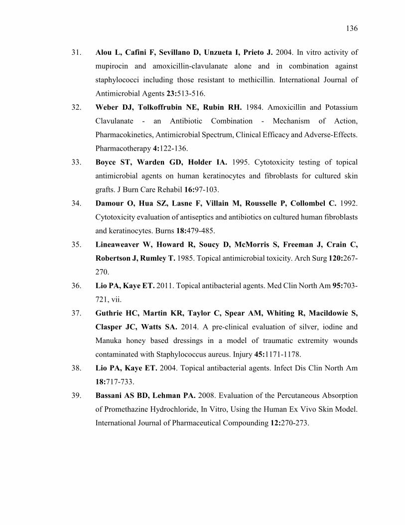
136
31. Alou L, Cafini F, Sevillano D, Unzueta I, Prieto J. 2004. In vitro activity of
mupirocin and amoxicillin-clavulanate alone and in combination against
staphylococci including those resistant to methicillin. International Journal of
Antimicrobial Agents 23:513-516.
32. Weber DJ, Tolkoffrubin NE, Rubin RH. 1984. Amoxicillin and Potassium
Clavulanate - an Antibiotic Combination - Mechanism of Action,
Pharmacokinetics, Antimicrobial Spectrum, Clinical Efficacy and Adverse-Effects.
Pharmacotherapy 4:122-136.
33. Boyce ST, Warden GD, Holder IA. 1995. Cytotoxicity testing of topical
antimicrobial agents on human keratinocytes and fibroblasts for cultured skin
grafts. J Burn Care Rehabil 16:97-103.
34. Damour O, Hua SZ, Lasne F, Villain M, Rousselle P, Collombel C. 1992.
Cytotoxicity evaluation of antiseptics and antibiotics on cultured human fibroblasts
and keratinocytes. Burns 18:479-485.
35. Lineaweaver W, Howard R, Soucy D, McMorris S, Freeman J, Crain C,
Robertson J, Rumley T. 1985. Topical antimicrobial toxicity. Arch Surg 120:267-
270.
36. Lio PA, Kaye ET. 2011. Topical antibacterial agents. Med Clin North Am 95:703-
721, vii.
37. Guthrie HC, Martin KR, Taylor C, Spear AM, Whiting R, Macildowie S,
Clasper JC, Watts SA. 2014. A pre-clinical evaluation of silver, iodine and
Manuka honey based dressings in a model of traumatic extremity wounds
contaminated with Staphylococcus aureus. Injury 45:1171-1178.
38. Lio PA, Kaye ET. 2004. Topical antibacterial agents. Infect Dis Clin North Am
18:717-733.
39. Bassani AS BD, Lehman PA. 2008. Evaluation of the Percutaneous Absorption
of Promethazine Hydrochloride, In Vitro, Using the Human Ex Vivo Skin Model.
International Journal of Pharmaceutical Compounding 12:270-273.
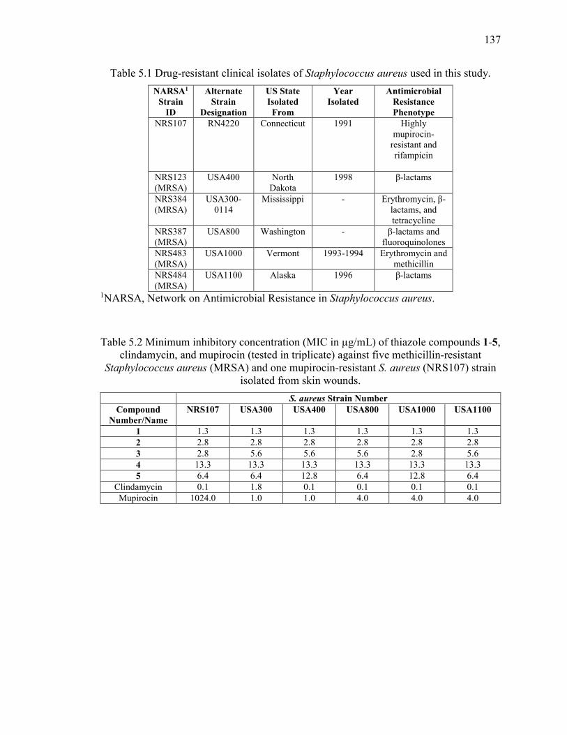
137
Table 5.1 Drug-resistant clinical isolates of Staphylococcus aureus used in this study.
NARSA1 Strain
ID
Alternate Strain
Designation
US State Isolated
From
Year Isolated
Antimicrobial Resistance Phenotype
NRS107 RN4220 Connecticut 1991 Highly mupirocin-
resistant and rifampicin
NRS123 (MRSA)
USA400 North Dakota
1998 β-lactams
NRS384 (MRSA)
USA300-0114
Mississippi - Erythromycin, β-lactams, and tetracycline
NRS387 (MRSA)
USA800 Washington - β-lactams and fluoroquinolones
NRS483 (MRSA)
USA1000 Vermont 1993-1994 Erythromycin and methicillin
NRS484 (MRSA)
USA1100 Alaska 1996 β-lactams
1NARSA, Network on Antimicrobial Resistance in Staphylococcus aureus.
Table 5.2 Minimum inhibitory concentration (MIC in µg/mL) of thiazole compounds 1-5, clindamycin, and mupirocin (tested in triplicate) against five methicillin-resistant
Staphylococcus aureus (MRSA) and one mupirocin-resistant S. aureus (NRS107) strain isolated from skin wounds.
S. aureus Strain Number Compound
Number/Name NRS107 USA300 USA400 USA800 USA1000 USA1100
1 1.3 1.3 1.3 1.3 1.3 1.3
2 2.8 2.8 2.8 2.8 2.8 2.8
3 2.8 5.6 5.6 5.6 2.8 5.6
4 13.3 13.3 13.3 13.3 13.3 13.3
5 6.4 6.4 12.8 6.4 12.8 6.4
Clindamycin 0.1 1.8 0.1 0.1 0.1 0.1
Mupirocin 1024.0 1.0 1.0 4.0 4.0 4.0
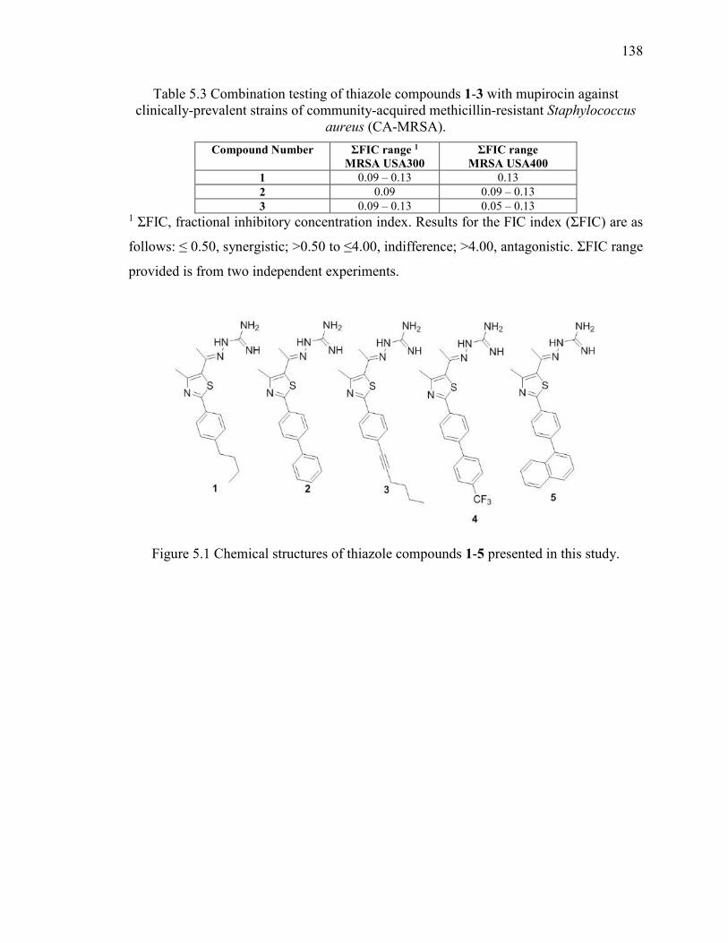
138
Table 5.3 Combination testing of thiazole compounds 1-3 with mupirocin against clinically-prevalent strains of community-acquired methicillin-resistant Staphylococcus
aureus (CA-MRSA).
Compound Number ƩFIC range 1 MRSA USA300
ƩFIC range MRSA USA400
1 0.09 – 0.13 0.13
2 0.09 0.09 – 0.13
3 0.09 – 0.13 0.05 – 0.13 1 ƩFIC, fractional inhibitory concentration index. Results for the FIC index (ƩFIC) are as
follows: ≤ 0.50, synergistic; >0.50 to ≤4.00, indifference; >4.00, antagonistic. ƩFIC range
provided is from two independent experiments.
Figure 5.1 Chemical structures of thiazole compounds 1-5 presented in this study.
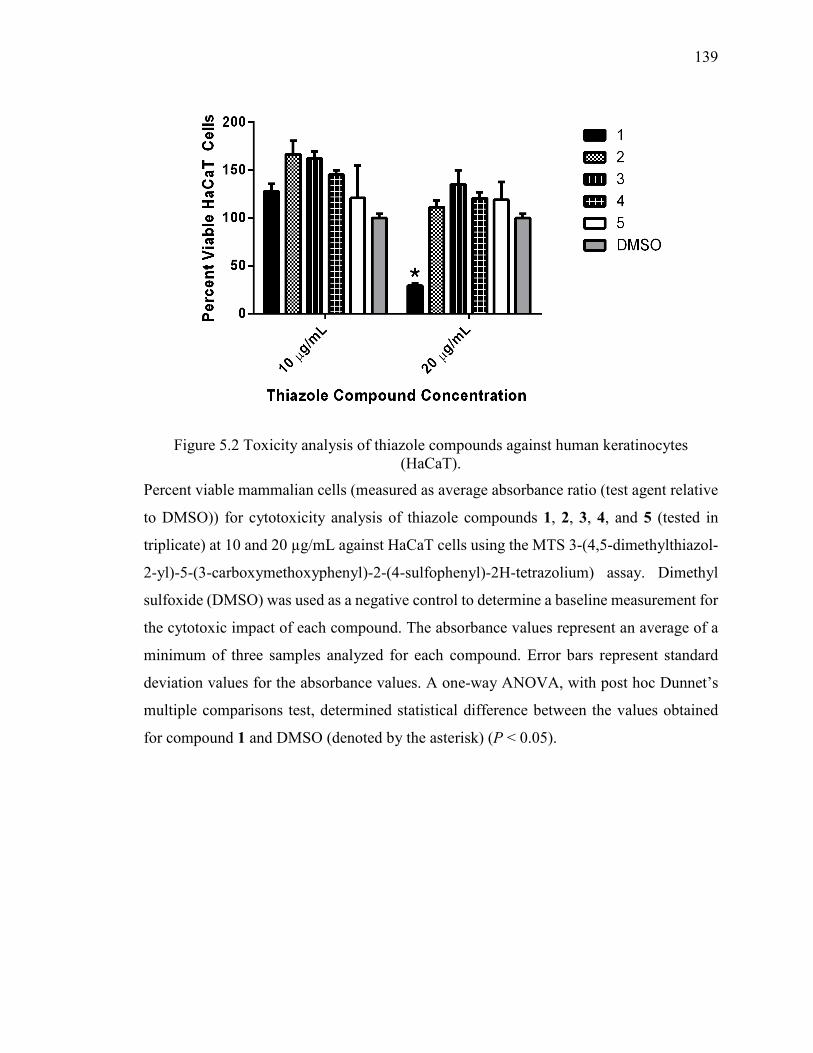
139
Figure 5.2 Toxicity analysis of thiazole compounds against human keratinocytes (HaCaT).
Percent viable mammalian cells (measured as average absorbance ratio (test agent relative
to DMSO)) for cytotoxicity analysis of thiazole compounds 1, 2, 3, 4, and 5 (tested in
triplicate) at 10 and 20 µg/mL against HaCaT cells using the MTS 3-(4,5-dimethylthiazol-
2-yl)-5-(3-carboxymethoxyphenyl)-2-(4-sulfophenyl)-2H-tetrazolium) assay. Dimethyl
sulfoxide (DMSO) was used as a negative control to determine a baseline measurement for
the cytotoxic impact of each compound. The absorbance values represent an average of a
minimum of three samples analyzed for each compound. Error bars represent standard
deviation values for the absorbance values. A one-way ANOVA, with post hoc Dunnet’s
multiple comparisons test, determined statistical difference between the values obtained
for compound 1 and DMSO (denoted by the asterisk) (P < 0.05).
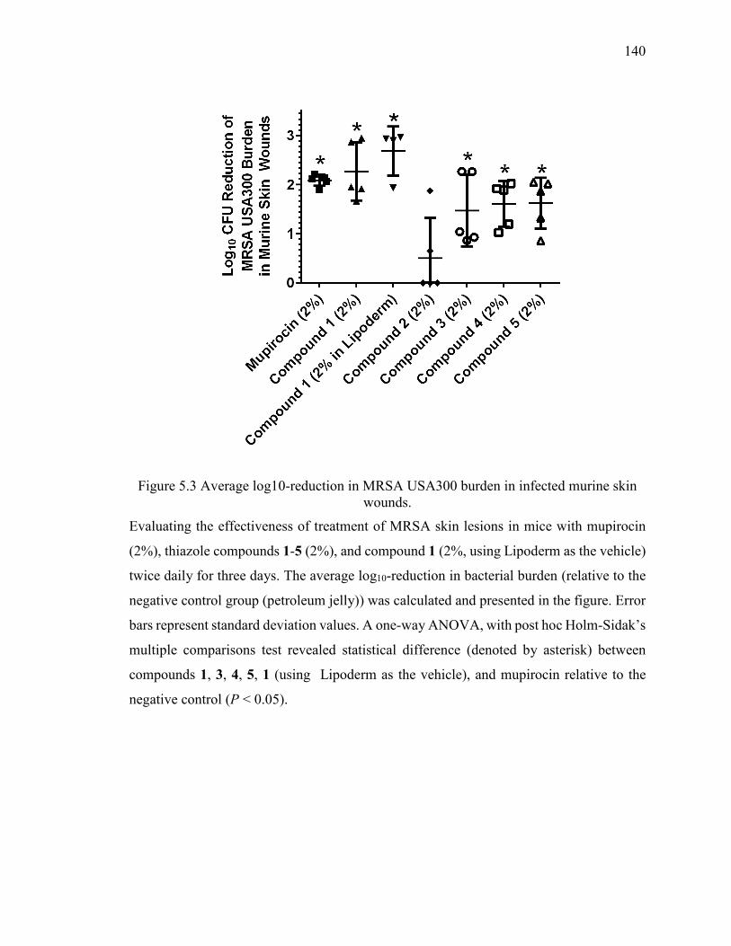
140
Figure 5.3 Average log10-reduction in MRSA USA300 burden in infected murine skin wounds.
Evaluating the effectiveness of treatment of MRSA skin lesions in mice with mupirocin
(2%), thiazole compounds 1-5 (2%), and compound 1 (2%, using Lipoderm as the vehicle)
twice daily for three days. The average log10-reduction in bacterial burden (relative to the
negative control group (petroleum jelly)) was calculated and presented in the figure. Error
bars represent standard deviation values. A one-way ANOVA, with post hoc Holm-Sidak’s
multiple comparisons test revealed statistical difference (denoted by asterisk) between
compounds 1, 3, 4, 5, 1 (using Lipoderm as the vehicle), and mupirocin relative to the
negative control (P < 0.05).
141
CHAPTER 6. ANTIBACTERIAL CHARACTERIZATION OF NOVEL SYNTHETIC THIAZOLE COMPOUNDS AGAINST
METHICILLIN-RESISTANT STAPHYLOCOCCUS
PSEUDINTERMEDIUS
THIS IS A PUBLISHED JOURNAL ARTICLE. Reprinted with permission from
Antibacterial characterization of novel synthetic thiazole compounds against
methicillin-resistant Staphylococcus pseudintermedius. Haroon Mohammad, P. V.
Narasimha Reddy, Dennis Monteleone, Abdelrahman S. Mayhoub, Mark Cushman, G.
Kenitra Hammac, and Mohamed N. Seleem. PLoS ONE (2015) 10, e0130385. doi:
10.1371/journal.pone.0130385 Copyright 2015 PLoS ONE
6.1 Introduction
Staphylococcus pseudintermedius is a significant problem in veterinary medicine as it is a
major source of opportunistic infections in companion animals and the leading causative
agent of canine pyoderma (1). It has also been linked to other severe infections in
companion animals including urinary tract infections, skin wounds, surgical site infections,
and otitis (2-4). The challenge to combat S. pseudintermedius infections has become more
difficult with the emergence of clinical isolates (primarily methicillin-resistant S.
pseudintermedius) exhibiting resistance to multiple antibiotic classes including β-lactams,
lincosamides, fluoroquinolones, macrolides, sulfonamides, aminoglycosides, tetracyclines,
and chloramphenicol (1, 5, 6). In several cases, patients that contracted an infection caused
by S. pseudintermedius, in particular newborn puppies, have died or been euthanized due
to the lack of effective treatment to remedy the medical condition (4, 7, 8). Thus there is a
critical need for the discovery and characterization of novel antimicrobials to treat
infections caused by methicillin-sensitive and methicillin-resistant S. pseudintermedius.
Thiazole compounds have been shown to be useful in multiple therapeutic
applications including as anticancer, antiviral, and anticonvulsant agents (9-11) but their
potential use as antibacterials has not been fully examined. Previous investigation into
thiazole compounds synthesized by our research group has revealed these compounds
exhibit potent antimicrobial activity against multidrug-resistant strains of Staphylococcus
142
aureus, including methicillin-resistant S. aureus (MRSA) (12, 13). MRSP bears similar
genetic and phenotypic traits to MRSA, including expression of the mecA gene that
encodes a modified penicillin-binding protein that confers resistance to β-lactam antibiotics
(14). Additionally, S. pseudintermedius has been shown to express surface proteins similar
to S. aureus that play an important role in bacterial colonization of host tissues (15).
Furthermore, both staphylococcal species secrete similar virulence factors, including
exfoliative toxins and leukocidins, that may play an important role in promoting
pathogenesis of disease in infected hosts (16-18).
Given the genetic and phenotypic similarities between S. pseudintermedius and S.
aureus, we suspected that the thiazole compounds we have found to be potent inhibitors of
MRSA would also be active against MRSP. The objectives of the present study were to
characterize the antibacterial activity of six of the most potent thiazole compounds (against
MRSA) (Fig. 1) against clinical isolates of MSSP and MRSP, to ascertain the likelihood
of MRSP acquiring rapid resistance to these novel compounds, and to determine if the
compounds could be used to re-sensitize MRSP to the effect of β-lactam antibiotics.
Additionally, we assessed the physicochemical profile of the most promising analogue and
examined the ability of MRSP to recover after exposure to the thiazole antibiotics, via a
post-antibiotic effect assay. The results garnered lend valuable insight into the
pharmacological utility of thiazole compounds as a possible future therapeutic option for
the treatment of S. pseudintermedius infections.
6.2 Materials and Methods
6.2.1 Bacterial isolates and chemical reagents
Fifteen isolates of S. pseudintermedius (eight MSSP and seven MRSP), identified at the
Indiana Animal Disease Diagnostic Laboratory from specimens collected from dogs
admitted to the small animal teaching hospital at Purdue University, were included in the
study. The specimens were not collected specifically for this research study but were
obtained from patients admitted to the hospital for treatment. Clinical specimens were
inoculated onto 5% sheep blood agar and incubated at 35 °C for 18-24 hours. Standard
methods including examination of colony morphology and hemolysis and biochemical
143
tests such as tube coagulase, Voges-Proskauer (VP) and fermentation tests for maltose,
trehalose and lactose, were used in addition to matrix-assisted laser desorption ionization
time of flight mass spectrometry (19) to identify isolates (19, 20). Antimicrobial
susceptibility was determined by broth microdilution using the SensiTitre (Thermofisher
Scientific). Isolates demonstrating resistance to oxacillin, a surrogate for methicillin, with
a minimum inhibitory concentration (MIC) value greater than or equal to 0.5 μg/mL were
tested for the presence of mecA by PCR as previously described (21). Bacterial isolates
used in this study are presented in Table 6.1.
Clindamycin hydrochloride monohydrate (Tokyo Chemical Industry, Portland, OR,
USA), oxacillin sodium salt monohydrate (Tokyo Chemical Industry, Portland, OR, USA),
rifampicin (Sigma-Aldrich, St. Louis, MO, USA), and vancomycin hydrochloride (Gold
Biotechnology, St. Louis, MO, USA) were purchased commercially. All antibiotics were
dissolved in dimethyl sulfoxide (DMSO) to obtain a stock 10 mM solution.
6.2.2 Synthesis of thiazole compounds 1-6
The detailed synthetic protocols and spectral data of final products 1-6 as well as all
intermediates have been previously reported (12, 22, 23). Chemical structures of
compounds 1-6 are presented in Figure 6.1. All compounds were dissolved in DMSO to
obtain a stock 10 mM solution.
6.2.3 Determination of minimum inhibitory concentration and minimum bactericidal concentration (MBC) against S. pseudintermedius
The MICs of the thiazole compounds, clindamycin, and rifampicin against eight clinical
isolates of MSSP and seven clinical isolates of MRSP were determined using the broth
microdilution method, in accordance with the recommendations contained in the CLSI
guidelines (24). Bacteria were prepared in phosphate-buffered saline (PBS) until a
McFarland standard of 0.5 was achieved. The solution was subsequently diluted 1:300 in
Mueller-Hinton broth (MHB) to reach a starting inoculum of 1 × 10⁵ colony-forming units
(CFU/mL). Bacteria were then transferred to a 96-well microtiter plate. Thiazole
compounds and antibiotics were added (in triplicate) to wells in the first row of the
microtiter plate and then serially diluted along the vertical axis. The plate was incubated at
144
37 °C for 22-24 hours before the MIC was determined. The MIC was categorized as the
concentration where there was no visible growth of bacteria observed.
The MBC was determined by plating 5 µL from wells on the 96-well microtiter plate
(where the MIC was determined) where no growth was observed onto tryptic soy agar
(TSA) plates. The TSA plates were then incubated at 37 °C for 22-24 hours before the
MBC was determined. The MBC was categorized as the concentration where ≥99%
reduction in bacterial cell count was observed.
6.2.4 Time-kill analysis of thiazole compounds and antibiotics against MRSP
MRSP cells in late logarithmic growth phase were diluted to ~1 × 10⁷CFU/mL and exposed
to concentrations equivalent to 4 × MIC (in triplicate) of thiazole compounds 1-6 and
rifampicin in MHB. Samples (20 µL) were collected after 0, 2, 4, 6, 8, 10, 12, and 24 hours
of incubation at 37 °C and subsequently serially diluted in PBS. Bacteria were then
transferred to TSA plates and incubated at 37 °C for 18-20 hours before viable CFU/mL
values were determined.
6.2.5 Cell membrane disruption analysis
In order to investigate the antimicrobial effect of the thiazole compounds on the integrity
of the bacterial cell membrane, the release of 260 and 280 nm absorbing components was
determined spectrophotometrically (25). The cell suspension of 1.2 × 10⁹ CFU/mL MRSP
was incubated with 4 × MIC of compound 2 at 37 °C for 30 minutes. Untreated MRSP
cells or cells treated with vancomycin (inhibits cell wall synthesis in bacterial cells) served
as negative controls. For the release of 260 and 280 nm absorbing material, the bacterial
suspension (control) was treated with lysostaphin (in 50 mM Tris-HCl, pH 8.00) for 30
minutes. Lysostaphin was used as a positive control due to its mode of action being the
disruption of the cross-linking of the pentaglycin bridges in the cell wall of staphylococci
bacteria (26). The absorbance of cell supernatant at 260 and 280 nm was determined using
a spectrophotometer (Jenway 6305). The average OD260 and OD280 values of duplicates of
each treatment option were calculated and expressed as the proportion of average OD260
(or OD280) for each treatment option compared to the average OD260 (or OD280) for the
positive control (lysostaphin).
145
6.2.6 In vitro cytotoxicity analysis
Compounds were assayed at concentrations of 5 µg/mL, 10 µg/mL, 20 µg/mL, and 40
µg/mL against a murine macrophage cell line (J774.A1) (ATCC® TIB-67™, American
Type Culture Collection (ATCC), Manassas, VA, USA) to determine the potential toxic
effect in vitro. Cells were cultured in Dulbeco’s modified Eagle’s medium (Sigma-Aldrich,
St. Louis, MO, USA) with 10% fetal bovine serum (USA Scientific, Inc.) at 37 °C with 5%
CO2. Controls received DMSO alone at a concentration equal to that in drug-treated cell
samples. The cells were incubated with the compounds in a 96-well plate at 37 ºC and 5.0%
CO2 for two hours prior to addition of the assay reagent MTS 3-(4,5-dimethylthiazol-2-yl)-
5-(3-carboxymethoxyphenyl)-2-(4-sulfophenyl)-2H-tetrazolium) (Promega, Madison, WI,
USA). Absorbance readings were taken using a kinetic ELISA microplate reader
(Molecular Devices, Sunnyvale, CA, USA). The quantity of viable cells after treatment
with each compound was expressed as a percentage of the control, DMSO.
6.2.7 Multi-step resistance selection
To assess the ability of MRSP to develop resistance to the thiazole compounds, a multi-
step resistance selection experiment was performed, as described elsewhere (27). The broth
microdilution method for MIC determination against a clinical isolate of MRSP was
repeated for ten passages over a period of ten days. The initial inoculum was prepared to a
McFarland standard of 0.5. The solution was subsequently diluted 1:300 in MHB to reach
a starting inoculum of 1 × 10⁵ CFU/mL. For each subsequent passage, the inoculum for the
MIC determination was adjusted to a final density of approximately 5 × 10⁵ CFU/mL using
the contents of a well containing a subinhibitory concentration of the compound (where
bacterial growth was observed from the previous passage). Bacteria were then transferred
to a new 96-well microtiter plate. Thiazole compounds 3-6 and clindamycin were added
(in triplicate) to wells in the first row of the microtiter plate and then serially diluted along
the ordinate. The plate was incubated at 37 °C for 22 hours before the MIC was determined
by visual inspection. Resistance was classified as a greater than four-fold increase in the
initial MIC, as reported elsewhere (28).
146
6.2.8 Combination therapy assessment of thiazole compounds with oxacillin
The relationship between the thiazole compounds and oxacillin was assessed via a standard
checkerboard assay (29). Bacteria equivalent to a McFarland standard of 0.5 were prepared
in PBS. The bacteria were next diluted in MHB to achieve a starting cell density of 1 × 10⁵
CFU/mL. MHB was transferred to all wells of a 96-well microtiter plate. The thiazole
compounds and oxacillin were diluted in MHB to achieve a starting concentration
equivalent to 2 × or 4 × MIC, respectively. Oxacillin was serially diluted along the abscissa
of the microtiter plate while the thiazole compound was serially diluted along the ordinate.
The plate was incubated for 22-24 hours at 37 °C. The MIC of the test compound in
combination with oxacillin was determined as the lowest concentration of each
compound/antibiotic where no visible growth of bacteria was observed. The fractional
inhibitory concentration index (ƩFIC) was calculated for each combination as described
previously (13). A synergistic relationship was classified as an FIC index less than or equal
to 0.5. FIC values above 0.5 but less than 4.0 were characterized as indifference while FIC
values above 4.0 were classified as antagonistic.
6.2.9 Re-sensitization of MRSP to oxacillin using broth microdilution method
MHB was inoculated with MRSP (5×10⁵ CFU/mL), as has been previously described (30).
Aliquots (5 mL) of the bacterial suspension were divided into microcentrifuge tubes. The
thiazole compounds tested (at ½ × MIC) were introduced into each tube. After sitting at
room temperature for 30 minutes, 1 mL samples from each tube were transferred to a new
centrifuge tube prior to addition of oxacillin (at a concentration equivalent to its MIC).
Using a 96-well microtiter plate, rows 2-12 were filled with the remaining 4 mL bacterial
suspension (containing the thiazole compound). Aliquots (200 µL) from tubes containing
both the thiazole compound and oxacillin were transferred to row 1 of the 96-well plate.
After aspirating contents in the first row 4-6 times, 100 µL was transferred from wells in
row 1 to row 2. This process was repeated to dilute the remaining wells containing no
antibiotic. Untreated bacteria served as a control. The plate was incubated at 37 °C for 22
hours before the MIC was recorded. The MIC was categorized as the concentration at
which no visible growth of bacteria was observed in a particular well. A fold reduction was
147
calculated by comparing the MIC of the antibiotic alone compared to the MIC of the
antibiotic given in combination with the thiazole compounds.
6.2.10 Kinetic solubility determination of compound 3
Serial dilutions of compound 3 were prepared in DMSO at 100× the final concentration.
Compound 3 was then diluted 100-fold into PBS in a 96-well plate and mixed. The
absorbance of the PBS-containing plate was measured prior to addition of the test agents
to determine the background absorbance. After two hours, the presence of precipitate was
detected by turbidity (absorbance at 540 nm). An absorbance value of greater than (mean
+ 3× standard deviation of the blank), after subtracting the pre-experiment background,
was indicative of turbidity. The solubility limit is reported as the highest experimental
concentration for compound 3 with no evidence of turbidity as described previously (12).
6.2.11 Microsomal stability analysis
Compound 3 was incubated in duplicate with dog liver microsomes at 37 ºC. The reaction
contained microsomal protein in 100 mM potassium phosphate, 2 mM NADPH, 3 mM
MgCl2, pH 7.4. A control was run for each test agent omitting NADPH to detect NADPH-
free degradation. At 0, 10, 20, 40, and 60 minutes, an aliquot was removed from each
experimental and control reaction and mixed with an equal volume of ice-cold Stop
Solution (methanol containing haloperidol, diclofenac, or other internal standard). Stopped
reactions were incubated at least 10 minutes at -20 ºC, and an additional volume of water
was subsequently added. The samples were centrifuged to remove precipitated protein, and
the supernatants were analyzed by LC/MS/MS to quantitate the remaining parent. Data
were converted to % remaining by dividing by the time zero concentration value. Data
were then fitted to a first-order decay model to determine half-life. Intrinsic clearance was
calculated from the half-life and the protein concentrations, as has been described
elsewhere (12).
6.2.12 Post-antibiotic effect
To assess if the thiazole compounds exhibit a post-antibiotic effect (PAE) against MRSP,
MRSP cells in late logarithmic growth phase (~1×10⁸ CFU/mL) were incubated with 4 ×
MIC of thiazole compounds 1-6, clindamycin, or rifampicin for one hour at 37 °C. A tube
148
containing untreated bacterial cells served as a control. Afterward, the compound/antibiotic
was washed out by diluting bacteria 1:1000 in MHB. Counts of CFU for all cultures were
obtained after washing. Aliquots (100 µL) of bacteria were removed every hour (for 10
hours), serially diluted in PBS, and plated on TSA plates. TSA plates were incubated for
20 hours at 37 °C before CFU values were determined. The PAE was calculated using the
same formula described elsewhere (31): PAE = T − C, where T is the time required for the
count of CFU values in the test culture to increase one log10 above the count observed
immediately after removal of the test agent and C is the time required for the count of CFU
in the untreated control culture to increase one log10 above the count observed immediately
after completion of the same procedure used on the test culture for removal of test agent.
6.2.13 Statistical analysis
All statistical analyses were performed with GraphPad Prism 6.0 (GraphPad Software, La
Jolla, CA). Data generated from cytotoxicity analysis of the thiazole compounds against
J774.A1 cells and the 260 and 280 nm cell leakage analysis were analyzed using one-way
ANOVA, with post hoc Tukey's multiple comparisons test (P < 0.05).
6.3 Results
6.3.1 MICs and MBCs of thiazole compounds and antibiotics against S.
pseudintermedius
All six thiazole compounds exhibited potent antimicrobial activity against all S.
pseudintermedius isolates tested (Table 6.2). The MIC50 values obtained for the compounds
against methicillin-sensitive S. pseudintermedius were in close proximity to one another
ranging from 0.30 µg/mL for compound 2 to 0.80 µg/mL for compound 6. These values
mimicked the results obtained for clindamycin (MIC50 of 0.48 µg/mL), a first-line
antibiotic recommended for use in the treatment of pyoderma infections (32). The MBC50
values matched or were up to three-fold higher than the MIC50 values determined for the
thiazole compounds; this indicates that these compounds exhibit bactericidal activity
against methicillin-sensitive S. pseudintermedius. The compounds retained their
antimicrobial activity against the isolates of methicillin-resistant S. pseudintermedius
tested. Interestingly, compounds 3 and 6 showed a nearly two-fold improvement in the
149
MIC50 value obtained against MRSP isolates as compared to the MSSA isolates. The
thiazoles retained their bactericidal activity against MRSP isolates with MBC50 values
ranging from 0.42 µg/mL for compound 5 to 1.47 µg/mL for compound 4.
6.3.2 Time-kill analysis of thiazole compounds and rifampicin
To confirm the thiazole compounds were bactericidal against MRSP, a time-kill assay was
performed using 4 × MIC of each compound and rifampicin. As Figure 6.2 demonstrates,
the thiazole compounds are bactericidal but the rate of killing varies. The lead compound
(1) required four hours to completely eliminate MRSP. Derivatives 3 and 6 showed
improved killing kinetics and mimic rifampicin’s rapid bactericidal activity, completely
eliminating MRSP within two hours. Compounds 2 and 5 require eight hours to achieve
the same result while compound 4 exhibits the slowest rate of bacterial killing, requiring
12 hours to completely eliminate MRSP.
6.3.3 MRSP cell membrane disruption assessment
Disruption of the physical integrity of the bacterial cell membrane (such as formation of
pores in the membrane) has been associated with antimicrobials that exhibit rapid
bactericidal activity. To assess if the thiazole compounds’ mode of action is disruption of
the integrity of the MRSP cell membrane, the leakage of intracellular contents at 260 and
280 nm was analyzed after exposure of bacterial cells to a high concentration of compound
2 (4 × MIC) for 30 minutes. Figure 6.3 demonstrates that the thiazole compounds do not
match the action of lysostaphin (a known membrane-disrupting agent). Less than 20% of
the intracellular content (at 260 nm) is released after treatment with the thiazole compound
as compared to cells treated with lysostaphin. This result confirms that the thiazole
compounds do not act in a manner that involves disruption of the physical integrity of the
MRSP cell membrane.
6.3.4 Toxicity analysis of thiazole compounds
Toxicity to host tissues is an important characteristic to assess with new compounds early
in the drug discovery process. To determine if the thiazole compounds were toxic to
eukaryotic cells, the viability of murine macrophage cells (J774.A1) exposed to each
thiazole compound was assessed using the MTS assay. The lead 1 and compounds 2 and 6
150
proved to be toxic at a concentration of 10 µg/mL (Figure 6.4). However, derivatives 3, 4,
and 5 exhibited an improved toxicity profile over the lead compound (matching the results
obtained with clindamycin), demonstrating they were not toxic to mammalian cells at 10
µg/mL. This is more than 20-fold higher than the MIC50 values obtained for these three
compounds against clinical isolates of MRSP.
6.3.5 Multi-step resistance selection of MRSP to thiazole compounds
To assess the potential for rapid emergence of resistance of MRSP to the thiazole
compounds, a multi-step resistance selection experiment was performed. The initial MICs
of compounds 3, 4, 5, and 6 were determined via the broth microdilution method and were
found to be 1.41 µg/mL (compound 3), 1.47 µg/mL (compound 4), 1.67 µg/mL (compound
5), and 1.60 µg/mL (compound 6). Bacteria were then subcultured for ten serial passages
to determine if a shift in the MIC of each compound tested would be observed against
MRSP. After the second serial passage of compound 5, there was a two-fold shift in the
MIC; the MIC remained stable at 3.34 µg/mL until the seventh passage where a second
increase in the MIC was observed to 6.68 µg/mL (Figure 6.5). Compounds 4 and 5
followed a similar course to one another; the MICs of both compounds remained stable for
three passages before a two-fold shift was observed in both compounds after the fourth
passage. The MIC did not increase again for both compounds after six additional passages.
MRSP was not able to develop resistance to compound 3 even after ten passages.
6.3.6 Combination therapy and re-sensitization of MRSP to oxacillin in the presence of the thiazole compounds
As MRSP strains exhibit resistance to the effect of β-lactam antibiotics, such as oxacillin,
we assessed whether exposure of MRSP to a subinhibitory concentration of the thiazole
compounds could re-sensitize the bacteria to the effect of these antibiotics. After initial
exposure of MRSP to a subinhibitory concentration (½ × MIC) of the thiazole compound,
the bacteria were next treated with oxacillin. Using the broth microdilution assay, the MIC
of oxacillin needed to inhibit MRSP growth in the presence and absence of each thiazole
compound was determined. As Table 6.3 presents, MRSP was re-sensitized to the effect of
oxacillin in the presence of a subinhibitory concentration of all six thiazole compounds.
There was a 128-fold reduction in the MIC of oxacillin observed in the presence of ½ ×
151
MIC of compounds 1, 2, 3, 4, and 6 and a 64-fold reduction in the MIC observed with
compound 5. The checkerboard assay was used to assess if these compounds could be used
in combination with oxacillin against MRSP. The assay revealed four compounds (1, 3, 5,
and 6) exhibited a synergistic relationship with oxacillin with ƩFIC values ranging from
0.19 to 0.38.
6.3.7 Solubility and metabolic stability assessment of compound 3
The solubility of novel compounds in aqueous solutions and stability to host metabolic
processes are important to analyze to determine if promising compounds possess suitable
drug-like properties. To assess the ability of the thiazole compounds to dissolve in aqueous
solutions, a turbidimetric solubility analysis was performed with compound 3 in phosphate-
buffered saline. As Table 6.4 demonstrates, the compound is soluble in PBS up to a
concentration of 5.51 µg/mL. This resembles the result obtained for the poorly aqueous
drug tamoxifen, indicating compound 3 possesses limited aqueous solubility.
To analyze the stability of compound 3 to metabolic processes present in the liver,
the compound was incubated with dog liver microsomes. As Table 6.5 shows, this
compound is metabolized very slowly (similar to the drug warfarin) with a NADPH-
dependent intrinsic clearance rate of 18.7 µL/min-mg and a half-life of over two hours. The
data from Table 6.5 indicate that this compound is a substrate of a NADPH-dependent
metabolic process. There is a nearly three-fold reduction in the intrinsic clearance rate
(down to 6.6 µL/min-mg) and a marked increase in the half-life (nearly six hours) of
compound 3 in the absence of the co-factor NADPH.
6.3.8 Post-antibiotic effect of thiazole compounds and antibiotics
In vitro pharmacodynamic analysis can provide valuable information regarding
establishing a proper dosing regimen for drug candidates. One method to obtain this
information is to determine if a compound/drug exhibits a post-antibiotic effect. The PAE
for the thiazole compounds, clindamycin, and rifampicin was determined against a clinical
isolate of MRSP. Table 6.6 reveals that all six thiazoles exhibit a long PAE ranging from
8 hours (for compounds 2 and 5) to > 9 hours (for the remaining four compounds). This is
152
similar to what was observed with rifampicin (PAE > 9 hours) and superior to what was
observed with clindamycin (PAE of only two hours).
6.4 Discussion
S. pseudintermedius infections have become a growing problem in veterinary medicine;
until fairly recently, the vast majority of infections observed in small animal veterinary
facilities could be treated with an array of efficacious antimicrobials (32, 33). However,
the rapid emergence and global spread of multidrug-resistant S. pseudintermedius (namely
MRSP) in the past ten years has presented a significant challenge to veterinary practitioners
(3, 34). Clinical isolates have been identified that exhibit resistance to numerous antibiotic
classes, limiting the treatment options available for veterinarians. This underscores the
critical need to identify and develop new antibiotics and unique therapeutic strategies to
combat this growing medical challenge.
The present study examines the antibacterial potential of novel synthetic thiazole
compounds against clinical isolates of MSSP and MRSP. We have previously
demonstrated the lead compound and derivative 2 possess potent antimicrobial activity
against important strains of multidrug-resistant S aureus (primarily MRSA) of concern to
both humans and animals (12). Four additional derivatives (compounds 3-6) of the lead
compound were subsequently constructed in an attempt to enhance the antibacterial activity
of the lead while mitigating potential toxicity to host tissues. Structural variation
constructed focused on modification of the lipophilic alkane side chain of the lead
compound, resulting in the butyne analogue 3, fluorobiphenyl derivative 4, trifluoromethyl
analogue 5, and the naphthyl derivative 6. All four were previously found to exhibit potent
activity against MRSA so they were also included in this study. As S. pseudintermedius
and S. aureus share similar genetic and phenotypic characteristics, we hypothesized that
these thiazole compounds would possess potent antibacterial activity against MSSP and
MRSP. This conjecture was confirmed via the broth microdilution method; all six thiazole
compounds exhibited potent antibacterial activity against clinical isolates of both MSSP
(MIC50 ranged from 0.30-0.80 µg/mL) and MRSP (MIC50 ranged from 0.40-1.47 µg/mL).
These results proved similar to what was found with clindamycin (MIC50 of 0.48 µg/mL
against both MSSP and MRSP), an antibiotic of choice for treatment of pyoderma
153
infections (32). Interestingly, the thiazole compounds retained their antibacterial activity
against nine MSSP and MRSP isolates that were found to be resistant to clindamycin and
other antibiotics; this indicates there is no cross-resistance present between these antibiotics
and the thiazole compounds. This further supports the notion that these thiazole compounds
have potential to be used as novel antibacterial agents, particularly against S.
pseudintermedius infections resistant to treatment with other antibiotics.
We were curious to find out if the thiazole compounds possessed bacteriostatic or
bactericidal activity. It has been suggested bactericidal antimicrobials have several
advantages over their bacteriostatic counterparts, including helping patients recover more
rapidly from infection, improving the clinical outcome of disease, reducing the potential
emergence of bacterial resistance to the antibiotic, and limiting the spread of infection (35).
Preliminary analysis indicated the thiazole compounds were bactericidal as they possessed
MBC50 values identical to or two- to three-fold higher than their MIC50 values against both
MSSP and MRSP isolates. While structural modifications made to the lead thiazole
compound did not significantly impact the MIC50 and MBC50 values found for the
subsequently constructed derivatives, there was a significant difference observed in the
bacterial killing kinetics against MRSP. A time-kill assay revealed that the alkyne
derivative 3 and the naphthyl derivative 6 exhibited superior activity to the lead 1, rapidly
eliminating MRSP within two hours (the lead compound required double the time to
achieve the same effect). This was similar to the result obtained with rifampicin, an
antibiotic of last resort for pyoderma infections (32).
Rapid bactericidal activity has been shown to be important in the treatment of
diseases caused by staphylococci such as endocarditis, meningitis, and osteomyelitis (35,
36). Thus these thiazole compounds possess a selective advantage over bacteriostatic
agents in their ability to be used for treatment of more severe clinical diseases. However,
one pitfall of antimicrobials that are rapidly bactericidal is many tend to work as
membrane-disrupting agents (35, 37). Such agents have limited therapeutic applications,
almost exclusively being restricted to use as topical ointments (37). As the thiazole
compounds were found to exhibit rapid bactericidal activity, we examined if the mode of
action of the thiazole compounds was via disruption of the MRSP cell membrane. A cell
leakage analysis confirmed that the thiazole compounds do not physically disrupt the
154
integrity of the bacterial membrane similar to the positive control lysostaphin. The exact
mechanism of action of these thiazole compounds against staphylococci is being
investigated and will be the subject of the next chapter.
After confirming the thiazole compounds do in fact possess potent antibacterial
activity and are capable of rapidly eliminating MRSP (in a mechanism that does not involve
physical disruption of the bacterial cell membrane), we next focused our attention to
assessing potential toxicity concerns with these compounds against mammalian cells.
Structural modifications made to the lead thiazole compound played an important role in
enhancing the toxicity profile of the thiazoles. The lead compound and biphenyl derivative
2 were found to be toxic to murine macrophage cells at a concentration of 10 µg/mL.
Surprisingly, replacement of the alkyl moiety in the lead with an alkyne, monofluoro, or
trifluoromethyl group (as in compounds 3-5, respectively) significantly improved the
toxicity profiles of the compounds. These three derivatives were not toxic to murine
macrophage cells at 10 µg/mL which represents a greater than 20-fold difference over the
MIC50 values determined against MRSP.
The ability of bacteria to develop resistance rapidly to antimicrobial compounds is
important to assess early in drug discovery. Previously, we have reported results of a
single-step resistance selection experiment that demonstrated MRSA is unlikely to develop
rapid resistance to thiazole compounds 1 and 2 (13). We decided to extend this analysis to
the newest thiazole derivatives (compounds 3-6) against MRSP but with an additional twist
– testing if bacterial resistance could be induced after repeated exposure to each compound
over 10 serial passages. There was no change observed in the MIC for compound 3, a two-
fold increase in the MIC for compounds 4 and 5, and a four-fold increase in the MIC of
compound 6 after 10 passages. Collectively, the results provide data supporting a low
probability of MRSP-resistance developing rapidly to these thiazole compounds (as a
greater than four-fold increase, as compared to the initial MIC, was not observed for any
of the compounds tested).
While discovery of novel antimicrobials for use in monotherapy is one important
avenue to address the burden of multidrug-resistant bacterial infections, other therapeutic
strategies must be explored. Recently, suppression of MRSA resistance to β-lactam
antibiotics by using these agents in combination with other antimicrobial compounds has
155
been explored as an alternative therapeutic strategy (38, 39). This has the potential to
prolong the usage of β-lactam antibiotics (particularly those that are less susceptible to
degradation by β-lactamase such as first-generation cephalosporins) in the clinical setting.
As first-generation cephalosporins are frequently used as first-line agents to treat
staphylococcal infections present in small animal veterinary practices, β-lactam antibiotics
still play a very integral role in the clinic (40). Prolonging the ability to use these antibiotics
against resistant strains of staphylococci, such as MRSP, is extremely important. No studies
have been reported thus far testing the ability of antimicrobial compounds to suppress
MRSP resistance to β-lactam antibiotics. In an earlier study, we demonstrated that thiazole
compound 2 can re-sensitize vancomycin-resistant Staphylococcus aureus (VRSA) to the
effect of vancomycin (13). As glycopeptide antibiotics (such as vancomycin) and β-lactam
antibiotics both target cell wall synthesis in bacteria, we hypothesized that the thiazole
compounds would be able to re-sensitize MRSP to the effect of β-lactam antibiotics.
Bacterial susceptibility to oxacillin is used as a standard to determine if bacteria are
sensitive or resistant to β-lactam antibiotics (as resistant strains can appear sensitive to
other β-lactam antibiotics in vitro but exhibit resistance to these antibiotics in vivo) (40).
Using a clinical isolate identified as MRSP, we used the broth microdilution assay to first
confirm that the isolate was resistant to oxacillin (MIC = 128 µg/mL). Next, the isolate
was exposed to a subinhibitory concentration (½ × MIC) of each thiazole compound for 30
minutes; afterward, the broth microdilution assay was used to determine the sensitivity of
the isolate to oxacillin. All six compounds demonstrated the ability to re-sensitize MRSP
to oxacillin (a 64 to 128-fold reduction in the MIC of oxacillin was observed after pre-
treatment with the thiazole compounds). Furthermore, the checkerboard assay confirmed
that compounds 1, 3, 5, and 6 exhibited a synergistic relationship with oxacillin with ƩFIC
values ranging from 0.19 to 0.38. This analysis confirmed that in addition to being used as
antimicrobial agents alone in the treatment of S. pseudintermedius infections, the thiazole
compounds have the potential to be used i.) in combination with β-lactam antibiotics
against MRSP or ii.) to suppress resistance of MRSP to β-lactam antibiotics. This expands
the potential therapeutic applications of these compounds beyond just use in monotherapy.
Additionally, the finding that thiazole compounds can be effectively combined with
oxacillin, an inhibitor of cell wall biosynthesis, against MRSP paves the way for further
156
investigation of combination therapy of thiazole compounds with other cell wall synthesis
inhibitors.
After confirming the thiazole compounds have potential use as antibacterial agents
for the treatment of S. pseudintermedius infections, it was important to assess if the newly
constructed derivatives exhibited suitable drug-like properties (such as aqueous solubility
for drug absorption and metabolic stability). As compound 3 appeared the most promising
drug candidate (due to its rapid bactericidal activity, improved toxicity profile, low
induction of MRSP resistance, and ability to suppress MRSP resistance to β-lactam
antibiotics), it was selected for further analysis. Previously, it was found that the lead
compound has moderate aqueous solubility in phosphate-buffered saline (solubility limit
of 20.56 µg/mL) (12). Substitution of the alkane in the lead compound with an alkyne (as
in compound 3) resulted in a reduction in the aqueous solubility observed. However, this
substitution significantly enhanced the metabolic stability of compound 3, when compared
to the lead compound. Previously the lead compound was cleared by human liver
microsomes at a rate of 80.3 µL/min-mg and had a half-life just under 30 minutes (12).
When the same analysis in human liver microsomes was performed for the modified
derivative 3, a significant improvement in the metabolic stability profile of this compound
was observed (clearance rate decreased to 3.7 µL/min-mg and half-life was more than 240
minutes (as detailed in chapter 5). In this study, compound 3 was analyzed using dog liver
microsomes (to compare if the results found in human liver microsomes could be
confirmed, given that metabolic processes in dogs and humans differ). Compound 3 was
found to have a metabolic clearance rate of 18.7 µL/min-mg (> four-fold improvement in
how rapidly the compound is metabolized and cleared from liver cells compared to the lead
compound) in dog liver microsomes. Additionally, the half-life of 123 minutes for
compound 3 is a significant improvement over the result found for the lead compound.
This result is important as it ensures this compound is unlikely to be rapidly metabolized
and excreted from the patient’s body, thus decreasing the size and frequency of doses
needed to be administered to treat a patient afflicted with a bacterial infection attributed to
S. pseudintermedius. Though compound 3 possesses poor aqueous solubility, formulation
technology has been shown to be an effective strategy to employ to overcome this
limitation and advance promising compounds to the market (41). Identification of this
157
limitation early in the drug discovery process provides an area for formulation scientists
and medicinal chemists to address to propel compound 3 into further drug discovery stages.
The metabolic stability analysis performed with compound 3 provided valuable
evidence that fewer doses of this compound would need to be administered to treat a patient
dealing with an infection. The post-antibiotic effect analysis performed further validated
this observation. PAE analysis has been shown to be an important parameter to establish
an optimal dosing regimen (size and frequency of doses given to patients) (42). As
compound 3 exhibits a long PAE (> 9 hours) against MRSP, this indicates bacteria are very
slow to recover after exposure to this compound. Thus, patients would need to be subjected
to fewer doses of this particular compound (as compared to clindamycin, for example,
where the PAE against MRSP was found to be only two hours). This is clinically significant
as antimicrobials that demonstrate a PAE (in particular an extended PAE as is observed
with the thiazole compounds) possess several advantages including reduced costs (fewer
doses needed for treatment), limited toxicity to host tissues, and greater patient cooperation
in sticking to the prescribed treatment regimen (43).
6.5 Conclusion
In this study we have demonstrated novel thiazole compounds synthesized by our research
group do in fact possess potent antibacterial activity against clinical isolates of both
methicillin-sensitive and methicillin-resistant S. pseudintermedius. The lead compound
and five derivatives are capable of inhibiting bacterial growth at concentrations similar to
clindamycin, a drug of choice in canine pyoderma infections. Though all six compounds
are bactericidal, two derivatives (3 and 6) exhibit superior killing kinetics by completely
eliminating MRSP within two hours (similar to rifampicin). Compound 3 appears to be the
most suitable derivative to continue with further studies involving S. pseudintermedius as
it is not toxic to mammalian cells at a concentration 20-fold higher than its MIC50 value
against MRSP. Additionally, MRSP is predicted not to develop rapid resistance to this
compound even after multiple exposures/doses. Furthermore, this compound exhibits a
markedly improved metabolic stability profile compared to the lead compound. While the
thiazole compounds show promise for use alone to treat S. pseudintermedius infections,
these compounds also demonstrate the ability to re-sensitize MRSP to the effect of oxacillin;
158
this opens the door for the potential use of these compounds to prolong the utility of β-
lactam antibiotics for treatment of infections caused by MRSP.
159
6.6 References
1. Eckholm NG, Outerbridge CA, White SD, Sykes JE. 2013. Prevalence of and
risk factors for isolation of meticillin-resistant Staphylococcus spp. from dogs with
pyoderma in northern California, USA. Vet Dermatol 24:154-161 e134.
2. Rubin JE, Gaunt MC. 2011. Urinary tract infection caused by methicillin-
resistant Staphylococcus pseudintermedius in a dog. Can Vet J 52:162-164.
3. Ruscher C, Lubke-Becker A, Wleklinski CG, Soba A, Wieler LH, Walther B.
2009. Prevalence of Methicillin-resistant Staphylococcus pseudintermedius
isolated from clinical samples of companion animals and equidaes. Vet Microbiol
136:197-201.
4. Weese JS, Faires MC, Frank LA, Reynolds LM, Battisti A. 2012. Factors
associated with methicillin-resistant versus methicillin-susceptible Staphylococcus
pseudintermedius infection in dogs. Javma-Journal of the American Veterinary
Medical Association 240:1450-1455.
5. Perreten V, Kadlec K, Schwarz S, Gronlund Andersson U, Finn M, Greko C,
Moodley A, Kania SA, Frank LA, Bemis DA, Franco A, Iurescia M, Battisti
A, Duim B, Wagenaar JA, van Duijkeren E, Weese JS, Fitzgerald JR, Rossano
A, Guardabassi L. 2010. Clonal spread of methicillin-resistant Staphylococcus
pseudintermedius in Europe and North America: an international multicentre study.
J Antimicrob Chemother 65:1145-1154.
6. Weese JS, van Duijkeren E. 2010. Methicillin-resistant Staphylococcus aureus
and Staphylococcus pseudintermedius in veterinary medicine. Vet Microbiol
140:418-429.
7. Haenni M, Targant H, Forest K, Sevin C, Tapprest J, Laugier C, Madec JY.
2010. Retrospective study of necropsy-associated coagulase-positive staphylococci
in horses. J Vet Diagn Invest 22:953-956.
8. Milani C, Corro M, Drigo M, Rota A. 2012. Antimicrobial resistance in bacteria
from breeding dogs housed in kennels with differing neonatal mortality and use of
antibiotics. Theriogenology 78:1321-1328.
160
9. Hays SJ, Rice MJ, Ortwine DF, Johnson G, Schwarz RD, Boyd DK, Copeland
LF, Vartanian MG, Boxer PA. 1994. Substituted 2-Benzothiazolamine as Sodium
Flux Inhibitors - Quantitative Structure-Activity-Relationships and Anticonvulsant
Activity. Journal of Pharmaceutical Sciences 83:1425-1432.
10. Paget CJ, Kisner K, Stone RL, Delong DC. 1969. Heterocyclic Substituted Ureas
.2. Immunosuppressive and Antiviral Activity of Benzothiazole- and
Benzoxazoleureas. J Med Chem 12:1016-&.
11. Hutchinson I, Bradshaw TD, Matthews CS, Stevens MF, Westwell AD. 2003.
Antitumour benzothiazoles. Part 20: 3'-cyano and 3'-alkynyl-substituted 2-(4'-
aminophenyl)benzothiazoles as new potent and selective analogues. Bioorg Med
Chem Lett 13:471-474.
12. Mohammad H, Mayhoub AS, Ghafoor A, Soofi M, Alajlouni RA, Cushman
M, Seleem MN. 2014. Discovery and Characterization of Potent Thiazoles versus
Methicillin- and Vancomycin-Resistant Staphylococcus aureus. J Med Chem
57:1609-1615.
13. Mohammad H, Mayhoub AS, Cushman M, Seleem MN. 2014. Anti-biofilm
activity and synergism of novel thiazole compounds with glycopeptide antibiotics
against multidrug-resistant Staphylococci. J Antibiot (Tokyo)
doi:10.1038/ja.2014.142.
14. Chambers HF. 1997. Methicillin resistance in staphylococci: molecular and
biochemical basis and clinical implications. Clin Microbiol Rev 10:781-791.
15. Geoghegan JA, Smith EJ, Speziale P, Foster TJ. 2009. Staphylococcus
pseudintermedius expresses surface proteins that closely resemble those from
Staphylococcus aureus. Vet Microbiol 138:345-352.
16. Ben Zakour NL, Bannoehr J, van den Broek AH, Thoday KL, Fitzgerald JR.
2011. Complete genome sequence of the canine pathogen Staphylococcus
pseudintermedius. J Bacteriol 193:2363-2364.
17. Koosha RZ, Fooladi AA, Hosseini HM, Aghdam EM. 2014. Prevalence of
exfoliative toxin A and B genes in Staphylococcus aureus isolated from clinical
specimens. J Infect Public Health doi:10.1016/j.jiph.2013.11.003.
161
18. Chi CY, Lin CC, Liao IC, Yao YC, Shen FC, Liu CC, Lin CF. 2014. Panton-
Valentine leukocidin facilitates the escape of Staphylococcus aureus from human
keratinocyte endosomes and induces apoptosis. J Infect Dis 209:224-235.
19. Murugaiyan J, Walther B, Stamm I, Abou-Elnaga Y, Brueggemann-Schwarze
S, Vincze S, Wieler LH, Lubke-Becker A, Semmler T, Roesler U. 2014. Species
differentiation within the Staphylococcus intermedius group using a refined
MALDI-TOF MS database. Clinical Microbiology and Infection 20:1007-1014.
20. Versalovic J, American Society for Microbiology. 2011. Manual of clinical
microbiology, 10th ed. ASM Press, Washington, DC.
21. Vannuffel P, Gigi J, Ezzedine H, Vandercam B, Delmee M, Wauters G, Gala
JL. 1995. Specific Detection of Methicillin-Resistant Staphylococcus Species by
Multiplex Pcr. Journal of Clinical Microbiology 33:2864-2867.
22. Mayhoub AS, Khaliq M, Botting C, Li Z, Kuhn RJ, Cushman M. 2011. An
investigation of phenylthiazole antiflaviviral agents. Bioorg Med Chem 19:3845-
3854.
23. Mohammad H, Reddy PV, Monteleone D, Mayhoub AS, Cushman M, Seleem
MN. 2015. Synthesis and antibacterial evaluation of a novel series of synthetic
phenylthiazole compounds against methicillin-resistant Staphylococcus aureus
(MRSA). Eur J Med Chem 94:306-316.
24. Institute NCLS. 2012. Methods for dilution antimicrobial susceptibility tests for
bacteria that grow aerobically. NCCLS approved standard M7-A9. , NCCLS,
Wayne, PA.
25. O'Neill AJ, Miller K, Oliva B, Chopra I. 2004. Comparison of assays for
detection of agents causing membrane damage in Staphylococcus aureus. J
Antimicrob Chemother 54:1127-1129.
26. Maria Freire Bastos BGC, Marcus Lívio Varella Coelho. 2010. Lysostaphin: A
Staphylococcal Bacteriolysin with Potential Clinical Applications. Pharmaceuticals
3:1139-1161.
162
27. Singh MP, Petersen PJ, Weiss WJ, Janso JE, Luckman SW, Lenoy EB,
Bradford PA, Testa RT, Greenstein M. 2003. Mannopeptimycins, new cyclic
glycopeptide antibiotics produced by Streptomyces hygroscopicus LL-AC98:
antibacterial and mechanistic activities. Antimicrob Agents Chemother 47:62-69.
28. Farrell DJ, Robbins M, Rhys-Williams W, Love WG. 2011. Investigation of the
potential for mutational resistance to XF-73, retapamulin, mupirocin, fusidic acid,
daptomycin, and vancomycin in methicillin-resistant Staphylococcus aureus
isolates during a 55-passage study. Antimicrob Agents Chemother 55:1177-1181.
29. Orhan G, Bayram A, Zer Y, Balci I. 2005. Synergy tests by E test and
checkerboard methods of antimicrobial combinations against Brucella melitensis.
J Clin Microbiol 43:140-143.
30. Furlani RE, Yeagley AA, Melander C. 2013. A flexible approach to 1,4-di-
substituted 2-aminoimidazoles that inhibit and disperse biofilms and potentiate the
effects of beta-lactams against multi-drug resistant bacteria. Eur J Med Chem
62:59-70.
31. Giguere S, Lee EA, Guldbech KM, Berghaus LJ. 2012. In vitro synergy,
pharmacodynamics, and postantibiotic effect of 11 antimicrobial agents against
Rhodococcus equi. Vet Microbiol 160:207-213.
32. Beco L, Guaguere E, Lorente Mendez C, Noli C, Nuttall T, Vroom M. 2013.
Suggested guidelines for using systemic antimicrobials in bacterial skin infections:
part 2-- antimicrobial choice, treatment regimens and compliance. Vet Rec
172:156-160.
33. LLoyd DH, Lamport. A.I., Feeney, C. 1996. Sensitivity to antibiotics amongst
cutaneous and mucosal isolates of canine pathogenic staphylococci in the UK,
1980–96. Veterinary Dermatology doi:10.1111/j:171-175.
34. Jones RD, Kania SA, Rohrbach BW, Frank LA, Bemis DA. 2007. Prevalence
of oxacillin- and multidrug-resistant staphylococci in clinical samples from dogs:
1,772 samples (2001-2005). J Am Vet Med Assoc 230:221-227.
35. French GL. 2006. Bactericidal agents in the treatment of MRSA infections--the
potential role of daptomycin. J Antimicrob Chemother 58:1107-1117.
163
36. Pankey GA, Sabath LD. 2004. Clinical relevance of bacteriostatic versus
bactericidal mechanisms of action in the treatment of Gram-positive bacterial
infections. Clin Infect Dis 38:864-870.
37. Oliva B, Miller K, Caggiano N, O'Neill AJ, Cuny GD, Hoemann MZ, Hauske
JR, Chopra I. 2003. Biological properties of novel antistaphylococcal quinoline-
indole agents. Antimicrob Agents Chemother 47:458-466.
38. Su Z, Peng L, Worthington RJ, Melander C. 2011. Evaluation of 4,5-
disubstituted-2-aminoimidazole-triazole conjugates for antibiofilm/antibiotic
resensitization activity against MRSA and Acinetobacter baumannii.
ChemMedChem 6:2243-2251.
39. Su ZM, Yeagley AA, Su R, Peng LL, Melander C. 2012. Structural Studies on
4,5-Disubstituted 2-Aminoimidazole-Based Biofilm Modulators that Suppress
Bacterial Resistance to beta-Lactams. ChemMedChem 7:2030-2039.
40. May ER. 2006. Bacterial skin diseases: current thoughts on pathogenesis and
management. Vet Clin North Am Small Anim Pract 36:185-202, viii.
41. Kwong AD, Kauffman RS, Hurter P, Mueller P. 2011. Discovery and
development of telaprevir: an NS3-4A protease inhibitor for treating genotype 1
chronic hepatitis C virus. Nat Biotechnol 29:993-1003.
42. Levison ME. 2004. Pharmacodynamics of antimicrobial drugs. Infect Dis Clin
North Am 18:451-465, vii.
43. Spivey JM. 1992. The postantibiotic effect. Clin Pharm 11:865-875.
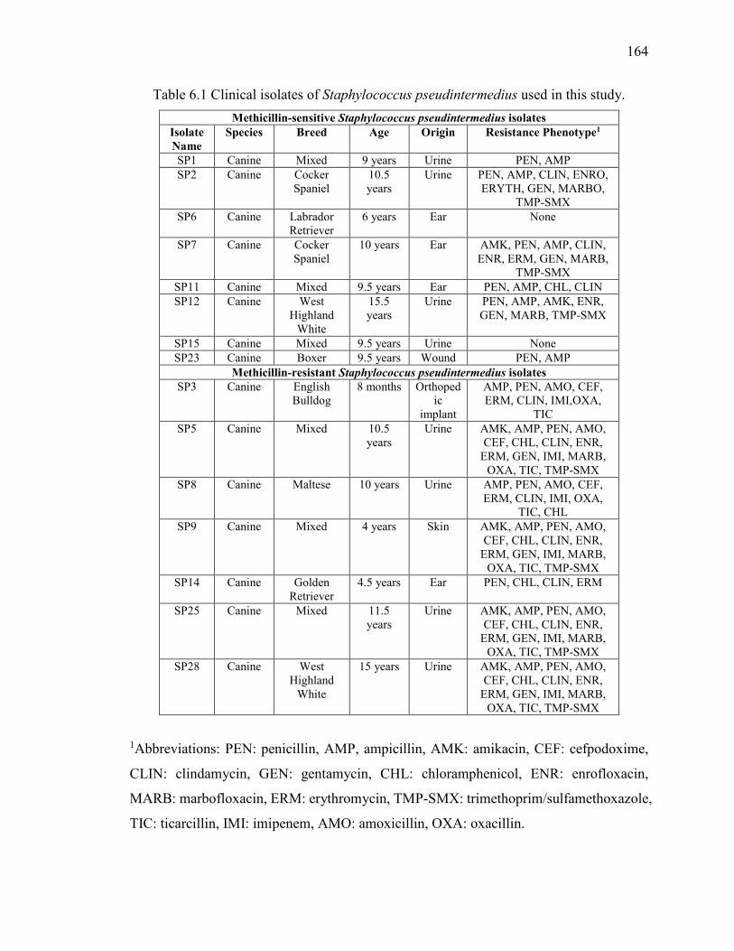
164
Table 6.1 Clinical isolates of Staphylococcus pseudintermedius used in this study.
Methicillin-sensitive Staphylococcus pseudintermedius isolates Isolate Name
Species Breed Age Origin Resistance Phenotype1
SP1 Canine Mixed 9 years Urine PEN, AMP
SP2 Canine Cocker Spaniel
10.5 years
Urine PEN, AMP, CLIN, ENRO, ERYTH, GEN, MARBO,
TMP-SMX
SP6 Canine Labrador Retriever
6 years Ear None
SP7 Canine Cocker Spaniel
10 years Ear AMK, PEN, AMP, CLIN, ENR, ERM, GEN, MARB,
TMP-SMX
SP11 Canine Mixed 9.5 years Ear PEN, AMP, CHL, CLIN
SP12 Canine West Highland
White
15.5 years
Urine PEN, AMP, AMK, ENR, GEN, MARB, TMP-SMX
SP15 Canine Mixed 9.5 years Urine None
SP23 Canine Boxer 9.5 years Wound PEN, AMP
Methicillin-resistant Staphylococcus pseudintermedius isolates SP3 Canine English
Bulldog 8 months Orthoped
ic implant
AMP, PEN, AMO, CEF, ERM, CLIN, IMI,OXA,
TIC
SP5 Canine Mixed 10.5 years
Urine AMK, AMP, PEN, AMO, CEF, CHL, CLIN, ENR,
ERM, GEN, IMI, MARB, OXA, TIC, TMP-SMX
SP8 Canine Maltese 10 years Urine AMP, PEN, AMO, CEF, ERM, CLIN, IMI, OXA,
TIC, CHL
SP9 Canine Mixed 4 years Skin AMK, AMP, PEN, AMO, CEF, CHL, CLIN, ENR,
ERM, GEN, IMI, MARB, OXA, TIC, TMP-SMX
SP14 Canine Golden Retriever
4.5 years Ear PEN, CHL, CLIN, ERM
SP25 Canine Mixed 11.5 years
Urine AMK, AMP, PEN, AMO, CEF, CHL, CLIN, ENR,
ERM, GEN, IMI, MARB, OXA, TIC, TMP-SMX
SP28 Canine West Highland
White
15 years Urine AMK, AMP, PEN, AMO, CEF, CHL, CLIN, ENR,
ERM, GEN, IMI, MARB, OXA, TIC, TMP-SMX
1Abbreviations: PEN: penicillin, AMP, ampicillin, AMK: amikacin, CEF: cefpodoxime,
CLIN: clindamycin, GEN: gentamycin, CHL: chloramphenicol, ENR: enrofloxacin,
MARB: marbofloxacin, ERM: erythromycin, TMP-SMX: trimethoprim/sulfamethoxazole,
TIC: ticarcillin, IMI: imipenem, AMO: amoxicillin, OXA: oxacillin.
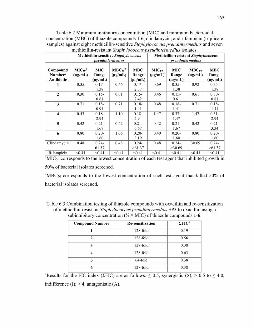
165
Table 6.2 Minimum inhibitory concentration (MIC) and minimum bactericidal concentration (MBC) of thiazole compounds 1-6, clindamycin, and rifampicin (triplicate samples) against eight methicillin-sensitive Staphylococcus pseudintermedius and seven
methicillin-resistant Staphylococcus pseudintermedius isolates. Methicillin-sensitive Staphylococcus
pseudintermedius
Methicillin-resistant Staphylococcus
pseudintermedius
Compound Number/ Antibiotic
MIC50
1 (µg/mL)
MIC
Range (µg/mL)
MBC50
2 (µg/mL)
MBC Range
(µg/mL)
MIC50
(µg/mL)
MIC
Range (µg/mL)
MBC50
(µg/mL)
MBC Range
(µg/mL) 1 0.35 0.17-
1.38 0.46 0.17-
2.77 0.69 0.35-
1.38 0.92 0.35-
1.38
2 0.30 0.15-0.61
0.61 0.15-2.42
0.46 0.15-0.61
0.61 0.30-0.81
3 0.71 0.18-0.94
0.71 0.18-1.41
0.48 0.18-1.41
0.71 0.18-1.41
4 0.43 0.18-2.94
1.10 0.18-2.94
1.47 0.37-1.47
1.47 0.31-2.94
5 0.42 0.21-1.67
0.42 0.21-6.67
0.42 0.21-1.67
0.42 0.21-3.34
6 0.80 0.20-1.60
1.06 0.20-3.19
0.40 0.20-1.60
0.80 0.20-1.60
Clindamycin 0.48 0.24-61.37
0.48 0.24- >61.37
0.48 0.24- >30.69
30.69 0.24- >61.37
Rifampicin <0.41 <0.41 <0.41 <0.41 <0.41 <0.41 <0.41 <0.41 1MIC50 corresponds to the lowest concentration of each test agent that inhibited growth in
50% of bacterial isolates screened.
2MBC50 corresponds to the lowest concentration of each test agent that killed 50% of
bacterial isolates screened.
Table 6.3 Combination testing of thiazole compounds with oxacillin and re-sensitization of methicillin-resistant Staphylococcus pseudintermedius SP3 to oxacillin using a
subinhibitory concentration (½ × MIC) of thiazole compounds 1-6.
Compound Number Re-sensitization ƩFIC1
1 128-fold 0.19
2 128-fold 0.56
3 128-fold 0.38
4 128-fold 0.63
5 64-fold 0.38
6 128-fold 0.38
1Results for the FIC index (ƩFIC) are as follows: ≤ 0.5, synergistic (S); > 0.5 to ≤ 4.0,
indifference (I); > 4, antagonistic (A).
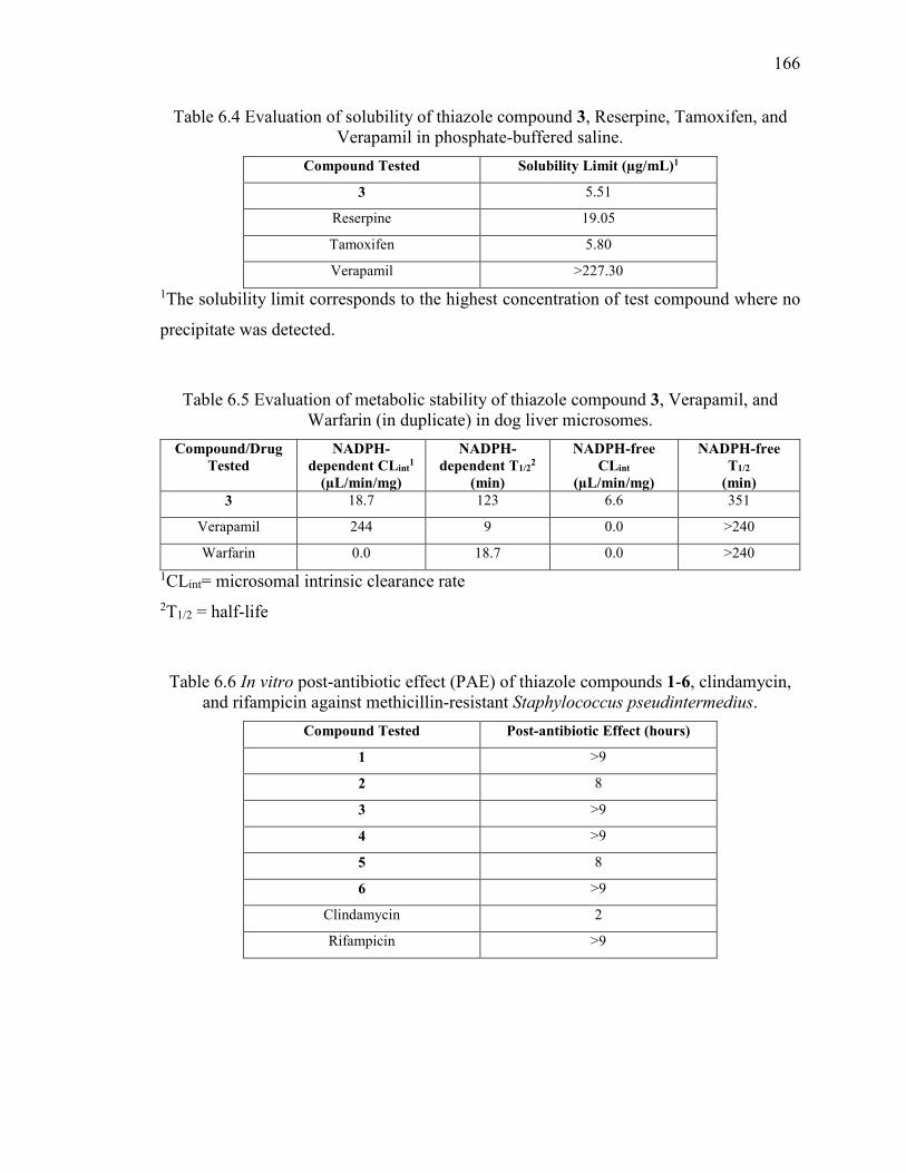
166
Table 6.4 Evaluation of solubility of thiazole compound 3, Reserpine, Tamoxifen, and Verapamil in phosphate-buffered saline.
Compound Tested Solubility Limit (µg/mL)1
3 5.51
Reserpine 19.05
Tamoxifen 5.80
Verapamil >227.30
1The solubility limit corresponds to the highest concentration of test compound where no
precipitate was detected.
Table 6.5 Evaluation of metabolic stability of thiazole compound 3, Verapamil, and Warfarin (in duplicate) in dog liver microsomes.
Compound/Drug Tested
NADPH-dependent CLint
1
(µL/min/mg)
NADPH-dependent T1/2
2 (min)
NADPH-free CLint
(µL/min/mg)
NADPH-free T1/2
(min) 3 18.7 123 6.6 351
Verapamil 244 9 0.0 >240
Warfarin 0.0 18.7 0.0 >240
1CLint= microsomal intrinsic clearance rate
2T1/2 = half-life
Table 6.6 In vitro post-antibiotic effect (PAE) of thiazole compounds 1-6, clindamycin, and rifampicin against methicillin-resistant Staphylococcus pseudintermedius.
Compound Tested Post-antibiotic Effect (hours)
1 >9
2 8
3 >9
4 >9
5 8
6 >9
Clindamycin 2
Rifampicin >9
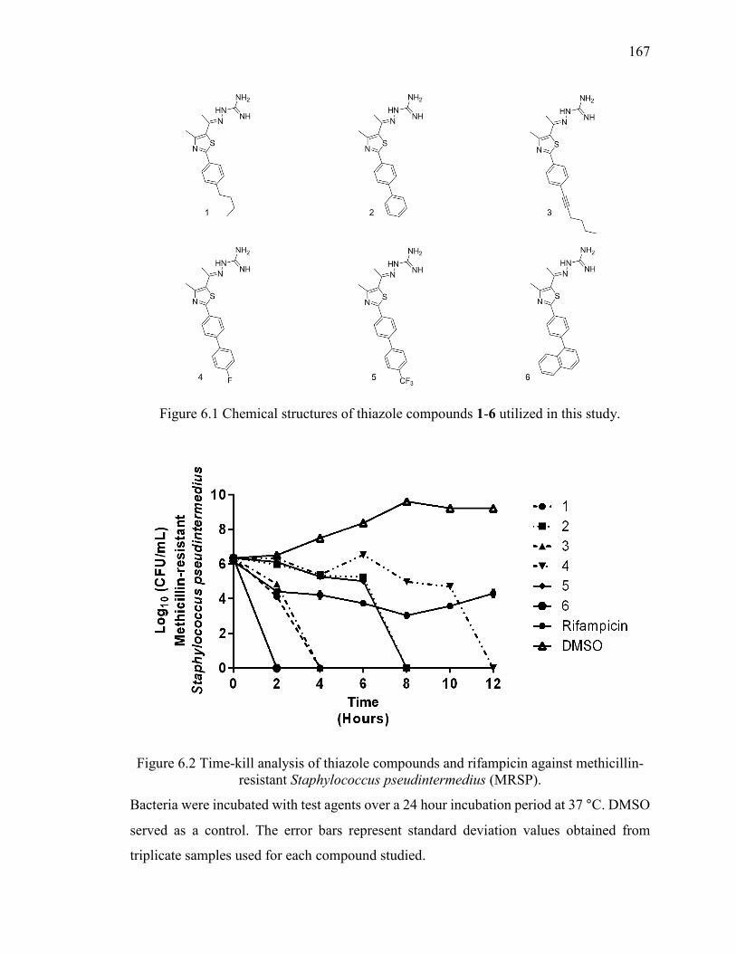
167
Figure 6.1 Chemical structures of thiazole compounds 1-6 utilized in this study.
Figure 6.2 Time-kill analysis of thiazole compounds and rifampicin against methicillin-resistant Staphylococcus pseudintermedius (MRSP).
Bacteria were incubated with test agents over a 24 hour incubation period at 37 °C. DMSO
served as a control. The error bars represent standard deviation values obtained from
triplicate samples used for each compound studied.
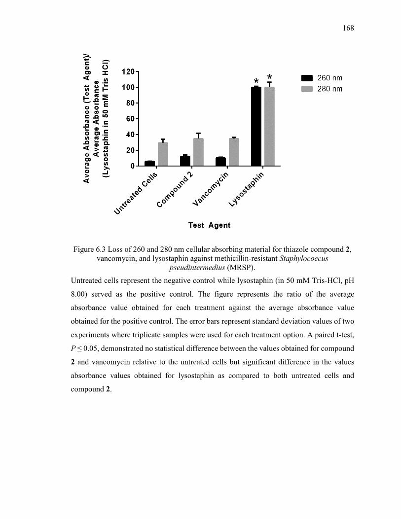
168
Figure 6.3 Loss of 260 and 280 nm cellular absorbing material for thiazole compound 2, vancomycin, and lysostaphin against methicillin-resistant Staphylococcus
pseudintermedius (MRSP).
Untreated cells represent the negative control while lysostaphin (in 50 mM Tris-HCl, pH
8.00) served as the positive control. The figure represents the ratio of the average
absorbance value obtained for each treatment against the average absorbance value
obtained for the positive control. The error bars represent standard deviation values of two
experiments where triplicate samples were used for each treatment option. A paired t-test,
P ≤ 0.05, demonstrated no statistical difference between the values obtained for compound
2 and vancomycin relative to the untreated cells but significant difference in the values
absorbance values obtained for lysostaphin as compared to both untreated cells and
compound 2.
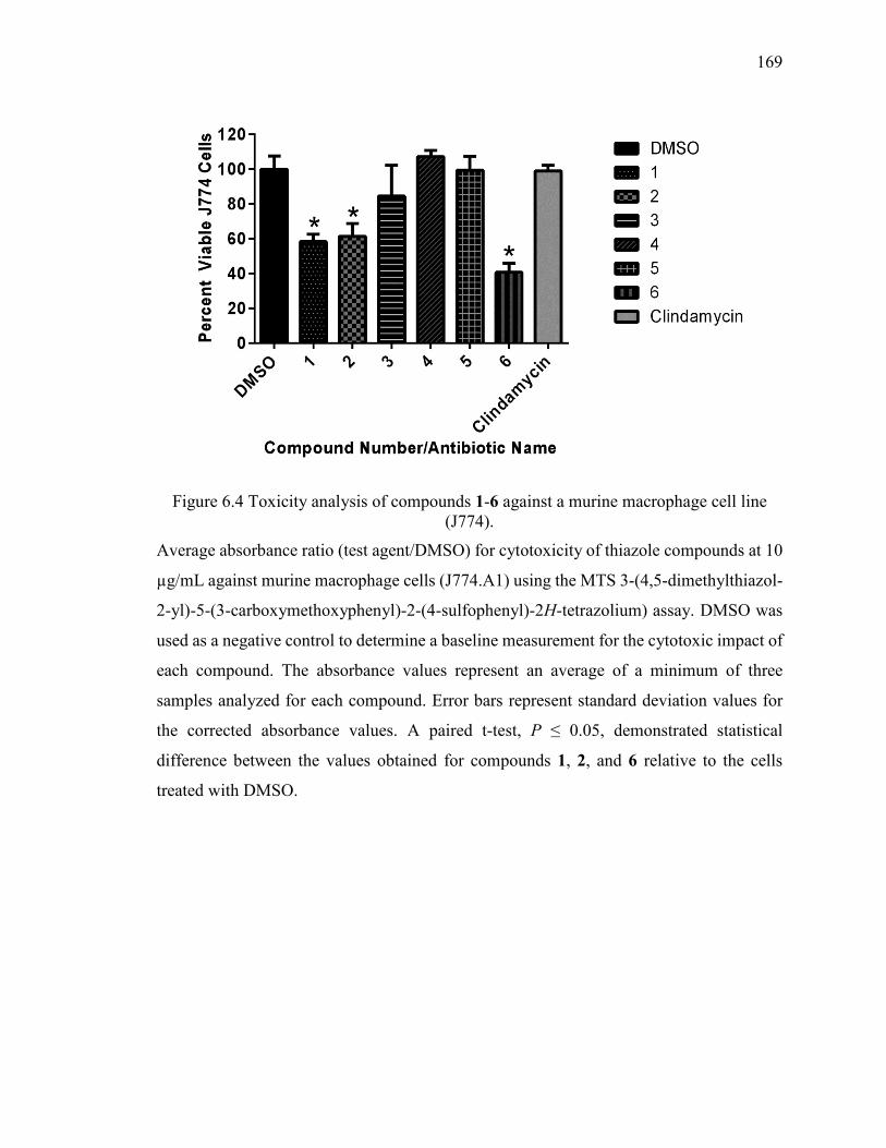
169
Figure 6.4 Toxicity analysis of compounds 1-6 against a murine macrophage cell line (J774).
Average absorbance ratio (test agent/DMSO) for cytotoxicity of thiazole compounds at 10
µg/mL against murine macrophage cells (J774.A1) using the MTS 3-(4,5-dimethylthiazol-
2-yl)-5-(3-carboxymethoxyphenyl)-2-(4-sulfophenyl)-2H-tetrazolium) assay. DMSO was
used as a negative control to determine a baseline measurement for the cytotoxic impact of
each compound. The absorbance values represent an average of a minimum of three
samples analyzed for each compound. Error bars represent standard deviation values for
the corrected absorbance values. A paired t-test, P ≤ 0.05, demonstrated statistical
difference between the values obtained for compounds 1, 2, and 6 relative to the cells
treated with DMSO.
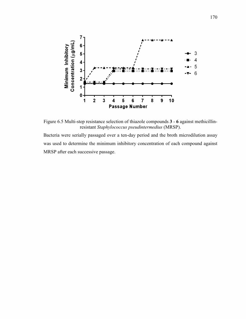
170
Figure 6.5 Multi-step resistance selection of thiazole compounds 3 - 6 against methicillin-resistant Staphylococcus pseudintermedius (MRSP).
Bacteria were serially passaged over a ten-day period and the broth microdilution assay
was used to determine the minimum inhibitory concentration of each compound against
MRSP after each successive passage.
171
CHAPTER 7. PHENYLTHIAZOLE ANTIBACTERIAL AGENTS TARGETING CELL WALL SYNTHESIS EXHIBIT POTENT
ACTIVITY IN VITRO AND IN VIVO AGAINST VANCOMYCIN-RESISTANT ENTEROCOCCI
7.1 Introduction
According to the US CDC, there are approximately 1.7 million hospital-acquired infections
(HAIs) in the US each year resulting in nearly 100,000 deaths and an estimated $20 billion
in healthcare costs (1). Many of the organisms responsible have become resistant to most
antibiotics, contributing to prolonged illness, high treatment costs, increased treatment
failure, and death (2, 3). Bacterial pathogens such as vancomycin-resistant enterococci
(VRE), in particular isolates of Enterococcus faecalis and Enterococcus faecium, are of
particular concern and have been identified as leading sources of nosocomial infections
(4). These range from skin infections to intra-abdominal infections to urinary tract
infections, with immunocompromised individuals—including the elderly and patients
undergoing organ transplants and cancer chemotherapy—being at particular risk (4).
Enterococcal infections were initially susceptible to many therapeutic agents including
β-lactams (in particular ampicillin), glycopeptides (vancomycin), fluoroquinolones, and
aminoglycosides (5). However, the ability of enterococci to colonize the gastrointestinal
tract of patients hospitalized for long periods has permitted these organisms to acquire
resistance, particularly after repeated drug exposure (4), severely limiting the number of
effective therapeutic options available. Moreover, VRE strains have been isolated that
exhibit resistance to newer antibacterial agents, including linezolid and daptomycin (6, 7).
The problem of antibiotic resistance is compounded by the diminishing number of new
antibiotics being approved. From 1980–1984, 19 new antibiotics were approved by the US
FDA and this number plummeted to one new approval from 2010–2012 (8). The approval
of three new antibiotics in 2014 indicates regulatory agencies understand the urgent need
for new treatment options (9) but the ever-present nature of resistance development
necessitates the continuous search for new drugs, and new drug leads.
Our group recently explored the antimicrobial activity of a broad range of
phenylthiazoles against drug-resistant staphylococci (10-14), three of which (1-3, Figure
172
1) had potent activity against both E. faecalis and E. faecium. We examine here the activity
of these compounds against clinical isolates of VRE; their mechanisms of action; potential
synergies with other antibiotics; toxicity, and activity in an in vivo model of VRE infection,
in Caenorhabditis elegans.
7.2 Materials and Methods
7.2.1 Synthesis of Thiazole Compounds 1-3
Synthetic schemes, spectral data, and purity (>95%, determined by HPLC) of compounds
1-3 (Figure 1), in addition to all intermediates, have been reported elsewhere (12, 14).
7.2.2 Bacterial Strains and Reagents Used
Clinical isolates of E. faecalis and E. faecium were obtained through BEI Resources (Table
7.1). Klebsiella pneumoniae, Acinetobacter baumannii, Pseudomonas aeruginosa, and
Enterobacter cloacae were obtained from the American Type Culture Collection
(Manassas, VA, USA). Escherichia coli OP50, E. coli 1411 and E. coli ΔAcrAB were
described before (15, 16). The human colorectal cell line (HRT-18) was obtained from the
American Type Culture Collection (Manassas, VA, USA). Ampicillin (IBI Scientific,
Peosta, IA), chloramphenicol (Sigma-Aldrich, St. Louis, MO, USA), ciprofloxacin (Enzo
Life Sciences, Farmingdale, NY, USA), doxycycline monohydrate (Alfa Aesar, Ward Hill,
MO, USA), linezolid (Chem-Impex International Inc., Wood Dale, IL, USA), and
vancomycin hydrochloride (Gold Biotechnology Inc., St. Louis, MO, USA) were from the
vendors noted above. Compounds were dissolved in dimethyl sulfoxide (DMSO) (for
ampicillin, doxycycline, linezolid, and vancomycin), ethanol (for chloramphenicol), or 0.1
N HCl (for ciprofloxacin), in order to prepare stock solutions (10 mg/mL). Tryptic soy
broth (TSB), tryptic soy agar (TSA), and brain heart infusion broth (BHI) were purchased
from Becton, Dickinson and Company (Franklin Lakes, NJ, USA). Phosphate-buffered
saline (PBS) (Sigma-Aldrich, St. Louis, MO, USA), RPMI-1640 medium (American Type
Culture Collection, Manassas, VA, USA), fetal horse serum (American Type Culture
Collection, Manassas, VA, USA), and 96-well plates (CellTreat Scientific Products,
Shirley, MA, USA) were all purchased from the vendors listed above. Nematode growth
medium (NGM) and M9 medium were prepared as described in the literature (17).
173
7.2.3 Determination of Minimum Inhibitory Concentration (MIC) and Minimum Bactericidal Concentration (MBC)
The MICs of thiazole compounds 1-3 and control antibiotics (linezolid, vancomycin) were
determined against all bacterial strains tested using a broth microdilution method,
following the guidelines outlined by the Clinical and Laboratory Standards Institute, as
described elsewhere (14, 18). Strains of E. faecalis, A. baumannii, E. cloacae, E. coli and
P. aeruginosa were cultured in TSB, while E. faecium strains were cultured in BHI. For
gram-negative bacterial strains, the MIC was also tested in the presence of a sub-inhibitory
concentration (¼ × MIC) of colistin (to permeabilize the outer membrane). Concentrations
of colistin used were 0.25 µM for A. baumanii and E. cloacae, 0.0625 µM for E. coli and
K. pneumoniae, and 1 µM for P. aeruginosa. Plates containing test agents and bacteria
were incubated at 37 °C for 18-22 hours prior to determining the MIC. The MIC was taken
to be the lowest concentration of each test agent where bacterial growth could not be
visualized.
The MBC for each test agent against E. faecalis and E. faecium was determined using
basically the methods described previously (14), with the following modifications.
Aliquots (5 µL) of E. faecalis were transferred to TSA plates while aliquots of E. faecium
were transferred onto BHI agar plates. Plates were incubated at incubated at 37 °C for 18-
22 hours before the MBC, the concentration where >99% reduction in bacterial cell count
was observed, was determined.
7.2.4 Time-Kill Analysis of Compounds 1-3 and Linezolid against VRE
Vancomycin-resistant E. faecalis ATCC HM-201 and vancomycin-resistant E. faecium
ATCC 700221 cells in late logarithmic growth phase were diluted to ~5 × 10⁶ colony-
forming units (CFU)/mL and (in triplicate) exposed to concentrations equivalent to 4 ×
MIC of 1-3 or linezolid in either TSB (for E. faecalis) or BHI (for E. faecium). 100 µL
samples were collected after 0, 2, 4, 6, 8, 10, 12, and 24 hours of incubation at 37 ºC, and
subsequently serially diluted in PBS. Bacteria were then transferred to either TSA (for E.
faecalis) or BHI agar plates (for E. faecium) and were incubated at 37 °C for 18-22 hours
before viable CFU/mL values were determined.
174
7.2.5 Cytotoxicity Analysis of Thiazole Compounds in Cell Culture
Compounds 1-3 were assayed at concentrations of 5 µg/mL, 10 µg/mL, 20 µg/mL, and 40
µg/mL against a human colorectal (HRT-18) cell line to determine their effects to
mammalian cells in vitro, as described elsewhere (19). Cells were cultured in RPMI-1640
medium with 10% fetal horse serum at 37 °C with 5% CO2. Cells were incubated with
compounds in 96-well plates at 37 ºC and 5% CO2 for either 2 or 24 hours prior to addition
of the assay reagent MTS 3-(4,5-dimethylthiazol-2-yl)-5-(3-carboxymethoxyphenyl)-2-(4-
sulfophenyl)-2H-tetrazolium) (Promega, Madison, WI, USA). Absorbance readings (at
OD490) were taken using a kinetic microplate reader (Molecular Devices, Sunnyvale, CA,
USA). The quantity of viable cells after treatment with each compound are expressed as a
percentage of the viability of untreated cells.
7.2.6 Single-Step Resistance Selection
The frequency of spontaneous single-step resistance of E. faecalis ATCC 31975 to the
phenylthiazole compounds was carried out as described elsewhere (20, 21). Briefly,
bacterial cultures (ranging from 5 × 10⁸ CFU/mL to 5 × 10¹⁰ CFU/mL) were spread onto
TSA plates (10-mm diameter) containing compound 1, 2, or 3 at either 2 ×, 4 ×, or 8 ×
MIC. Plates were incubated aerobically at 37 °C for 48 hours. The MIC of colonies present
on each plate was checked (to determine any shift relative to the wild-type strain) using the
broth microdilution method outlined above. The frequency of resistance was calculated as
the number of resistant colonies per inoculum (21).
7.2.7 Multi-Step Resistance Selection of VRE to Thiazole Compounds
To assess the ability of VRE to develop resistance to the thiazole compounds after repeated
exposure, a multi-step resistance selection experiment was performed, as described
elsewhere (22). The broth microdilution method for MIC determination against a clinical
isolate of vancomycin-resistant E. faecalis (ATCC 51299) and an isolate of vancomycin-
resistant E. faecium (ATCC HM-968) was repeated for 14 passages over a period of two
weeks. The initial inoculum was prepared to a McFarland standard of 0.5. The solution was
subsequently diluted 1:300 in either TSB (for E. faecalis) or BHI (for E. faecium) to reach
a starting inoculum of 1 × 10⁵ CFU/mL. For each subsequent passage, the inoculum for the
175
MIC determination was adjusted to a final density of approximately 5 × 10⁵ CFU/mL using
the contents of a well containing a sub-inhibitory concentration of the compound (where
bacterial growth was observed from the previous passage). Bacteria were then transferred
to a new 96-well microtiter plate. Phenylthiazole compounds 1-3, ampicillin, daptomycin,
and linezolid were added (in triplicate) to wells in the first row of the microtiter plate, and
then serially diluted along the vertical axis. The plate was incubated at 37 °C for a minimum
of 18 hours before the MIC was determined, by visual inspection. Resistance was classified
as a greater than four-fold increase in the initial MIC, as reported elsewhere (23).
7.2.8 Bacterial Cytological Profiling of Thiazole Compounds against Bacillus subtilis and E. coli
Cells were grown in Luria Bertani (LB) medium at 30 °C (E. coli) or 37 °C (B. subtilis)
until the optical density at 600 nm (OD600
) was ~0.2. Cells were then left untreated, treated
with compounds, or treated with compounds in the presence of 0.5 M sucrose (E. coli) or
MSM (B. subtilis) as described previously (24-26). After 30 minutes or two hours, cells
were stained with FM 4−64 (1 µg/mL) to visualize the membranes; DAPI (2 µg/mL E. coli,
1 µg/ml B. subtilis) to visualize the DNA, and SYTOX Green (1 µg/mL), a vital stain which
is normally excluded from cells with an intact membrane but brightly stains cells that are
lysed (26). Images were collected using a Delta Vision Spectris Deconvolution microscope,
as described previously (26).
7.2.9 Inhibition of Cell Wall Synthesis in Enterococci by Compound 1 via UDP-N-acetylmuramyl-pentapeptide Accumulation
To investigate whether the phenylthiazole compounds exhibit their effects on enterococci
by inhibiting cell wall synthesis, as suggested by previous work in which we found
synergistic activity of thiazoles with known cell-wall biosynthesis inhibitors, we
determined the accumulation of the final soluble cell wall precursor (UDP-N-
acetylmuramyl-pentapeptide) inside bacterial cells. We used the procedure described
previously (27), with the following modifications: E. faecalis NR-31975, in early
logarithmic growth stage (OD600 ~ 0.5), was incubated with 130 µg/mL chloramphenicol
for 15 minutes at 37 ºC. Bacteria were subsequently incubated with either 10 × MIC of
compound 1 or vancomycin (positive control) for 30 minutes at 37 ºC. Untreated samples
176
served as a negative control. Samples were then centrifuged at 10,000 rpm, the supernatant
discarded, and the pellet re-suspended in 1 mL of sterile deionized water. The cell pellet
was boiled at 100 ºC for 30 minutes before samples were chilled on ice for 10 minutes.
UDP-N-acetylmuramyl-pentapeptide levels were measured using an Agilent High
Performance Liquid Chromatograph coupled to a time-of-flight mass spectrometer (HPLC-
MS). A Waters XBridge Phenyl (2.1 x 100 mm, 3.5 um) column was used, with mobile
phases of water, 0.1% formic acid (Buffer A) and acetonitrile, 0.1% formic acid (Buffer
B). A gradient of 5-20% Buffer B over 14 minutes was then employed at a flow rate of 0.3
mL/min, with an electrospray source in positive ionization mode. Extracted ion
chromatograms (EICs) were generated at a m/z of 1150.3588 (20 ppm window). The mass
error for UDP-N-acetylmuramyl-pentapeptide was less than 1 ppm.
7.2.10 Molecular Target Identification Using Genomic Insertion of a Transposon with a Strong Outward-Oriented Promoter
Overexpression of the target/resistance mechanism was carried out using a transposon with
a strong outward-oriented promoter for the random overexpression of neighboring genes,
in Bacillus subtilis. The pEP26 delivery vector carrying the transposon with the promoter
(TnHyJump) was transformed into B. subtilis, as described (28). For transposon integration
into bacterial DNA, cells were grown for 10 hours at 25 ºC, serially diluted, sub-cultured
in dual-selection TSA plates containing 5 µg/ml chloramphenicol (for transposon selection)
and 3 × MIC compound 1 (for selection of compound resistance), then incubated overnight
at 42 ºC. Growth at 42 ºC is non-permissive for the maintenance of the delivery vector, so
chloramphenicol/compound 1 resistance arises mainly from the chromosomal insertion of
the transposon. 12 colonies out of 142 colonies on 3 × MIC compound 1 were screened for
MIC shift (resistance) against compound 1 using the broth microdilution method to confirm
resistance. Genomic DNA was extracted from resistant colonies (recombinants that were
capable of growth at concentrations that were inhibitory to the control) and were sent to
the Purdue Genomics Core Facility for sequencing. Insertion sites were identified by
sequencing. Transposon location within the resistant B. subtilis genome, orientation, and
flanking genes were determined by performing a BLASTN search on the NCBI public
BLAST server.
177
7.2.11 HsFPPS, EcUPPS and EcUPPP Inhibition Assays
Human FPPS, EcUPPS and EcUPPP were purified as described previously (29-31).
Compound 1 was prepared as a 20 mM stock solution in DMSO and then serially diluted
from 200 μM to 0.2 μM. 0.8 mg EcUPPS was incubated with compound 1 at room
temperature for 30 minutes in HEPES buffer (100 mM HEPES, 50 mM NaCl, 0.5 mM
MgCl2, 0.02% DDM (w/v), pH 7.5) before adding the reaction mixture with 10 μM IPP
and FPP, 0.375 U/mL inorganic phosphatase. The 100 μL reaction was quenched by the
same volume of the malachite green mixture from a malachite green phosphate assay kit
(BioAssay Systems). For the EcUPPP inhibition assay, the 20 mM stock solution of
compound 1 was serially diluted from 300 μM to 0.8 μM. Compound 1 was incubated with
0.125 μM EcUPPP at room temperature for 15 minutes in HEPES buffer (50 mM HEPES,
150 mM NaCl, 10 mM MgCl2, 0.02% DDM (w/v), pH 7.5), followed by the addition of 35
µM FPP and incubation at 37 °C for 20 minutes. The 100 μL reaction was then mixed with
the same volume of the malachite green mixture. The released phosphate in EcUPPS and
EcUPPP assays was monitored by absorbance at 620 nm after 30 minutes development.
Dose response curves were constructed using GraphPad PRISM (Graphpad Software, San
Diego, CA).
7.2.12 Uncoupler Assays
Proton translocation out of EcIMVs was measured by the fluorescence increase of ACMA.
The excitation and emission wavelengths were 410 and 480 nm, respectively. IMVs (0.1
mg/mL membrane protein), 2 μM ACMA and 0.5 mM ATP/succinate were added in
HEPES buffer (10 mM HEPES-KOH, 5 mM MgSO4, 100 mM KCl, pH 7.5). The
uncoupler CCCP and compound 1 were serially diluted in the reaction mixture. Dose
response curves were constructed using GraphPad PRISM (Graphpad Software, San Diego,
CA).
7.2.13 Re-sensitization of VRE to Vancomycin and Aminoglycoside Antibiotics
TSB (for E. faecalis ATCC HM-201 and E. faecalis ATCC 51299) or BHI (for E. faecium
ATCC 700221 and E. faecium HM-968) were inoculated with VRE (5×10⁵ CFU/mL), as
described previously (13), with the following modifications. Aliquots (5 mL) of the
178
bacterial suspensions were divided into micro-centrifuge tubes and compounds 1, 2, or 3
(at ½ × MIC) were introduced into each tube. After sitting at room temperature for 30
minutes, 1 mL samples from each tube were transferred to a new micro-centrifuge tube,
prior to addition of the antibiotic (either vancomycin or gentamicin at concentrations
equivalent to their MIC). Plates containing the test agents and bacteria were then incubated
at 37 °C for 18-21 hours after which the MIC values were measured. A fold-reduction was
calculated by comparing the MIC of the antibiotic alone compared to the MIC of the
antibiotic given in combination with the phenylthiazole compound.
7.2.14 Combination Therapy of Phenylhiazole Compounds With Conventional Antibiotics
Possible synergistic interactions between the thiazole compounds and ampicillin,
ciprofloxacin, doxycycline, and linezolid were determined via checkerboard assay (32).
Initially, compound 1 was examined in combination with the four antibiotics against a
single strain of E. faecalis (ATCC 51299) prior to investigating compounds 1-3 in
combination with ciprofloxacin against E. faecalis ATCC 49532 and E. faecalis ATCC
49533. Bacteria equivalent to a McFarland standard of 0.5 were prepared in PBS. The
bacteria were then diluted in TSB to achieve a starting cell density of 1 × 10⁵ CFU/mL.
TSB was transferred to all wells of a 96-well micro-titer plate. The phenylthiazole
compounds and antibiotics were diluted in TSB to achieve a starting concentration
equivalent to 2 × or 4 × the MIC. Compounds were serially diluted along the horizontal
axis of the microtiter plate while the antibiotics were serially diluted along the vertical axis.
Plates were incubated for 20 hours at 37 °C. The MIC of the test compound in combination
with each antibiotic studied was taken to be the lowest concentration of each
compound/antibiotic where no visible growth of bacteria was observed. The fractional
inhibitory concentration index (ΣFIC) was calculated for each combination, as described
previously (11). A synergistic relationship was classified as an FIC index less than or equal
to 0.5. FIC values above 0.5 but less than 2.0 were characterized as additive, values
between 2.0 and 4.0 characterized as indifference, while FIC values above 4.0 were
classified as antagonistic.
179
7.2.15 In vivo Analysis of Toxicity and Efficacy of Phenylthiazole Compounds
To examine the toxicity of the thiazole compounds in vivo and to examine their efficacy in
treating a VRE infection in vivo, we used the C. elegans animal model. The temperature-
sensitive sterile mutant strain C. elegans AU37 [sek-1(km4); glp-4(bn2) I] was used
because this strain is sterile at room temperature and capable of laying eggs only at 15 °C.
Additionally, the strain is quite susceptible to infection, due to a mutation in the sek-1 gene
of the p38 mitogen-activated protein kinase pathway (33, 34). Briefly, worms were grown
for 5 days at 15 °C (permitting worms to lay eggs) on NGM agar plates seeded with a lawn
of E. coli OP50. Eggs were harvested by bleaching (35) and maintained for 24 hours at
room temperature with gentle agitation, for hatching. Hatched larvae were transferred to a
new NGM plate seeded with E. coli OP50 and were kept at room temperature for 4-5 days
until the worms reached their adult growth stage. Adult worms were then collected and
washed three times with PBS in a 1:10 (worm:PBS) ratio to remove E. coli.
In order to examine the toxicity of the thiazole compounds to C. elegans, 15-20 adult
worms were transferred to wells of a 96-well microtiter plate. Worms were incubated with
either 10 or 20 µg/mL of compounds 1, 3, linezolid (positive control), or sterile water
(negative control) (in triplicate). After 24, 48, 72, 96, and 120 hours, worms were examined
microscopically to examine viability. The number of worms that survived each treatment
regimen were counted and results are presented as percent viable worms.
To test the antibacterial activity of the thiazole compounds against VRE in vivo, adult
worms were transferred to TSA agar plates seeded with a lawn of E. faecalis HM-201, for
infection (36). After two hours of infection, worms were collected and washed with M9
buffer, three times, before transferring 20-25 worms to wells in a 96-well microtiter plate.
Worms were incubated with 20 µg/mL of compounds 1, 3, linezolid (positive control), or
sterile water (negative control) (in triplicate). After treatment for 20 hours, worms were
washed three times with M9 buffer and then examined microscopically to examine
morphological changes, and viability. They were subsequently lysed in micro-centrifuge
tubes containing 200 mg of 1.0-mm silicon carbide particles (Biospec Products,
Bartlesville, OK) that were vortexed for one minute. Samples were serially diluted and
plated onto TSA plates containing 50 µg/mL gentamicin to select for VRE. Plates were
incubated at 37 °C for 18 hours before viable CFUs were determined.
180
7.2.16 In silico Pharmacokinetic Analysis
The pharmacokinetic profile (oral and intravenous dose of 600 mg) was simulated using
chemPK version 2.0 (Cyprotex Inc., Cheshire, United Kingdom), for compounds 1, 3, and
linezolid.
7.3 Results
7.3.1 Antibacterial Activity of Compounds 1-3 Against the ESKAPE Pathogens Enterococcus faecium, Klebsiella pneumoniae, Acinetobacter baumannii,
Pseudomonas aeruginosa, and Enterobacter spp
We first investigated the antibacterial activity of phenylthiazole compounds 1-3 against a
panel of ESKAPE pathogens using the broth micro-dilution method. Examination of the
spectrum of activity of 2-3 revealed that they were inactive against most Gram-negative
pathogens (minimum inhibitory concentration, MIC > 128 µg/mL), Table 7.2, the
exception being with A. baumannii where the MIC was 8 µg/mL, similar to that found with
erythromycin. Compound 1 also exhibited limited activity against the same pathogens
(MIC ranges from 8-64 µg/mL), indicating that all three compounds are generally
ineffective against Gram-negative bacteria. These results are in contrast to our earlier
results on S. aureus, differences that could be due to the presence of the outer membrane
(OM) in Gram-negative bacteria, and/or efflux pumps. We thus next investigated whether
the presence of the OM and/or efflux pumps did in fact contribute to the lack of
antibacterial activity observed for 1-3 against Gram-negative bacteria.
In the presence of a sub-inhibitory concentration of the membrane-disrupting
antibiotic colistin, which permeabilizes the outer membrane, the MIC of compounds 1-3
against Gram-negative bacteria decreased dramatically. For example, 2 and 3 were inactive
against K. pneumoniae, P. aeruginosa and E. coli when tested alone (MIC > 128 µg/mL),
but in the presence of sub-inhibitory levels of colistin, both compounds exhibited potent
antibacterial activity (MIC from 1 to 4 µg/mL). This behavior is similar to that seen with
the antibiotic erythromycin, whose activity is known to be impeded by the presence of the
OM. For example, with K. pneumoniae, P. aeruginosa, and E. coli, we find that
erythromycin alone lacks activity (MIC > 128 µg/mL), but in the presence of colistin,
erythromycin has potent activity against each organism (MIC from 0.5 to 1 µg/mL). It thus
181
appears likely that the OM impedes both phenylthiazole as well as erythromycin entry into
Gram-negative bacteria.
When the antibacterial activity of compounds 1-3 was examined against E. coli
1411 and a mutant strain containing a deletion of the gene encoding the AcrAB efflux
pump, there was a major decrease in the MIC in the mutant. Against E. coli 1411,
compound 1 inhibited growth at 64 µg/mL, and compounds 2 and 3 were inactive (MIC >
128 µg/mL). However, against the mutant strain (Escherichia coli 1411 ΔacrAB), all three
compounds were potent inhibitors of bacterial growth (MIC of 4 µg/mL). This behavior is
similar to that observed with linezolid, a known substrate of the AcrAB efflux pump (37).
It thus appears that the lack of activity of the phenylthiazoles against Gram-negative
bacterial pathogens is due both to the presence of the OM as well as drug efflux pumps.
Fortunately, in earlier work we found that compounds 1-3 exhibited potent antibacterial
activity against the Gram-positive pathogen, methicillin-resistant S. aureus (MIC values
ranged from 1.3 to 5.6 µg/mL) (10), and as shown in Table 7.2, all three compounds are
potent inhibitors of the growth of another important Gram-positive pathogen, vancomycin
resistant E. faecium (MIC = 0.5 µg/mL). Thus, the phenylthiazole compounds appear to be
potent leads against clinically-relevant Gram-positive pathogens, including MRSA and
VRE. Plus, the fact that they synergize with cell wall biosynthesis inhibitors in S. aureus
suggests, perhaps, a similar target area in enterococci.
7.3.2 Phenylthiazole Compounds Retain Their Potent Activity against Clinical Isolates of Drug-Resistant Enterococci
To further evaluate the antibacterial activity of compounds 1-3 against enterococci, we
determined MIC values against 24 strains of E. faecalis and E. faecium (including 16
strains resistant to vancomycin), isolated from diverse sources including blood, urine,
peritoneal fluid, sputum, and feces (Table 7.1), from infected patients. All three compounds
exhibited potent antibacterial activity against all isolates tested (Table 7.2). Interestingly,
1-3 were most active against isolates of vancomycin-resistant E. faecium, inhibiting growth
at concentrations ranging from 0.5 to 4 µg/mL (Table 7.3), with MIC50 values of 1 (for
compounds 2 and 3) to 4 µg/mL (for compound 1). These values are similar to those we
find with the antibiotic linezolid (MIC range from 0.5 to 2 µg/mL). Against E. faecalis, 1
and 3 retained their potent antibacterial activity (MIC range from 1-8 µg/mL), but there
182
was an increase in the MIC of compound 2 (MIC = 16 µg/mL, against multiple strains).
However, all compounds proved quite effective at inhibiting the growth of VRE. The
compounds also retained activity against isolates exhibiting resistance to ampicillin,
ciprofloxacin, gentamicin, streptomycin, erythromycin, and doxycycline. This indicates
that cross-resistance between these antibiotics and 1-3, against enterococci, is unlikely to
occur.
We next investigated whether the antibacterial effects were bacteriostatic, or
bactericidal. To address this, we determined the minimum bactericidal concentrations,
MBCs. Against most strains of E. faecalis and E. faecium, the MBC values were equal to
or higher than the MIC values for compounds 1 and 3, indicating that the two compounds
are bactericidal. A similar trend was observed for compound 2 against E. faecalis.
However, against E. faecium, the MBC for 2 was more than four-fold higher than the MIC,
indicating 2 may be bacteriostatic, particularly against vancomycin-resistant isolates. As
expected, ampicillin exhibited bactericidal activity against enterococci strains sensitive to
this antibiotic, while linezolid exhibited bacteriostatic activity against both E. faecalis and
E. faecium (MBC was more than four-fold higher than the MIC).
7.3.3 Compounds 1 and 3 Rapidly Eradicate Vancomycin-Resistant Enterococci as Determined by Time-Kill Analysis
In order to confirm the bactericidal activity of the phenylthiazole compounds against VRE,
we carried out time-kill assays. As shown in Figure 7.2, compounds 1 and 3 exhibited rapid
bactericidal activity against both vancomycin-resistant E. faecium as well as vancomycin-
resistant E. faecalis. Compound 1 (at 4 × MIC) was capable of completely eradicating both
strains of VRE within two hours, and no bacterial re-growth was observed over the
following 22 hours. Compound 3 matched compound 1 in completely eliminating
vancomycin-resistant E. faecalis within two hours (Figure 7.2B), but required four hours
to achieve the same effect against vancomycin-resistant E. faecium (Figure 7.2A).
Compound 2 also exhibited rapid bactericidal activity against E. faecalis, completely
eradicating bacterial growth within two hours. However, although 2 produced a gradual
reduction in CFU against vancomycin-resistant E. faecium after 24 hours, the compound
was not capable of completely eradicating the bacteria. This supports the MBC results for
2 against vancomycin-resistant E. faecium, indicating that 2 is bacteriostatic. The
183
bacteriostatic activity of linezolid against both E. faecium and E. faecalis is also confirmed
since the antibiotic was not able to generate a 103 × reduction in VRE CFU over 24 hours.
7.3.4 Compounds 1-3 Exhibit Limited Toxicity to Human Colorectal Cells
Enterococci are commensal organisms of the human gastrointestinal tract. We thus
next examined the toxicity of all three compounds against a human colorectal (HRT-18)
cell line. When the compounds were incubated with cells for a short period (two hours),
compound 1 was non-toxic up to 20 µg/mL (Figure 7.3A). Compounds 2 and 3 exhibited
an improved toxicity profile since both were not toxic up to 40 µg/mL. When compounds
were incubated with HRT-18 cells for 24 hours (Figure 7.3B), the toxicity profile of 1
remained the same (not toxic up to 20 µg/mL), while 2 and 3 were toxic at 40 µg/mL, but
non-toxic at 20 µg/mL.
7.3.5 Single-Step and Multi-Step Resistance Selection of Enterococci to Compounds 1-
3
Given the promising results described above, we next sought to examine the likelihood that
enterococci will develop resistance to 1-3, using two different methods. First, we attempted
to isolate spontaneous mutants exhibiting resistance using a single-step resistance selection
experiment. In the presence of a high inoculum of E. faecalis NR-31975 (5 × 10⁸ CFU/mL),
no resistant mutants to 1-3 were isolated at concentrations of 2 ×, 4 ×, or 8 × MIC. When
the inoculum size was increased (to 5 × 10¹⁰ CFU/mL), the same result was obtained,
indicating a frequency of resistance > 2 × 10-11. We next attempted to isolate resistant
mutants to 1-3 via a multi-step resistance selection experiment using two strains of VRE:
E. faecalis ATCC 51299 and E. faecium ATCC HM-968. A four-fold increase in MIC was
categorized as resistance. Against E. faecium (Figure 7.4A), there was no shift in the MIC
observed for compounds 1 and 3 over 14 serial passages (similar to results obtained with
linezolid). A one-fold increase (that is, the MIC increases from x → 2x) in MIC was
observed for linezolid after the third passage and for compound 2, after the eleventh
passage. However, no additional increase in the MIC of either agent was observed
thereafter. Against E. faecalis (Figure 7.4B), a one-fold increase in MIC for compounds 1
and 3 was observed after the sixth passage; no further increase in MIC was observed until
the last passage. For compound 2, a one-fold increase in MIC was observed after the sixth
184
passage and an additional two-fold increase was observed after the eleventh passage. No
additional increase was observed thereafter. No increase in MIC was observed for linezolid
over the 14 serial passages. These results indicate a low likelihood for enterococci to
develop rapid resistance to, in particular, 1 and 3.
7.3.6 Compound 1 Exerts its Antibacterial Activity by Inhibiting Cell Wall Synthesis
In order to investigate the mechanism of action of the phenylthiazoles, compound 1 was
subjected to Bacterial Cytological Profiling (BCP) in representative Gram-positive
(Bacillus subtilis) and Gram-negative (E. coli) bacteria. BCP identifies the likely pathway
targeted by novel antibiotics by comparing their cytological effects with those found using
a library of cytological profiles generated by using antibacterials with known mechanisms
of action (MOAs), or by the rapid proteolytic depletion of essential proteins (24-26). When
E. coli ΔtolC (which lacks an effective efflux pump) was treated with compound 1, cells
lysed and formed spheroplasts after two hours (Figure 7.5). Spheroplasts and misshapen
cells were very prevalent in the presence of 0.5 M sucrose, which osmotically stabilizes
cells lacking a functional cell wall. Lysis and cell shape defects were observed as early as
30 minutes after addition of compound 1 to the medium. In contrast, cells incubated with
the cell wall biosynthesis inhibitor D-cycloserine formed misshapen cells and spheroplasts
after 30 minutes (Figure 7.5), and cells were completely lysed after two hours (data not
shown). These results suggest that compound 1 inhibits cell wall biosynthesis in E. coli
ΔtolC.
To determine whether 1 had the same effect in a Gram-positive bacterium, we
examined the effects of 1 in B. subtilis, again using BCP. B. subtilis incubated with 1 at 5
× MIC rapidly lysed, with 95% (n = 131) of cells being permeable to Sytox Green (a nucleic
acid stain that is impermeable to live cells) within 30 minutes of treatment. Since disruption
of either the cell wall or the cell membrane can result in permeabilized cells (Figure 7.6),
we next investigated the effects of 1 with control agents in the presence of dimethylsulfone
(a.k.a. methylsulfonyl methane, MSM), which osmotically stabilizes cells for better
observation of cell shape defects. Cells treated with 1 for two hours in the presence of
MSM were slightly misshapen or bent and contained pools of membrane (Figures 7.7 and
Figure 7.8). Figure 7.8 shows four examples of cells containing a small bulge at the site of
185
the bend that could be visualized with phase contrast microscopy. These cells appeared
very similar to vancomycin-treated cells, which also show subtle cell shape-defects,
forming bends, bulges, and pools of membrane (Figure 7.7 and Figure 7.4A). Unlike
compound 1 and vancomycin, D-cycloserine generated very obvious cell shape defects
(Figure 7.7), and Triton X-100 detergent-treated cells were lysed without affecting overall
cell shape. MSM suppresses cell lysis and permeability defects for cell wall active
antibiotics, but not for membrane active compounds (Figure 7.8 & Figure 7.9) (24). We
found MSM dramatically suppressed the permeability phenotypes of compound 1, as well
as vancomycin and D-cycloserine, but had no effect on Triton X-100 treated cells,
suggesting that 1 inhibits cell wall/peptidoglycan synthesis (Figure 7.8) in both B. subtilis
and E. coli ΔtolC, implicating broadly conserved target(s) and MOA. But what are the
actual targets of 1?
7.3.7 Target Identification
Peptidoglycan biosynthesis involves numerous enzymes and a simplified metabolic
pathway is shown in Figure 7.10. The first step involves the sequential addition of two
molecules of isopentenyl diphosphate (IPP, 4) to dimethylallyl diphosphate (DMAPP, 5)
to form the (C15) isoprenoid farnesyl diphosphate (FPP, 6) in a reaction catalyzed by
farnesyl diphosphate synthase (FPPS), with IPP/DMAPP being produced by the
mevalonate pathway in S. aureus and the non-mevalonate (methylerythritol phosphate,
MEP) pathway in B. subtilis and E. coli. FPP then reacts with 8 additional IPP molecules
to form the (C55) isoprenoid undecaprenyl diphosphate (UPP, 7) in a reaction catalyzed by
undecaprenyl diphosphate synthase (UPPS). UPP is converted to undecaprenyl
monophosphate (UP, 8) by undecaprenyl diphosphate phosphatase (UPPP), then UP reacts
with UDP-N-acetylmuramyl pentapeptide (9) to form Lipid I (10) in a reaction catalyzed
by MraY, followed by conversion to Lipid II, and after several more steps, peptidoglycan
(11) is formed. Drugs such as ampicillin and vancomycin inhibit these later stages in cell
wall synthesis as they interfere with peptidoglycan crosslinking.
We first sought to see if 1 resulted in changes in the concentration of compound 9,
the final soluble cell wall precursor in peptidoglycan biosynthesis, in E. faecalis NR-31975
cells. We found that treatment of cells with compound 1 resulted in a similar LC-MS result
186
to treatment of cells with vancomycin, a known inhibitor of bacterial cell wall synthesis.
This can be seen in the results shown in Figure 7.11 in which there are large increases in
compound 9 accumulation with 1 or vancomycin treatment, implicating inhibition of
peptidoglycan biosynthesis. A peak was present in the chromatograms at the same retention
time (~8.76 minutes) for both 1 and vancomycin-treated samples, and had the correct m/z
for the pentapeptide, m/z = 1150.3588, a <1 ppm error. These results support inhibition of
a target in the peptidoglycan biosynthesis pathway, but they do not suggest a specific
molecular target.
We next tried to identify the molecular target of the phenylthiazole 1 by using a
target overexpression experiment, in Bacillus subtilis. The metabolic function(s) in B.
subtilis inhibited by the compound should in principle be restored by over-expression of
the targeted protein(s) via genomic insertion of a transposon with a strong outward-oriented
promoter. That is, resistance to compound 1 should be achieved by over-expression of the
drug-resistance gene—which could be a molecular target in peptidoglycan biosynthesis—
but also perhaps an efflux pump (28). In the presence of a high concentration of compound
1, only bacterial colonies where the transposon successfully inserts adjacent to the
biological target/resistance mechanism survive, due to overexpression of the
target/resistance mechanism by the bacterium. Using this approach we identified three
possible targets: 1) yubA, locus tag BSU31160, a putative inner membrane AI-2E
(autoinducer-2-exporter) family protein; 2) yubB, locus tag BSU31150, undecaprenyl
diphosphate phosphatase (UPPP) and 3) yubD, locus tag BSU31130, a putative major
facilitator superfamily transporter. Clearly, these results strongly suggest UPPP as a likely
target since UPPP is in the peptidoglycan biosynthesis pathway, Figure 7.10. If UPPP
(YubB) is inhibited by 1, there would be a decrease in UPP and UP levels and this would
be expected to lead to accumulation of the MraY substrate 9 (since it would not have a
substrate to react with).
To test this hypothesis we used the E. coli UPPP expression system described
previously (30, 38) and determined the IC50 for inhibition by 1. We found a 6 µM IC50
(corresponding to 2 µg/mL), consistent with a UPPP target, Figure 7.12A. We also tested
for HsFPPS and EcUPPS inhibition. There was no inhibition of FPPS (data not shown),
but UPPS was inhibited with a 19 µM IC50 (corresponding to 6.3 µg/mL), Figure 7.12B.
187
What is interesting here is that UPPS as well as UPPP are both inhibited at low µM levels
and since these two enzymes are adjacent to each other in the biosynthetic pathway, this
multi-target inhibition is expected to contribute to their activity, in cells, and is very similar
to the dual UPPS/UPPP inhibition we have reported with other inhibitors (38).
What, then—if anything—is the involvement of YubA and YubD in the activity of
1 in cells? At present, these proteins have not been characterized in detail but both are
annotated as transporters, raising the question: do the phenythiazoles also target membrane
transporters? Upon inspection of the structure of 1 (as well as 2, 3) it is clear that each
compound has a polar aminoguanidine “headgroup” (pKa ~7) and a lipophilic “tail”. In
other work, we and others have shown that many such compounds—lipophilic bases—can
act as protonophore uncouplers, collapsing the proton motive force (PMF) in cells, as
determined by using fluorescence probes as well as by 31P NMR spectroscopy (39, 40).
That work led to a re-appraisal of the mechanism of action (MOA) of the tuberculosis (TB)
drug lead SQ109, as well as the MOA of TB drugs in clinical use such as bedaquiline and
clofazimine (40). More importantly, many TB drug leads that had been thought to target
the trehalose monomycolate transporter MmpL3 (mycobacterial membrane protein large
3) in a direct fashion are now thought to actually function by collapsing the PMF, inhibiting
the function of PMF-driven transporters. If 1 were to also collapse the PMF, this could
indirectly inhibit the transporters YubA and YubD, identified in the transposon
mutagenesis experiments.
To determine whether 1 is a protonophore uncoupler, we used the E. coli inverted
membrane vesicle (IMV) system described previously (41). Results with 1 and the potent,
known uncoupler CCCP (m-chlorophenyl carbonyl cyanide phenylhydrazone) are shown
in Figure 7.12C and Figure 7.12D with both ATP-powered PMF generation as well as
succinate/O2-powered PMF generation. The IMVs have their ATPase on the outside of the
vesicle so ATP hydrolysis through the ATPase, or succinate/O2, drives H+ into the vesicles,
the fluorophore ACMA (9-amino-6-chloro-2-methoxyacridine) accumulates and its
fluorescence is self-quenched (the signal goes down). Addition of CCCP or 1 collapses the
PMF and fluorescence increases (back to normal). For CCCP, the IC50 is 0.4 µM in ATP
and 0.2 µM in succinate; for 1, the EC50 for PMF collapse is 3.8 µg/mL in ATP and 6.6
188
µg/mL in succinate, Figure 7.12C and Figure 7.12D. This is relatively weak uncoupling
but could inhibit some transporters, including drug efflux pumps.
At present, the three-dimensional structures of YubA, YubB (UPPP) and YubD
have not been reported. However, a structural model for UPPP (together with site-directed
mutagenesis results) for UPPP has been reported (30) and is shown in Figure 7.13, together
with RaptorX predicted structural models for YubA and YubD, and an X-ray structure for
UPPS (42) (PDB ID code 1X06 (43)). YubA and YubD are both membrane proteins and
proteins with known structures were used to create these homology models are all
transporters, including PMF-driven multi-drug efflux pumps. We thus propose that 1
inhibits both UPPS and UPPP by directly binding to these proteins, in addition to
potentially affecting YubA and/or YubD function, by acting as a protonophore uncoupler,
reducing ∆pH, with such multi-targeting contributing to the very low rate of resistance that
we observe.
7.3.8 Resensitization of Enterococci to the Effects of Other Antibiotics
The discovery that the phenylthiazole 1 inhibits peptidoglycan synthesis led us to
investigate its ability to resensitize VRE to the effects of known antibiotics. Compounds
1-3 have thus far been shown to be potent single-agent inhibitors of the growth of drug-
resistant strains of S. aureus, E. faecium and E. faecalis. However, combination therapy
using two or more antibiotics has multiple potential advantages over monotherapy
including reducing the size and frequency of doses needed to resolve infection while
mitigating toxicity issues associated with single agents (such as vancomycin).
Additionally, pairing a bactericidal agent (such as cell wall synthesis inhibitors) with an
aminoglycoside is already known to be necessary for treating certain enterococcal
infections, such as endocarditis.
Previously, we demonstrated that 1 resensitizes vancomycin-resistant S. aureus
(VRSA) to vancomycin (11). With aminoglycoside antibiotics, resistance is due to an
inability to cross the cell wall to reach the bacterial ribosome (5), and increased
accumulation of such antibiotics has been observed in enterococci in the presence of a cell
wall synthesis inhibitor (5). We therefore next examined whether VRE exposed to a sub-
inhibitory concentration (½ × MIC) of compound 1 were re-sensitized to the effects of
189
vancomycin and gentamicin. Phenylthiazole 1 was able to re-sensitize E. faecium ATCC
700221 to the effect of vancomycin —a 256-fold decrease in the MIC, Table 7.5. However,
against E. faecium HM-968 and two strains of E. faecalis (HM-201 and HM-934), no
resensitization to vancomycin was observed. Likewise, a sub-inhibitory concentration of 1
was unable to resensitize either E. faecium (ATCC 700221) or E. faecalis (ATCC 51299)
to the effect of gentamicin (data not shown). Nevertheless, the results found with
vancomycin were of interest, so we next examined whether the thiazole compounds would
exhibit synergistic relationships with antibiotics frequently used to treat enterococcal
infections. Using checkerboard assays, we first tested compound 1 in combination with
ampicillin, ciprofloxacin, doxycycline, or linezolid against E. faecalis ATCC 51299. While
ampicillin (FIC index = 3.00), doxycycline (FIC index = 2.00), and linezolid (FIC index =
2.00) exhibited indifferent or additive relationships, ciprofloxacin demonstrated synergy
with compound 1 (FIC index = 0.50). We then examined if synergisms between compounds
1-3 and ciprofloxacin would be observed when tested against additional strains of E.
faecalis (ATCC 49532 and ATCC 49533), but all three compounds exhibited an additive
relationship against both strains (FIC index ranging from 0.63 to 1.00, Table 7.6).
7.3.9 Compounds 1 and 3 Retain Their Potent Antibacterial Activity in vivo Against VRE
The finding that the phenylthiazole compounds possess good in vitro activity against many
VRE strains and exert their antibacterial effect by targeting cell wall synthesis prompted
us to investigate the efficacy of these compounds in vivo in a C. elegans animal model.
Vancomycin-resistant E. faecalis HM-201, a highly pathogenic strain, was examined with
compounds 1 and 3 since these compounds exhibited rapid bactericidal activity, in vitro.
Based upon the results from the HRT-18 cell growth inhibition experiments, we chose a
dose of 20 µg/mL. To verify that this concentration was not toxic to C. elegans, worms
were exposed to compounds 1, 3, and a control antibiotic (linezolid), and viability was
observed over a 24-hour period. All worms survived when exposed to compound 1 (Figure
7.14A) as well as linezolid, and 90% survived after exposure to compound 3. C. elegans
AU37 were then infected with VRE HM-201 and subsequently treated for 18 hours with
20 µg/mL 1, 3, linezolid, or PBS. After treatment, worms were lysed and the VRE burden
inside infected worms determined. Compounds 1 (89% decrease) and 3 (94% decrease)
190
produced significant decreases in VRE burden in infected C. elegans (Figure 7.14B). In
contrast, the bacteriostatic antibiotic linezolid was unable to reduce the burden of VRE in
infected worms at this concentration.
7.3.10 In silico Examination of the Pharmacokinetic Profile of Compounds 1 and 3
The promising results obtained in the C. elegans infection experiment led us to examine
the pharmacokinetic profiles of compounds 1 and 3 in order to identify appropriate routes
of administration. Utilizing computer modeling, the pharmacokinetic profiles of both
compounds, in addition to linezolid, were simulated utilizing a dose of 600 mg (as is
commonly administered for linezolid in human patients for the treatment of enterococcal
infections). As shown in Table 7.7, the results indicate that neither compound 1 nor
compound 3 would be suitable for oral use for the treatment of systemic enterococcal
infections since neither is predicted to reach a concentration in plasma/blood sufficient to
inhibit bacterial growth. The maximum plasma concentration (Cmax) predicted for
compound 1 is 1.02 µg/mL, whereas the MIC50 ranges from 2 to 4 µg/mL. Similarly for
compound 3, the Cmax is predicted to be 1.83 µg/mL, while the MIC50 ranges from 1 to 8
µg/mL. Intravenous administration of compounds 1 and 3 is predicted to result in slow
rates of clearance (8.22 mL/min-kg and 8.71 mL/min-kg, respectively) and moderate half-
lives (6.42 and 8.38 hours, respectively) which could alleviate the need for multiple daily
dosing. The low values obtained for the volume of distribution at steady-state (2.10 L/kg
for compound 1 and 2.55 L/kg for compound 3) are similar to the value obtained for
linezolid (1.12 L/kg), indicating that 1 and 3 are not expected to distribute extensively into
tissues. These pharmacokinetic simulations clearly indicate, then, that intravenous
administration of 1, 3 would be required for treatment of systemic enterococcal infections.
7.4 Discussion
The burden of resistance to currently available antibiotics necessitates the development of
new antibacterial agents, targeting in particular the ESKAPE microorganisms, a significant
threat given their ability to evade many antibiotics. One member of this group are the
vancomycin-resistant Enterococci, which are commensal microorganisms of the human
gastrointestinal tract. Their intrinsic resistance (or reduced susceptibility) towards multiple
191
antibiotics (including penicillin-based antibiotics, cephalosporins, aminoglycosides,
fluoroquinolones, and trimethoprim-sulfamethoxazole) limits the number of therapeutic
agents available (44). In addition, some species exhibit differing levels of sensitivity to
specific antibiotics, which further complicates treatment options. For example, although E.
faecium is typically susceptible to clindamycin and quinupristin-dalfopristin, some strains
of E. faecalis are resistant to both agents (45).
The present study identifies three phenylthiazole compounds that exhibit potent
activity against both drug-resistant E. faecalis and E. faecium. When examined against a
clinically-relevant panel of E. faecalis, the MIC50 of compounds 1-3 ranged from 4 to 8
µg/mL. Against a panel of E. faecium, the MIC50 of compounds 1-3 ranged from 1 to 8
µg/mL. The compounds maintain their activity against strains exhibiting resistance to
numerous antibiotics including ampicillin, linezolid, and vancomycin, an important finding
given the emergence of ampicillin-resistant strains of E. faecium. Although vancomycin
has been frequently used to treat infections caused by these strains, more than 80% of
ampicillin-resistant E. faecium in the United States now exhibit resistance to glycopeptide
antibiotics like vancomycin. Additionally, these strains exhibit high-level resistance to
aminoglycoside antibiotics such as gentamicin and streptomycin (45). Given that
compounds 1-3 exhibit potent activity against both ampicillin-resistant and vancomycin-
resistant enterococci, they represent potentially important leads for the treatment of drug-
resistant infections caused by both E. faecium and E. faecalis.
As noted above, enterococci are intrinsically resistant to many antibiotics. However,
enterococci also have the ability to acquire genetic material via horizontal gene transfer.
Indeed, more than 25% of the genome of E. faecalis is composed of DNA acquired
externally (46). This ability to acquire exogenous genomic material has contributed in part
to the rapid development of resistance to newer antibacterial agents, such as daptomycin
and tigecycline (45). We thus examined if we could isolate enterococcal mutants exhibiting
resistance to the phenylthiazoles. Initially, a single-step resistance selection experiment
using E. faecalis NR-39175 was conducted in order to isolate mutants exhibiting resistance
to compounds 1-3. Even at an inoculum size > 1010 CFU/mL, no resistant mutants were
isolated, which corresponds to a low frequency of mutation, > 2 × 10-11. Next, we serially
passaged compounds 1-3 against two vancomycin-resistant strains (one E. faecalis and one
192
E. faecium strain) over a 14-day period. Once again, no resistant mutants (categorized as a
>four-fold increase in MIC) were isolated, indicating that rapid resistance to these thiazole
agents is unlikely to occur.
To further understand the antibacterial activity of these phenylthiazoles, Bacterial
Cytological Profiling (BCP) was employed to narrow down the compounds’ mechanism
of action. BCP suggests the mechanism of action of an antibiotic by comparing changes in
cytological parameters for new compounds to those observed using a library of known
drugs. Since BCP has not yet been developed for E. faecalis or E. faecium, we examined
the effect of compound 1 in B. subtilis, as well as in an efflux pump-defective mutant of E.
coli that is sensitive to many antibiotics that are typically ineffective against Gram-negative
bacteria. In both organisms, we obtained results that suggested that inhibition of cell wall
synthesis was a likely target. In E. coli, cells lysed or formed spheroplasts as early as 30
minutes after exposure to compound 1. In B. subtilis, notable cell shape defects were
observed, similar to the effects seen with vancomycin in osmotically buffered media. We
then found an increased accumulation of the final soluble precursor of peptidoglycan
synthesis in the bacterial cytoplasm (9, UDP-N-acetylmuramyl pentapeptide) both in the
presence of compound 1 and vancomycin, again implicating inhibition of peptidoglycan
(cell wall) biosynthesis.
We next utilized a transposon mutagenesis assay that suggested three possible
targets: the isoprenoid biosynthesis protein undecaprenyl diphosphate phosphatase (UPPP,
a.k.a. yubB) as well as two transporters, yubA and yubD. We found that 1 inhibited an
expressed UPPP (YubB) at low µg/mL concentrations, in addition to inhibiting the
previous enzyme in the pathway, UPPS (at 6.3 µg/mL). The roles of YubA and YubD are
currently unknown, but based on bioinformatics and computational modeling they are both
predicted to be transporters (with some template models being PMF-driven multi-drug
efflux pumps). This suggested the possibility that 1 might be a protonophore uncoupler,
which (using E. coli inverted membrane vesicles) was found to be the case.
Taken together, these results suggested that the phenylthiazoles might resensitize
VRE to the effects of vancomycin and the aminoglycoside antibiotic gentamicin since in
earlier work we reported that compounds 1 and 2 were capable of resensitizing
vancomycin-resistant S. aureus to the effects of vancomycin. When similar experiments
193
were conducted with compound 1 against VRE, only one strain of vancomycin-resistant E.
faecium (ATCC 700221) was resensitized with a large improvement in the MIC being
observed in the presence of a sub-inhibitory concentration of 1. Sensitivity to vancomycin
was not restored in the remaining VRE strains. Closer inspection of the susceptibility data
of these strains to vancomycin suggested one reason for this behavior: the MIC of
vancomycin against E. faecium ATCC 700221 was 256 µg/mL, but in the remaining strains,
the MIC of vancomycin was 512 µg/mL or higher. So, strains of VRE that exhibit high-
level resistance to vancomycin may not be amenable to resensitization, and a similar
pattern has been observed with high-level resistance to aminoglycoside antibiotics (47).
As noted above, recent reports have demonstrated that antibacterial agents that
inhibit peptidoglycan biosynthesis can lead to increased uptake of aminoglycoside
antibiotics, by enterococci (5). Thus, enterococci become susceptible to aminoglycoside
antibiotics in the presence of a sub-inhibitory concentrations of cell wall biosynthesis
inhibitors. However, we did not observe this when we examined our compounds, at sub-
inhibitory concentrations, with gentamicin. This is likely due to the fact that the isolates
tested exhibited high level resistance to aminoglycosides (MIC > 128 mg/L), which has
been shown to nullify the effectiveness of pairing a cell wall biosynthesis inhibitor with an
aminoglycoside (45). High level resistance to aminoglycosides occurs through enterococci
acquiring mutations in the target (ribosome) or enzymes (such as aminoglycoside
acetyltransferases, AACs) that transfer acetyl groups to the amino groups of
aminoglycoside antibiotics, rendering them ineffective (48). Thus, the ability of the
phenylthiazole compounds to resensitize VRE to the effects of vancomycin (and potentially,
aminoglycosides) may be strain-specific, and be limited to isolates exhibiting low-to-
moderate resistance.
The final step in our study was to determine if the thiazole compounds retained
antibacterial activity in vivo against VRE in an animal model of infection. We used C.
elegans, a well-established in vivo model for early-stage drug discovery (33, 49, 50). Both
compounds 1 and 3 (at 20 µg/mL) proved superior to the bacteriostatic antibiotic, linezolid,
in reducing the burden of VRE in infected worms. However, in silico pharmacokinetic
analysis revealed that oral administration of these compounds (simulating a dose of 600
mg) would not achieve plasma concentrations sufficient to inhibit VRE growth, due to
194
limited permeability across the gastrointestinal tract. Two reasons behind the expected
limited oral bioavailability are that the aminoguanidine is charged at physiological pH and
might be a substrate for the P-gp efflux system (51) and previously, a Caco-2 bidirectional
permeability analysis of compounds 1 and 3 revealed limited ability to cross from the apical
to basolateral surface of a polarized monolayer of cells. While this may limit the utility of
these compounds for oral treatment of systemic enterococcal infections, it may open the
door for use intravenously (for systemic infections) or for use as decolonizing agents. That
is, an alternative approach to quelling infection is to reduce or eliminate VRE from the
gastrointestinal tract of colonized patients at high risk of infection, including organ
transplant recipients and patients undergoing chemotherapy (4, 52).
For example, VRE colonization in liver transplant patients has been linked to an
increased risk of infection and death (53). However, decolonization of VRE is very difficult
given that enterococci in the gastrointestinal tract can range from 1–10 million CFU/g of
stool (53). Bacteriostatic antibiotics such as linezolid are unable to significantly reduce this
burden. Other antibacterial agents examined for use as decolonizing agents including
bacitracin and gentamicin are problematic since many patients are unable to tolerate the
side-effects associated with treatment (53). The ability of the phenylthiazole compounds
to rapidly eradicate a high inoculum of VRE within two hours (as shown with the time-kill
assay) suggests that they warrant further investigation as decolonizing agents. Specifically,
their inability to cross the GI tract and reach the bloodstream may prove beneficial.
Ramoplanin, an orally administered lipoglycodepsipeptide antibiotic that inhibits cell wall
synthesis was recently approved for the treatment of Clostridium difficile infection. As with
our compounds, ramoplanin is not absorbed systemically, but when administered orally,
was shown to suppress gastrointestinal colonization of VRE in up to 90% of patients in a
phase II clinical trial. A phase III clinical trial validated the efficacy of ramoplanin as a
decolonizing agent to prevent bloodstream infections caused by VRE (53).
An alternative application for the phenylthiazoles would of course be to examine
their ability to treat skin infections caused by enterococci. Though not as frequent as
infections caused by S. aureus, enterococci have been associated with 8% of all
complicated skin and soft tissue infections, particularly in the lower extremities, and in
polymicrobial infections (54, 55). Polymicrobial skin infections caused by S. aureus,
195
enterococci, and other bacterial species were present in nearly one half of all affected
patients, in one study (55). We previously demonstrated that compounds 1 and 3 were
potent topical antibacterial agents in a murine model of MRSA skin infection with both
compounds reducing the burden of MRSA in infected skin wounds by 96% (10). Future
studies with these phenylthiazole compounds will aim to address their potential application
as decolonizing agents, as well as topical antibacterial agents for the treatment of
enterococcal skin infections in animal models.
7.5 Conclusion
In this study, we demonstrate that three phenylthiazole compounds exhibit potent
antimicrobial activity in vitro against clinically-relevant strains of vancomycin-resistant
enterococci. These agents are rapidly bactericidal against both E. faecalis and E. faecium
and are not toxic to mammalian tissues, at 20 µg/mL, both in cell culture (HRT-18) and in
an animal model (C. elegans). We were not able to isolate VRE mutants exhibiting
resistance to these compounds. The phenylthiazoles exert their antibacterial effect at least
in part by inhibiting isoprenoid biosynthesis, targeting undecaprenyl diphosphate
phosphatase (UPPP) and undecaprenyl diphosphate synthase (UPPS). The results of
transposon mutagenesis identified UPPP (YubB) as well as YubA and YubD as possible
targets. The latter two are putative transporters and we find that in addition to inhibiting
UPPP and UPPS, 1 collapses the PMF (in membrane vesicles), suggesting it may also block
YubA and/or YubD function. The antimicrobial activity of 3 against VRE was confirmed
in vivo in a C. elegans whole animal model (the compounds reduced the bacterial load of
infected worms by ~ 90%). Collectively, the results provide valuable information to be
used in the development of this class of compounds as antimicrobial and decolonizing
agents for infections caused by vancomycin-resistant enterococci.
196
7.6 References
1. Centers for Disease Control and Prevention. Preventing Healthcare-Associated
Infections. http://www.cdc.gov/washington/~cdcatWork/pdf/infections.pdf.
2. World Health Organization. 2014. Antimicrobial Resistance Global Report on
Surveillance WHO Press, France.
3. Centers for Disease Control and Prevention. 2013. Antibiotic Resistance Threats
in the United States, 2013.
4. Patel R. 2003. Clinical impact of vancomycin-resistant enterococci. J Antimicrob
Chemother 51 Suppl 3:iii13-21.
5. Arias CA, Contreras GA, Murray BE. 2010. Management of multidrug-resistant
enterococcal infections. Clin Microbiol Infect 16:555-562.
6. Kelesidis T, Humphries R, Uslan DZ, Pegues DA. 2011. Daptomycin
nonsusceptible enterococci: an emerging challenge for clinicians. Clin Infect Dis
52:228-234.
7. Bonora MG, Solbiati M, Stepan E, Zorzi A, Luzzani A, Catania MR, Fontana
R. 2006. Emergence of linezolid resistance in the vancomycin-resistant
Enterococcus faecium multilocus sequence typing C1 epidemic lineage. J Clin
Microbiol 44:1153-1155.
8. Hede K. 2014. Antibiotic resistance: An infectious arms race. Nature 509:S2-3.
9. Thangamani S, Mohammad H, Younis W, Seleem MN. 2015. Drug Repurposing
for the Treatment of Staphylococcal Infections. Current Pharmaceutical Design
21:2089-2100.
10. Mohammad H, Cushman M, Seleem MN. 2015. Antibacterial Evaluation of
Synthetic Thiazole Compounds In Vitro and In Vivo in a Methicillin-Resistant
Staphylococcus aureus (MRSA) Skin Infection Mouse Model. PLoS One
10:e0142321.
11. Mohammad H, Mayhoub AS, Cushman M, Seleem MN. 2015. Anti-biofilm
activity and synergism of novel thiazole compounds with glycopeptide antibiotics
against multidrug-resistant staphylococci. J Antibiot (Tokyo) 68:259-266.
197
12. Mohammad H, Mayhoub AS, Ghafoor A, Soofi M, Alajlouni RA, Cushman
M, Seleem MN. 2014. Discovery and characterization of potent thiazoles versus
methicillin- and vancomycin-resistant Staphylococcus aureus. J Med Chem
57:1609-1615.
13. Mohammad H, Reddy PV, Monteleone D, Mayhoub AS, Cushman M,
Hammac GK, Seleem MN. 2015. Antibacterial Characterization of Novel
Synthetic Thiazole Compounds against Methicillin-Resistant Staphylococcus
pseudintermedius. PLoS One 10:e0130385.
14. Mohammad H, Reddy PV, Monteleone D, Mayhoub AS, Cushman M, Seleem
MN. 2015. Synthesis and antibacterial evaluation of a novel series of synthetic
phenylthiazole compounds against methicillin-resistant Staphylococcus aureus
(MRSA). Eur J Med Chem 94:306-316.
15. Abushahba MF, Mohammad H, Seleem MN. 2016. Targeting Multidrug-
resistant Staphylococci with an anti-rpoA Peptide Nucleic Acid Conjugated to the
HIV-1 TAT Cell Penetrating Peptide. Mol Ther Nucleic Acids 5:e339.
16. Thangamani S, Mohammad H, Abushahba MF, Sobreira TJ, Hedrick VE,
Paul LN, Seleem MN. 2016. Antibacterial activity and mechanism of action of
auranofin against multi-drug resistant bacterial pathogens. Sci Rep 6:22571.
17. Stiernagle T. 2006. Maintenance of C. elegans. WormBook
doi:10.1895/wormbook.1.101.1:1-11.
18. Clinical and Laboratory Standards Institute. 2012. Methods for Dilution
Antimicrobial Susceptibility Tests for Bacteria That Grow Aerobically—Ninth
Edition: Approved Standard M07-A9, Wayne, PA.
19. Allen CN, Harpur ES, Gray TJB, Hirst BH. 1991. Toxic Effects of Nonsteroidal
Antiinflammatory Drugs in a Human Intestinal Epithelial-Cell Line (Hct-8), as
Assessed by the Mtt and Neutral Red Assays. Toxicology in Vitro 5:183-191.
20. Baldoni D, Haschke M, Rajacic Z, Zimmerli W, Trampuz A. 2009. Linezolid
alone or combined with rifampin against methicillin-resistant Staphylococcus
aureus in experimental foreign-body infection. Antimicrob Agents Chemother
53:1142-1148.
198
21. Nagai K, Davies TA, Pankuch GA, Dewasse BE, Jacobs MR, Appelbaum PC.
2000. In vitro selection of resistance to clinafloxacin, ciprofloxacin, and
trovafloxacin in Streptococcus pneumoniae. Antimicrob Agents Chemother
44:2740-2746.
22. Singh MP, Petersen PJ, Weiss WJ, Janso JE, Luckman SW, Lenoy EB,
Bradford PA, Testa RT, Greenstein M. 2003. Mannopeptimycins, new cyclic
glycopeptide antibiotics produced by Streptomyces hygroscopicus LL-AC98:
antibacterial and mechanistic activities. Antimicrob Agents Chemother 47:62-69.
23. Farrell DJ, Robbins M, Rhys-Williams W, Love WG. 2011. Investigation of the
potential for mutational resistance to XF-73, retapamulin, mupirocin, fusidic acid,
daptomycin, and vancomycin in methicillin-resistant Staphylococcus aureus
isolates during a 55-passage study. Antimicrob Agents Chemother 55:1177-1181.
24. Lamsa A, Lopez-Garrido J, Quach D, Riley EP, Pogliano J, Pogliano K. 2016.
Rapid Inhibition Profiling in Bacillus subtilis to Identify the Mechanism of Action
of New Antimicrobials. ACS Chem Biol 11:2222-2231.
25. Lamsa A, Liu WT, Dorrestein PC, Pogliano K. 2012. The Bacillus subtilis
cannibalism toxin SDP collapses the proton motive force and induces autolysis.
Mol Microbiol 84:486-500.
26. Nonejuie P, Burkart M, Pogliano K, Pogliano J. 2013. Bacterial cytological
profiling rapidly identifies the cellular pathways targeted by antibacterial
molecules. Proc Natl Acad Sci U S A 110:16169-16174.
27. Schmitt P, Wilmes M, Pugniere M, Aumelas A, Bachere E, Sahl HG, Schneider
T, Destoumieux-Garzon D. 2010. Insight into invertebrate defensin mechanism of
action: oyster defensins inhibit peptidoglycan biosynthesis by binding to lipid II. J
Biol Chem 285:29208-29216.
28. Pozsgai ER, Blair KM, Kearns DB. 2012. Modified mariner transposons for
random inducible-expression insertions and transcriptional reporter fusion
insertions in Bacillus subtilis. Appl Environ Microbiol 78:778-785.
29. Baykov AA, Evtushenko OA, Avaeva SM. 1988. A Malachite Green Procedure
for Ortho-Phosphate Determination and Its Use in Alkaline Phosphatase-Based
Enzyme-Immunoassay. Analytical Biochemistry 171:266-270.
199
30. Chang HY, Chou CC, Hsu MF, Wang AH. 2014. Proposed carrier lipid-binding
site of undecaprenyl pyrophosphate phosphatase from Escherichia coli. J Biol
Chem 289:18719-18735.
31. Durrant JD, Cao R, Gorfe AA, Zhu W, Li J, Sankovsky A, Oldfield E,
McCammon JA. 2011. Non-bisphosphonate inhibitors of isoprenoid biosynthesis
identified via computer-aided drug design. Chem Biol Drug Des 78:323-332.
32. Orhan G, Bayram A, Zer Y, Balci I. 2005. Synergy tests by E test and
checkerboard methods of antimicrobial combinations against Brucella melitensis.
J Clin Microbiol 43:140-143.
33. Moy TI, Ball AR, Anklesaria Z, Casadei G, Lewis K, Ausubel FM. 2006.
Identification of novel antimicrobials using a live-animal infection model.
Proceedings of the National Academy of Sciences of the United States of America
103:10414-10419.
34. Jakobsen H, Bojer MS, Marinus MG, Xu T, Struve C, Krogfelt KA, Lobner-
Olesen A. 2013. The Alkaloid Compound Harmane Increases the Lifespan of
Caenorhabditis elegans during Bacterial Infection, by Modulating the Nematode's
Innate Immune Response. PLoS One 8.
35. Powell JR, Ausubel FM. 2008. Models of Caenorhabditis elegans infection by
bacterial and fungal pathogens. Methods Mol Biol 415:403-427.
36. Stiernagle T. 2006. Maintenance of C. elegans. Accessed
37. Bohnert JA, Kern WV. 2005. Selected arylpiperazines are capable of reversing
multidrug resistance in Escherichia coli overexpressing RND efflux pumps.
Antimicrob Agents Chemother 49:849-852.
38. Wang Y, Desai J, Zhang Y, Malwal SR, Shin CJ, Feng X, Sun H, Liu G, Guo
RT, Oldfield E. 2016. Bacterial Cell Growth Inhibitors Targeting Undecaprenyl
Diphosphate Synthase and Undecaprenyl Diphosphate Phosphatase.
ChemMedChem 11:2311-2319.
39. Feng X, Zhu W, Schurig-Briccio LA, Lindert S, Shoen C, Hitchings R, Li J,
Wang Y, Baig N, Zhou T, Kim BK, Crick DC, Cynamon M, McCammon JA,
Gennis RB, Oldfield E. 2015. Antiinfectives targeting enzymes and the proton
motive force. Proc Natl Acad Sci U S A 112:E7073-7082.
200
40. Li W, Upadhyay A, Fontes FL, North EJ, Wang YH, Crans DC,
Grzegorzewicz AE, Jones V, Franzblau SG, Lee RE, Crick DC, Jackson M.
2014. Novel Insights into the Mechanism of Inhibition of MmpL3, a Target of
Multiple Pharmacophores in Mycobacterium tuberculosis. Antimicrobial Agents
and Chemotherapy 58:6413-6423.
41. Li K, Schurig-Briccio LA, Feng X, Upadhyay A, Pujari V, Lechartier B,
Fontes FL, Yang H, Rao G, Zhu W, Gulati A, No JH, Cintra G, Bogue S, Liu
YL, Molohon K, Orlean P, Mitchell DA, Freitas-Junior L, Ren F, Sun H, Jiang
T, Li Y, Guo RT, Cole ST, Gennis RB, Crick DC, Oldfield E. 2014. Multitarget
drug discovery for tuberculosis and other infectious diseases. J Med Chem
57:3126-3139.
42. Kallberg M, Wang HP, Wang S, Peng J, Wang ZY, Lu H, Xu JB. 2012.
Template-based protein structure modeling using the RaptorX web server. Nature
Protocols 7:1511-1522.
43. Guo RT, Ko TP, Chen APC, Kuo CJ, Wang AHJ, Liang PH. 2005. Crystal
structures of undecaprenyl pyrophosphate synthase in complex with magnesium,
isopentenyl pyrophosphate, and farnesyl thiopyrophosphate - Roles of the metal ion
and conserved residues in catalysis. Journal of Biological Chemistry 280:20762-
20774.
44. Hollenbeck BL, Rice LB. 2012. Intrinsic and acquired resistance mechanisms in
enterococcus. Virulence 3:421-433.
45. Kristich CJ, Rice LB, Arias CA. 2014. Enterococcal Infection-Treatment and
Antibiotic Resistance. In Gilmore MS, Clewell DB, Ike Y, Shankar N (ed),
Enterococci: From Commensals to Leading Causes of Drug Resistant Infection,
Boston.
46. Manson JM, Hancock LE, Gilmore MS. 2010. Mechanism of chromosomal
transfer of Enterococcus faecalis pathogenicity island, capsule, antimicrobial
resistance, and other traits. Proc Natl Acad Sci U S A 107:12269-12274.
47. Lorian V. 1996. Antibiotics in laboratory medicine, 4th ed. Williams & Wilkins,
Baltimore.
201
48. Arias CA, Murray BE. 2008. Emergence and management of drug-resistant
enterococcal infections. Expert Rev Anti Infect Ther 6:637-655.
49. Squiban B, Kurz CL. 2011. C. elegans: An All in One Model for Antimicrobial
Drug Discovery. Current Drug Targets 12:967-977.
50. Ewbank JJ, Zugasti O. 2011. C. elegans: model host and tool for antimicrobial
drug discovery. Disease Models & Mechanisms 4:300-304.
51. Seelig A. 1998. A general pattern for substrate recognition by P-glycoprotein. Eur
J Biochem 251:252-261.
52. Tran TT, Palmer HR, Weimar MR, Arias CA, Cook GM, Murray BE. 2015.
Oral Bacitracin: A Consideration for Suppression of Intestinal Vancomycin-
Resistant Enterococci (VRE) and for VRE Bacteremia From an Apparent
Gastrointestinal Tract Source. Clin Infect Dis 60:1726-1728.
53. Cheng VC, Chen JH, Tai JW, Wong SC, Poon RW, Hung IF, To KK, Chan
JF, Ho PL, Lo CM, Yuen KY. 2014. Decolonization of gastrointestinal carriage
of vancomycin-resistant Enterococcus faecium: case series and review of literature.
BMC Infect Dis 14:514.
54. File TM, Jr., Tan JS. 1991. Treatment of bacterial skin and soft tissue infections.
Surg Gynecol Obstet 172 Suppl:17-24.
55. Giordano P, Weber K, Gesin G, Kubert J. 2007. Skin and skin structure
infections: treatment with newer generation fluoroquinolones. Ther Clin Risk
Manag 3:309-317.
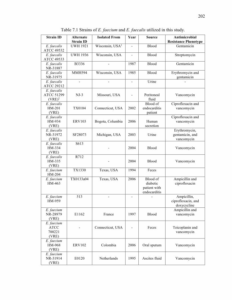
202
Table 7.1 Strains of E. faecium and E. faecalis utilized in this study.
Strain ID Alternate Strain ID
Isolated From Year Source Antimicrobial Resistance Phenotype
E. faecalis ATCC 49532
UWH 1921 Wisconsin, USA1 - Blood Gentamicin
E. faecalis ATCC 49533
UWH 1936 Wisconsin, USA - Blood Streptomycin
E. faecalis NR-31887
B3336 - 1987 Blood Gentamicin
E. faecalis NR-31975
MMH594 Wisconsin, USA 1985 Blood Erythromycin and gentamicin
E. faecalis ATCC 29212
- - - Urine -
E. faecalis
ATCC 51299 (VRE)2
NJ-3
Missouri, USA
-
Peritoneal
fluid
Vancomycin
E. faecalis
HM-201 (VRE)
TX0104
Connecticut, USA
2002
Blood of endocarditis
patient
Ciprofloxacin and vancomycin
E. faecalis
HM-934 (VRE)
ERV103
Bogota, Columbia
2006
Human
secretion
Ciprofloxacin and vancomycin
E. faecalis NR-31972
(VRE)
SF28073
Michigan, USA
2003
Urine
Erythromycin, gentamicin, and
vancomycin
E. faecalis HM-334 (VRE)
S613 -
2004
Blood
Vancomycin
E. faecalis HM-335 (VRE)
R712 -
2004
Blood
Vancomycin
E. faecium
HM-204
TX1330 Texas, USA 1994 Feces
E. faecium
HM-463 TX0133a04 Texas, USA 2006 Blood of
diabetic patient with endocarditis
Ampicillin and ciprofloxacin
E. faecium
HM-959 513 - - - Ampicillin,
ciprofloxacin, and doxycycline
E. faecium
NR-28979 (VRE)
E1162
France
1997
Blood
Ampicillin and vancomycin
E. faecium
ATCC 700221 (VRE)
-
Connecticut, USA
-
Feces
Teicoplanin and
vancomycin
E. faecium
HM-968 (VRE)
ERV102
Colombia
2006
Oral sputum
Vancomycin
E. faecium
NR-31914 (VRE)
E0120
Netherlands
1995
Ascites fluid
Vancomycin
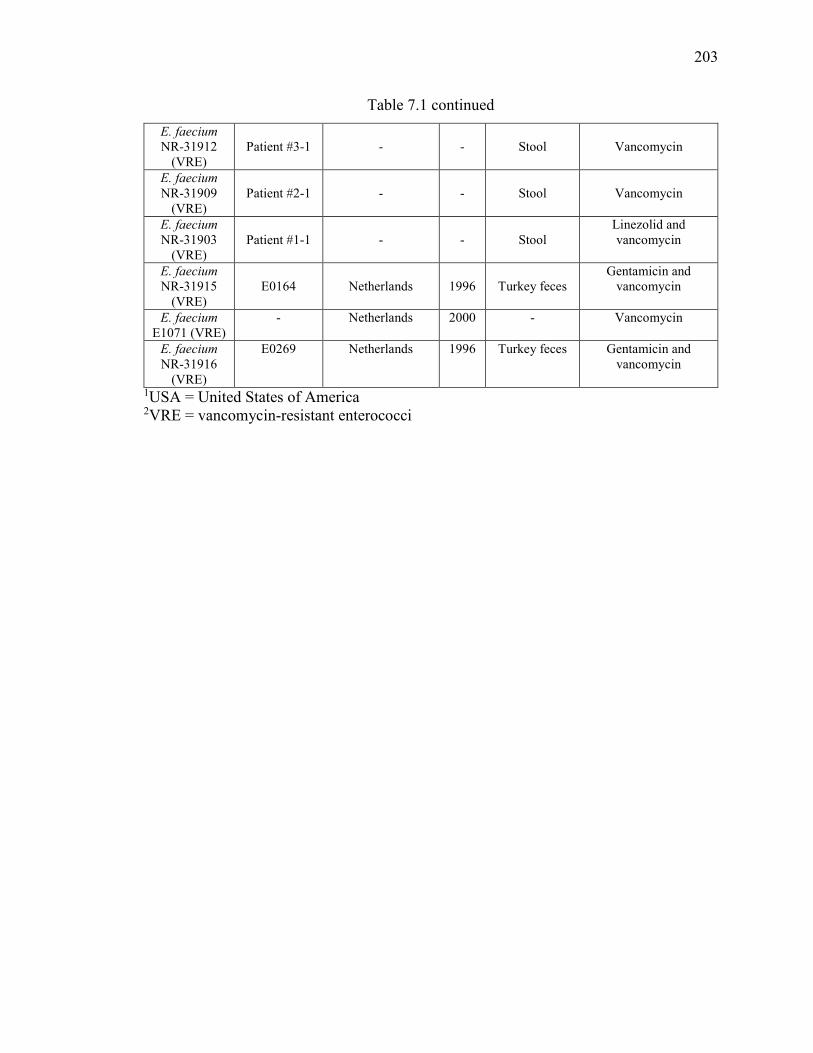
203
Table 7.1 continued
E. faecium
NR-31912 (VRE)
Patient #3-1
-
-
Stool
Vancomycin
E. faecium
NR-31909 (VRE)
Patient #2-1
-
-
Stool
Vancomycin
E. faecium
NR-31903 (VRE)
Patient #1-1
-
-
Stool
Linezolid and vancomycin
E. faecium
NR-31915 (VRE)
E0164
Netherlands
1996
Turkey feces
Gentamicin and vancomycin
E. faecium
E1071 (VRE) - Netherlands 2000 - Vancomycin
E. faecium
NR-31916 (VRE)
E0269 Netherlands 1996 Turkey feces Gentamicin and vancomycin
1USA = United States of America 2VRE = vancomycin-resistant enterococci
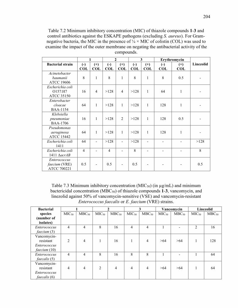
204
Table 7.2 Minimum inhibitory concentration (MIC) of thiazole compounds 1-3 and control antibiotics against the ESKAPE pathogens (excluding S. aureus). For Gram-negative bacteria, the MIC in the presence of ¼ × MIC of colistin (COL) was used to
examine the impact of the outer membrane on negating the antibacterial activity of the compounds.
1 2 3 Erythromycin Linezolid Bacterial strain (-)
COL (+)
COL (-)
COL (+)
COL (-)
COL (+)
COL (-)
COL (+)
COL Acinetobacter
baumanii ATCC 19606
8
1
8
1
8
1
8
0.5
-
Escherichia coli O157:H7
ATCC 35150
16
4
>128
4
>128
1
64
1
-
Enterobacter
cloacae BAA-1154
64
1
>128
1
>128
1
128
1
-
Klebsiella
pneumoniae BAA-1706
16
1
>128
2
>128
1
128
0.5
-
Pseudomonas
aeruginosa ATCC 15442
64
1
>128
1
>128
1
128
1
-
Escherichia coli 1411
64 - >128 - >128 - - - >128
Escherichia coli 1411 ΔacrAB
4 - 4 - 8 - - - 8
Enterococcus
faecium (VRE) ATCC 700221
0.5
-
0.5
-
0.5
-
-
-
0.5
Table 7.3 Minimum inhibitory concentration (MIC50) (in µg/mL) and minimum bactericidal concentration (MBC50) of thiazole compounds 1-3, vancomycin, and linezolid against 50% of vancomycin-sensitive (VSE) and vancomycin-resistant
Enterococcus faecalis or E. faecium (VRE) strains.
Bacterial species
(number of isolates)
1 2 3 Vancomycin Linezolid MIC50 MBC50 MIC50 MBC50 MIC50 MBC50 MIC50 MBC50 MIC50 MBC50
Enterococcus
faecium (3) 4 4 8 16 4 4 1 - 2 16
Vancomycin-resistant
Enterococcus
faecium (10)
2
4
1
16
1
4
>64
>64
1
128
Enterococcus
faecalis (5) 4 4 8 16 8 8 1 - 1 64
Vancomycin-resistant
Enterococcus
faecalis (6)
4
4
2
4
4
4
>64
>64
1
64
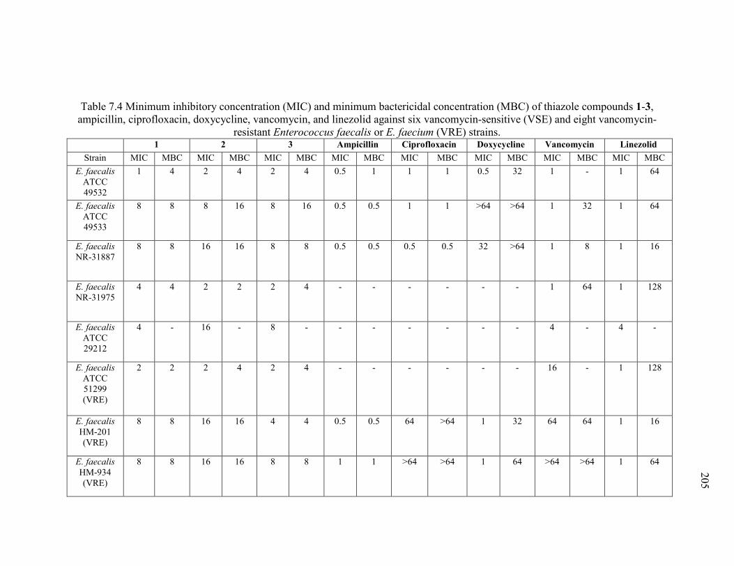
205
Table 7.4 Minimum inhibitory concentration (MIC) and minimum bactericidal concentration (MBC) of thiazole compounds 1-3, ampicillin, ciprofloxacin, doxycycline, vancomycin, and linezolid against six vancomycin-sensitive (VSE) and eight vancomycin-
resistant Enterococcus faecalis or E. faecium (VRE) strains. 1 2 3 Ampicillin Ciprofloxacin Doxycycline Vancomycin Linezolid
Strain MIC MBC MIC MBC MIC MBC MIC MBC MIC MBC MIC MBC MIC MBC MIC MBC
E. faecalis ATCC 49532
1 4 2 4 2 4 0.5 1 1 1 0.5 32 1 - 1 64
E. faecalis ATCC 49533
8 8 8 16 8 16 0.5 0.5 1 1 >64 >64 1 32 1 64
E. faecalis NR-31887
8 8 16 16 8 8 0.5 0.5 0.5 0.5 32 >64 1 8 1 16
E. faecalis NR-31975
4 4 2 2 2 4 - - - - - - 1 64 1 128
E. faecalis ATCC 29212
4 - 16 - 8 - - - - - - - 4 - 4 -
E. faecalis
ATCC 51299 (VRE)
2 2 2 4 2 4 - - - - - - 16 - 1 128
E. faecalis
HM-201 (VRE)
8 8 16 16 4 4 0.5 0.5 64 >64 1 32 64 64 1 16
E. faecalis
HM-934 (VRE)
8 8 16 16 8 8 1 1 >64 >64 1 64 >64 >64 1 64
205
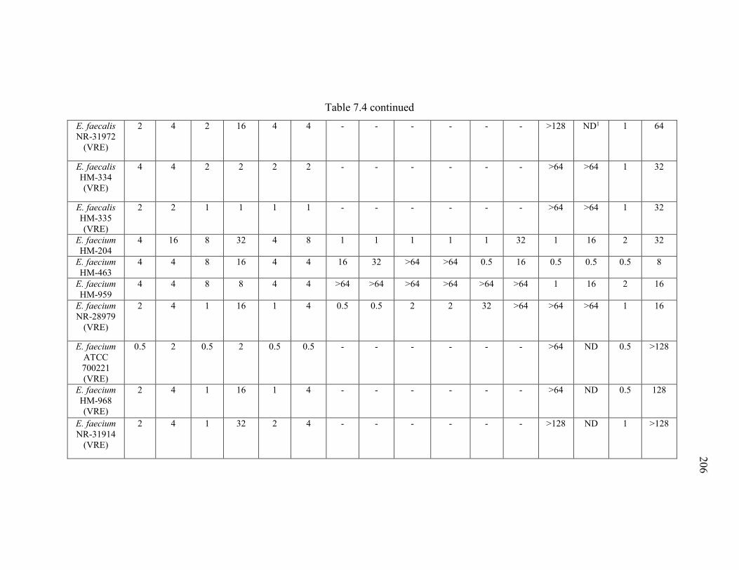
206
Table 7.4 continued
E. faecalis NR-31972
(VRE)
2 4 2 16 4 4 - - - - - - >128 ND1 1 64
E. faecalis HM-334 (VRE)
4 4 2 2 2 2 - - - - - - >64 >64 1 32
E. faecalis HM-335 (VRE)
2 2 1 1 1 1 - - - - - - >64 >64 1 32
E. faecium
HM-204
4 16 8 32 4 8 1 1 1 1 1 32 1 16 2 32
E. faecium
HM-463
4 4 8 16 4 4 16 32 >64 >64 0.5 16 0.5 0.5 0.5 8
E. faecium
HM-959 4 4 8 8 4 4 >64 >64 >64 >64 >64 >64 1 16 2 16
E. faecium
NR-28979 (VRE)
2 4 1 16 1 4 0.5 0.5 2 2 32 >64 >64 >64 1 16
E. faecium
ATCC 700221 (VRE)
0.5 2 0.5 2 0.5 0.5 - - - - - - >64 ND 0.5 >128
E. faecium
HM-968 (VRE)
2 4 1 16 1 4 - - - - - - >64 ND 0.5 128
E. faecium
NR-31914 (VRE)
2 4 1 32 2 4 - - - - - - >128 ND 1 >128
206
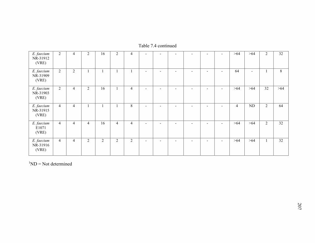
207
Table 7.4 continued
E. faecium
NR-31912 (VRE)
2 4 2 16 2 4 - - - - - - >64 >64 2 32
E. faecium
NR-31909 (VRE)
2 2 1 1 1 1 - - - - - - 64 - 1 8
E. faecium
NR-31903 (VRE)
2 4 2 16 1 4 - - - - - - >64 >64 32 >64
E. faecium
NR-31915 (VRE)
4 4 1 1 1 8 - - - - - - 4 ND 2 64
E. faecium
E1071 (VRE)
4 4 4 16 4 4 - - - - - - >64 >64 2 32
E. faecium
NR-31916 (VRE)
4 4 2 2 2 2 - - - - - - >64 >64 1 32
1ND = Not determined
207
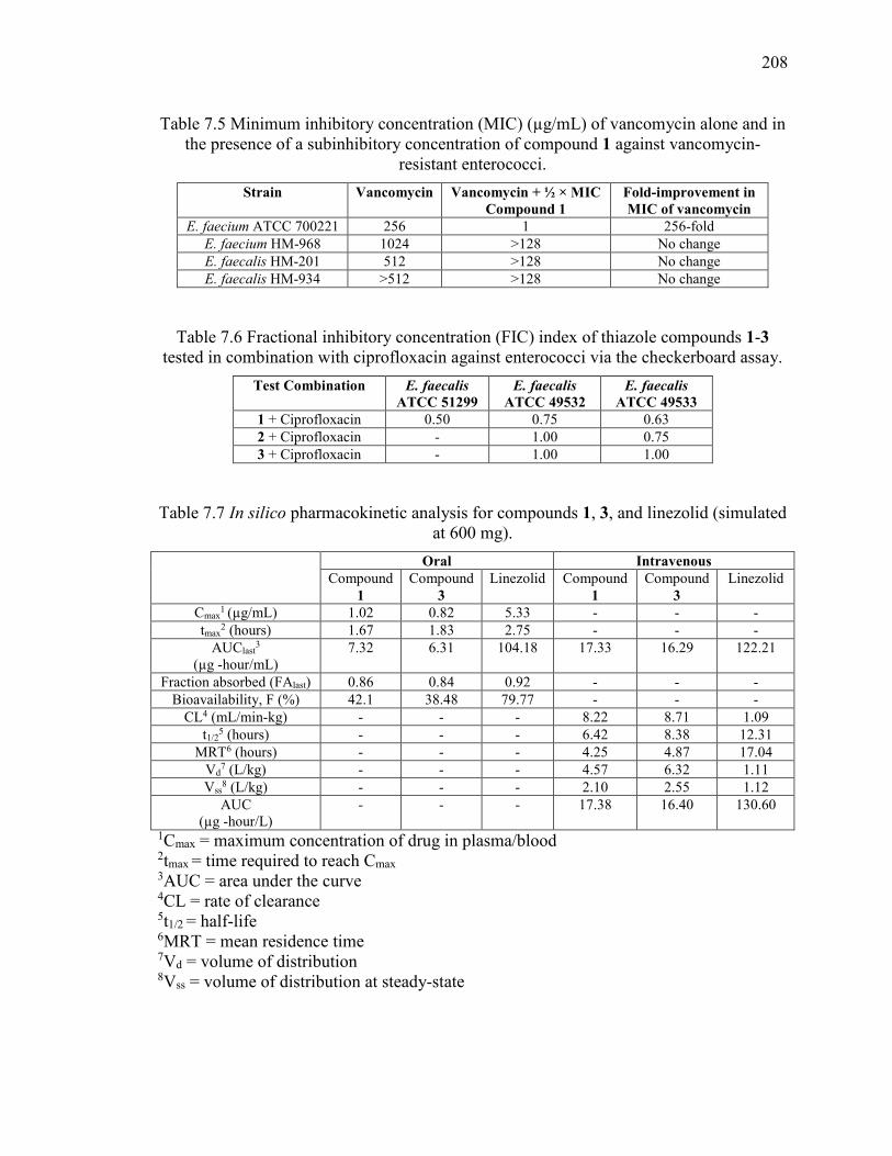
208
Table 7.5 Minimum inhibitory concentration (MIC) (µg/mL) of vancomycin alone and in the presence of a subinhibitory concentration of compound 1 against vancomycin-
resistant enterococci.
Strain Vancomycin
Vancomycin + ½ × MIC Compound 1
Fold-improvement in MIC of vancomycin
E. faecium ATCC 700221 256 1 256-fold
E. faecium HM-968 1024 >128 No change
E. faecalis HM-201 512 >128 No change
E. faecalis HM-934 >512 >128 No change
Table 7.6 Fractional inhibitory concentration (FIC) index of thiazole compounds 1-3 tested in combination with ciprofloxacin against enterococci via the checkerboard assay.
Test Combination E. faecalis ATCC 51299
E. faecalis ATCC 49532
E. faecalis ATCC 49533
1 + Ciprofloxacin 0.50 0.75 0.63
2 + Ciprofloxacin - 1.00 0.75
3 + Ciprofloxacin - 1.00 1.00
Table 7.7 In silico pharmacokinetic analysis for compounds 1, 3, and linezolid (simulated at 600 mg).
Oral Intravenous Compound
1
Compound
3
Linezolid Compound
1
Compound
3
Linezolid
Cmax1
(µg/mL) 1.02 0.82 5.33 - - -
tmax2 (hours) 1.67 1.83 2.75 - - -
AUClast3
(µg -hour/mL) 7.32 6.31 104.18 17.33 16.29 122.21
Fraction absorbed (FAlast) 0.86 0.84 0.92 - - -
Bioavailability, F (%) 42.1 38.48 79.77 - - -
CL4 (mL/min-kg) - - - 8.22 8.71 1.09
t1/25 (hours) - - - 6.42 8.38 12.31
MRT6 (hours) - - - 4.25 4.87 17.04
Vd7 (L/kg) - - - 4.57 6.32 1.11
Vss8 (L/kg) - - - 2.10 2.55 1.12
AUC (µg -hour/L)
- - - 17.38 16.40 130.60
1Cmax = maximum concentration of drug in plasma/blood 2tmax = time required to reach Cmax 3AUC = area under the curve 4CL = rate of clearance 5t1/2 = half-life 6MRT = mean residence time 7Vd = volume of distribution 8Vss = volume of distribution at steady-state
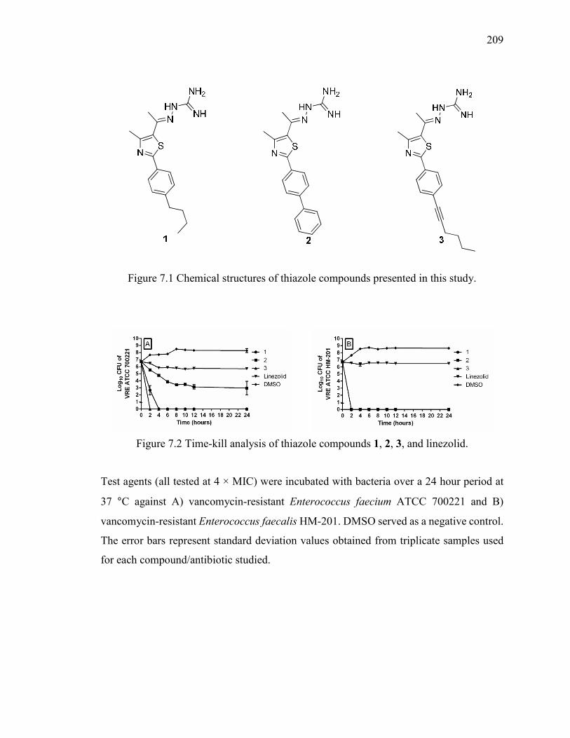
209
Figure 7.1 Chemical structures of thiazole compounds presented in this study.
Figure 7.2 Time-kill analysis of thiazole compounds 1, 2, 3, and linezolid.
Test agents (all tested at 4 × MIC) were incubated with bacteria over a 24 hour period at
37 °C against A) vancomycin-resistant Enterococcus faecium ATCC 700221 and B)
vancomycin-resistant Enterococcus faecalis HM-201. DMSO served as a negative control.
The error bars represent standard deviation values obtained from triplicate samples used
for each compound/antibiotic studied.
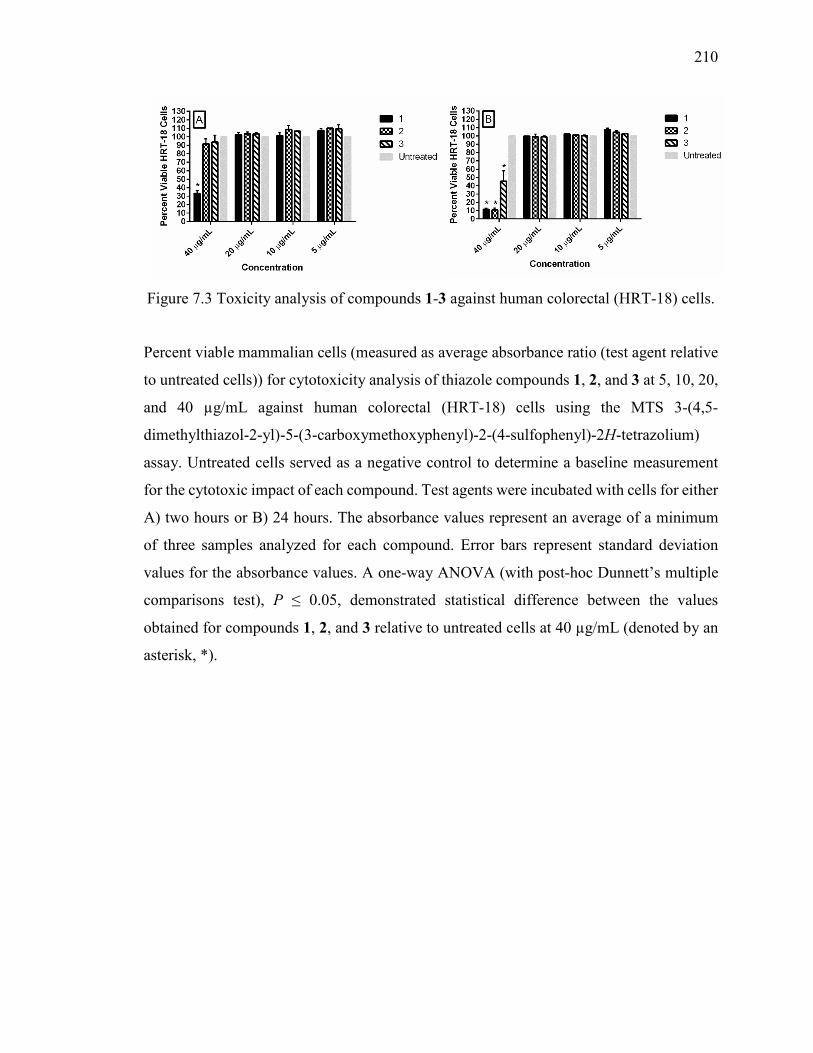
210
Figure 7.3 Toxicity analysis of compounds 1-3 against human colorectal (HRT-18) cells.
Percent viable mammalian cells (measured as average absorbance ratio (test agent relative
to untreated cells)) for cytotoxicity analysis of thiazole compounds 1, 2, and 3 at 5, 10, 20,
and 40 µg/mL against human colorectal (HRT-18) cells using the MTS 3-(4,5-
dimethylthiazol-2-yl)-5-(3-carboxymethoxyphenyl)-2-(4-sulfophenyl)-2H-tetrazolium)
assay. Untreated cells served as a negative control to determine a baseline measurement
for the cytotoxic impact of each compound. Test agents were incubated with cells for either
A) two hours or B) 24 hours. The absorbance values represent an average of a minimum
of three samples analyzed for each compound. Error bars represent standard deviation
values for the absorbance values. A one-way ANOVA (with post-hoc Dunnett’s multiple
comparisons test), P ≤ 0.05, demonstrated statistical difference between the values
obtained for compounds 1, 2, and 3 relative to untreated cells at 40 µg/mL (denoted by an
asterisk, *).
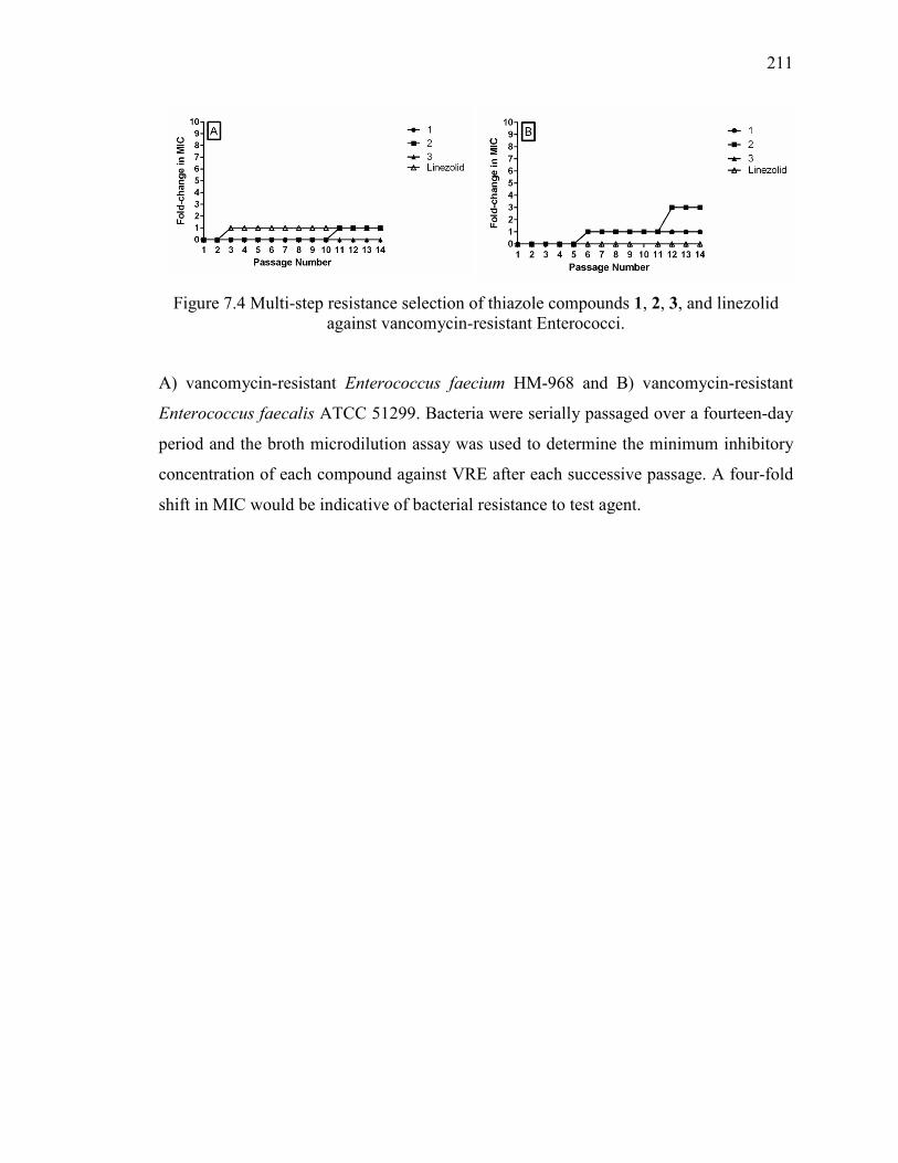
211
Figure 7.4 Multi-step resistance selection of thiazole compounds 1, 2, 3, and linezolid against vancomycin-resistant Enterococci.
A) vancomycin-resistant Enterococcus faecium HM-968 and B) vancomycin-resistant
Enterococcus faecalis ATCC 51299. Bacteria were serially passaged over a fourteen-day
period and the broth microdilution assay was used to determine the minimum inhibitory
concentration of each compound against VRE after each successive passage. A four-fold
shift in MIC would be indicative of bacterial resistance to test agent.
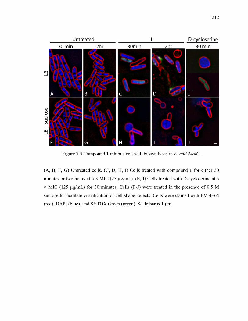
212
Figure 7.5 Compound 1 inhibits cell wall biosynthesis in E. coli ΔtolC.
(A, B, F, G) Untreated cells. (C, D, H, I) Cells treated with compound 1 for either 30
minutes or two hours at 5 × MIC (25 µg/mL). (E, J) Cells treated with D-cycloserine at 5
× MIC (125 µg/mL) for 30 minutes. Cells (F-J) were treated in the presence of 0.5 M
sucrose to facilitate visualization of cell shape defects. Cells were stained with FM 4−64
(red), DAPI (blue), and SYTOX Green (green). Scale bar is 1 μm.
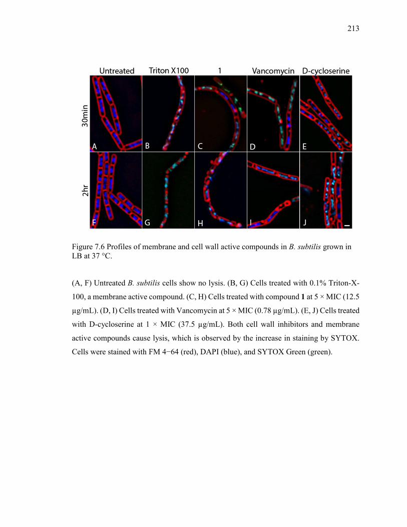
213
Figure 7.6 Profiles of membrane and cell wall active compounds in B. subtilis grown in LB at 37 °C.
(A, F) Untreated B. subtilis cells show no lysis. (B, G) Cells treated with 0.1% Triton-X-
100, a membrane active compound. (C, H) Cells treated with compound 1 at 5 × MIC (12.5
µg/mL). (D, I) Cells treated with Vancomycin at 5 × MIC (0.78 µg/mL). (E, J) Cells treated
with D-cycloserine at 1 × MIC (37.5 µg/mL). Both cell wall inhibitors and membrane
active compounds cause lysis, which is observed by the increase in staining by SYTOX.
Cells were stained with FM 4−64 (red), DAPI (blue), and SYTOX Green (green).
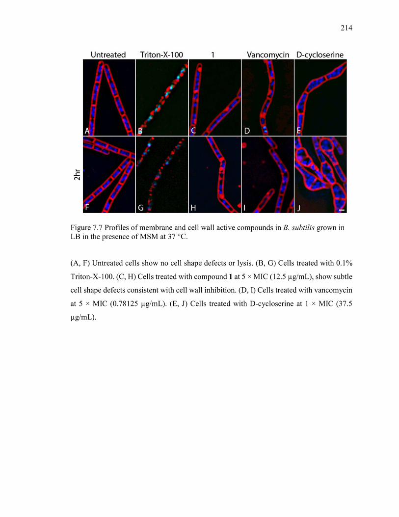
214
Figure 7.7 Profiles of membrane and cell wall active compounds in B. subtilis grown in LB in the presence of MSM at 37 °C.
(A, F) Untreated cells show no cell shape defects or lysis. (B, G) Cells treated with 0.1%
Triton-X-100. (C, H) Cells treated with compound 1 at 5 × MIC (12.5 µg/mL), show subtle
cell shape defects consistent with cell wall inhibition. (D, I) Cells treated with vancomycin
at 5 × MIC (0.78125 µg/mL). (E, J) Cells treated with D-cycloserine at 1 × MIC (37.5
µg/mL).
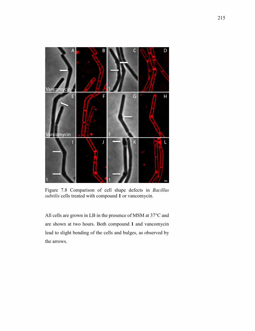
215
Figure 7.8 Comparison of cell shape defects in Bacillus
subtilis cells treated with compound 1 or vancomycin.
All cells are grown in LB in the presence of MSM at 37°C and
are shown at two hours. Both compound 1 and vancomycin
lead to slight bending of the cells and bulges, as observed by
the arrows.
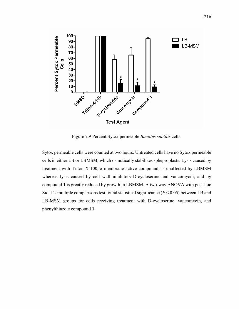
216
Figure 7.9 Percent Sytox permeable Bacillus subtilis cells.
Sytox permeable cells were counted at two hours. Untreated cells have no Sytox permeable
cells in either LB or LBMSM, which osmotically stabilizes spheproplasts. Lysis caused by
treatment with Triton X-100, a membrane active compound, is unaffected by LBMSM
whereas lysis caused by cell wall inhibitors D-cycloserine and vancomycin, and by
compound 1 is greatly reduced by growth in LBMSM. A two-way ANOVA with post-hoc
Sidak’s multiple comparisons test found statistical significance (P < 0.05) between LB and
LB-MSM groups for cells receiving treatment with D-cycloserine, vancomycin, and
phenylthiazole compound 1.
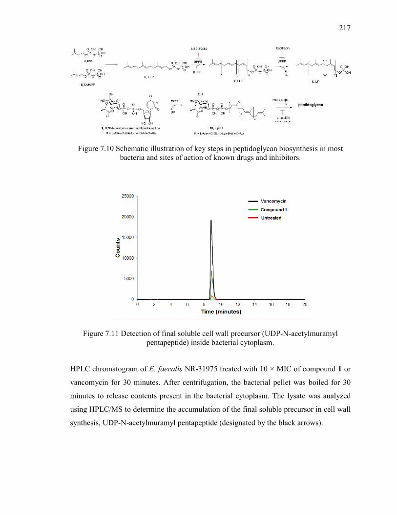
217
Figure 7.10 Schematic illustration of key steps in peptidoglycan biosynthesis in most bacteria and sites of action of known drugs and inhibitors.
Figure 7.11 Detection of final soluble cell wall precursor (UDP-N-acetylmuramyl pentapeptide) inside bacterial cytoplasm.
HPLC chromatogram of E. faecalis NR-31975 treated with 10 × MIC of compound 1 or
vancomycin for 30 minutes. After centrifugation, the bacterial pellet was boiled for 30
minutes to release contents present in the bacterial cytoplasm. The lysate was analyzed
using HPLC/MS to determine the accumulation of the final soluble precursor in cell wall
synthesis, UDP-N-acetylmuramyl pentapeptide (designated by the black arrows).
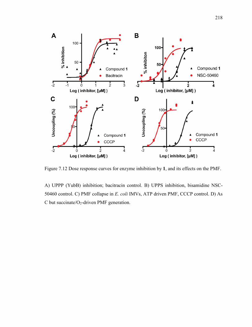
218
Figure 7.12 Dose response curves for enzyme inhibition by 1, and its effects on the PMF.
A) UPPP (YubB) inhibition; bacitracin control. B) UPPS inhibition, bisamidine NSC-
50460 control. C) PMF collapse in E. coli IMVs, ATP driven PMF, CCCP control. D) As
C but succinate/O2-driven PMF generation.
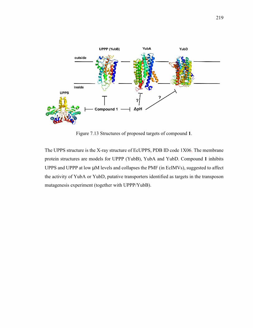
219
Figure 7.13 Structures of proposed targets of compound 1.
The UPPS structure is the X-ray structure of EcUPPS, PDB ID code 1X06. The membrane
protein structures are models for UPPP (YubB), YubA and YubD. Compound 1 inhibits
UPPS and UPPP at low µM levels and collapses the PMF (in EcIMVs), suggested to affect
the activity of YubA or YubD, putative transporters identified as targets in the transposon
mutagenesis experiment (together with UPPP/YubB).
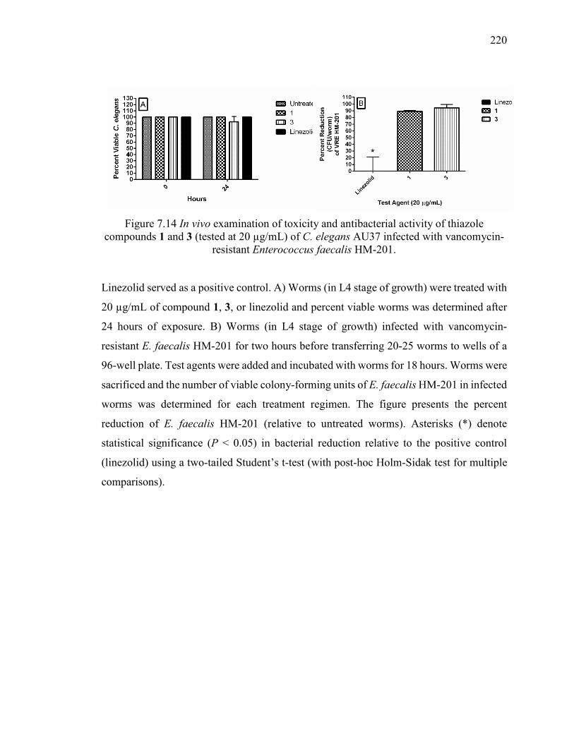
220
Figure 7.14 In vivo examination of toxicity and antibacterial activity of thiazole compounds 1 and 3 (tested at 20 µg/mL) of C. elegans AU37 infected with vancomycin-
resistant Enterococcus faecalis HM-201.
Linezolid served as a positive control. A) Worms (in L4 stage of growth) were treated with
20 µg/mL of compound 1, 3, or linezolid and percent viable worms was determined after
24 hours of exposure. B) Worms (in L4 stage of growth) infected with vancomycin-
resistant E. faecalis HM-201 for two hours before transferring 20-25 worms to wells of a
96-well plate. Test agents were added and incubated with worms for 18 hours. Worms were
sacrificed and the number of viable colony-forming units of E. faecalis HM-201 in infected
worms was determined for each treatment regimen. The figure presents the percent
reduction of E. faecalis HM-201 (relative to untreated worms). Asterisks (*) denote
statistical significance (P < 0.05) in bacterial reduction relative to the positive control
(linezolid) using a two-tailed Student’s t-test (with post-hoc Holm-Sidak test for multiple
comparisons).
221
VITA
Haroon Mohammad
Education
Purdue University, West Lafayette, Indiana Ph.D. Candidate in Microbiology (December 2016) B.S. in Biological and Food Process Engineering (December 2008) B.S. in Pharmaceutical Sciences (December 2008)
Publications
Refereed Journal Articles
1.) Thangamani, S., Eldesouky, H., Mohammad, H., Pascuzzi, P., Avramova, L., Hazbun, T., Seleem, M.N. Just Accepted (September 28, 2016). Ebselen exerts antifungal activity by regulating glutathione (GSH) and reactive oxygen species (ROS) production in fungal cells. BBA-General Subjects. Manuscript ID: BBAGEN-16-748R1.
2.) Abushahba, M.F.*, Mohammad, H.*, and Seleem, M.N. Targeting Multidrug-resistant
Staphylococci with an anti-rpoA Peptide Nucleic Acid Conjugated to the HIV-1 TAT Cell Penetrating Peptide. June 2016. Molecular Therapy – Nucleic Acids. 5(7):e339. doi: 10.1038/mtna.2016.53. (*Co-first authors)
3.) Seleem, M.A., Disouky, A.M., Mohammad, H., Abdelghany, T.M., Mancy, A. S.,
Bayoumi, S.A., Elshafeey, El-Morsy, A., A., Seleem, M. N., and Mayhoub, A. Second Generation Phenylthiazole Antibiotics with Enhanced Pharmacokinetic Properties. May 2016. Journal of Medicinal Chemistry. 59 (10): 4900-4912. doi: 10.1021/acs.jmedchem.6b00233
4.) Thangamani, S., Mohammad, H., Abushahba, M.F., Sobreira, T.J., Hedrick, V.E., Paul,
L.N., and Seleem, M.N. Antibacterial activity and mechanism of action of auranofin against multi-drug resistant bacterial pathogens. March 2016. Scientific Reports. 6:22571. doi: 10.1038/srep22571.
5.) Thangamani, S., Mohammad, H., Sobreira, T.J., Abushahba, M.F., and Seleem, M.N.
Repurposing auranofin for the treatment of cutaneous staphylococcal infections. March 2016. International Journal of Antimicrobial Agents. 47(3):195-201. doi: 10.1016/j.ijantimicag.2015.12.016.
222
6.) Abushahba, M.F., Mohammad, H., Thangamani, S., Hussein, A., and Seleem, M.N. Impact of different cell penetrating peptides on the efficacy of antisense therapeutics for targeting intracellular pathogens. February 2016. Scientific Reports. 6:20832. doi: 10.1038/srep20832.
7.) Mohammad, H., Cushman, M., and Seleem, M.N. Antibacterial Evaluation of Novel
Synthetic Thiazole Compounds in vitro and in vivo Against A Methicillin-resistant Staphylococcus aureus (MRSA) Murine Skin Infection Model. November 2015. PLoS ONE. 10(11):e0142321. doi: 10.1371/journal.pone.0142321.
8.) Thangamani, S., Mohammad, H., Sobreira T.J., Paul, L.N., Hedrick, V.E., and Seleem,
M.N. Exploring simvastatin, an antihyperlipidemic drug, as a potential topical antibacterial agent. November 2015. Scientific Reports. 5:16407. doi: 10.1038/srep16407.
9.) Davis, D.C.*, Mohammad, H.*, Younis, W., Creemer, C.N., Seleem, M.N., Dai, M.
Discovery and Characterization of Aryl Isonitriles as A New Class of Compounds versus Methicillin- and Vancomycin-resistant Staphylococcus aureus. August 2015. European
Journal of Medicinal Chemistry.101: 384-390. doi: 10.1016/j.ejmech.2015.06.031. (*Co-first authors)
10.) Mohammad, H., Reddy, P.V.N., Monteleone, D., Mayhoub, A.S., Cushman, M.,
Hammac, G.K., and Seleem, M.N. Antibacterial Characterization of Novel Synthetic Thiazole Compounds Against Methicillin-resistant Staphylococcus pseudintermedius.
June 2015. PLoS ONE. 18:10(6):e0130385. doi: 10.1371/journal.pone.0130385.
11.) Mohammad, H., Reddy, P.V.N., Monteleone, D., Mayhoub, A.S., Cushman, M., and Seleem, M.N. Synthesis and Antibacterial Evaluation of A Novel Series of Synthetic Phenylthiazole Compounds Against Methicillin-resistant Staphylococcus aureus (MRSA). March 2015. European Journal of Medicinal Chemistry. 94: 306-316. doi: 10.1016/j.ejmech.2015.03.015.
12.) Mohammad, H., Mayhoub, A.S., Cushman, M., and Seleem, M. N. Anti-Biofilm Activity
and Synergism of Novel Thiazole Compounds with Glycopeptide Antibiotics Against
Multidrug-Resistant Staphylococci. April 2015. Journal of Antibiotics. 68 (4):259-266. doi: 10.1038/ja.2014.142.
13.) Mohammad, H., Thangamani, S., and Seleem, M. N. Antimicrobial Peptides and
Peptidomimetics – Potent Therapeutic Allies for Staphylococcal Infections. 2015. Current Pharmaceutical Design. 21(16):2073-88.
14.) Thangamani, S., Mohammad, H., Younis, W., and Seleem, M.N. Repurposing Non-
antimicrobial Drugs for Treatment of Staphylococcal Infections. 2015. Current Pharmaceutical Design. 21(16):2089-100.
15.) Mohammad, H., Mayhoub, A.S., Ghafoor, A., Soofi, M., Alajlouni, R.A., Cushman, M.
and Seleem, M.N. Discovery and Characterization of Potent Thiazoles Versus Methicillin- and Vancomycin-Resistant Staphylococcus aureus. February 2014. Journal of Medicinal Chemistry. 57(4): 1609-1615. doi: 10.1021/jm401905m.
223
Journal Articles Under Review
1.) Eassa, I.,* Mohammad, H.,* Qassem, O., Younis, W., Abdelghany, T., Elshafeey, A.,
Moustafa, M., Seleem, M. N., Mayhoub, A. S. Diphenylurea: a Novel Discovery for Combating Methicillin- and Vancomycin-Resistant Staphylococcus aureus. Journal of Medicinal Chemistry. Manuscript ID: jm-2016-01664f. (*Co-first authors)
2.) Yahia, E., Mohammad, H., Abdelghany, T. M., Fayed, I., Seleem, M. N., and Mayhoub1,
A. S. Phenylthiazole Antibiotics: A Metabolism-Guided Approach to Overcome Short Duration of Action. European Journal of Medicinal Chemistry. Submitted August 2016.
3.) Mohamed, M., Brezden, A., Mohammad, H., Chmielewski, J., Seleem, M. N. A short D-
enantiomeric antimicrobial peptide with potent immunomodulatory and antibiofilm activity against multidrug-resistant Pseudomonas aeruginosa and Acinetobacter
baumannii. Antimicrobial Agents and Chemotherapy. Manuscript ID: AAC01840-16R1.
4.) Ghosh, C., Yadav, V., Younis, W., Mohammad, H., Hegazy, Y., Seleem, M. N., Sanyal, K., Haldar, J. Aryl-alkyl-lysines: Membrane-active fungicides that act against biofilms of Candida albicans. ACS Infectious Diseases. Manuscript ID: id-2016-00192v.
5.) Thangamani, S., Hazbun, T., Maland, M., Mohammad, H., Pascuzzi, P., Avramova, L.,
Koehler, C., Seleem, M. N. Auranofin exhibits antifungal activity by targeting mitochondrial protein(s). Frontiers in Cellular and Infection Microbiology. Submitted August 2016.
Selected Presentations Invited Presentations
1.) H. Mohammad. Antibacterial Characterization of Novel Synthetic Thiazole
Compounds Against Methicillin-Resistant Staphylococcus pseudintermedius. 29th Annual Phi Zeta Research Day. West Lafayette, IN. April 11, 2016.
2.) H. Mohammad. New Drugs for Bad Bugs - Development of Novel Synthetic
Antimicrobials for Treatment of MRSA Infections. Purdue University Office of Interdisciplinary Graduate Programs (OIGP) Spring Reception, Keynote Address. West Lafayette, IN, April 1, 2015.
3.) H. Mohammad. Bad Bugs Need Good Drugs. Purdue MSA RADx Talks, West
Lafayette, IN, November 19, 2014.
Oral Presentations
1.) H. Mohammad. Repurposing Auranofin and Ebselen for Treatment of Invasive
Fungal Infections. Purdue Institute for Drug Discovery Postdoctoral Award Presentation, West Lafayette, IN, November 8, 2016.
2.) H. Mohammad. From Contamination to the Discovery of a Novel Broad-spectrum
Antibacterial Agent. Purdue Comparative Pathobiology Department Seminar, West Lafayette, IN, September 29, 2016.
224
3.) H. Mohammad & Mohamed N. Seleem. Phenylthiazole Antibiotics for Multidrug-
resistant Staphylococcus aureus (MRSA) Infections. Purdue Institute for Immunology, Inflammation and Infectious Disease Charting a Roadmap for Pre-clinical Drug Development and Beyond Workshop, West Lafayette, IN, September 2, 2016.
4.) H. Mohammad. Gut Check: Discovery of Phenylthiazole Antibiotics Effective
Against Vancomycin-resistant Enterococci. Hitchhiker’s Guide to the Biomolecular Galaxy Symposium, West Lafayette, IN, May 11, 2016.
5.) H. Mohammad. Saving Fido – Unearthing a novel topical antimicrobial for treatment
of multidrug-resistant staphylococcal skin infections in companion animals. Conference of Research Workers in Animal Diseases, Chicago, IL, December 7, 2015.
6.) H. Mohammad. Saving Fido – Unearthing a novel topical antimicrobial for treatment
of multidrug-resistant staphylococcal skin infections in companion animals. Purdue Comparative Pathobiology Department Seminar, West Lafayette, IN, November 5, 2015.
7.) H. Mohammad. Bad Bugs Need Good Drugs, 3MT Thesis Competition, Purdue
University Graduate School, West Lafayette, IN, April 15, 2015.
8.) H. Mohammad. MRSA on the Rise, Therapeutics in Demise, Thiazoles Emerge as
Future Allies. Purdue Comparative Pathobiology Department Seminar, West Lafayette, IN, October 30, 2014.
9.) H. Mohammad. Bad Bugs Need Good Drugs: Characterization of Substituted
Thiazole Compounds for Treatment of Methicillin-resistant Staphylococcus aureus (MRSA) Infections. Purdue Comparative Pathobiology Department Seminar, West Lafayette, IN, November 14, 2013.
10.) H. Mohammad. A Super Drug for A Superbug. 5th Annual Regional Population,
Health, and Environmental Economics Graduate Student Summit, West Lafayette, IN, April 14, 2012.
11.) H. Mohammad. Old Bug vs. New Drug: Novel Substituted Thiazoles for Treatment
of Methicillin-resistant Staphylococcus aureus (MRSA) Infections. Purdue Comparative Pathobiology Department Seminar, West Lafayette, IN, November 29, 2012.
225
International Conference Poster Presentations
1.) E. Mostafa, H. Mohammad, M.N. Seleem & A.S. Mayhoub. Diphenylureas: a Novel
Discovery for Beating Methicillin and Vancomycin Resistant Staphylococcus aureus (MRSA & VRSA). FUE 3rd International Conference of Pharmaceutical Sciences, Cairo, Egypt, February 9-11, 2015.
2.) M. A. Seleem, H. Mohammad, M.N. Seleem & A.S. Mayhoub. Design & Synthesis
of New Phenylthiazole Derivatives with Enhanced Pharmacokinetic Profile for Treatment of Methicillin and Vancomycin Resistant Staphylococcus aureus (MRSA & VRSA). FUE 3rd International Conference of Pharmaceutical Sciences, Cairo, Egypt, February 9-11, 2015.
3.) W.M. Townsend, H. Mohammad, and M.N. Seleem. Equine Ulcerative Keratitis:
Comparison of Histology, Cytology, and Mycotic Culture with Results of Polymerase Chain Reaction using Universal Fungal Primers, 2013 IEOC/Acrivet Symposium, Jackson Hole, WY, June 8, 2013.
4.) H. Mohammad, A.S., Mayhoub, A. Ghafoor, M. Soofi, R.A. Alajlouni, M. Cushman,
and M.N. Seleem. A New Drug for an Old Bug: Antimicrobial Activity of Novel Substituted Thiazoles Against Methicillin-Resistant Staphylococcus aureus, Conference of Research Workers in Animal Diseases, Chicago, IL, December 2, 2012.
National Conference Poster Presentations
1.) H. Mohammad, M. Cushman, and M.N. Seleem. Skin Deep - Novel Thiazole
Compounds Exhibit Potent Antibacterial Activity in vitro and in vivo in a Methicillin-resistant Staphylococcus aureus (MRSA) Skin Infection Mouse Model. ASM Microbe & Interscience Conference on Antimicrobial Agents and Chemotherapy 2016, Boston, MA, June 16 – 20, 2016.
2.) H. Mohammad, M. Sedlak, N.W.Y. Ho, and N.S. Mosier. Effect of Na+, K+, NH4
+, and Glycerol on the Glucose and Xylose Co-fermentation of Glucose and Xylose by S.
cerevisiae424A(LNH-ST), 31st Symposium on Biotechnology for Fuels and Chemicals, San Francisco, CA, May 3 – 6, 2009.
3.) M. Sedlak, H. Mohammad, N.W.Y. Ho, and N.S. Mosier. Combined Effect of Acetic
Acid and pH on the Co-fermentation of Glucose and Xylose by Recombinant Yeast, 29th Symposium on Biotechnology for Fuels and Chemicals, Denver, CO, April 29 – May 2, 2007.
Regional Conference Poster Presentations
1.) H. Mohammad, M. Cushman, and M.N. Seleem. Attacking the Multidrug-resistance Puzzle with Phenylthiazole Antibiotics. Purdue Institute for Immunology, Inflammation and Infectious Disease Graduate Student/Postdoc Symposium, West Lafayette, IN, August 10, 2016.
226
2.) H. Mohammad, W. Younis, M. Cushman, and M.N. Seleem. Discovery of Thiazole
Compounds with Potent Antimicrobial Activity Against Drug-resistant Enterococci. The 8th Annual Interdisciplinary Graduate Program Spring Reception, Purdue Office of Interdisciplinary Graduate Programs, West Lafayette, IN, May 2, 2016.
3.) H. Mohammad, W. Younis, M. Cushman, and M.N. Seleem. Evaluation of Phenylthiazole Antibacterial Agents Targeting Drug-resistant Enterococci. The 33rd Herbert C. Brown Lectures in Organic Chemistry, West Lafayette, IN, April 15, 2016.
4.) H. Mohammad, W. Younis, M. Cushman, and M.N. Seleem. Evaluation of
Phenylthiazole Antibacterial Agents Targeting Drug-resistant Enterococci. The 29th Annual Phi Zeta Research Day, Purdue College of Veterinary Medicine, West Lafayette, IN, April 11, 2016.
5.) H. Mohammad*, W. Younis, M. Cushman, and M.N. Seleem. Gut Check:
Identification of Phenylthiazole Antibacterial Agents Effective Against Vancomycin-resistant Enterococci (VRE). Indiana Branch of the American Society for Microbiology (ASM) Annual Meeting 2016, Fort Wayne, IN, April 2, 2016.
*Awarded second-place in PhD Division
6.) H. Mohammad*, W. Younis, M. Cushman, and M.N. Seleem. In vitro and in vivo examination of novel phenylthiazole antibacterial compounds against vancomycin-resistant enterococci (VRE). 2016 Health and Disease: Science, Culture, Policy Research Poster Session, West Lafayette, IN, March 28, 2016. *Awarded first-place in Inflammation, Immunology and Infectious Diseases Division
7.) H. Mohammad*, W. Younis, M. Cushman, and M.N. Seleem. In vitro and in vivo
examination of novel phenylthiazole antibacterial compounds against vancomycin-resistant enterococci (VRE). 2016 Sigma Xi Graduate Student Research Poster Award Competition, West Lafayette, IN, March 2, 2016. *Awarded first-place in Life Sciences Division
8.) H. Mohammad*, M. Cushman, and M.N. Seleem. Antibacterial Effect of Synthetic Thiazole Compounds in vitro and in vivo in a Methicillin-resistant Staphylococcus
aureus (MRSA) Skin Infection Mouse Model. The 28th Annual Phi Zeta Research Day, Purdue College of Veterinary Medicine. West Lafayette, IN, April 14, 2015. *Awarded first-place in Basic Research Division
9.) H. Mohammad, A.S. Mayhoub, P.V.N. Reddy, D. Monteleone, M. Cushman, G.K.
Hammac, and M.N. Seleem. Good Drugs for Bad Bugs –Thiazole Compounds As New Partners Against Methicillin-resistant Staphylococcus pseudintermedius (MRSP) Infections. The 7th Annual Interdisciplinary Graduate Program Spring Reception, Purdue Office of Interdisciplinary Graduate Programs, West Lafayette, IN, April 1, 2015.
10.) H. Mohammad, A.S. Mayhoub, P.V.N. Reddy, M. Cushman, and M.N. Seleem. Skin
Deep – Synthetic Thiazole Compounds Emerge as New Allies for Drug-resistant Staphylococcal Skin Infections. 2015 Health and Disease: Science, Culture, Policy Research Poster Session, West Lafayette, IN, March 5, 2015.
227
11.) H. Mohammad, A.S. Mayhoub, P.V.N. Reddy, M. Cushman, and M.N. Seleem.
Skinning Staph: Therapeutic Applications for Novel Thiazole Compounds Against Drug-resistant Staphylococcal Skin Infections. 2015 Sigma Xi Graduate Student Research Poster Award Competition, West Lafayette, IN, February 18, 2015.
12.) H. Mohammad, A.S. Mayhoub, P.V.N. Reddy, M. Cushman, and M.N. Seleem.
Therapeutic Applications for Novel Thiazole Compounds Against Multidrug-resistant Staphylococci. ESE Symposium. West Lafayette, IN, October 20, 2014.
13.) H. Mohammad, A.S. Mayhoub, P.V.N. Reddy, M. Cushman, and M.N. Seleem. New
Drugs for Bad Bugs: Antimicrobial Versatility of Novel Synthetic Thiazole Compounds Against Multidrug-resistant Staphylococci. Purdue/Lilly Tech Day. West Lafayette, IN, May 8, 2014.
14.) H. Mohammad, A.S. Mayhoub, P.V.N. Reddy, M. Cushman, and M.N. Seleem.
Antimicrobial Versatility of Novel Thiazole Compounds Against Multidrug-Resistant Staphylococci. The 27th Annual Phi Zeta Research Day, Purdue College of Veterinary Medicine. West Lafayette, IN, April 14, 2014.
15.) H. Mohammad, A.S. Mayhoub, M. Cushman, and M.N. Seleem. Antimicrobial
Versatility and Anti-Biofilm Activity of Novel Synthetic Thiazole Compounds Against Multidrug-Resistant Staphylococci. The 6th Annual Interdisciplinary Graduate Program Spring Reception, Purdue Office of Interdisciplinary Graduate Programs, West Lafayette, IN, April 2, 2014.
16.) H. Mohammad, A.S. Mayhoub, P.V.N. Reddy, M. Cushman, and M.N. Seleem.
Antimicrobial Versatility of Novel Thiazole Compounds Against Multidrug-Resistant Staphylococci. 2014 Health and Disease: Science, Culture, Policy Research Poster Session, West Lafayette, IN, March 31, 2014.
17.) H. Mohammad, A.S. Mayhoub, M. Cushman, and M.N. Seleem. Antimicrobial
Versatility and Anti-Biofilm Activity of Novel Synthetic Thiazole Compounds Against Multidrug-Resistant Staphylococci. 2014 Sigma Xi Graduate Student Research Poster Award Competition, West Lafayette, IN, February 12, 2014.
18.) M. Ali, H. Mohammad, A.S. Mayhoub, A. Shehata, M. Cushman, and M.N. Seleem.
In vitro Activity and Cell Toxicity of Novel Modified Thiazole Compounds. 2014 Sigma Xi Graduate Student Research Poster Award Competition, West Lafayette, IN, February 12, 2014.
19.) H. Mohammad, A.S. Mayhoub, R.A. Alajlouni, M. Cushman, and M.N. Seleem. Bad
Bugs Need New Drugs – Antimicrobial Activity of Novel Substituted Thiazoles Against Methicillin-Resistant Staphylococcus aureus (MRSA). The 26th Annual Phi Zeta Research Day, Purdue College of Veterinary Medicine. West Lafayette, IN, April 15, 2013.
20.) H. Mohammad, A.S. Mayhoub, R.A. Alajlouni, M. Cushman, and M.N. Seleem.
MRSA on the Rise, Therapeutics in Demise, Thiazoles Emerge As Future Allies. The 5th Annual Interdisciplinary Graduate Program Spring Reception, Purdue Office of Interdisciplinary Graduate Programs, West Lafayette, IN, April 1, 2013.
228
21.) H. Mohammad, A.S. Mayhoub, A. Ghafoor, M. Soofi, R.A. Alajlouni, M. Cushman,
and M.N. Seleem. A Super Drug for A Superbug – Modified Thiazole Compounds Show Promise In Battle Against Methicillin-Resistant Staphylococcus aureus (MRSA). The 25th Annual Phi Zeta Research Day, Purdue College of Veterinary Medicine, West Lafayette, IN, April 16, 2012.
22.) H. Mohammad, A.S. Mayhoub, A. Ghafoor, M. Soofi, R.A. Alajlouni, M. Cushman,
and M.N. Seleem. Has MRSA Met Its Match? – Modified Thiazole Compounds Show Promise Against MRSA, 5th Annual Regional Population, Health, and Environmental Economics Graduate Student Summit, West Lafayette, IN, April 14, 2012.
23.) H. Mohammad, A.S. Mayhoub, A. Ghafoor, M. Soofi, R. A. Alajlouni, M. Cushman,
and M.N. Seleem. Modified Thiazole Compounds Show Promise As A New Therapeutic Tool Against MRSA, 2012 Sigma Xi Graduate Student Research Poster Award Competition, West Lafayette, IN, February 15, 2012.
Awards
Purdue Institute for Drug Discovery Post-Doctoral Fellowship (January – December 2017) Bilsland Dissertation Fellowship (August – December 2016) American Society for Microbiology Travel Award (ASM Microbe 2016) (June 2016) Purdue Graduate Student Government Travel Grant (ASM Microbe 2016) (April 2016) Phi Zeta Omicron Chapter Graduate Student Research Award (April 2016) First-place (Life Sciences Division), Purdue Society of Sigma Xi Poster Competition
(April 2016) Second-place (PhD Division), Indiana Branch of American Society for Microbiology Annual Meeting Poster Presentation (April 2016) First-place (Inflammation, Immunology and Infectious Diseases Division), Purdue Health & Disease Research Symposium (March 2016) First-place (Basic Research), Phi Zeta (Omicron Chapter) Poster Competition (April 2015) Purdue University Office of Interdisciplinary Graduate Programs (OIGP) Most Outstanding Interdisciplinary Graduate Project Award (April 2015) Purdue Graduate School 3-Minute Thesis Competition, Audience Choice Award
(April 2015) Purdue Graduate Student Government (PGSG) Graduate Student Excellence Award
(April 2014)
Patent
“Aryl Isonitriles as A New Class of Antimicrobial Compounds” Dai, M. J., Seleem, M., Davis, D. C., Mohammad, H. T. US Provisional Application filed (US, 62/143,031).
Professional Memberships American Association for the Advancement of Science (AAAS) (2015 – Present) American Society for Microbiology (ASM) (2015 – Present)
229
Ad-hoc Reviewer for Journals Current Pharmaceutical Design (Special edition, November to December 2014) PLoS ONE (August 2015 – current) Scientific Reports (November 2015 – current)
Research Experience
Laboratory of Molecular Microbiology, Purdue University Graduate Research Assistant/Lab Manager/Lab Organization Officer (August 2011-Present) PhD Advisor: Dr. Mohamed N. Seleem, Department of Comparative Pathobiology
• Identified four new series of synthetic chemical compounds with potent antibacterial activity against methicillin-resistant Staphylococcus aureus (MRSA) and vancomycin-resistant enterococci (VRE) using whole-cell screening
• Investigated the antibacterial mechanism of action of synthetic compounds utilizing target overexpression in Bacillus subtilis (via transposon mutagenesis) and biochemical assays
• Evaluated toxicity of promising antibacterial compounds against multiple human cell lines (HRT-18, HaCaT, HeLa, HEK-293)
• Examined ability of synthetic compounds to inhibit biofilm formation and toxin production
• Verified the antibacterial activity of phenylthiazole compounds in a murine model of MRSA skin infection and Caenhorhabditis elegans model of enterococcal infection
• Trained new graduate, undergraduate, and professional students proper safety procedures and protocols for conducting experiments with biosafety level 2 microorganisms
• Reviewed grant proposals and journal articles of lab members prior to submission Laboratory of Renewable Resources Engineering (LORRE), Purdue University Undergraduate Engineering Research Assistant (May 2006 – May 2009) Research Advisor: Dr. Nathan Mosier, Department of Agricultural & Biological Engineering
• Discovered effect of various inhibitors (acetic acid, excess sugar, excess ethanol, salts) on production of fuel-grade ethanol from pure sugars (glucose and xylose)
• Designed process to produce liquefied corn mash and remove antimicrobial agent added to corn mash prior to fermentation
Summer Undergraduate Research Fellowship (SURF), Purdue University Undergraduate Research Assistant (May 2005 – August 2005) Research Advisor: Dr. Arif Ghafoor, Department of Electrical & Computer Engineering
230
Leadership & Community Outreach Briarwood Apartments HEADS UP Program Youth mentor and homework tutor (December 2015 – current) Co-organizer, “Yes We Can! Making the College Dream A Reality” Spring Fest Tour*
(April 2016) *Awarded $1200 grant from Purdue University Office of Engagement Indiana University School of Medicine – Lafayette Briarwood Health Fair Co-organizer, Microbial Infection and Prevention Information Table (August 2016) Purdue Graduate Student Government Academic and Professional Development Committee (August 2015 – May 2016) Advancement Committee* (August 2015 – May 2016) *Awarded $500 for organization of “Restaurant Hope” service project Career Fair Committee (August 2015 – December 2015) Purdue Veterinary Medicine Family Program Series Youth career presentation “Microbes here, microbes there, microbes everywhere”
(March 2016) Science Fair Judge Lafayette Regional Science & Engineering Fair (March 2016) Lafayette Catholic Schools Education Fair (February 2013)