Embed Size (px)
Citation preview

Antimicrobial Activity of Tea Tree Oil
A report for the Rural Industries Research and Development Corporation by C F Carson & T V Riley
July 1998 RIRDC Publication No 98/70 RIRDC Project No UWA-24A
LASZLO – http://www.laszlo.com.br

LASZLO – http://www.laszlo.com.br
© 1998 Rural Industries Research and Development Corporation. All rights reserved. ISBN 0 642 54099 3 ISSN 1440-6845
"Antimicrobial Activity of Tea Tree Oil”
Publication no. 98/70 Project no. UWA-24A
The views expressed and the conclusions reached in this publication are those of the author and not necessarily those of persons consulted. RIRDC shall not be responsible in any way whatsoever to any person who relies in whole or in part on the contents of this report.
This publication is copyright. However, RIRDC encourages wide dissemination of its research, providing the Corporation is clearly acknowledged. For any other enquiries concerning reproduction, contact the Communications Manager on phone 02 6272 3186.
Researcher Contact Details Associate Professor Thomas V. Riley Department of Microbiology, The University of Western Australia and Division of Microbiology and Infectious Diseases The Western Australian Centre for Pathology and Medical Research Queen Elizabeth II Medical Centre, Nedlands, WA 6009 Phone: 08 9346 3690 Fax: 08 9346 2912 E-mail [email protected] RIRDC Contact Details Rural Industries Research and Development Corporation Level 1, AMA House 42 Macquarie Street BARTON ACT 2600 PO Box 4776 KINGSTON ACT 2604 Phone: 02 6272 4539 Fax: 02 6272 5877 email: [email protected] Internet: http://www.rirdc.gov.au
Published in July 1998 Printed on environmentally friendly paper by the DPIE Copy Centre

iii
Foreword Tea tree (Melaleuca alternifolia) oil has become an increasingly popular product in recent years and production has continued to expand in an effort to meet the growing demand. Despite mounting interest in the oil for therapeutic purposes, the vast majority of information available about it was, until now, anecdotal in nature. There is enormous potential to expand the tea tree oil industry if claims about its medicinal properties are verified. Scientific evidence regarding the antimicrobial activity of tea tree oil may assist in the accessing of international markets and the registration of the oil with national regulatory bodies. The aim of this project was to produce that scientific evidence and, to that end, it has been extremely successful. This publication describes work which confirms and characterises the antimicrobial activity of the oil. It looks at the methods used to evaluate the oil as well as the spectrum of activity and mechanism of action. The project is part of RIRDC’s Tea Tree Oil Program which aims to support the continued development of a profitable tea tree oil industry. The results will undoubtedly enhance the profile of this unique Australian commodity. Peter Core Managing Director Rural Industries Research and Development Corporation
LASZLO – http://www.laszlo.com.br
Acknowledgements The authors thank Australian Plantations Pty. Ltd., Wyrallah, NSW for the provision of tea tree oil samples throughout the course of this project. We are grateful for the technical, financial and institutional support of the Department of Microbiology, The University of Western Australia and the Division of Microbiology and Infectious Diseases, The Western Australian Centre for Pathology and Medical Research. In particular, the Infection Control Unit of the Sir Charles Gairdner Hospital provided strains for testing. Finally, we are also grateful for the technical assistance of Ms Kate Hammer during much of the project, and for the contribution of the various students who have worked on tea tree oil projects over the last 3 years: Ms Indra Grohmann, Ms Jennifer Love and Ms Siew-Lee Thoo.

v
Table of Contents
Executive Summary
Chapter 1 Introduction
1.1 Background to the project
1.2 Objectives
Chapter 2 Materials and methods
2.1 Bacteria
2.2 Susceptibility testing
2.3 Mechanism of action
2.4 Statistical analysis
Chapter 3 Microdilution method
3.1 Introduction
3.2 Results
3.3 Discussion
Chapter 4 Activity of tea tree oil components
4.1 Introduction
4.2 Results
4.3 Discussion
Chapter 5 Activity against methicillin-resistant Staphyloccus aureus
5.1 Introduction
5.2 Results
5.3 Discussion

LASZLO – http://www.laszlo.com.br
Chapter 6 Activity against streptococci
6.1 Introduction
6.2 Results
6.3 Discussion
Chapter 7 Activity against transient and commensal flora
7.1 Introduction
7.2 Results
7.3 Discussion
Chapter 8 Activity against anaerobic bacteria
8.1 Introduction
8.2 Results
8.3 Discussion
Chapter 9 Mechanism of action of tea tree oil
9.1 Introduction
9.2 Results
9.3 Discussion
Chapter 10 Miscellaneous
10.1 Toxicity
10.2 Podiatry
Chapter 11 Implications, recommendations and intellectual property
Chapter 12 Communication strategy
Chapter 13 References

vii
Executive Summary Despite the increasing interest in tea tree oil for therapeutic purposes, the vast majority
of reports of its efficacy in treating a variety of infections are anecdotal and there is a
paucity of information published in appropriate peer-reviewed journals This situation
constitutes a significant dilemma for the tea tree oil industry. Early submissions to the
Food and Drug Administration in the United States of America for tea tree oil to be
registered as an over-the-counter topical antimicrobial have not been successful. One
reason for this is that published in vitro efficacy data were lacking
In 1990, the results of a clinical trial examining the efficacy of tea tree oil in the treatment
of acne were published (Bassett et al., 1990) and created considerable interest. The trial
demonstrated quite clearly that similar trials in this and other areas should be attempted.
First, however, adequate data regarding the susceptibility of various pathogens to tea
tree oil are required. Indeed, this suggestion has been made by the FDA. Therefore, one
of the tea tree industry’s first priorities has been to accumulate substantial susceptibility
data on isolates from infections potentially treatable with tea tree oil.
The second area of research requires a focus on the mechanism of action of tea tree oil as
no information on this area is available. Since the mechanism of action has implications
for the selectivity and safety of antimicrobial agents, this issue was becoming increasingly
important. In addition, investigations into the antimicrobial activity of the individual
components were required. Not only was this information necessary for our
understanding of how the oil works, but it may also have implications for distillation
methods and the manipulation of oil composition.
The objectives of this project therefore were to increase the acceptability of tea tree oil
as a naturally-occurring antimicrobial agent, both nationally and internationally, by:
• producing scientifically valid data relating to the antimicrobial activity of tea tree oil
against bacteria.
• determining which components of tea tree oil are responsible largely for antimicrobial
activity of the oil.

LASZLO – http://www.laszlo.com.br
• elucidating the mechanism of action of tea tree oil.
• publishing results of this work in peer-review medical journals.
As a first step methods were developed and validated and finally, a broth micro-dilution
method was used to examine the susceptibility of Escherichia coli (n=110) and
Staphylococcus aureus (n=105) to tea tree oil. The detergent Tween 80 was used
successfully to enhance the solubility of tea tree oil in the test medium and the minimum
inhibitory concentration (MIC) inhibiting 90% of strains for E. coli was 0.25%, while for
S. aureus it was 0.50%.
The antimicrobial activity of eight components of tea tree oil was evaluated using disc
diffusion and broth microdilution methods. After assessing media with and without
solubilizing agents, the disc diffusion method was used to determine the susceptibility of
a range of micro-organisms to 1,8-cineole, 1-terpinen-4-ol, ρ-cymene, linalool, α-
terpinene, γ-terpinene, α-terpineol and terpinolene. While the disc diffusion method
lacked reproducibility, it was considered useful as a procedure for screening for
antimicrobial activity. Terpinen-4-ol was active against all the test organisms while ρ-
cymene demonstrated no antimicrobial activity. Linalool and α-terpineol were active
against all organisms with the exception of Pseudomonas aeruginosa. Minimum
inhibitory and minimum cidal concentrations of each component against Candida
albicans, E. coli and S. aureus were determined using a broth microdilution method.
Modifications to this method previously developed overcame solubility and turbidity
problems associated with the oil components and allowed the antimicrobial activity of
each of the components to be quantified reproducibly.
Methicillin-resistant strains of S. aureus (MRSA) are a cause of important hospital-
acquired infections. They are usually carried in the nose and may be amenable to topical
treatment with tea tree oil. The susceptibility of a further 66 isolates of S. aureus to tea
tree oil was determined using disc diffusion and modified broth microdilution methods.
The addition of Tween 80 detergent to the test system adequately enhanced the solubility
of tea tree oil in the aqueous test medium. Dissolution in ethanol further assisted
solubilisation of the tea tree oil in the test broth. Of the isolates tested, 64 were MRSA

ix
and 33 were resistant to mupirocin the most commonly prescribed topical antibiotic. All
the isolates tested were susceptible to tea tree oil using both methods. The MIC and
minimum bactericidal concentration (MBC) for 60 Australian isolates were 0.25% and
0.50%, respectively. Co-workers in Britain using similar methods, determined the MIC
and MBC for the remaining 6 isolates as 0.312% and 0.625%, respectively. These in
vitro results suggest tea tree oil may be useful in the treatment of MRSA carriage.
Streptococci may also cause skin infections that could possibly be treated with a tea tree
oil product and therefore a number of strains was tested. For 19 Streptococcus pyogenes
isolates, the MIC90 was 0.12%, while the MBC90 was 0.25%. The most susceptible
streptococci were S. dysgalactiae and one of the S. pyogenes isolates with an MIC and
MBC of 0.03%. S. equi, S. equisimilis and the Lancefield’s group G streptococcus had
an MIC and MBC of 0.12%. The MIC for S. zooepidemicus was 0.06% while the MBC
was 0.12%.
The Food and Drug Administration requires that the in vitro antimicrobial spectrum of
compounds intended for use as a health-care antiseptic be determined. In particular they
are interested in the susceptibility of normal flora to intended active compounds. We
therefore tested a range of normal and commensal isolates of the type found on skin
Serratia marcescens had the lowest MIC90 of 0.25%. The highest MIC90 was 3% for Ps.
aeruginosa. The lowest MBC90 was 0.25% for S marcescens while the highest was 8%
for S. capitis. S aureus and most Gram-negative bacteria tested were more susceptible
to tea tree oil than the coagulase negative staphylococci and micrococci. These results
suggest that tea tree oil may be useful in removing transient skin flora while suppressing
but maintaining resident flora.
The in vitro activity tea tree oil against 105 isolates of anaerobic and microaerophilic
vaginal bacteria was determined. MICs and MBCs of oil were determined by agar and
broth dilution methods. By agar dilution, 90% of isolates of Bacteroides, Prevotella,
Fusobacterium and Peptostreptococcus were inhibited by concentrations of ≤0.5% (v/v)
tea tree oil. In contrast, 2.0% tea tree oil was required to inhibit 90% of the lactobacilli

LASZLO – http://www.laszlo.com.br
tested. This difference in susceptibility suggests that tea tree oil, when formulated into
appropriate products, may be useful in the treatment of bacterial vaginal infections.
Treatment of E. coli suspensions with tea tree oil or components resulted in significant
reductions in optical density. These results suggested that the membrane was a site of
action in E. coli. None of the treatments affected the optical density of S. aureus
suspensions suggesting that action of the oils on the cell membrane is not the prime
mechanism of action in S. aureus cells.
The second manner in which the putative membrane action of tea tree oil and its
components was examined was by observing the leakage of nucleic acids from treated
cells. While only S. aureus suspensions were examined in this manner, significant results
were obtained. Treatment with MICs of α-terpineol, 1, 8-cineole, terpinen-4-ol and
linalool all resulted in the appearance of 260nm-absorbing material outside the cells.
This suggests that genetic material is being lost from the cell through a damaged
membrane.
Electron microscopy of terpinen-4-ol treated cells provided further information. C.
albicans cells were not lysed by terpinen-4-ol treatment and by electron microscopy,
appeared unaltered. In contrast, electron microscopy provided useful information about
terpinen-4-ol-induced damage to E. coli. These cells had been sensitive to lysis by oil
treatment and the appearance of empty “ghost” cells by electron microscopy confirmed
this effect. The appearance of mesosome-like structures in terpinen-4-ol treated S.
aureus also suggests damage to the cell membrane or wall.
The original premise was that tea tree oil and/or its components act on the cell membrane
or wall. These results indicate that this is a site of action. While further evidence is
required to corroborate these observations, the possibility that other sites of action may
exist, must be considered.
This project has firmly established that tea tree oil has significant antimicrobial activity.
These results have been published in mainstream medical and scientific journals and
generated considerable interest in Australia and, in particular, Europe and the United

xi
States. The industry now has a firm basis for the next step in the promotion of tea tree
oil as a bona fide topical antibiotic. The next step in the process is to establish that tea
tree oil products work in the clinical setting. To do this randomised clinical trials will
need to be conducted at appropriate testing centres. This is not an inexpensive exercise,
however, the potential benefits to the industry should justify the outlay.


1
1.0 Introduction 1.1 Background to the project Renewed interest in tea tree oil as a naturally-occurring antimicrobial agent has fostered
substantial expansion of the tea tree oil industry. As additional growers are attracted to
the industry, production has increased. In the absence of a corresponding increase in
demand, it is possible that a surplus of oil on the world market will occur. There is
enormous potential to expand the tea tree oil industry if the United States and European
markets are more accessible. To enable this, approval from regulatory authorities, such
as the Food and Drug Administration in the United States, is required.
Despite the increasing interest in tea tree oil for therapeutic purposes, the vast majority
of reports of its efficacy in treating a variety of infections are anecdotal and there is a
paucity of information published in appropriate peer-reviewed journals. There are
industry reports containing some data but these are often treated as confidential and are
not generally available. In addition, they have not been subject to peer review. This
situation constitutes a significant dilemma for the tea tree oil industry. Early submissions
to the FDA in the US for tea tree oil to be registered as an over-the-counter topical
antimicrobial have not been successful. One reason for this is that published in vitro
efficacy data were lacking. This lack of information is compounded by the fact that some
of the methods required by the FDA for testing purposes, are inappropriate for tea tree
oil. Approval is unlikely to be obtained unless tea tree oil is investigated using scientific
methods currently acceptable by regulatory authorities.
In addition, for pharmaceutical and medical communities to accept tea tree oil as a bona
fide antimicrobial agent, the results of investigations need to be published in international
refereed journals acceptable to these groups. Only when this occurs will tea tree oil
move out of the realms of “quackery” and “alternative medicine.” In 1990 the results of
a clinical trial examining the efficacy of tea tree oil in the treatment of acne were
published (Bassett et al., 1990) and created considerable interest. It demonstrated quite
clearly that similar trials in this and other areas should be attempted. First, however,
adequate data regarding the susceptibility of various pathogens to tea tree oil are
required. Indeed, this was suggested by the FDA after one of the tea tree oil

LASZLO – http://www.laszlo.com.br
submissions. Therefore, one of the tea tree industry’s first priorities has been to
accumulate substantial susceptibility data on isolates from infections potentially treatable
with tea tree oil. While data on all infectious agents, including bacteria, fungi and viruses
were required, this project concentrated on bacteria. Following industry requests some
additional work with fungi was also included.
The second area of research focussed on the mechanism of action of tea tree oil. No
previous investigations reporting on this were available. Since the mechanism of action
has implications for the selectivity and safety of antimicrobial agents, this issue was
becoming increasingly important.
In addition, investigations into the antimicrobial activity of the individual components
were required. Not only was this information necessary for our understanding of how
the oil works, but it may also have implications for distillation methods and the
manipulation of oil composition.
1.2 Objectives At present tea tree oil is sold mainly on the basis of its alleged antimicrobial properties.
If the markets for this commodity are to be increased, then scientific evidence of this
activity must be produced. It is imperative that this evidence be of a calibre suitable for
international regulatory authorities.
At the outset of this project, very little contemporary data of an adequate standard were
available. The objectives of this project were to increase the acceptability of tea tree oil
as a naturally occurring antimicrobial agent, both nationally and internationally, by:
• producing scientifically valid data relating to the antimicrobial activity of tea tree oil
against bacteria.
• determining which components of tea tree oil are responsible largely for antimicrobial
activity of the oil.
• elucidating the mechanism of action of tea tree oil.
• publishing results of this work in peer-review medical journals.

3
2.0 Materials and methods 2.1 Bacteria It was important to test large numbers of isolates of various species and to include a
selection of clinical and reference isolates. Tests with recent clinical isolates provide
contemporary data about the susceptibility of organisms to tea tree oil while the inclusion
of reference isolates allow other workers to repeat the work and corroborate the results.
The bacteria (n = 682) examined were as follows: Acinetobacter baumannii (60),
Bacteroides spp. (12), Corynebacterium spp. (10), Escherichia coli (113),
Fusobacterium spp. (2), Klebsiella spp. (63), Lactobacillus spp. (26), Micrococcus
luteus (4), M. varians (2), Micrococcus spp. (5), miscellaneous anaerobic Gram positive
cocci (11), Peptostreptococcus anaerobius (12), Prevotella spp. (24), Pseudom onas
aeruginosa (53), Serratia m arcescens (30), Staphy lococcus aureus (105),
methicillin-resistant Staphy lococcus aureus (66), S. capitis (10), S. epiderm idis
(15), S. haem oly ticus (10), S. hom inis (10), S. saprophy ticus (4), S. w arneri (9),
S. xy losus (2), Streptococcus dysgalactiae (1), S. equi (1), S. equisim ilis (1), S.
pyogenes (19), S. zooepidem icus (1), Lancefield-s Group G streptococcus (1).
2.2 Susceptibility tests 2.2.1 Broth microdilution tests
The methodology for testing the susceptibility of microorganisms to tea tree oil has been
the subject of some controversy. The problems have been largely due to the
immiscibility of tea tree oil with various water-based testing media. However, this can
be over come by the use of detergents such as Tween 80 (Beylier, 1979; Walsh and
Longstaff, 1987) in broth procedures. This creates another problem in that uninoculated
media are quite turbid thereby precluding the visual reading of endpoints.
A number of approaches were employed to overcome this problem. In preliminary
experiments a growth indicator, triphenyl tetrazolium chloride (TTC), was added to the
broths. TTC changes from colourless to red in the presence of bacterial growth. This
provided clearly defined and easily readable endpoints with some organisms.

LASZLO – http://www.laszlo.com.br
Another point considered during the selection of methods for susceptibility tests was the
overall acceptability of the chosen method. In our diagnostic laboratory most
susceptibility testing of bacteria follows the procedures recommended by the National
Committee for Clinical Laboratory Standards (NCCLS) (NCCLS, 1991). While these
guidelines do not provide a solution to the problems of oil miscibility and turbidity, they
do allow control over all the other aspects of the testing procedure. More important,
they are recognised in the United States and Europe. Their use would also facilitate the
publication of the results in peer-reviewed medical journals, one of the aims of the
project.
One of the aims of the project was to obtain quantitative data, rather than qualitative
data. Information about the concentrations of oil that inhibit and kill bacteria was
sought. These concentrations were defined as minimum inhibitory concentrations
(MICs) and minimum bactericidal concentrations (MBCs). These values were also
sought because they could give valuable insight as to the mechanism of action of tea tree
oil.
With conventional antimicrobial agents, the correlation between MIC/MBC data and disc
diffusion data is well understood and conclusions about the susceptibility of organisms
may be made from disc diffusion tests. Since disc diffusion methods are much simpler to
perform than MIC/MBC tests, considerable effort was put into determining if this
relationship was similar for tests with tea tree oil.
To quantitate the antibacterial activity of M . alternif olia oil, MICs and
MBCs were determined using a broth microdilution method. Initially tests
were performed in heart infusion broth. Later, the medium was changed
to Mueller-Hinton broth to comply with NCCLS guidelines. In some cases,
nutritional requirements of the test organisms necessitated the use of
alternative growth media. Where NCCLS guidelines were available, these
were followed. Tween 80 detergent was added to enhance the solubility of the tea
tree oil in the broth system. At first, the concentration of Tween 80 detergent was 0.5%
5
(v/v) but later work demonstrated that non-ionic detergents such as Tween 80
compromised the activity of the oil and the concentration was reduced to 0.001% (v/v).
In some tests, triphenyl tetrazolium chloride (TTC) (Aldrich Chemical
Company Inc., Milwaukee, Wis., USA) was also added to the culture
medium as a growth indicator to overcome the problem of turbidity due to
the solubilised oil. The final concentration of TTC after inoculation was
0.005% (w/v).
For organisms that grow aerobically, serial doubling dilutions of M .
alternif olia oil were prepared in a 96-well tray (Falcon, Becton-Dickinson &
Co., Lincoln Park, New Jersey, USA) over the range 0.03-4.0% (v/v).
Overnight broth cultures of test organisms were prepared by inoculating
Mueller-Hinton broth with 1-2 colonies from a blood agar plate and
incubating for 18h at 35-C with shaking. The concentration of organisms
was adjusted using a nephelometer (model # 41100-71, Biomerieux Vitek, Hach
Company, Loveland, USA) so that, upon inoculation, each well contained
approximately 5.0 x 10⁵ cfu/ml. Positive and negative growth controls were
included in every test. The concentration of each inoculum was confirmed
using viable counts on blood agar plates (Oxoid). Trays were incubated
aerobically at 35-C for 24h, and the MICs and MBCs determined.
In tests with TTC, the reduction of this compound from the colourless
solution to red precipitate served as an indicator of bacterial growth
although this did not always correlate exactly with either the MIC or MBC.
To confirm MICs and to establish MBCs, 10µL of broth was removed
from each well and spot inoculated onto blood agar. After aerobic
incubation at 35-C overnight, the number of surviving organisms was
determined. The MIC was the lowest concentration that resulted in the
maintenance of reduction of the inoculum, while the MBC was the point
where 99.9% of the inoculum was killed. Since sub-culture was required to

LASZLO – http://www.laszlo.com.br
determine the MBC, the TTC was omitted from subsequent tests and the
MIC determined from the sub-culture results.
For anaerobic organisms, the method was similar except that inocula were
usually prepared from cultures of the organism grown on blood agar for
48h. One to two colonies were emulsified in sterile distilled water, adjusted
using the nephelometer and used to inoculate the tea tree oil dilutions.
Trays were incubated in an anaerobic chamber (Don Whitley Scientific Ltd.)
for 48h before being sub-cultured to determine the MIC and MBC.
Once the MIC and MBC of S . aureus NCTC 6571 and E . coli (NCTC
10418) had been determined, one of these organisms was included in all
susceptibility tests as a control. If the resulting MIC/MBC for the control
organism differed by more than one dilution in either direction from the
known MIC/MBC, the results were rejected and the test repeated.
Broth microdilution methods were also used to examine eight of the
individual components of tea tree oil. The eight individual components
tested were- 1,8-cineole (Sigma Chemical Co., St. Louis, MO, USA), 1-
terpinen-4-ol (Aldrich Chemical Co. Inc., Milwaukee, WI, USA), ρ-cymene
(Aldrich), linalool (Sigma), α-terpinene (Sigma), γ-terpinene (Sigma), α-
terpineol (Aldrich) and terpinolene (Keith Harris and Co. Ltd., Thornleigh,
NSW, Australia). These components were tested because they were readily
available and, apart from linalool, are major individual components of tea
tree oil. The test organisms were C. albicans (ATCC 10231), E . coli (NCTC
10418) and S . aureus (NCTC 6571). MICs and minimum cidal concentrations
(MCCs) were determined.
2.2.2 Disc diffusion tests
In order to determine the correlation between broth microdilution and disc diffusion
tests, the individual components of M. alternifolia oil, were tested using both methods.

7
Broth microdilution tests were conducted as described above and the disc diffusion
method is described here.
In preliminary experiments, the effect of solubilising agents on the diffusion of
components through the agar was examined. Five sets of media were prepared. MH
agar was used as the standard test medium. MH agar supplemented with Tween 80
detergent (Atlas Chemical Industries Inc.) (0.5% or 1.0% v/v) or dimethyl sulphoxide
(DMSO) (1.0% or 2.0% v/v) was also prepared. All agar plates were prepared in 90mm
Petri dishes with 22ml of agar giving a final depth of 4mm. Escherichia coli and Staph.
aureus were tested on this range of media. Overnight broth cultures were prepared,
adjusted in sterile distilled water to yield approximately 1 x 10⁸ cfu/ml and used to lawn
inoculate the range of solid media. Paper discs (diameter 12.7mm) were placed on the
inoculated agar surfaces and impregnated with 30µl of undiluted oil component. The
components tested were 1,8-cineole, ρ-cymene, linalool, α-terpinene, γ-terpinene,
terpinen-4-ol, α-terpineol and terpinolene. Each individual oil component was tested on
a separate plate. A duplicate set of plates was prepared. Once the plates were
inoculated and the M. alternifolia oil component placed on the paper disc on the agar
surface, one set was left at 4°C for 2h to allow pre-diffusion of the components through
the medium prior to incubation. The second set was incubated immediately. All plates
were incubated aerobically at 35°C for 24h. After incubation, zones of inhibition were
determined. Each test was performed in triplicate and the results analysed for statistical
significance (see below).
Following these experiments, MH was selected as the most appropriate medium and the
remaining organisms were tested. The pre-diffusion step was omitted. Overnight broth
cultures of the organisms, except Lact. acidophilus, were prepared; Bact. fragilis and
Cl. perfringens were grown in pre-reduced anaerobic brain heart infusion broth (Unipath
Ltd.), Mor. catarrhalis in MH broth (Unipath Ltd.) and the remainder in HIB.
Suspensions of Lact. acidophilus were prepared in saline (0.85%) from blood agar
(Unipath Ltd.) plate cultures. All cultures/suspensions were adjusted as before and
inoculated onto MH agar except for Bact. fragilis and Mor. catarrhalis which were
inoculated onto MH agar supplemented with horse blood (5%) and Lact. acidophilus

LASZLO – http://www.laszlo.com.br
which was inoculated onto blood agar. After placement of the component-impregnated
disc, plates were incubated immediately for 24h. Each test was performed in triplicate
and the mean size of the zone of inhibition determined. The disc diffusion method was
also used to estimate the susceptibility of 60 isolates of MRSA to M. alternifolia oil.
2.3 Mechanism of action Most antimicrobial agents function by inhibiting the synthesis of macromolecules, such as
protein or DNA, or by affecting the cell wall. The mechanism of action of M.
alternifolia oil and some of its major components was investigated.
2.3.1 Optical density of microbial suspensions treated with Melaleuca
alternifolia oil or components
The capacity of M. alternifolia oil and several of the individual components to cause cell
lysis was examined spectrophotometrically. Fresh BA cultures of E. coli, S. aureus or C.
albicans were prepared and used to inoculate MHB which was incubated at 35°C for
18h with shaking. For E. coli, 500ml of MHB was inoculated while for C. albicans and
S. aureus, 200ml volumes were inoculated. Organisms were separated from the growth
medium by centrifugation (20min 10K rpm JA-10 rotor), washed twice with PBS and
resuspended finally in PBS/Tw. All suspensions were adjusted so that the OD620 of a 1
in 100 dilution was 0.1-0.3. Viable counts were performed on the stock suspensions to
determine the number of organisms.
Volumes of 4.6ml of test organism were prepared in 20ml McCartney bottles. The
optical density of each suspension was confirmed by removing 0.1ml, diluting 1 in 100 in
PBS/Tw and measuring the optical density (λ=620nm) (OD620). Stock dilutions of M.
alternifolia oil or component, at 10 times the desired final concentration, were prepared
and 0.5ml was added to each 4.5ml test suspension. Each organism was tested against
M. alternifolia oil, terpinen-4-ol, 1,8-cineole and α-terpineol at 1 x and 2 x the MIC.
Untreated controls were included in each experiment. For tests with E. coli, each agent
was tested with and without ethylene dinitrilo tetraacetic acid (EDTA) at a final
concentration of 0.02%. All tests were conducted at room temperature (22°C).

9
At 0, 30, 60, 90 and 120min, the suspensions were mixed thoroughly using a vortex
mixer and 0.1ml was removed. This was diluted 1 in 100 in PBS/Tw and the OD620
measured. The 1 in 100 dilution effectively reduced M. alternifolia oil or component
concentration to a level where its contribution to absorbance was negligible.
Corresponding dilutions of test agents were used as blanks in each case. Results were
expressed as a ratio of the OD620 at 120min versus the OD620 at 0min (%). The effect
over time of each treatment on the test organisms was graphed.
2.3.2 Leakage of 260nm-absorbing material from microbial suspensions
treated with Melaleuca alternifolia oil or components
The capacity of several of the individual components of M. alternifolia oil to cause
leakage of 260nm-absorbing material from microbial cells was examined
spectrophotometrically. Attempts were made to evaluate the effect of M. alternifolia oil
on leakage. However, M. alternifolia oil absorbed too strongly at 260nm and dilution of
the samples could not be increased to remove this effect.
Fresh BA cultures of E. coli, S. aureus or C. albicans were prepared and used to
inoculate 50ml of MHB which was incubated at 35°C for 18h with shaking. Growth
medium was removed by centrifugation (20min 10K rpm JA-10 rotor), the organisms
washed twice with PBS buffer and resuspended finally in PBS/Tw. Suspensions of E.
coli and S. aureus were adjusted to a final concentration of ~109 cfu/ml which
corresponded to an OD620 of a 1 in 10 dilution of 0.33 and 0.18 respectively.
Suspensions of C. albicans were adjusted to ~108 cfu/ml which corresponded to an
OD620 of a 1 in 100 dilution of 0.14.
Volumes of 13.6ml were placed in McCartney bottles. The OD of each separate
suspension was confirmed by removing 0.1ml, diluting in PBS/Tw and measuring the
OD620. Stock solutions of the test agents were prepared at 10 x the desired final
concentrations. 1,8-Cineole, linalool, terpinen-4-ol and α-terpineol were tested at 0.1 x
MIC, 0.5 x MIC and MIC. In some cases, 2 x MIC was tested. Volumes of 1.5ml of the
test solutions were added to the microbial suspensions and mixed thoroughly using a
vortex mixer. At 0, 5, 10, 15, 30, 60, 120 and 240 and 300min, 1ml samples were
removed using a syringe and needle. Samples were filtered immediately through a

LASZLO – http://www.laszlo.com.br
0.22µm filter to remove organisms and the filtrate collected. The optical density of each
filtrate was measured at 260nm. Filtrates of PBS/Tw supplemented with the appropriate
concentration of test agent were used as blanks.
2.3.3 Electron microscopy of terpinen-4-ol treated cells
Stationary phase cultures of C. albicans, E. coli and S. aureus were prepared by O/N
incubation of 30ml HIB broths. Each culture was divided into 3 separate 10ml plastic V-
bottom test tubes and centrifuged. The supernatant was discarded and the pellets
resuspended in HIB/Tw.
Each organism was treated with terpinen-4-ol at a concentration just in excess of the
MIC. C. albicans and S. aureus were treated at 0.3% and E. coli at 0.15%. The
treatment times for S. aureus and E. coli were 4 and 10min respectively, while C.
albicans was treated for 1 and 2h. Control tubes of each culture were allowed to stand
in HIB/Tw for the maximum treatment time.
After centrifugation (Hettich Universal) at 1500g for 5min, the pellet was fixed in 2.5%
glutaraldehyde (TAAB Laboratories, Reading, England) in 0.1M cacodylate buffer
(TAAB). Each tube was mixed gently by inversion for approximately 2min and allowed
to fix O/N at room temperature. The suspensions were washed twice with 0.1M
cacodylate buffer and placed in microfuge tubes. The supernatant was discarded,
replaced with 10% bovine serum albumin (BSA) and the pellet resuspended. After
centrifugation, the 10% BSA was removed, without disturbing the pellet surface, and
replaced with 2.5% glutaraldehyde in cacodylate buffer. The preparations were allowed
to stand for at least 6h or until the pellets were fixed firmly.
Processing, embedding, sectioning and staining procedures were performed by the
Department of Pathology, SCGH. Semi-thin (approx. 0.5-1.0mm thick) and ultra-thin
sections (50-60nm) were cut on an LKB III (LKB, Sweden) ultramicrotome using glass
knives. Semi-thin sections were placed on glass slides and stained with 0.2% toluidine
blue in 5% borax on a hot plate at 60°C. After mounting in DePeX they were viewed on
a light microscope (Carl Zeiss Research).

11
Ultra-thin sections were picked up on thin bar 200 mesh copper grids (SPI Supplies,
West Chester, USA). A Polaron P650A vacuum evaporator (Polaron, USA) was used
to coat with 20nm of carbon. Thin sections were stained with modified lead citrate (Kay,
1965) for 5min, washed thoroughly in filtered double distilled water for 20 seconds. The
stained copper grids were examined with a Philips 410 transmission electron microscope
(Philips Scientific, Eindhoven, Netherlands) at an accelerating voltage of 80kV and
exposures taken on Kodak Electron Microscope Film (No 4489). All prints were
prepared using a Devere 504 enlarger (Devere, England).
2.3 Statistical analysis The two-tailed student's t-test was used to determine the statistical significance of
differences observed between zones of inhibition with and without pre-diffusion. To
determine if there were any significant differences between the media used, zones of
inhibition recorded from the plates which were either incubated immediately or allowed
to pre-diffuse were compared using a one-way analysis of variance (ANOVA). Where a
statistically significant difference occurred (P≤0.05), zones of inhibition were further
compared using the student's t-test.
3.0 Microdilution method for E. coli and Staph.
aureus 3.1 Introduction Standard methods to evaluate the antimicrobial activity of tea tree oil are urgently
required as are susceptibility data for large numbers of isolates. The purpose of this part
of the project was to develop a broth micro-dilution method to determine the
susceptibility of multiple isolates of E.coli and S.aureus to tea tree oil in vitro.
3.2 Results The MICs of tea tree oil for the organisms tested are shown in Table 3.1. The MIC and
MBC of tea tree oil for E. coli ATCC 10536 were both 0.12% after repeated testing.
This was also the MIC and MBC for 23 of the clinical isolates while for the remaining 87

LASZLO – http://www.laszlo.com.br
isolates the MIC and MBC were 0.25%. The MIC and MBC of tea tree oil for S. aureus
NCTC 6571 were 0.25% and 0.50%, respectively, after repeated testing. Of the 105
clinical isolates of S. aureus, 3 had a MIC of 0.12%, 78 had a MIC of 0.25% while the
remaining 24 had a MIC of 0.50%. Cumulative MBCs of tea tree oil are shown in Table
3.2.
Table 3.1. Susceptibility of E. coli and S. aureus to tea tree oil
MIC (% v/v)
Organism (No. tested) 50% 90% Range
E.coli (110) 0.25 0.25 0.12-0.25
S. aureus (105) 0.25 0.50 0.12-0.5
Table 3.2. Cumulative % of MBCs of tea tree oil for E. coli and S. aureus
Cumulative MBC (% v/v)
Organism (No. tested) 0.12 0.25 0.50 1.0 2.0
E.coli (110) 20.1 100 100 100 100
S. aureus (105) 0 7.6 62.9 80 100
3.3 Discussion A convenient and accurate method to determine the susceptibility of micro-organisms to
tea tree oil is required urgently as failure to substantiate manufacturers' claims and
anecdotal evidence of its efficacy with reliable scientific work will result ultimately in the
decline of the tea tree oil industry. In addition, attempts to have tea tree oil recognised by
national regulatory authorities will not succeed until suitable scientific evidence has been
generated. In many cases, although susceptibility data have been published, insufficient
information on methodology was provided (Altman, 1988; Bassett et al., 1990). In
addition, the range of organisms tested often did not include a specified reference strain

13
to allow comparison of results between investigators or only one isolate of each species
was tested (Low et al., 1974; Walsh and Longstaff, 1987; Williams et al., 1990).
Overall, E. coli was more susceptible to tea tree oil than S. aureus with all isolates
having an MIC and MBC of ≤0.25%. In contrast, 77% of the S. aureus tested had a MIC
of ≤0.25% while the MBCs spanned the range 0.25-2.0%. These results correlate with
those obtained in disc-diffusion tests where E. coli was more susceptible to tea tree oil
than S. aureus (Carson and Riley, 1994a).
The development of methods to determine the susceptibility of specific organisms to tea
tree oil has been difficult, due partly to the inherent problems associated with testing an
oil; tea tree oil is lipophilic. The insolubility of tea tree oil in standard test media has
represented a serious impediment in the evaluation of its antimicrobial activity. Attempts
to overcome this problem have included the use of surfactants, such as the non-ionic
detergent Tween 80 (Atkinson and Brice, 1955; Low et al., 1974; Beylier, 1979; Walsh
and Longstaff, 1987; Carson and Riley, 1994b).
Although the incorporation of Tween 80 detergent into the test broth did not result in the
formation of a completely homogeneous solution, it did enhance the solubility of tea tree
oil in broth to the point where consistent results could be obtained thereby allowing the
antimicrobial activity to be evaluated. Tween 80 does not possess antibacterial activity of
its own (Beylier, 1979) and the presence of Tween 80 in the HIB at a concentration of
0.5%, did not inhibit the growth of E. coli or S. aureus. While the incorporation of an
extraneous compound into the test method is undesirable, it appears to represent a
reasonable compromise between method integrity and expediency.
Although Tween 80 enhanced the solubility of the tea tree oil, the turbidity of the
resulting solution hampered visual determination of the MICs. While this problem was
overcome in part by the inclusion of TTC as a growth indicator, this remedy would not
be universally applicable as many micro-organisms fail to reduce TTC. TTC colour
development corresponded closely to inoculum survival or growth in tests with E. coli.
However, in tests with S. aureus a distinct endpoint was less evident with viable
inoculum persisting 1-2 wells further than colour development. Since subculture is

LASZLO – http://www.laszlo.com.br
required to establish the MBCs, TTC can be omitted and the presumptive visual MIC can
be confirmed in this step.
We have previously used similar modifications successfully with a broth macro-dilution
system to determine the MBC of tea tree oil for 32 isolates of Propionibacterium acnes
(Carson and Riley, 1994b). The application of these modifications to a broth micro-
dilution system should allow the rapid determination of MICs and MBCs of tea tree oil
for a larger number of isolates.
4.0 Activity of tea tree oil components
4.1 Introduction The antimicrobial activity of the terpinen-4-ol component of tea tree oil, with a Rideal-
Walker (RW) coefficient of 13 (Penfold and Grant, 1925), is not disputed. This
observation, along with anecdotal evidence about the enhanced antimicrobial activity of
tea tree oils with high terpinen-4-ol and low cineole content, has resulted in terpinen-4-ol
being regarded as the main antimicrobial component (Penfold and Morrison, 1937;
Lassak and McCarthy, 1983). Other major components include ρ-cymene, linalool, α-
and γ-terpinene, α-terpineol and terpinolene and, together with cineole and terpinen-4-ol,
these components constitute approximately 80-90% of tea tree oil (Brophy et al. 1989;
Williams and Home, 1989) . The contribution of individual components to the overall
antimicrobial activity is unknown.
The purpose of this section of the work was to examine the antimicrobial activity of eight
individual components of tea tree oil using modified disc diffusion and broth
microdilution methods to overcome the problems of oil dispersion and turbidity.
4.2 Results For both E. coli and S. aureus, zones of inhibition on media containing Tween 80 were
consistently smaller than on either MH or MH containing DMSO. These differences
usually occurred in tests with α-terpineol, linalool or terpinen-4-ol. Zones of inhibition
on both MH and DMSO containing media were similar with only three statistically

15
significant differences in zone sizes occurring. As a result, MH agar was used for the
remaining tests.
Terpinen-4-ol was the only component that was significantly affected by the pre-diffusion
step when tested on MH agar. For E. coli and S. aureus, the mean zone size for pre-
diffused plates was significantly larger than that for plates incubated immediately. No
other combinations of components and test media were affected.
Representative results of the disc diffusion method are shown in Table 4.1. Using this
method, each of the individual components demonstrated some antimicrobial activity.
The growth of Ps. aeruginosa was inhibited by terpinen-4-ol only. Bacteroides fragilis,
C. albicans and Cl. perfringens were inhibited by all of the components tested. While
terpinen-4-ol inhibited all 12 of the test organisms, linalool and α-terpineol failed to
inhibit Ps. aeruginosa only. The least inhibitory component was ρ-cymene which
inhibited four of the test organisms. While there was little variation between triplicate
zone diameter measurements, reproducability was a problem with mean zones of
inhibition varying by several millimetres on occasions.
Table 4.1. Disc diffusion susceptibility of various micro-organisms to eight components
of tea tree oil
Organism Component
1,8-
cine
ole
Lina
lool
terp
inen
-4-o
l
α-te
rpin
eol
ρ-cy
men
e
α-te
rpin
ene
γ-te
rpin
ene
terp
inol
ene
B. subtilis (NCTC 3610) 0* 33 28 23 0 19.3 0 18.3
Bact. fragilis (NCTC 9343) 19 42 36.7 27.7 15 21.3 15 17.7
C. albicans (ATCC 10231) 16 40.3 41 33.7 15 17 15 18
Cl. perfringens (NCTC 8359) 18.7 6.3 38.7 29.3 20 32.3 22 28.7
Ent. faecalis (NCTC 8213) 0 23 19.7 17 0 15 14 19.7

LASZLO – http://www.laszlo.com.br
E. coli (NCTC 10418) 25 42.5 37 33 0 16 0 23
L. acidophilus (NCTC 2949) 0 44 25 19.3 0 0 0 14.7
Mor. catarrhalis (NCTC 3622) 22 >90 32 28 0 20.3 16 20.7
M. smegmatis (NCTC 333) 14.3 27.7 35.7 41 0 19.7 0 36
Ps. aeruginosa (NCTC 10622) 0 0 14.6 0 0 0 0 0
S. marcescens (NCTC 1377) 15.3 22 32 20 0 0 0 15.7
Staph. aureus (NCTC 6571) 0 27.7 23 20.7 0 15 0 17.7
*mean zone size (mm)
In contrast, the broth microdilution method gave highly reproducible results. The MICs
and MCCs determined by broth microdilution are shown in Table 4.2. Linalool, terpinen-
4-ol and α-terpineol exhibited the greatest antimicrobial activity, with MICs and MCCs
≤0.25%. The lowest dilution of ρ-cymene (8.0%) failed to inhibit the growth of any of
the test organisms, while 8% γ-terpinene did not inhibit C. albicans or E. coli. Eight
percent α-terpinene inhibited the growth of C. albicans, but was not fungicidal. MIC and
MCC results for C. albicans were, with the exception of α-terpinene, the same for each
of the components. Similar results were obtained for E. coli, although MICs and MCCs
were consistently lower. Similar MICs and MCCs of linalool, terpinen-4-ol and α-
terpineol were also observed against S. aureus. In contrast, the MICs of 1,8-cineole, α-
terpinene, γ-terpinene and terpinolene for S. aureus were one dilution higher than the
MCCs.
Table 4.2. Minimum inhibitory concentrations (MICs) and minimum cidal
concentrations (MCCs) (expressed as % v/v) of individual components of M. alternifolia
oil against Candida albicans, E scherichia coli and Staphy lococcus aureus.
Component Organism
C. albicans E. coli S. aureus

17
MIC MFC MIC MBC MIC MBC
1,8-cineole 1 1 0.25 0.25 0.50 1
linalool 0.25 0.25 0.06 0.06 0.25 0.25
terpinen-4-ol 0.25 0.25 0.06 0.06 0.25 0.25
α-terpineol 0.25 0.25 0.06 0.06 0.25 0.25
ρ-cymene >8.0# >8.0 >8.0 >8.0 >8.0 >8.0
α-terpinene 8.0 >8.0 2.0 2.0 4.0 8.0
γ-terpinene >8.0 >8.0 >8.0 >8.0 4.0 8.0
terpinolene 8.0 8.0 4.0 4.0 2.0 4.0
#8% was the maximum concentration tested
4.3 Discussion
There is a paucity of information about the individual components of tea tree oil and their
antimicrobial activity. Several properties of tea tree oil and its components would appear
to render them unsuitable for examination in conventional susceptibility testing systems.
A comparison of five media types demonstrated that, despite the limited solubility of the
test components, MH agar could be used for the disc diffusion assay and the addition of
solubilizing agents was not required. Pre-diffusion was discontinued since terpinen-4-
ol/MH was the only component/medium combination that showed increased zones of
inhibition following pre-diffusion. The reduction in zone size seen when Tween 80
detergent was added to the MH agar may be due to the Tween 80 allowing better
distribution of components through the agar, resulting in a lower overall concentration.
Alternatively, the Tween 80 may have enhanced the growth of the test organisms as it is
a source of oleic acid (Carson and Riley, 1994b). Antagonism between Tween 80 and the
oil components is also possible. Despite its initial promise, however, the disc diffusion
method lacked reproducibility.
The inclusion of Tween 80 detergent in the broth dilution method was more important
and facilitated a relatively stable and even dispersion of the oil components in the test

LASZLO – http://www.laszlo.com.br
medium. While the incorporation of such extraneous compounds into susceptibility
testing media is undesirable, the need to adequately solubilize the hydrophobic tea tree
oil components, in order to accurately quantify their antimicrobial activity, was greater.
Although the detergent possesses negligible antimicrobial activity of its own, the
possibility that it may act synergistically with the oil components or enhance the growth
of test organisms cannot be disregarded.
The addition of TTC to the HIB provided a simple method of detecting the growth of E.
coli and overcoming the problem of oil turbidity. Growth and colour development
correlated well and MICs could be determined accurately based on the presence or
absence of a colour change. However, with S. aureus, growth often occurred in the
absence of a colour change and so this method was not as useful. While a similar
technique for C. albicans would be useful, the tendency for this organism to form pellets
on the well floors allowed adequate growth detection. Since subculture was required in
each test to determine the MCC, the provisional MIC may be confirmed in this step and
the TTC omitted.
The disc diffusion method was useful for the preliminary examination of the antimicrobial
properties of tea tree oil and oil components. All components demonstrated some
antimicrobial activity confirming the suggestion that no one single component is
responsible for the activity of tea tree oil. The lack of reproducibility of the disc diffusion
method is however, a distinct disadvantage and it should only be used as a screening test.
Antimicrobial activity was better qualified by MIC and MCC determinations using a
broth dilution technique. Despite previous reports attributing the antimicrobial activity of
tea tree oil almost entirely to terpinen-4-ol, two other components, linalool and α-
terpineol, demonstrated antimicrobial activity equivalent to that of terpinen-4-ol.
Although the MIC and MCC results varied between test organisms, for linalool,
terpinen-4-ol and α-terpineol, each MIC was equivalent to the MCC indicating that these
components were biocidal. A cidal effect was also exerted by 1,8-cineole against C.
albicans and E. coli while the MIC/MCC results against S. aureus suggest a
bacteriostatic action. Linalool has previously been reported as having antibacterial
(Onawunmi et al. 1984; Ross et al. 1980), antifungal (Maruzzella et al. 1961; Reuveni et

19
al. 1984) and anthelmintic (McKern and Parnell, 1964) activity with a RW coefficient of
13 (Penfold and Grant, 1925; Lassak and McCarthy, 1983). In tea tree oil, linalool levels
vary from a trace to 0.2% (Swords and Hunter, 1978; Brophy et al. 1989; Williams and
Home 1989). The antimicrobial activity demonstrated by α-terpineol was not unexpected
as it has a RW coefficient of 16 (Penfold and Grant, 1925; Lassak and McCarthy, 1983).
Anthelmintic activity of this component has been reported before (McKern and Parnell
,1964). Levels of α-terpineol vary from 2.4 to 5.3% in tea tree oil (Swords and Hunter,
1978; Brophy et al. 1989; Williams and Home, 1989).
The antimicrobial activity demonstrated by cineole contrasted with previous reports that
it had negligible antimicrobial activity (Low et al. 1974; Lassak and McCarthy, 1983;
Cruz et al. 1989; Hayes et al. 1993). Since it appears to contribute to the antimicrobial
activity of the oil, the low level stipulated by the Australian Standard (<15%) may be
inappropriate. However, this level is also a function of cineole's reputation as a skin
irritant (Lassak and McCarthy, 1983). This reputation is based largely on anecdotal
evidence, although contact dermatitis to tea tree oil has been reported (Apted, 1991; de
Groot and Weyland, 1992) with cineole being confirmed as the allergen in the latter case.
Adverse skin reactions have also followed ingestion of the oil (Elliott, 1993) .
The growth of Ps. aeruginosa was inhibited by terpinen-4-ol only. In previous work
using the same disc diffusion method, whole oil failed to inhibit the growth of Ps.
aeruginosa (Carson and Riley, 1994a) suggesting there may be antagonism between
components in the oil. This may also account for the conflicting reports regarding the
susceptibility of Ps. aeruginosa to tea tree oil (Altman, 1988) given the variation in
terpinen-4-ol content allowed by the Australian standard. The complexity of the oil, with
approximately 100 components, increases the likelihood that synergistic or antagonistic
interactions are occurring between components.
Although only C. albicans, E. coli and S. aureus were tested by both MIC/MCC and
disc diffusion methods, and although the disc diffusion method lacked reproducibility,
there appeared to be reasonable correlation obtained between MICs and zones of
inhibition for most components. The only exception was 1,8-cineole which produced no
zone of inhibition against S. aureus despite having a relatively low MIC of 0.5%. The

LASZLO – http://www.laszlo.com.br
size of the zone of inhibition produced by a compound in a disc diffusion test is
determined by the inherent antimicrobial activity of the compound, its solubility in and
diffusion through the medium and the various characteristics of the organism. For
mainstream antimicrobial agents the relationships between these parameters have been
well studied, however, for tea tree oil components further investigations are required.
This study has identified several components which appear to contribute significantly to
the antimicrobial activity of tea tree oil, although the possibility that other oil
components possess antimicrobial activity remains. Disc diffusion methods are suitable
for screening purposes only and quantitative data should be sought using a broth dilution
technique. In addition, the role of synergistic or antagonistic interactions needs to be
evaluated. Finally, little is known about the mechanism of action of tea tree oil or the
potential for the emergence of resistance. If tea tree oil continues to be marketed as an
antimicrobial agent, investigation of these and other issues is essential.
5.0 Activity against methicillin-resistant
Staphylococcus aureus 5.1 Introduction The carriage and subsequent dissemination of methicillin-resistant Staphylococcus aureus
(MRSA) by hospital staff and patients is a recognised risk for nosocomial infection.
Attempts to eliminate MRSA carriage often include the application of topical
preparations such as the antistaphylococcal agent mupirocin which has been useful in
eliminating MRSA carriage previously (Kauffman et al., 1993; Naguib et al., 1993). The
emergence of mupirocin-resistant MRSA (Rahman et al., 1987; Janssen et al., 1993;
Riley et al., 1994) represents a serious threat to our capacity to manage MRSA carriage
and suitable alternatives to mupirocin are required (Maple et al., 1992).
Walsh and Longstaff (1987) reported the minimum inhibitory concentration (MIC) of tea
tree oil for S. aureus was 0.08%, while Altman (1988) found that MICs for a range of
common pathogens, including S. aureus, were between 0.5 and 1.0%. Little additional
susceptibility data for tea tree oil are available, due partly to methodological problems

21
experienced in testing the oil. Tea tree oil has limited solubility in aqueous media and this
may prejudice its performance in some susceptibility tests. The purpose of our study was
to examine the susceptibility of MRSA, both mupirocin-susceptible and mupirocin-
resistant, to tea tree oil, by disc diffusion and broth dilution methods.
5.2 Results All 60 MRSA tested against tea tree oil by the disc diffusion method were susceptible,
with a mean zone size of 27.0mm (range=21.3-33.3mm; σ=2.9mm). The MICs and
MBCs for the 60 isolates, as determined by the broth microdilution method, were all
0.25% and 0.50%, respectively. There was no difference in tea tree oil susceptibility
between mupirocin-susceptible and mupirocin-resistant MRSA. Values obtained at the
LHI were similar, with the MIC and MBC values determined being 0.312% and 0.625%,
respectively. Again, there was no difference between mupirocin-susceptible and
mupirocin-resistant strains.
5.3 Discussion The incidence of high-level mupirocin resistance in MRSA has been reported as 0%
(Maple et al., 1992), 1.3% (Naguib et al., 1993) and 1.5% (Kauffman et al., 1993).
These rates contrast sharply with the rate of high-level mupirocin resistance (15.0%)
exhibited in a recent study of MRSA isolated in WA (Riley et al., 1994). In that study,
mupirocin resistance occurred mainly in WA MRSA corroborating the suggestion made
by Udo et al., (1994) that mupirocin resistance has emerged rapidly within the WA
MRSA population. Given that high-level mupirocin-resistance, in MRSA in general
(Janssen et al., 1993) and in WA MRSA specifically (Udo et al., 1994), appears to be
transmissable, mupirocin may soon be of little value in the control and treatment of
MRSA. The universal susceptibility to tea tree oil of all MRSA and MSSA isolates
tested, including those which were mupirocin-resistant, represents a significant result
which may find application in the control of MRSA.
Despite the insolubility of tea tree oil in aqueous media, disc diffusion testing proved to
be a rapid and convenient method of assessing the susceptibility of MRSA to tea tree oil.
All isolates produced zones of inhibition within a range of 21.3-33.3mm and these zone

LASZLO – http://www.laszlo.com.br
diameters corresponded to low MIC and MBC results. Based on the MIC and MBC
results, none of the isolates could be considered resistant. Unfortunately, in the absence
of a range of MIC/MBC results it was not possible to determine the extent of correlation
between zone diameter and MIC/MBC. However, the MICs and MBCs were within
previously reported ranges (Walsh and Longstaff, 1987; Altman, 1988) and are well
below the concentrations of tea tree oil found in most currently available product
formulations (2-5%). What remains to be established is the efficacy of these products in
clinical settings. Although the occurrence of dermal irritation upon exposure to M.
alternifolia oil has been reported, it appears to be rare (Apted, 1991) and requires
further investigation.
6.0 Activity against streptococci 6.1 Introduction Chapter 5 describes the antibacterial activity of tea tree oil against S. aureus, both
methicillin-susceptible and resistant (Carson et al., 1995a; Carson et al., 1995b). S.
aureus, along with Streptococcus pyogenes, are the main aetiological agents of the
common childhood infection impetigo (Shriner et al., 1995). In light of suggestions that
the oil may be useful in the topical treatment of impetigo, data regarding the
susceptibility of Streptococcus spp. were sought.
6.2 Results For the 19 S. pyogenes isolates, the MIC90 was 0.12%, while the MBC90 was 0.25%.
The most susceptible organisms were S. dysgalactiae and one of the S. pyogenes isolates
with an MIC and MBC of 0.03%. S. equi, S. equisimilis and the Lancefield’s group G
streptococcus had an MIC and MBC of 0.12%. The MIC for S. zooepidemicus was
0.06% while the MBC was 0.12%.
6.3 Discussion Several reports on the in vitro susceptibility of bacteria and fungi to tea tree oil have
been published in recent years (Altman, 1988; Carson et al., 1995a, b). However, none
has given data on the susceptibility of S. pyogenes to tea tree oil except for Altman’s

23
reported MICs of about 1% (v/v). The MIC90 of 0.12% demonstrated in our study
suggests that tea tree oil may be effective against streptococci when used topically as a
wound disinfectant.
Although systemic treatment is usually indicated for impetigo, topical treatment may also
be of value (Barnett and Frieden, 1992; Shriner et al., 1995). Mupirocin has been used
successfully for this problem and offers the advantage that it is effective against both S.
aureus and S. pyogenes (Barnett and Frieden, 1992). However, the development of
mupirocin-resistant S. aureus poses a problem (Riley et al., 1994). Tea tree oil has
demonstrated antibacterial activity against S. aureus, MRSA and mupirocin-resistant S.
aureus (Carson and Riley, 1995a,b) and development of resistance to the oil has not been
observed. Given that tea tree oil exhibits antibacterial activity against both of the
organisms implicated in impetigo, it may prove useful in topical treatment. Careful
product formulation and clinical trials are now required to evaluate the potential of this
natural product.
7.0 Activity against transient and commensal
flora 7.1 Introduction The Food and Drug Administration of the USA requires that the in vitro antimicrobial
spectrum of compounds intended for use as a health-care antiseptic be determined. In
particular they are interested in the susceptibility of normal flora to intended active
compounds. The purpose of this part of the project was to determine the susceptibility of
a range of transient and commensal skin flora to tea tree oil using a broth microdilution
method.
7.2 Results
MIC and MBC results are given in Tables 7.1 and 7.2 respectively. S marcescens had the
lowest MIC90 of 0.25%. The highest MIC90 was 3% for P aeruginosa. Isolates of A
baumannii, Corynebacterium spp., Micrococcus spp., M luteus and S capitis had the
lowest MIC of 0.06% while the highest MIC was 5% for one isolate of P aeruginosa. S

LASZLO – http://www.laszlo.com.br
marcescens had the lowest MBC90 of 0.25% while the highest was 8% for S capitis. The
lowest MBC was 0.12% for isolates of A baumannii, Corynebacterium spp.,
Micrococcus spp. and S epidermidis. Isolates of M luteus, Micrococcus spp., S capitis
and S warneri all had MBCs equal to or greater than 6%. The highest MBC was 10% for
one S capitis isolate. In the present study, with Gram-negative organisms, the MIC often
corresponded to the MBC and this may be indicative of a bactericidal effect. In contrast,
Gram-positive organisms often exhibited trailing endpoints with a large difference
between the MIC and MBC, suggesting tea tree oil may exert a bacteriostatic effect on
these organisms.
7.3 Discussion There was wide variation in the susceptibility of the staphylococci to tea tree oil with
MBCs for the coagulase negative staphylococci (CNS) being higher than those for S
aureus. Lower concentrations of tea tree oil were required for cidal activity against the
Gram-negative organisms, A baumannii, K pneumoniae and S marcescens. In previous
studies we obtained similar MIC90 and MBC90 results for E coli (Carson et al., 1995) and
demonstrated that methicillin-resistant S aureus has an MIC90 of 0.25% and MBC90 of
0.5% (Carson et al., 1995). The results suggest that S aureus and most of the Gram-
negative bacteria tested, are more susceptible to tea tree oil than CNS and micrococci.
This may afford tea tree oil a use in removing transient skin flora, while suppressing but
maintaining resident flora as a protective measure against colonisation by multiresistant
pathogenic bacteria.
Table 7.1. MICs of tea tree oil for various bacteria obtained by the broth microdilution
method.
_____________________________________________________________________
MIC (%v/v)
Organism
(no. of isolates) ______________________________
Range 50% 90%
_____________________________________________________________________
A baumannii (23) 0.06-1 0.25 1

25
Corynebacterium spp.(10) 0.06-2 0.5 2
K pneumoniae (14) 0.12-0.5 0.25 0.25
M luteus (4) 0.06-0.5 nda nd
M varians (2) 0.5-1 nd nd
Micrococcus spp. (5) 0.06-0.5 nd nd
P aeruginosa (10) 2-5 2 3
S marcescens (11) 0.25-0.5 0.25 0.25
S aureus (69) 0.12-0.5 0.25 0.5
S capitis (10) 0.06-1 0.5 1
S epidermidis (15) 0.12-1 0.5 1
S haemolyticus (10) 0.5-1 0.5 0.5
S hominis (10) 0.12-0.5 0.5 0.5
S saprophyticus (4) 0.25-1 nd nd
S warneri (9) 0.5-3 nd nd
S xylosus (2) 0.25-0.5 nd nd
____________________________________________________________________ anot determined
Table 7.2. MBCs of tea tree oil for various bacteria by the broth microdilution method.
_____________________________________________________________________
MBC (%v/v)
Organism
(no. of isolates) ______________________________
Range 50% 90%
_____________________________________________________________________
A baumannii (23) 0.12-1 0.25 1
Corynebacterium spp.(10) 0.12-3 2 2
K pneumoniae (14) 0.12-0.5 0.25 0.25
M luteus (4) 0.25-6 nda nd
M varians (2) 1-3 nd nd
Micrococcus spp. (5) 0.12-6 nd nd
P aeruginosa (10) 2-5 2 3
S marcescens (11) 0.25-1 0.25 0.25

LASZLO – http://www.laszlo.com.br
S aureus (69) 0.25-2 1 2
S capitis (10) 1-10 6 8
S epidermidis (15) 0.12-4 2 4
S haemolyticus (10) 1-4 1 2
S hominis (10) 1-4 2 4
S saprophyticus (4) 2-3 nd nd
S warneri (9) 2-8 nd nd
S xylosus (2) 1-3 nd nd
_____________________________________________________________________ anot determined
The MIC and MBC values obtained in our study may not be comparable to those of
other studies due to methodological differences. Previously published MICs for S aureus,
determined by agar or broth dilution methods, included 0.5% (Beylier, 1979) and 0.08%
(Walsh and Longstaff, 1987). MBCs were not determined. Altman (1988) reported
MICs in the range 0.5-1.0% for a range of bacteria including E coli, S aureus and P
aeruginosa. Despite possible methodological differences, these results are all similar to
those obtained in this study with the exception of the MIC of 0.08% for S aureus (Walsh
and Longstaff, 1987) and the MIC of 1% for P aeruginosa (Altman, 1988). Variation
between study results also highlights the value of including reference strains in any
chosen method, both as an internal control and for the purposes of external comparison.
The hands of staff are one of the main modes of transmission of hospital infection, and
handwashing remains the principle method for reducing cross-infection in hospital wards.
Unfortunately, frequent handwashing may result in dry, cracked skin and this is a major
impediment to compliance. Consequently, the refinement of existing handwashing
products and the development of novel products continues. The natural product
renaissance of the last decade has seen a plethora of claims regarding the usefulness of
many essential oils, including tea tree oil, in numerous products. Given the antimicrobial
activity of tea tree oil seen in this study, particularly against transient bacterial pathogens,
potential exists for tea tree oil to be used in hygienic hand disinfection. In addition, the
lipophilic nature of tea tree oil means that it lends itself readily to incorporation in
surfactant preparations. Tea tree oil is already incorporated into a number of moisturising

27
products and this property may constitute another benefit of this natural product. The
potential of this lipophilic compound to penetrate the outer layers of skin may also
enhance its antimicrobial activity against transient and commensal flora by means of a
residual effect.
Tea tree oil has demonstrated potentially useful antimicrobial activity in vitro and its
potential as an active ingredient in handwash preparations warrants further examination.
In addition to its antibacterial activity, the antifungal and antiviral activity of tea tree oil
also requires investigation. When the full spectrum and extent of the antimicrobial
activity of tea tree oil is known, in vivo work will be able to proceed on a sound basis.
8.0 Activity against anaerobic bacteria 8.1 Introduction Bacterial vaginosis (BV) is a polymicrobial vaginal infection affecting women of
reproductive age (Cook et al., 1992) . The numbers of normally predominant lactobacilli
are reduced, with a corresponding increase in the numbers of anaerobic and
microaerophilic vaginal bacteria. These bacteria include Gardnerella vaginalis,
Mobiluncus spp., Prevotella spp., Porphyromonas spp., Bacteroides spp., and anaerobic
cocci such as Peptostreptococcus. BV is commonly treated with oral metronidazole,
which usually results in the restoration of normal vaginal flora and alleviation of
symptoms. However, BV can recur within 3 months in up to 40% of women who
responded to the initial therapy (Cook et al., 1992).
Blackwell (1991) described a patient with typical signs and symptoms of BV who treated
herself with tea tree oil vaginal pessaries. After treatment, the patient was symptom free
and the vaginal flora was predominantly Gram-positive bacilli. It was suggested that the
application of tea tree oil for therapy of BV be further assessed and that in vitro data on
the susceptibility of organisms associated with BV be sought. The aim of this study was
to evaluate the activity of tea tree oil against lactobacilli and a range of organisms
associated with BV.
8.2 Results
LASZLO – http://www.laszlo.com.br
8.2.1 Broth dilution assay
All six Bacteroides isolates tested had MICs of 0.06%. Five isolates had MBCs of 0.06%
while the remaining isolate had a MBC of 0.12%. Two Prevotella isolates had MICs and
MBCs of 0.03%. Of five Peptostreptococcus anaerobius isolates, one and four isolates
had MICs of 0.03% and 0.06%, respectively. MBCs were 0.03% for one isolate, 0.06%
for three isolates and 0.12% for the remaining isolate. MICs for the 26 lactobacilli
ranged from 0.12% to 2.0%. The MIC50 and MIC90 were 1.0% and 2.0%, respectively.
The range of MBCs for lactobacilli was 0.25% to 2.0% and the MBC90 was 2.0%. Both
MICs and MBCs were 0.12% for S. aureus and E. coli.
8.2.2 Agar dilution assay
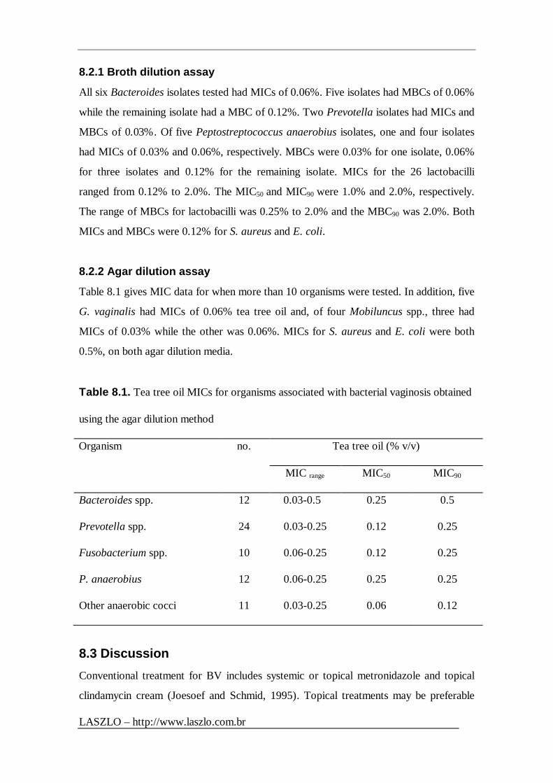
Table 8.1 gives MIC data for when more than 10 organisms were tested. In addition, five
G. vaginalis had MICs of 0.06% tea tree oil and, of four Mobiluncus spp., three had
MICs of 0.03% while the other was 0.06%. MICs for S. aureus and E. coli were both
0.5%, on both agar dilution media.
Table 8.1. Tea tree oil MICs for organisms associated with bacterial vaginosis obtained
using the agar dilution method
Organism no. Tea tree oil (% v/v)
MIC range MIC50 MIC90
Bacteroides spp. 12 0.03-0.5 0.25 0.5
Prevotella spp. 24 0.03-0.25 0.12 0.25
Fusobacterium spp. 10 0.06-0.25 0.12 0.25
P. anaerobius 12 0.06-0.25 0.25 0.25
Other anaerobic cocci 11 0.03-0.25 0.06 0.12
8.3 Discussion Conventional treatment for BV includes systemic or topical metronidazole and topical
clindamycin cream (Joesoef and Schmid, 1995). Topical treatments may be preferable

29
because they produce fewer systemic side-effects, such as gastrointestinal upset and an
unpleasant taste (Joesoef and Schmid, 1995). Tea tree oil has previously been advocated
for topical use in the treatment of vaginal infections, including BV, trichomonal vaginitis
and vaginal candidiasis (Humphrey, 1930; Pena, 1962; Belaiche, 1985). Intra-vaginal tea
tree oil products are available in many countries and may be purchased by women
wanting to treat vaginal symptoms, including BV. Self treatment with alternative
medicines appears to be common amongst women with chronic vaginal symptoms
(Nyirjesy et al., 1997), illustrating the need for in vitro and in vivo data pertaining to the
efficacy of alternative products, in particular, those containing tea tree oil.
MICs and MBCs of tea tree oil have been reported for anaerobic bacteria including
fusobacteria, Bacteroides, Prevotella and Peptostreptococcus, by agar and broth dilution
methods (Walsh and Longstaff, 1987; Shapiro et al., 1994). Despite differences in
methodology, our MIC data are similar to previous studies (Carson and Riley, 1993;
Shapiro et al., 1994). In addition, all groups of organisms showed a range of
susceptibilities to tea tree oil. However, all Lactobacillus spp. tested were appreciably
more resistant to tea tree oil than those organisms known to be associated with BV, with
at least a two-fold difference in MIC 90 results.
The clinical success reported by Blackwell (1991) may be due, in part, to this differential
susceptibility to tea tree oil between BV- associated organisms and commensal
lactobacilli. We demonstrated similar variation in susceptibility to tea tree oil previously
between commensal skin flora, such as coagulase negative staphylococci, and transiently
colonising potential pathogens, such as Staphylococcus aureus (Hammer et al., 1996).
These differences may allow products to be formulated that will selectively kill or inhibit
certain organisms while having a minimal effect on others. The susceptibility data
obtained in this work suggest that tea tree oil may be a useful treatment for BV and
further investigation in the form of a controlled clinical trial is warranted.
9.0 Mechanism of action of tea tree oil 9.1 Introduction

LASZLO – http://www.laszlo.com.br
A second area of research focussed on the mechanism of action of tea tree oil. No
previous investigations reporting on this were available. Since the mechanism of action
has implications for the selectivity and safety of antimicrobial agents, this issue was
becoming increasingly important.
9.2 Results 9.2.1 Optical density of microbial suspensions treated with Melaleuca
alternifolia oil or individual components
Treatment of C. albicans suspensions with MIC and 2xMIC of M. alternifolia oil did not
reduce the optical density. Similarly, after two hours treatment with the MIC of
terpinen-4-ol, the mean optical density of four separate replicates was 93% that of the
original suspensions. Treatment with 2xMIC terpinen-4-ol resulted in modest reductions
in the optical densities with a mean final density of 86% of the original densities. For α-
terpineol, the optical densities of untreated and MIC treated C. albicans suspensions
were not diminished.
Treatment of E. coli suspensions with MIC and 2xMIC of M. alternifolia oil, 1,8-
cineole, terpinen-4-ol and α-terpineol reduced the OD620. The only exception was
terpinen-4-ol treatment at the MIC. Treatment with M. alternifolia oil at 2xMIC
reduced the OD620 to approximately 30% of the original, irrespective of EDTA content.
1,8-Cineole treatment at MIC and 2xMIC also reduced the OD620 to this level but only in
the presence of EDTA. In the absence of EDTA, the OD620 was reduced, but not to the
same extent (53% and 42%, respectively). Terpinen-4-ol treatment at 2xMIC reduced
the OD620 in the presence and absence of EDTA. MIC treatment with α-terpineol
reduced the OD620 and EDTA addition did not enhance this effect. In contrast, 2xMIC
treatment in the presence of EDTA resulted in a larger reduction in OD620 than in the
absence.
The ODs620 of S. aureus suspensions were unaffected by MIC or 2xMIC treatment with
M. alternifolia oil, 1,8-cineole, α-terpineol or terpinen-4-ol.
9.2.2 Leakage of 260nm-absorbing material from microbial suspensions
treated with Melaleuca alternifolia oil or components

31
Treatment of S. aureus suspensions with 1,8-cineole at 0.1xMIC resulted in higher
absorbances compared to the control suspensions. Furthermore, 0.5xMIC, MIC and
2xMIC treatments all resulted in the appearance of approximately twice the level of
260nm-absorbing material in the filtrates compared to the control. Terpinen-4-ol
treatment at 0.5xMIC and MIC, resulted in approximately 3-fold and 4-fold absorbances
at 5h compared to control filtrates. Filtrates from S. aureus suspensions treated with
0.5xMIC and MIC of -terpineol,
9.2.3 Electron microscopy
When examined by electron microscopy, no discernible differences in cell morphology
were seen between C. albicans cells treated with terpinen-4-ol for up to 2h and untreated
control cells. In contrast, examination of terpinen-4-ol treated S. aureus cells revealed
some gross differences in morphology. Multi-lamellar, mesosome-like structures not
seen in untreated S. aureus cells were seen in treated cells. Furthermore, these structures
were more marked and appeared more frequently in cells treated for 10 min compared to
those treated for 4 min. In addition, the contents of some cells appeared depleted and
amorphous. There was no discernible differences between untreated E. coli cells and
cells treated for 4 min. However, cells treated for 10 min contained several gross
changes. Almost none contained the dark granules seen in control cells or cells treated
for 4 min. Many of these cells appeared empty and vacuous with only the exterior cell
membrane/wall structure remaining. In those cells that still had contents, they appeared
monochromatic and amorphous. In some cases the contents had contracted away from
the terminal ends of the cells.
9.3 Discussion The lipophilic walls and membranes of micro-organisms constitute a prime target for the
similarly-natured tea tree oil and its individual components. Consequently, the outer
microbial membranes were the first site to be investigated in work to identify the
mechanism of action of tea tree oil.
Some antimicrobial agents are capable of causing such gross membrane damage as to
provoke whole cell lysis. In microbial suspensions treated with the agent of interest, this

LASZLO – http://www.laszlo.com.br
lysis is reflected in reductions in optical density. Treatment of C. albicans suspensions
with tea tree oil or -terpineol did not significantly reduce the optical density over 2
hours while the higher concentration of terpinen-4-ol had a modest affect.
Treatment of E. coli suspensions with tea tree oil or components resulted in significant
reductions in optical density. In some cases, the presence of the chelating agent EDTA
further enhanced these reductions. EDTA chelates, or binds up, the cations present in
the cell membrane causing the membrane to be destabilised. Hence its presence would
enhance the action of a membrane-active substance. Collectively, these results suggested
that the membrane was a site of action in E. coli.
None of the treatments affected the optical density of S. aureus suspensions and this
would suggest that action of the oils on the cell membrane so as to cause gross cell lysis,
is not the prime mechanism of action in S. aureus cells.
The second manner in which the putative membrane action of tea tree oil and its
components was examined was by observing the leakage of nucleic acids from treated
cells. Nucleic acids are the building blocks of the genetic material, DNA and RNA,
contained in cells. In healthy cells with an intact cell membrane, these macromolecules
do not leak out of the cell in significant quantities. However, if the integrity of the
membrane has been compromised then these molecules may leak from the cell into the
surrounding fluid. These molecules may be detected by measuring the optical density at
260nm. While only S. aureus suspensions were examined in this manner, significant
results were obtained. Treatment with MICs of α-terpineol, 1, 8-cineole, terpinen-4-ol
and linalool all resulted in the appearance of 260nm-absorbing material outside the cells.
This suggests that genetic material is being lost from the cell through a damaged
membrane. While the first series of experiments demonstrated that cell membranes were
not being lysed by these treatment concentrations, this later work suggested that the
membranes were being compromised.
Electron microscopy of terpinen-4-ol treated cells provided further information. C.
albicans cells were not lysed by terpinen-4-ol treatment and by electron microscopy,
appeared unaltered. In contrast, electron microscopy provided useful information about

33
terpinen-4-ol-induced damage to E. coli. These cells had been sensitive to lysis by oil
treatment and the appearance of empty “ghost” cells by electron microscopy confirmed
this effect. The cytoplasm coagulation seen in E. coli cells is also suggestive of more
subtle membrane damage which is sufficient to allow oils to penetrate but insufficient to
cause whole cell lysis. Since cells observed by electron microscopy were treated with the
MIC of terpinen-4-ol, these microscopic results correlate well with the failure of MIC of
terpinen-4-ol to cause lysis. The appearance of mesosome-like structures in terpinen-4-
ol treated S. aureus also suggests damage to the cell membrane or wall. As with E. coli,
the otherwise intact nature of the S. aureus cells correlates well with the failure of
terpinen-4-ol to cause lysis.
The original premise was that tea tree oil and/or its components act on the cell membrane
or wall. These results indicate that this is a site of action. While further evidence is
required to corroborate these observations, the possibility that other sites of action may
exist, must be considered. Furthermore, it is possible that other components, not
examined thus far, may render significant contributions to the antimicrobial activity of
this oil.
10.0 Miscellaneous 10.1 Toxicity In 1994, a paper was published in the Journal of Toxicology – Clinical Toxicology
relating to a case of tea tree oil poisoning (Jacobs and Hornfeldt, 1994). This afforded
us an opportunity to comment on this issue and our response was published soon after
(Carson and Riley, 1995) and is reproduced below.
“Jacobs and Hornfeldt (1994) make several valid points in their recent article concerning
Melaleuca alternifolia or tea tree oil poisoning. Given the widespread availability of an
extensive range of tea tree oil products, the paucity of accurate toxicity data represents a
serious problem. This essential oil which has useful antimicrobial activity is a complex
mixture of approximately 100 components. Little is known of the toxicity of the
individual components or the whole oil. At present the levels of two components are
regulated by an Australian standard (AS 2782-1985) which stipulates that terpinen-4-ol,
the putative antimicrobial component, must comprise at least 30% of the oil, while 1,8-

LASZLO – http://www.laszlo.com.br
cineole, reputedly a skin irritant, must not exceed 15%. While the antimicrobial activity
of terpinen-4-ol has been confirmed, the role of 1,8-cineole is less clear (Carson and
Riley, 1993). The lipophilic nature of tea tree oil and its ability to penetrate skin may
potentiate its toxicity.
Apart from this report of oral toxicity in a 23-month-old boy, there have been three other
cases of oral toxicity reported in the literature. In the first, dermatitis associated with the
topical application of tea tree oil was exacerbated following ingestion of oil (de Groot
and Weyland, 1992). In the second, half a teaspoon of tea tree oil was ingested by a 60-
year-old man resulting in a "dramatic rash" and a general feeling of unwellness (Elliott,
1993). The patient had taken tea tree oil on a number of prior occasions with no
noticeable ill-effects. In the third case, a patient was comatose for 12h and semi-
conscious for a further 36h following ingestion of half a tea cup of neat tea tree oil
(Seawright, 1993). No data regarding the oral toxicity of tea tree oil in humans are
available although the acute oral toxicity in rats is 1.9-2.6 mL/kg (Altman, 1990). Tea
tree oil toxicity has also been reported in dogs and cats (Villar et al., 1994) with the main
symptoms including depression, weakness, incoordination and muscle tremors.
Contact dermatitis from tea tree oil is more frequently reported although accurate
estimates of its incidence are still required (Apted, 1991; de Groot and Weyland, 1992;
de Groot and Weyland, 1993; Moss, 1994; Selvaag et al., 1994; Van der Valk et al.,
1994). In most reports, patch tests with the major components of the oil were not
performed. In the two cases where patch tests with 1,8-cineole were performed in an
attempt to identify the allergen, the results were conflicting (de Groot and Weyland,
1992; Selvaag et al., 1994).
The limited toxicity data and few well-controlled studies that are currently available
require corroboration urgently. Since clinical presentation of toxicity associated with tea
tree oil products may become more common following the current interest in natural
products, it is imperative that our clinical knowledge of this essential oil and its related
products progresses accordingly.”

35
10.2 Podiatry In 1997, a paper was published in the Journal of British Podiatric Medicine on the use
of tea tree oil as adjunctive therapy. This also allowed us to comment on some
applications of tea tree oil.
“Rodger (1997) makes some interesting points about the use of tea tree oil as adjunct
therapy in chronic mucocutaneous candidiasis. Tea tree oil is widely available in
the United Kingdom, in products such as creams, toothpastes, soaps and
antiseptic lotions. The oil is produced by steam distillation of the leaves of the
Australian native plant Melaleuca alternifolia and is composed of approximately 100
individual components (Carson and Riley, 1993).
Tea tree oil is known to inhibit a wide range of fungi in vitro including Aspergillus niger,
A. flavus, Epidermophyton floccosum, Microsporum audonii, M. canis, Trichophyton
mentagrophytes and T. rubrum (Walker, 1972), however, few clinical trials have
examined the therapeutic efficacy of tea tree oil in cutaneous fungal infections (Tong et
al., 1992; Buck et al., 1994). Also, the commensal yeast Malassezia furfur
(Pityrosporum ovale) is also inhibited in vitro suggesting a possible role for tea tree oil in
the treatment of pityriasis versicolor (Hammer et al., 1996). Tea tree oil and several of
its major components have also been shown to inhibit the growth of Candida albicans
(Carson and Riley, 1993; Carson and Riley, 1995) and it is this activity which may have
contributed to the successful outcome reported by Rodger (1997).
Apart from inhibiting the growth of C. albicans, tea tree oil is known to inhibit a wide
range of organisms which may otherwise result in secondary infections such as
Staphylococcus aureus and Streptococcus spp (Walker, 1972; Carson and Riley, 1993).
It has also been shown to inhibit many transient skin organisms at concentrations lower
than those which inhibit commensal skin flora (Hammer et al., 1996). It is possible that
this differential inhibition may reduce disturbance of the patient’s commensal skin flora
and, in turn, enhance the protection offered by these commensal flora against
colonisation by other transient flora. Further work is required to examine this possibility.

LASZLO – http://www.laszlo.com.br
Although there is no scientific evidence as yet, anecdotal evidence suggests that tea tree
oil has mild analgesic properties and this is further suppo rted by Rodger’s observations.
In addition, the lipophilic nature of tea tree oil and the ability of some of its components
to penetrate skin (Obaata et al., 1991) may potentiate the antimicrobial activity and
result in a residual antimicrobial effect of the oil in the skin.
The in vitro antibacterial activity of tea tree oil is becoming well-established and
investigations into the antifungal activity are beginning. In contrast, too few clinical trials
have demonstrated the efficacy of tea tree oil in vivo and much further work on this
potentially useful natural product is required.”
11.0 Implications, recommendations and
intellectual property
11.1 Implications It is difficult to measure the impact of our research on the tea tree oil industry in
Australia. Although not directly assessable, several parameters give an indication of the
impact of our work.
First, the significant number of interviews and articles provided for the popular press,
both in Australia and overseas, has undoubtedly increased public awareness of tea tree
oil and tea tree oil products. Publicity regarding the scientific proof that tea tree oil
“works” directly enhances the profile and marketability of the oil.
Second, the publication of much of this project in peer-reviewed medical journals has had
a significant and profound impact on the industry in Australia. This is evident by a

37
number of effects. Publication has stimulated interest in the antimicrobial properties of
tea tree oil in international medical and scientific fora. One practical way in which this
has been manifested is the development of several collaborative projects between the
Project Team and other research groups including the Laboratory for Hospital Infection
at the Central Public Health Laboratory and a group at St. Mary’s Hospital at Imperial
College both in London. Numerous requests (~200) from other researchers for reprints
of our publications, also indicate that interest in the applications of tea tree oil has been
stimulated. Additional investigation into, and verification of, the potential uses of tea
tree oil will further strengthen the industry.
Another way in which publications indicate the impact of this project, is the citation of
these publications by other authors. During the course of the project, several
publications by other researchers regarding the antimicrobial activity of tea tree oil have
appeared and the results of this project have been cited (Jacobs & Hornfeldt, 1994;
Raman et al., 1995; Nenoff et al., 1996; Boon & Johnstone, 1997; Buchbauer, 1997;
Faoagali et al., 1997; Hili et al., 1997; Nelson, 1997; Southwell et al., 1997).
Furthermore, the work conducted by these independent investigators corroborates, to a
large extent, the results of this project.
Publications have also allowed the Project Team to clarify or elaborate on tea tree oil
work from other research groups. This has been done on at least two occasions (Carson
and Riley, 1995; Carson et al., 1997) and, apart from sustaining scientific interest in tea
tree oil, permits misconceptions or inaccuracies to be addressed.
Members of the Project Team have been asked to review research conducted by other
workers prior to acceptance for publication in medical and scientific journals and this
highlights the level of expertise attained by the group. More important, in an industry
that will soon come under increasing international competition, it helps consolidate the
impression of Australia as the leader in the tea tree oil industry.
Third, the impact of our work can be measured by the degree of interest shown in the
project results by companies in the tea tree oil industry. A significant number of reprints
of our publications have been purchased by several tea tree oil companies, presumably
for distribution to clients.

LASZLO – http://www.laszlo.com.br
In addition, results of our studies are cited frequently on tea tree oil company world wide
web sites, indicating that producers and distributors consider the project results beneficial
in the marketing of tea tree oil and tea tree oil products.
Finally, as part of the on-going attempts to register tea tree oil as an active antimicrobial
ingredient with the FDA in the United States, copies of our publications have been added
to the previous submissions. Our data have also been presented in submissions to other
national regulatory bodies, including those in Sweden and Germany.
11.2 Recommendations The antimicrobial activity of tea tree oil characterised in this project indicates clearly that
tea tree oil has the potential to become a clinically useful product. The in vitro data
suggest that tea tree oil products may have efficacy in the treatment of many cutaneous
skin infections. These data also provide a sound basis from which to proceed to in vivo
work. What is required now is in vivo data from well-designed, randomised, controlled
clinical trials.
11.3 Intellectual Property Apart from the impact of the project upon the profile and marketability of tea tree oil,
there are no commercially significant developments arising from the project.
12.0 Communications Strategy Since one of the aims of the project was to address the paucity of information available in
the literature, a substantial effort was put into publishing the results of the project in
peer-reviewed medical and scientific journals. A significant effort also went in to
presenting project results at scientific and industry conferences as well as in the popular
press.
Results were published as they became available. As a result of the project, the
following papers have been submitted to or published in peer-review journals:

39
Carson, C. F. and Riley, T. V. (1995) Antimicrobial activity of the major components of
the essential oil of Melaleuca alternifolia. Journal of Applied Bacteriology
78:264-269.
Carson, C. F., Cookson, B. D., Farrelly, H. D. and Riley, T. V. (1995) Susceptibility of
methicillin-resistant Staphylococcus aureus to the essential oil of Melaleuca
alternifolia. Journal of Antimicrobial Chemotherapy 35:421-424.
Carson, C. F. and Riley, T. V. (1995) Toxicity of the essential oil of Melaleuca
alternifolia or tea tree oil (Letter). Journal of Toxicology - Clinical Toxicology
33:193-194.
Carson, C. F., Hammer, K. A. and Riley, T. V. (1995) Broth micro-dilution method for
determining the susceptibility of Escherichia coli and Staphylococcus aureus to
the essential oil of Melaleuca alternifolia (tea tree oil). Microbios 82:181-185.
Carson, C. F., Hammer, K. A. and Riley, T. V. (1996) In vitro activity of the essential oil
of Melaleuca alternifolia against Streptococcus spp. (Letter). Journal of
Antimicrobial Chemotherapy 37:1177-1178.
Hammer, K .A., Carson, C. F. and Riley, T. V. (1996) Susceptibility of transient and
commensal skin flora to the essential oil of Melaleuca alternifolia (tea tree oil).
American Journal of Infection Control 24:186-189.
Carson, C. F., Hammer, K. A. and Riley, T. V. (1997) Use of the essential oil of
Melaleuca alternifolia (tea tree oil) in cutaneous fungal infections (Letter).
Journal of British Podiatric Medicine 52:iv-v.
Hammer, K .A., Carson, C. F. and Riley, T. V. In vitro susceptibility of lactobacilli and
organisms associated with bacterial vaginosis to Melaleuca alternifolia (tea tree)
oil. Submitted for publication
There are two more manuscripts in preparation; one discussing the interaction of tea tree
oil with various product excipients and the other detailing the mechanism of action work.
In addition to the above work, that was supported directly by RIRDC, two other
publications on tea tree oil were produced by members of our laboratory:

LASZLO – http://www.laszlo.com.br
Hammer, K .A., Carson, C. F. and Riley, T. V. In vitro susceptibility of Malassezia
furfur to the essential oil of Melaleuca alternifolia (tea tree oil). Journal of
Medical and Veterinary Mycology 35:375-377.
Hammer, K .A., Carson, C. F. and Riley, T. V. In vitro activity of Melaleuca alternifolia
(tea tree) oil, intra-vaginal tea tree oil products and other essential oils against
Candida spp. Submitted for publication
As well as papers published in medical and scientific journals, several oral and poster
presentations were published as conference abstracts. These were as follows:
Carson, C. F. (1995) Evaluating the antibacterial activity of the essential oil of
Melaleuca alternifolia and its major components. Proceedings of the National
Conference on Tea Tree Oil, 24-25 August 1995, Sydney, Australia.
Carson, C. F. (1996) Potential uses of Melaleuca alternifolia oil – the acne removal
company. Proceedings of the National Conference on Tea Tree Oil, 26 October
1996, Sydney, Australia.
Carson, C. F. and Riley, T. V. (1996) Working with and against tea tree oil – issues of
synergy and antagonism. Proceedings of the 19th IFSCC Congress, Sydney,
October 1996.
Hammer, K. A., Carson, C. F. and Riley, T. V. (1995) Susceptibility of transient and
commensal flora to the essential oil of Melaleuca alternifolia (tea tree oil).
Microbiology Australia 16: P30.4.
Riley, T. V. (1995) The anti-staphylococcal activity of tea tree oil and other research
opportunities. Proceedings of the National Conference on Tea Tree Oil, 24-25
August 1995, Sydney, Australia.
Riley, T. V. (1996) Antiseptic properties of tea tree oil and MRSA. Proceedings of the
National Conference on Tea Tree Oil, 26 October 1996, Sydney, Australia.
In addition, the following abstracts were submitted for presentation at the annual meeting
of the Australian Society for Microbiology held in Adelaide in September, 1997:
Carson, C. F. and Riley, T. V. Effect of the essential oil of Melaleuca alternifolia (tea
tree oil) on bacterial and yeast cell membranes.

41
Hammer, K. A., Carson, C. F. and Riley, T. V. Effect of organic matter, surfactants and
cations on the antimicrobial activity of Melaleuca alternifolia (tea tree) oil.
Other talks on aspects of the work with tea tree oil were as follows:
• A/Prof Riley presented results of the group’s work at the Australian Society for
Microbiology (WA Branch) Country Scientific Weekend Meeting in May, 1996.
• A talk entitled “Tea tree and golden staph” was delivered by Ms Christine Carson at
the July 1995 meeting of the Western Australian Essential Oils forum.
• Ms Christine F. Carson presented a talk entitled "Tea tree oil and product
formulations" to the Bacteriology Research Group in the Department of
Microbiology in November, 1994 .
• Ms C Carson delivered a lecture entitled "The antimicrobial activity of tea tree oil" to
the West Australian branch of the Australian Society for Microbiology in December,
1994.
Throughout the course of the project numerous interviews for newspaper, radio and
television were conducted. They are listed below.
• Television interviews for Australian evening news' programmes were conducted in
October 1994.
• A/Prof Thomas V. Riley addressed several international television and newspaper
groups and tea tree oil industry representatives in November 1994.
• Television interviews for Australian evening news' programmes were conducted in
January 1995.
• An article describing aspects of our work with tea tree oil appeared in The West
Australian (21/1/95) and resulted in many enquiries.
• An article entitled “Taking the ’quackery’ out of tea tree oil” appeared in June 1995
issue of Agriculture Today and resulted in several enquiries from primary producers.

LASZLO – http://www.laszlo.com.br
• The tea tree oil research group was featured on the ABC’s programme, “The 7.30
Report” (28/6/95). This report discussed the susceptibility of “golden staph” to tea
tree oil and resulted in several enquiries from the public and other media groups.
• An article discussing our work with tea tree oil appeared in the Albany Advertiser in
July 1995.
• A/Prof Riley was interviewed for radio and newspaper articles in September 1995 at
the annual Microbiology conference in Canberra.
• A radio interview discussing the recent findings about tea tree oil was given for the
Victorian ABC on October 5, 1995 by Ms Carson.
• GEO magazine prepared a story regarding tea tree oil and our work in November
1995.
• On December 2 1995, a group of Scandinavian journalists and medical practitioners
who had been brought to Australia by Australian Bodycare Pty. Ltd., visited our
research group for a series of lectures on the properties of tea tree oil and the results
of our work. Several interviews were prepared for national television and radio in
Sweden and Denmark.
• A radio interview was conducted by Ms Carson on Thursday 7 March for Radio
Australia (Melbourne).
• An article discussing our work with tea tree oil in the treatment of vaginal thrush
appeared in the March edition of Australian Pharmacist (15:104,109).
• A series of lectures and interviews on tea tree oil were given to a group of journalists
and medical practitioners from Denmark and the Czech Republic on 27 March 1996.
The group had been brought to Australia by the Bodycare Corporation Pty. Ltd.
• The West Australian (3 April 1996) published a story about our work with tea tree
oil.
• A radio interview was conducted by Ms Carson on Thursday 11 April for ABC
Regional Radio (WA).
• A radio interview was conducted by Ms Carson on Thursday 9 May for 6NR
(national).
• On Wednesday 29 May 1996, an article appeared in The West Australian newspaper
outlining A/Prof Riley’s research interests including the work on tea tree oil.

43
• In June 1996, Assoc/Prof Riley and Ms Carson visited the United Kingdom and met
with representatives from several British hospitals regarding tea tree oil and MRSA
and discussed collaborative clinical trials.
• Several interviews with UK print media journalists were conducted during the June
1996 visit. As a result, a multi-page supplement about tea tree oil was published by
the Daily Mail newspaper. This paper has a circulation of approximately 1 million.
Finally, scientific presentations were made in Copenhagen for a range of journalists
and medical professionals.
• Ms Carson was interviewed on Tuesday 18 June for ABC 6WF (WA).
• On Thursday 27 June, Ms Carson was interviewed for 6PR (WA).
• An article discussing our work appeared in the June/August edition of Earth Garden
• You magazine published a piece on our work in the 14 July, 1996 issue.
• Interviews for the BBC television programme “Tomorrow’s World” were conducted
with Assoc/Prof Riley and Ms Carson on 1 and 3 August 1996 and a segment dealing
with tea tree oil and methicillin-resistant Staphylococcus aureus was aired in the
United Kingdom in early October. Subsequently, approximately 100 enquiries
regarding tea tree oil were received from the UK, which have resulted in
collaborative research.
• On Saturday 1 February 1997, an article appeared in the West Australian newspaper
outlining our work on tea tree oil, in particular with MRSA.
• A/Prof Riley was interviewed for a South African magazine, Fair Lady, and an
article was published in May, 1997.
• A/Prof Riley gave several interviews in Poland in May 1997.
• Ms Christine Carson was invited to speak at a by the WA Venereology Society
seminar on June 8 1997.
13.0 References
Altman PM. Australian tea tree oil. Aust J Pharm 1988;69: 276-278.
Altman PM. Summary of safety studies concerning Australian tea tree oil. In Modern
Phytotherapy - The Clinical Significance of Tree Oil and other Essential Oils.

LASZLO – http://www.laszlo.com.br
Proceedings of a Conference on December 1-2, 1990 in Sydney and a Symposium on
December 8, 1990 in Surfers Paradise, II, 21-22.
Apted JH. Contact dermatitis associated with the use of tea tree oil (Letter). Australas J
Dermatol 1991;32: 177.
Atkinson N, Brice HE. Antibacterial substances produced by flowering plants. 2. The
antibacterial action of essential oils from some Australian plants. Aust J Exp Biol
1995;33: 547-554.
Balows A, ed. Manual of Clinical Microbiology. (5th ed.) American Society for
Microbiology, Washington D.C. 1991.
Barnett BO, Frieden IJ. Streptococcal skin diseases in children. Sem Dermatol 1992;11:
3-10.
Bassett IB, Pannowitz DL, Barnetson RStC. A comparative study of tea tree oil versus
benzoylperoxide in the treatment of acne. Med J Aust 1990;153: 455-458.
Belaiche P. Traitement des infections vaginales a Candida albicans par l'huile essentielle
de Melaleuca alternifolia Cheel. Phytother 1985;15: 13-14.
Beylier MF. Bacteriostatic activity of some Australian essential oils. Perfum Flavorist
1979;4: 23-25.
Blackwell AL. Tea tree oil and anaerobic (bacterial) vaginosis.(Letter) Lancet 1990;337:
300.
Boon P, Johnstone L. Organic matter decay in coastal wetlands- an
inhibitory role for essential oil from M elaleuca alternif olia leaves? A rch
Hydrobiol 1997-138- 433-449.

45
Brophy JJ, Davies NW, Southwell IA, Stiff IA, Williams LR. Gas chromatographic
quality control for oil of Melaleuca terpinen-4-ol type (Australian tea tree). J Ag Food
Chem 1989; 37: 1330-1335.
Buchbauer G. Über das teebaumöl. Eurocosmetics 1997 i: 21-24.
Buck DS, Nidorf DM, Addino JG. Comparison of two topical preparations for the
treatment of onychomycosis: Melaleuca alternifolia (tea tree) oil and clotrimazole. J
Fam Pract 1994;38: 601-605.
Carson CF, Hammer KA, Riley T V. In vitro activity of the essential oil of Melaleuca
alternifolia against Streptococcus spp. J Antimicrob Chemother 1996;37: 1177-1178.
Carson CF, Riley TV. Antimicrobial activity of the essential oil of Melaleuca
alternifolia. Letts Appl Microbiol 1993;16: 49-55.
Carson CF, Riley TV. Antimicrobial activity of the major components of the essential oil
of Melaleuca alternifolia. J Appl Bacteriol 1995;78: 264-269.
Carson CF, Cookson BD, Farrelly HD, Riley TV. Susceptibility of methicillin-resistant
Staphylococcus aureus to the essential oil of Melaleuca alternifolia. J Antimicrob
Chemother 1995;35: 421-424.
Carson CF, Hammer KA, Riley TV. Broth micro-dilution method for determining the
susceptibility of Escherichia coli and Staphylococcus aureus to the essential oil of
Melaleuca alternifolia (tea tree oil). Microbios 1995;82: 181-185.
Carson CF, Riley TV. The antimicrobial activity of tea tree oil. Med J Aust 1994a;160:
236.
Carson CF, Riley TV. Susceptibility of Propionibacterium acnes to the essential oil of
Melaleuca alternifolia. Letts Appl Microbiol 1994b;19: 24-25.

LASZLO – http://www.laszlo.com.br
Carson CF, Riley TV. Toxicity of the essential oil of Melaleuca alternifolia or tea tree
oil. (Letter). J Toxicol Clin Toxicol 1995;33: 193-194.
Carson CF, Hammer KA, Riley TV. Use of the essential oil of Melaleuca alternifolia
(tea tree oil) in cutaneous fungal infections. (Letter). J Brit Pod Med 1997;52: iv-v.
Cook RL, Redondo-Lopez V, Schmitt C, Meriwether C, Sobel JL. Clinical,
microbiological and biochemical factors in recurrent bacterial vaginosis. J Clin Microbiol
1992;30: 870-877.
Cruz T, Cabo MP, Cabo MM, Jimenez J, Cabo J, Ruiz C. In vitro effect of the essential
oil of Thymus longiflorus Boiss. Microbios 1989;60: 59-61.
de Groot AC, Weyland JW. Contact allergy to tea tree oil. (Letter). Contact Dermatitis
1993;28: 309.
de Groot AC, Weyland JW. Systemic contact dermatitis from tea tree oil. Contact
Dermatitis 1992;27: 279-280.
Dyke KGH, Curnock SP, Golding M,Noble WC. Cloning of the gene conferring
resistance to mupirocin in Staphylococcus aureus. FEMS Microbiol Letts 1991;77: 195-
198.
Elliott C. Tea tree oil poisoning. Med J Aust 1993;159: 830-831.
Faoagali J, George N, Leditschke JF. Does tea tree oil have a place in the topical
treatment of burns? Burns 1997;23: 349-351.
Feinblatt HM. Cajeput-type oil for the treatment of furunculosis. J Natl Med Assoc
1960;52: 32-34.

47
Hammer KA, Carson CF, Riley TV. In vitro susceptibility of Malassezia furfur to the
essential oil of Melaleuca alternifolia (tea tree oil). Proceedings of the 19th International
Federation of the Societies of Cosmetic Chemists Congress, Sydney, Australia, 1996.
Hammer KA, Carson CF, Riley TV. Susceptibility of transient and commensal skin flora
to the essential oil of Melaleuca alternifolia (tea tree oil). Amer J Infect Cont 1996;24:
186-189.
Hammer KA, Carson CF, Riley TV. Effect of organic matter, surfactants and cations on
the antimicrobial activity of Melaleuca alternifolia (tea tree) oil. Australian Society for
Microbiology Annual Scientific Meeting and Exhibition, Adelaide 1997. Abstract
P02.49, p. A111.
Hayes AJ, Markham JL, Leach DN, Southwell IA. Relationship between chemical
composition and antimicrobial activity of Australian tea tree oil. Australian Society for
Microbiology, Melbourne, Australia. In Program and Abstracts of the Annual Scientific
Meeting of the Australian Society for Microbiology, Perth Western Australia, 1993..
Abstract P2.2, p. A-4.
Humphery EM. A new Australian germicide. Med J Aust 1930;1: 417-418.
Jacobs MR, Hornfeldt CS. Melaleuca oil poisoning. J Toxicol Clin Toxicol 1994;32:
461-464.
Janssen DA, Zarins LT, Schaberg DR, Bradley SF, Terpenning MS, Kauffman CA.
Detection and characterisation of mupirocin resistance in Staphylococcus aureus.
Antimicrob Agents Chemother 1993;37: 2003-2006.
Joesoef MR, Schmid GP. Bacterial vaginosis: review of treatment options and potential
clinical indications for therapy. Clin Infect Dis 1995;20(Suppl 1): S72-79.

LASZLO – http://www.laszlo.com.br
Kauffman CA, Terpenning MS, Xiaogong H, Zarins LT, Ramsey MA, Jorgensen KA. et
al. Attempts to eradicate methicillin resistant Staphylococcus aureus from a long term
care facility with the use of mupirocin ointment. Amer J Med 1993;94: 371-8.
Kerr S, Kerr GE, MacKintosh CA, Marples RR. A survey of methicillin-resistant
Staphylococcus aureus affecting patients in England and Wales. J Hosp Infect 1990;16:
35-48.
Knight TE, Hausen BM. Melaleuca oil (tea tree oil) dermatitis. J Amer Acad Dermatol
1994;30: 423-427.
Kristofferson SS, Atkin PA, Shenfield GM. Uptake of alternative medicine. Lancet
1996;347: 972.
Lassak EV, McCarthy T. Australian Medicinal Plants. Sydney : Methuen.1983.
Low D, Rawal BD, Griffin WJ. Antibacterial action of the essential oils of some
Australian Myrtaceae with special references to the activity of chromatographic fractions
of oil of Eucalyptus citriodora. Planta Medica 1974;26: 184-189.
Maple PAC, Hamilton-Miller JMT, Brumfitt W. Comparison of the in-vitro activities of
the topical antimicrobials azelaic acid, nitrofurazone, silver sulphadiazine and mupirocin
against methicillin-resistant Staphylococcus aureus. J Antimicrob Chemother 1992;29:
661-8.
Maruzzella JC, Chiaramonte JS, Garofalo MM. Effects of vapors of aromatic chemicals
on fungi. J Pharmaceut Sci 1961;50: 665-668.
McKern HHG, Parnell IW. The larvicidal effect of various chemical compounds and
plant products on the free-living stages of Haemonchus contortus Rud. (Nematoda). J
Helminthol 1964;38: 223-244.
Moss A. Tea tree oil poisoning. Med J Aust 1994;160: 236.

49
Naguib MH, Naguib MT, Flournoy DJ. Mupirocin resistance in methicillin resistant
Staphylococcus aureus from a veterans hospital. Chemother 1993;39: 400-4.
National Committee for Clinical and Laboratory Standards. Methods for dilution,
antimicrobial susceptibility tests for bacteria that grow aerobically; Approved Standard
M7-A4. NCCLS, Wayne, PA. 1997.
National Committee for Clinical Laboratory Standards. . Methods for antimicrobial
susceptibility testing of aerobic bacteria. M7-A2 (2nd ed.) NCCLS, Villanova, PA. 1990
Nenoff P, Haustein U-F, Brandt W. Antifungal activity of the essential oil
of M elaleuca alternif olia (tea tree oil) against pathogenic fungi in vitro. Skin
Pharm acol 1996-9- 388-394.
Nyirjesy P, Weitz MV, Grody MHT,. Lorber B. Over-the-counter and alternative
medicines in the treatment of chronic vaginal symptoms. Obstet Gynecol 1997;90: 50-53.
Obata Y, Takayama K, Machida Y, Nagai T, Combined effect of cyclic monoterpenes
and ethanol on percutaneous absorption of diclofenac sodium. Drug Design and
Delivery 1991;8: 137-144.
Onawunmi GO, Yisak W-A, Ogunlana EO. Antibacterial constituents in the essential oil
of Cymbopogon citratus (DC.) Stapf. J Ethnopharmacol 1984;12: 279-286.
Peña EF. Melaleuca alternifolia oil. Its use for trichomonal vaginitis and other vaginal
infections. Obstet Gynecol 1962;19: 793-795.
Penfold AR, Grant R. The germicidal values of some Australian essential oils and their
pure constituents. J Proc Royal Soc NSW 1925;60: 167-70.

LASZLO – http://www.laszlo.com.br
Penfold AR, Grant R. The germicidal values of some Australian essential oils and their
pure constituents. Together with those for some essential oil isolates, and synthetics. Part
III. Journal and Proceedings of the Royal Society of New South Wales 1925;59: 346-
350.
Penfold AR, Morrison FR. Some notes on the essential oil of Melaleuca alternifolia.
Australas J Pharm 1937;18: 274-275.
Rahman M, Noble WC, Cookson B. Mupirocin-resistant Staphylococcus aureus. Lancet
1987;2: 387.
Raman A, Weir U, Bloomfield SF. Antimicrobial effects of tea-tree oil and
its major components on Staphy lococcus aureus, Staph. epiderm idis and
Propionibacterium acnes. L etts A ppl M icrobiol 1995-21- 242-245.
Reuveni R, Fleischer A, Putievsky E. Fungistatic activity of essential oils from Ocimum
basilicum chemotypes. Phytopathologische Zeitschrift 1984;110: 20-22.
Reynolds JEF. (Ed) (1993) Martindale - the extra pharmacopoeia, 30th edn, pp. 1385.
The Pharmaceutical Press, London.
Riley TV, Carson CF, Bowman RA, Mulgrave L, Golledge CL, Pearman JW, Grubb
WB. Mupirocin-resistant methicillin-resistant Staphylococcus aureus in Western
Australia. Med J Aust 1994;161: 397-398.
Rodger E. ‘Chronic mucocutaneous candidiasis - a case study’. J Brit Pod Med 1997;52:
9-10.
Ross SA, El-Keltawi NE, Megalla SE. Antimicrobial activity of some Egyptian aromatic
plants. Fitoterapia 1980;51: 201-205.
Seawright A. Comment: Tea tree oil poisoning. Med J Aust 1993;159: 831.

51
Selvaag E, Erikson B, Thune P. Contact allergy to tea tree oil and cross-sensitisation to
colophony. Contact Dermatitis 1994;31:124-125.
Shapiro S, Meier A, Guggenheim B. The antimicrobial activity of essential oils and
essential oil components towards oral bacteria. Oral Microbiol Immunol 1994;9: 202-
208.
Shemesh A, Mayo WL. Australian tea tree oil: a natural antiseptic and fungicidal agent.
Aust J Pharm 1991;72: 802-3.
Shriner DL, Schwartz RA, Janniger CK. Impetigo. Cutis 1995;56: 30-32.
S-derberg TA, Johansson A, Gref R. Toxic effects of some conifer resin
acids and tea tree oil on human epithelial and fibroblast cells. T ox icol
1996-107: 99-109.
Southwell IA, Freeman S, Rubel D. Skin irritancy of tea tree oil. J E ssent
O il Research 1997-9- 47-52.
Standards Association of Australia. Essential oils - oils of Melaleuca, terpinen-4-ol type.
Standards Australia, Sydney, 1985;2782.
Summanen P, Baron EJ, Citron DM, Strong CA., Wexler HM, Finegold SM. (1993)
Wadsworth Anaerobic Bacteriology Manual 5th ed. Star Publishing Company, Belmont,
California.
Swords G, Hunter GLK. Composition of Australian tea tree oil (Melaleuca alternifolia).
J Ag Food Chemistry 1978;26: 734-737.
Tong MM, Altman PM, Barnetson RStC, ‘Tea tree oil in the treatment of tinea pedis’.
Australas J Dermatol 1992;33: 145-149.

LASZLO – http://www.laszlo.com.br
Udo EE, Pearman, JW, Grubb WB. Genetic analysis of community isolates of
methicillin-resistant Staphylococcus aureus in Western Australia. J Hosp Infect 1993;25:
97-108.
Udo EE, Pearman JW, Grubb WB. Emergence of high-level mupirocin resistance in
methicillin-resistant Staphylococcus aureus in Western Australia. J Hosp Infect 1994;26:
101-9.
Van der Valk PGM, de Groot AC, Bruynzeel DP, Coenraads PJ, Weijland JW. Allergic
contact dermatitis from 'tea tree' oil. Ned Tijdschr Geneeskd 1994;138: 823-825.
Villar D, Knight MJ, Hansen SR, Buck WB. Toxicity of Melaleuca oil and related
essential oils applied topically on dogs and cats. Vet Human Toxicol 1994;36: 139-142.
Walker M, ‘Clinical investigation of Australian Melaleuca alternifolia oil for a variety of
common foot problems’. Curr Pod 1972;April: 7-15.
Walsh LJ, Longstaff J. The antimicrobial effects of an essential oil on selected oral
pathogens. Periodontol 1987;8: 11-15.
Williams LR, Home VN, Asre S. Antimicrobial activity of oil of Melaleuca (tea tree oil).
Its potential use in cosmetics and toiletries. Cosmetics, Aerosols and Toiletries in
Australia 1990;4: 12-3, 16-8, 22.
Williams LR, Home VN. (1989) Plantations of Melaleuca alternifolia - a revitalized
Australian tea tree industry. Proceedings of the 11th International Congress of Essential
Oils, Fragrances and Flavours 49-53.