Embed Size (px)
Citation preview
i
ANTIMICROBIAL ACTIVITY OF SOME MEDICINAL PLANT EXTRACTS
AGAINST BACTERIA CAUSING DIARRHOEA
by
NAOMI TOPE KOMOLAFE
Submitted in accordance with the requirements for
the degree of
MASTER OF SCIENCE
in the subject
Life Sciences
at the
UNIVERSITY OF SOUTH AFRICA
Supervisor: Dr MA Nyila
Co-supervisors: Dr FT Tabit & Dr TE Tshikalange
DECEMBER 2014
ii
DECLARATION
I declare that ANTIMICROBIAL ACTIVITY OF SOME MEDICINAL PLANT
EXTRACTS AGAINST BACTERIA CAUSING DIARRHOEA is my own work and
that all sources that I have used or quoted have been indicated and acknowledged
by means of complete references.
________________________________________
Signed by: NAOMI TOPE KOMOLAFE
Student Number: 49022547
Date: 05 December 2014

iii
DEDICATION
This dissertation is dedicated to my almighty father Jehovah God, through whom I
had the strength and vigour to start and complete this work.
iv
ACKNOWLEDGEMENTS
My heartfelt gratitude goes to my husband, Ayoola Komolafe, for all his love and
support throughout the duration of my studies. I would like to acknowledge my son
Micheal Jahtobiloba, who has brought me so much joy and happiness. Many thanks
also to my parents and siblings for their words of encouragement and moral support.
To my supervisors, Dr Tabit, Dr Nyila and Dr Tshikalange, I am very grateful for your
supervision and guidance during the course of my studies. Special thanks go to Dr
Tabit for his constant positive criticism and grilling to make me a better research
student.
My appreciation also goes to the post-graduate research students at the Department
of Plant Sciences, University of Pretoria. Thanks a lot for your kindness and
willingness to put me through some of the laboratory techniques.
v
ABSTRACT
Infectious diarrhoea is the second largest single cause of mortality in children under
the age of five globally. Bacteria are responsible for most diarrhoeal episodes
especially in developing countries, and progressive increase in antimicrobial
resistance has given rise to the need to investigate other sources of therapy such as
medicinal plants. Ten plant extracts were analysed for their antimicrobial activities
using the agar well diffusion and broth microdilution method. Their phytochemical
contents were screened, and their effect on 1, 1-diphenyl-2-picrylhydrazyl (DPPH)
was used to assess their antioxidant activities. Their toxicity profiles were evaluated
using the XTT Cytotoxicity Assay. Water and methanol extracts of Adansonia digitata
seeds and pulp showed no inhibition against all the test organisms, while water and
methanol extracts of A. digitata leaves showed inhibition, with minimum inhibitory
concentration (MIC) ranging from 0.39 to 6.25mg/ml. Water and methanol extracts of
Garcinia livingstonei and Sclerocarya birrea barks showed good activity against all
the test organisms, with MICs between 0.39 and 1.56 mg/ml. Alkaloids, phenols,
flavonoids, saponins, tannins, and terpenoids were found in one or more of the plant
extracts, and all the plant extracts demonstrated scavenging power against
DPPH.The cytotoxicity of extracts of Garcinia livingstonei, and Sclerocarya birrea
barks ranged between 105.9 µg/ml and 769.9 µg/ml. The results obtained in this
study validate the traditional use of A. digitata leaves, G. livingstonei and S. birrea
bark in treating bacteria causing diarrhoea.
vi
TABLE OF CONTENTS
DECLARATION ...................................................................................................................... ii
DEDICATION .......................................................................................................................... iii
ACKNOWLEDGEMENTS .................................................................................................... iv
ABSTRACT .............................................................................................................................. v
LIST OF FIGURES............................................................................................................... viii
LIST OF ABBREVIATIONS .................................................................................................. xi
CHAPTER 1: INTRODUCTION ............................................................................................ 1
1.1 BACKGROUND OF THE STUDY .......................................................................... 1
1.2 PROBLEM STATEMENT ........................................................................................ 3
1.3 PURPOSE OF THIS RESEARCH ......................................................................... 5
1.4 LAYOUT OF THE DISSERTATION ..................................................................... 6
CHAPTER 2: LITERATURE REVIEW ................................................................................. 7
2.1 ANTIMICROBIALS ................................................................................................... 7
2.1.1 TYPES AND SOURCES OF ANTIMICROBIALS .......................................... 7
2.2 MEDICINAL PLANTS .............................................................................................. 8
2.2.1 HISTORY OF MEDICINAL PLANTS ............................................................... 8
2.2.2 GENERAL USES OF MEDICINAL PLANTS ................................................. 9
2.3 ACTIVE COMPONENTS OF PLANT EXTRACTS ........................................... 10
2.4 MECHANISM OF ACTION OF PLANT SECONDARY COMPOUNDS ......... 11
2.5 SIGNIFICANCE OF ANTIMICROBIAL SUSCEPTIBILITY TESTING ............ 13
2.6 EXTRACTION TECHNIQUES OF PLANT EXTRACTS ................................... 14
2.7 ISOLATION AND IDENTIFICATION METHODS .............................................. 15
2.8 CURRENT TRENDS IN PHYTOCHEMISTRY AND MEDICINAL PLANT RESEARCH ........................................................................................................................... 16
2.9 RELEVANCE OF THIS RESEARCH .................................................................. 17
2.9.1 Adansonia digitata ............................................................................................ 18
2.9.2 Garcinia livingstonei ......................................................................................... 21
2.9.3 Sclerocarya birrea ............................................................................................ 23
2.10 HYPOTHESIS AND OBJECTIVES ................................................................. 27
CHAPTER 3: ANTIMICROBIAL AND CYTOTOXICITY ASSAYS ................................. 28
3.1 MATERIALS AND METHOD ................................................................................ 28
3.1.1 PLANT MATERIALS AND SAMPLE COLLECTION ..................................... 28
3.1.2 INSTRUMENTS AND REAGENTS USED ................................................... 28
3.1.3 PREPARATION OF PLANT EXTRACT ........................................................ 28
3.2 ANTIMICROBIAL ACTIVITY ................................................................................ 29
3.2.1 INSTRUMENTS AND REAGENTS/MATERIALS USED ........................... 29
3.2.2 PREPARATION OF BACTERIA CULTURE ................................................ 29
vii
3.2.3 AGAR WELL DIFFUSION ASSAY ................................................................ 30
3.2.4 BROTH MICRODILUTION ASSAY FOR MIC AND MBC .......................... 30
3.3 CYTOTOXICITY EXPERIMENT ......................................................................... 32
3.3.1 INSTRUMENTS AND REAGENTS/MATERIALS USED ........................... 32
3.4 RESULTS ................................................................................................................ 33
3.5 DISCUSSION .......................................................................................................... 41
CHAPTER 4: PHYTOCHEMICAL AND ANTIOXIDANT ASSAYS ................................ 44
4.1 MATERIALS AND METHODS ............................................................................. 44
4.1.1 PREPARATION OF PLANT EXTRACT ........................................................ 44
4.1.2 INSTRUMENTS AND REAGENTS/MATERIALS USED ........................... 44
4.2 PHYTOCHEMICAL SCREENING ....................................................................... 44
4.2.1 TEST FOR ALKALOIDS .................................................................................. 44
4.2.2 TEST FOR FLAVONOIDS .............................................................................. 45
4.2.3 TEST FOR PHENOLS ..................................................................................... 45
4.2.4 TEST FOR TANNINS ...................................................................................... 45
4.2.5 TEST FOR TERPENOIDS .............................................................................. 46
4.2.6 TEST FOR SAPONINS ................................................................................... 46
4.3 DETERMINATION OF ANTIOXIDANT ACTIVITY ............................................ 46
4.4 RESULTS ................................................................................................................ 47
4.5 DISCUSSION .......................................................................................................... 49
CHAPTER 5: GENERAL DISCUSSION AND CONCLUSION ....................................... 53
5.1 GENERAL DISCUSSION ...................................................................................... 53
5.2 CONCLUSION ........................................................................................................ 55
5.3 RECOMMENDATIONS ......................................................................................... 55
viii
LIST OF FIGURES
Fig 1.0 (a) Adansonia digitata tree, (b) Adansonia digitata fruit, (c) Adansonia
digitata seeds and (d) Adansonia digitata fruit pulp...........................................…….21
Fig 2.0 (a) Garcinia livingstonei tree and (b) Garcinia livingstonei tree bearing
fruit.............................................................................................................................23
Fig 3.0 (a) Sclerocarya birrea tree, (b) Sclerocarya birrea tree bearing fruits and (c)
Sclerocarya birrea bark..............................................................................................26
Figure 4.0 (a) Rotavapor used to concentrate the methanol extracts and (b) Freeze
dryer used to concentrate the water extracts.............................................................29
Figure 5.0 Images of micro titre plates with plant extracts and test organism before
incubation..................................................................................................................34
Figure 6.0 Images of micro titre plates with plant extracts and test organism after
incubation and addition of presto blue.......................................................................34
Figure 7.0 Chart showing the inhibitory activity of water extract of G. livingstonei
bark on Hek cells.......................................................................................................38
Figure 8.0 Chart showing the inhibitory activity of methanol extract of G. livingstonei
bark on Hek cells.......................................................................................................38
ix
Figure 9.0 Chart showing the inhibitory activity of water extract of S. birrea bark on
Hek cells....................................................................................................................39
Figure 10.0 Chart showing the inhibitory activity of methanol extract of S. birrea bark
on Hek cells...............................................................................................................39
Figure 11.0 Chart showing the inhibitory activity of the positive control Actinomycin
D on Hek cells............................................................................................................40
x
LIST OF TABLES
Table 1.0 Anti-diarrhoeic Uses of Different Plant Parts........................................18
Table 2.0: Minimum Inhibitory Concentration Results of Plant Extracts
(mg/ml).......................................................................................................................35
Table 3.0: Minimum Bactericidal Concentration Results of Plant Extracts
(mg/ml).......................................................................................................................36
Table 4.0: The Half Maximal Effective Concentration of Four Plant Extracts and
Actinomycin D on HEK Cell Lines..............................................................................37
Table 5.0: Cytotoxicity and Therapeutic Index of Three Medicinal Plant Extracts
against Five Bacterial Pathogens Calculated by Dividing Cytotoxicity by
MIC………………………………………………………………………...........................41
Table 6.0: Phytochemical Analysis of Ten Plant Extracts.....................................48
Table 7.0: 50% Inhibitory Concentration of Ten Plant Extracts and Vitamin C and
their Coefficient of Determination Values .................................................................49
xi
LIST OF ABBREVIATIONS
ATCC American Type Culture Collection
CLSI Clinical and Laboratory Standards Institute
DNA Deoxyribonucleic acid
DMEM Dulbecco’s Modified Eagle’s Medium
DMSO Dimethyl Sulphoxide
DPPH 1,1-diphenyl-2-picrylhydrazyl
EC50 Half Maximal Effective Concentration
ELISA Enzyme-linked Immunosorbent Assay
HEK Human Embryonic Kidney
HCL Hydrochloric Acid
HPLC High Performance Liquid Chromatography
IC50 50% Inhibitory Concentration
MS Mass Spectrometry
MBC Minimum Bactericidal Concentration
MIC Minimum Inhibitory Concentration
Mm Millimetres
NaOH Sodium Hydroxide
PS Penicillin/Streptomycin
xii
Rf Retention Factor
TLC Thin Layer Chromatography
XTT (2,3-Bis-(2-methoxy-4-Nitro-5-Sulfophenyl)-2H-Tetrazolium-5-
carboxanilide)
1
CHAPTER 1: INTRODUCTION
1.1 BACKGROUND OF THE STUDY
Diarrhoea and pneumonia together account for up to 29% of all child deaths globally,
with children living in remote communities being affected most often (WHO 2013).
Infectious diarrhoea is a major public health concern worldwide. It is still the second
largest single cause of mortality in children under the age of five globally, and the
largest cause in Sub-Saharan Africa (UNICEF 2010). Diarrhoea continues to kill
more children under the age of five years than AIDS, malaria and measles combined
(Black et al. 2010). In 2010, an estimated 1•731 billion episodes of diarrhoea (36
million of which progressed to severe episodes) occurred in children younger than
five years and in 2011, an estimated 700 000 episodes of diarrhoea led to death.
Most of the deaths occurred within the first two years of life with diarrhoea
accounting for 72% of these deaths (Walker et al. 2013).
In South Africa, of the 72 553 deaths recorded in children under the age of five in
2008, deaths caused by diarrhoea amounted to 6 293, making diarrhoea the third
largest cause of death in children in this age group (Black et al. 2010).
In developing countries, diarrhoea often results from food and water contaminated by
Salmonella typhi, Campylobacter jejuni and Shiga toxin-producing Escherichia coli
(STEC), and water sources contaminated by Giardia intestinalis and
Cryptosporidium parvum (Mathabe et al. 2006; Aboutalebet al. 2014). Shigella spp.,
Salmonella spp., Campylobacter jejuni and the protozoan Entamoeba histolytica are
notably responsible for acute bloody diarrhoea (Farthing and Kelly 2007).
2
Besides bacteria, infectious diarrhoea can be caused by viruses and parasites. The
prevalence of each pathogen varies with geographical region and population factors.
Bacteria, for instance, are responsible for 10–55% of diarrhoeal episodes, with the
highest number of episodes occurring in the developing world (Fhogartaigh and
Edgeworth2009). This is because sanitation, hygiene and safe drinking water are
directly related to economic development, and over time this has effectively defined
the incidence and prevalence of many of the bacterial agents of enteric infections.
For example, cholera, shigellosis and typhoid are most common in the most
underserved populations, with greater incidence at times of limited water supply and
flooding, during which water supplies can be contaminated by sewage (Petri et al.
2008). Viruses on the other hand, are more common in infants and children,
particularly in developed countries because some viruses are geographically
ubiquitous, such as rotavirus, which is estimated to infect 90% of the population of
the world younger than five years of age (Petri et al. 2008; Odiit et al. 2014).
Microbial infections followed by inflammation and oxidative stress are also
associated with gastrointestinal tract disorders (Ahmedet al. 2013). Reactive
metabolites of oxygen and nitrogen through oxidation and free radical reactions are
believed to be responsible for inflammatory bowel disease and contribute to causing
diarrhoea by acting as secretagogues (Gaginella et al. 1995).
Escherichia coli has been implicated as one of the major bacteria causing diarrhoea.
In an outbreak of haemolytic uraemic syndrome and bloody diarrhoea caused by a
virulent Escherichia coli strain O104:H4 in Germany (with some cases elsewhere in
Europe and North America), 810 cases of the syndrome and 39 deaths occurred in
May, 2011 (Bielaszewska et al. 2011).
3
As of June 2011, 15 cases of haemolytic uraemic syndrome or bloody diarrhoea was
also identified in Gironde, south-west France. Investigations suggest that the vehicle
of transmission was sprouts, served at an event in Bègles on 8 June 2011. A strain
of shiga toxin- producing Escherichia coli O104:H4 was also isolated from five of the
cases presented (Gault et al. 2011).
An outbreak of Shiga bacillus (Shigella dysenteriae type 1) infection was detected for
the first time in KwaZulu-Natal, South Africa in 1994. Forty-eight cases of this
epidemic that presented at a referral hospital were clinically evaluated and patients
of all age groups presented with dysentery. The isolates were demonstrated to be
pathogenic by in vitro testing for invasion and toxin production, and were found to be
resistant to first-line antibiotics used for the treatment of shigellosis, namely
ampicillin, cotrimoxazole, tetracycline and chloramphenicol. However, they were
susceptible to nalidixic acid, ceftriaxone and ciprofloxacin (Pillay et al. 1997).
1.2 PROBLEM STATEMENT
In South Africa, diarrhoeal disease accounts for 24% of deaths among children aged
one to five years (Statistics South Africa 2008). Diarrhoea is also a leading cause of
morbidity and mortality in HIV-infected children. HIV-infected children admitted with
diarrhoea are more likely to have prolonged diarrhoea and malnutrition and require a
longer hospital stay. They also have a higher frequency of recurrent diarrhoea and
recurrent hospital admissions (Chhagan and Kauchali 2006).
A major consequence of diarrhoea in young children is the need for medical and
parental care, clinic visits, hospitalisation, and loss of work by the parents or the
caregiver. In a study carried out to estimate the costs associated with diarrhoeal
4
disease of all aetiologies in children less than five years of age at Dr George Mukhari
Hospital, a tertiary level hospital in Gauteng, South Africa, the mean duration of
hospital stay was between 4.6 and 5.7 days, while the mean total in-patient cost was
between R5963 and R7256. This shows the significant burden of diarrhoea on the
South African health system (MacIntyre et al. 2010).
A joint statement by the World Health Organization and United Nations Children's
Fund in 2004 recommended the use of low osmolarity oral rehydration solution along
with zinc for 14 days as an adjunct therapy to decrease diarrhoeal deaths among the
world's most vulnerable children (Fischer et al. 2009). Antibiotics however are to be
used only for acute bloody diarrhoea (stools with visible blood) (Bhatnagar et al.
2004). The drug of choice is co-trimoxazole if local prevalence of resistance in
Shigella is less than 30%; nalidixic acid if resistance exceeds 30%, norfloxacin,
ciprofloxacin or a third-generation cephalosporin must be used as second and third
line drugs (Deepali et al. 2011).
The progressive increase in antimicrobial resistance among enteric pathogens in
developing countries is becoming a critical area of concern. Treatment of diarrhoea
caused by bacteria with antibiotics often results in drug resistance in both enteric and
non-enteric diarrhoea-causing bacteria (Fhogartaigh and Edgeworth2009). This is
most likely related to the frequent unrestricted use of over-the-counter drugs without
medical supervision.
Antibiotic resistance to trimethoprim-sulfamethoxazole, ampicillin, chloramphenicol,
and tetracycline was noted among Campylobacter spp, Shigella, Salmonella and E.
coli (Isenbarger et al. 2002). High resistance to antimicrobials, such as erythromycin,
ciprofloxacin, vancomycin, and fusidic acid by Campylobacter spp. isolated from
human diarrhoeal stools was demonstrated in Vhembe district, South Africa (Samie
5
et al. 2007). Samie et al. (2012) demonstrated that unsafe drinking water was the
cause of diarrhoea among HIV-infected patients in Makhado municipality of the
Limpopo province of South Africa. Acinetobacter lwoffii, Vibro fluvialis, Enterobacter
cloacae, Shigella spp., Pseudomonas spp. and Yersinia enterocolitica were isolated
from water samples taken and were found to be highly resistant to cefazolin (83.5%),
cefoxitin (69.2%), ampicillin (66.4%), and cefuroxime (66.2%). Intermediate
resistance was observed against gentamicin (10.6%), cefepime (13.4%), ceftriaxone
(27.6%), and cefotaxime (29.9%).
Antibiotics such as amoxicillin/clavulanate and co-trimoxazole, sometimes used in
the treatment of infectious diarrhoea of bacterial origin, may paradoxically cause
hepatotoxic reactions. Other side effects of antibiotics include cutaneous reactions,
gastrointestinal disorders and yeast overgrowth (Andrade et al. 2011).
At present, the continued development of new antimicrobials, other than antibiotic
ones, particularly those used for the treatment of diarrhoea in children, is critically
important.
1.3 PURPOSE OF THIS RESEARCH
The purpose of this study was to investigate the use of selected plant extracts,
Adansonia digitata, Garcinia livingstonei, and Sclerocarya birrea, in the treatment of
diarrhoea caused by some bacteria. This may lead to the discovery of an alternative
form of treatment other than antibiotics being used at present, to which many of the
bacteria are developing resistance. Many plants have been used traditionally in the
treatment of diarrhoeal diseases and researchers have noted that further
investigations on these plants might lead to the development of antimicrobial drugs
6
of natural origin that may combat the rapid development of multiple resistances to
the available antibiotics by pathogens.
1.4 LAYOUT OF THE DISSERTATION
This dissertation has five chapters, which are arranged as follows:
Chapter 1: Introduction
The introduction to the study provides an overview and background to the study.
This section also outlines the problem statement, purpose of the study and explains
the layout of the dissertation.
Chapter 2: Literature review
The literature review in chapter 2 provides an overview of existing literature on
medicinal plant extracts and their antimicrobial activities against diarrhoea-causing
bacteria.
Chapter 3 and 4: Research
These chapters outline the research outputs emanating from the different research
objectives that have been submitted for publication to peer-reviewed journals.
Chapter 5: General discussion and conclusions
This chapter discusses the results of all the experiments in relation to one another.
Conclusions of the study are provided and recommendations for improvements are
made.
7
CHAPTER 2: LITERATURE REVIEW
2.1 ANTIMICROBIALS
In an attempt to combat the various forms of disease that have continued to plague
humans from time immemorial to this day, different types of antimicrobials have been
developed to fight the pathogens responsible for these diseases. Antimicrobials,
which are substances that kill or inhibit the growth of microorganisms, could be in the
form of antibiotics, which are products of microorganisms or synthesised derivatives
(Cowan 1999), antimicrobial peptides produced by complex organisms as well as
some microbes (Jenssen et al. 2006) and medicinal plants, which appear to be the
focus of mainstream medicine today (Cowan 1999).
2.1.1 TYPES AND SOURCES OF ANTIMICROBIALS
Different types of antimicrobials exist: antibiotics, anti-viral, anti-fungal, anti-
protozoan etc. Antibiotics are used in the treatment of bacterial infections and can be
obtained from either natural or synthetic sources. Examples of those with a natural
origin are phenyl propanoids (chloramphenicol), polyketides (tetracycline),
aminoglycosides (streptomycin, gentamycin), macrolides (erythromycin),
glycopeptides (vancomycin) and second-generation β-lactams (cephalosporins).
Those from synthetic sources are sulphonamides, quinolones and oxazolidinones.
Most antibiotics exert their action either by inhibition of the bacterial cell wall or
protein synthesis. Exceptions are the quinolones that inhibit DNA synthesis, and the
sulphonamides that inhibit the synthesis of metabolites used for the synthesis of
deoxyribonucleic acid (DNA) (Singh and Barrett 2006). Most anti-viral, anti-fungal,
anti-protozoa and anti-cancer drugs however are obtained from synthetic sources.
8
Because of the re-occurring resistance of pathogenic microorganisms to antibiotics,
as well as the side effects presented by these antibiotics, investigation of other
sources of antimicrobials, such as medicinal plants, for their antimicrobial properties
is gaining ground. Plants produce secondary metabolites (phytochemicals), which
have demonstrated their potential as antibacterials when used alone and as
synergists or potentiators of other antibacterial agents. Phytochemicals frequently
act through different mechanisms than conventional antibiotics and could therefore
be of use in the treatment of resistant bacteria (Abreu et al. 2012).
2.2 MEDICINAL PLANTS
2.2.1 HISTORY OF MEDICINAL PLANTS
Human use of plants as medicines could be dated back to the Middle Paleolithic
Age, which is about 60 000 years ago, according to fossil records (Fabricant and
Farnsworth 2001). The first records written on clay tablets in cuneiform are from
Mesopotamia and date from about 2 600 BC. Some of the substances that were
used were oils of Cedrus species (cedar) and Cupressus sempervirens (cypress),
Glycyrrhiza glabra (licorice), Commiphora species (myrrh) and Papaver somniferum
(poppy juice), most of which are still in use today for treating ailments ranging from
coughs and colds to parasitic infections and inflammation (Gurib-Fakim 2006).
Health care in ancient times included the use of leaves, flowers, stems, berries and
roots of herbs for their therapeutic or medicinal value. These medicines initially took
the form of crude drugs such as tinctures, teas, poultices, powders, and other herbal
formulations (Balick and Cox, 1996; Samuelsson 2004). Knowledge of the specific
plants to be used and the methods of application for particular ailments were passed
9
down through oral history and information regarding medicinal plants was eventually
recorded in herbals (Balunasa and Kinghorn 2005).
2.2.2 GENERAL USES OF MEDICINAL PLANTS
Medicinal plants (otherwise referred to as herbs, herbal medicines,
pharmacologically active plants or phytomedicinals) remain the dominant form of
medicine in most countries. Over three fourth of the earth’s population depend
primarily on raw plant products to meet their daily health care needs (Barrett and
Kieffer 2001). Most of the plant materials collected are used fresh in order to obtain
the extract from the whole plant or parts of it, which could be leaves, roots, flowers or
fruit. In case of woody forms, mostly the bark, roots and other parts are used.
Carminatives such as ginger, cloves and coriander are also usually added as fresh
or dried materials (Rao and Arora 2004). For example, bearberry (Arctostaphylos
uva-ursi) and cranberry juice (Vaccinium macrocarpon) have been reported in
different manuals of phytotherapy to treat urinary tract infections while species such
as lemon balm (Melissa officinalis), garlic (Allium sativum) and tea tree (Melaleuca
alternifolia) are described as broad-spectrum antimicrobial agents (Heinrich et al.
2004).
In South Africa and Uganda, many individuals in the rural areas receive their health
education and health care from practitioners of traditional medicine (Jager et al.
1996; Light et al. 2005; Lewis, 2014). In the Eastern Cape Province, of South Africa,
Elephantorrhiza elephantina, Hermannia incana, Pelargonium reniforme, Alepidea
amatymbica and Bulbine latifolia are the plants recommended most frequently for the
treatment of diarrhoea by both traditional healers and rural dwellers. Roots, bark and
leaves are the common parts of plants used, while decoctions and infusions are the
main methods of preparation (Appidi et al. 2008).
10
Some plant extracts with great medicinal value are the stem bark decoction of Albizia
gummifera, which is used in the management of venereal diseases (Buwa and Van
Staden 2006), leaves of Glyphaea brevis, which are macerated in water and are
used to treat intestinal diseases and hepatitis (Noumi and Yomi 2001) and oil
extracts of the roots, seeds and stem barks of Monodora myristica, which are used
to cure scabies, helminthiasis, malaria and dysenteric syndromes (Okpekon et al.
2004).
Many plants used as traditional medicines are now being validated through scientific
research by isolation of bioactive compounds for direct use in medicines. For
instance, drug discovery from medicinal plants led to the isolation of early drugs such
as morphine from opium, cocaine, codeine, digitoxin and quinine, some of which are
still in use (Balunasa and Kinghorn 2005; Samuelsson 2004).
More recently a drug, β-methoxypsoralen, has been produced from the plant Ammi
majus (bishop’s weeds), which was reported by Egyptian medical practitioners to
treat vitiligo, a skin condition characterised by the loss of pigments. This drug is now
used to treat psoriasis and other skin disorders, as well as T-cell lymphoma (Gurib-
Fakim 2006).
2.3 ACTIVE COMPONENTS OF PLANT EXTRACTS
The beneficial medicinal effects of plant materials typically result from the
combination of secondary products present in plants. These compounds are mostly
secondary metabolites such as alkaloids, steroids, tannins, and phenol compounds,
which are synthesised and deposited in specific parts or in all parts of the plant
(Joseph and Raj 2010). Generally, leaves are the favourable storage site for desired
11
compounds. Fruits also contain a substantial amount of active ingredients, and thus
are often consumed as juice via oral administration to obtain the desired compounds.
Other parts of plants that can be extracted for therapeutic compounds are roots,
aerial parts, flowers, seeds, stem barks, etc. (Chan et al. 2012).
Plant secondary metabolites are used as the basis for the production of valuable
synthetic compounds such as pharmaceuticals, cosmetics, or more recently
nutraceuticals (Bourgaud et al. 2001). These secondary metabolites are largely
viewed as potential sources of new drugs, antibiotics, insecticides and herbicides
(Crozieret al. 2006). This is because of their biological significance and potential
health effects, such as antioxidant, anticancer, anti-aging, anti-atherosclerotic,
antimicrobial and anti-inflammatory activities.
2.4 MECHANISM OF ACTION OF PLANT SECONDARY COMPOUNDS
Plant secondary compounds are usually classified according to their biosynthetic
pathways. Three large molecular families are generally considered: phenolics,
terpenes and steroids, and alkaloids. A good example of a widespread metabolite
family is the phenolics, because these molecules are involved in lignin synthesis,
they are common to all higher plants. Phenolic compounds are potent antioxidants
and free radical scavengers which can act as hydrogen donors, reducing agents,
metal chelators and singlet oxygen quenchers (Chew et al. 2009). Studies have
shown that phenolic compounds such as catechin and quercetin are very efficient in
stabilising phospholipid bilayers against peroxidation induced by reactive oxygen
species (Gülçin et al. 2010; Gülçin 2010). Flavonoids, which are a subclass of
phenolics, are known to be synthesised by plants in response to microbial infection
and they have been found in vitro to be effective antimicrobial substances against a
12
wide array of microorganisms. Their activity is probably due to their ability to complex
with extracellular and soluble proteins and to complex with bacterial cell walls
(Cowan 1999). Tannins and flavonoids are thought to be responsible for
antidiarrhoeal activity by increasing colonic water and electrolyte reabsorption
(Palombo 2006).
Terpenoids are condensation products of C5 isoprene units which are important
constituents of essential oils (Pichersky and Gershenzon 2002). They have been
shown to be active against bacteria, fungi, viruses, and protozoa. The mechanism of
action of terpenes is not fully understood but is speculated to involve membrane
disruption by the lipophilic compounds (Cowan 1999).
Alkaloids are the best known nitrogen-containing metabolites of plants and are
sparsely distributed in the plant kingdom, but much more specific to defined plant
genera and species. This is probably due to the limited supply of nitrogen in plants
(Harborne 1999). Alkaloids have been found to have antimicrobial properties with
microbicide effects against Giardia and Entamoeba species as well as antidiarrhoeal
effects, which are probably due to their effects on transit time in the small intestine
(Cowan 1999).
In research carried out by Nitta et al. (2002), the active extracts obtained from the
bark of Shorea hemsleyana and roots of Cyphostemma bainessi were separated into
their components and these exhibited strong inhibitory activity against methicillin-
resistant Staphylococcus aureus. These active compounds were identified as
stilbene derivatives.
13
2.5 SIGNIFICANCE OF ANTIMICROBIAL SUSCEPTIBILITY TESTING
In screening new antimicrobials or antibiotics, evaluation of biological activity is
essential for the assessment of susceptibility of pathogens to the antimicrobial agent.
Antimicrobial susceptibility testing is used in pathology to determine the resistance of
certain microbial strains to different antimicrobials and in pharmacology research it is
used to determine the efficacy of novel antimicrobials from biological extracts against
different microorganisms (Das et al. 2010). Microbial growth or its inhibition can be
measured in a number of ways, e.g. viable counts, direct microscopic counts,
turbidity measurement, bioluminescence and fluorimetry (Grare et al. 2008). Of the
various antimicrobial susceptibility methods employed, the disk diffusion method and
the broth microdilution method are commonly used to evaluate the effect of the plant
extracts or any other antimicrobial on disease-causing pathogens.
The disk diffusion method is used in determining the zones of inhibition exhibited by
the plant extracts, while the broth microdilution method, which has been
recommended by the Clinical and Laboratory Standards Institute (2003), is used in
determining the minimum inhibitory concentration (MIC) of plant extracts. This
method is less cumbersome, less expensive and quite reproducible when compared
with the disk diffusion method. The use of microplates allows large amounts of data
to be generated quickly. Bacterial growth could be assessed either visually by
grading turbidity or better spectrophotometrically by measuring optical density (Grare
et al. 2008). The disadvantage of visual assessment of bacterial growth is that it
lacks objectivity and precision; whereas the accuracy of spectrophotometric readings
may be hampered by (i) additives or antibacterial compounds that affect the spectral
characteristics of growth media, (ii) the aggregation of bacteria, or (iii) bacterial
pigments (Eloff 1998). Colorimetric methods therefore could represent an alternative
14
approach, using tetrazolium salts as indicators, since bacteria convert them to
coloured formazan derivatives that can be quantified (Grare et al. 2008).
2.6 EXTRACTION TECHNIQUES OF PLANT EXTRACTS
In the analysis of medicinal plants, extraction is the crucial first step because it is
necessary to extract the desired chemical components from the plant materials for
further separation and characterisation (Sasidharan et al. 2011). Different extraction
techniques are available, but the most common ones used in plants extraction are
the conventional techniques. In conventional extraction, the release of the desired
compounds traditionally requires soaking and maceration in mild solvents (Chan et
al. 2012). Decoction in water is broadly employed in traditional Chinese medicinal
practices and is an effective method that can be considered in cases where the
presence of a chemical solvent is undesirable (Das et al. 2010). Other solvents that
can be used in conventional extraction are acetone, petroleum ether and hexane.
Liquid nitrogen has also been used as a form of extraction in some research work
(Karuna et al. 2000). Techniques such as lyophilization (Chen et al. 2003; Grover et
al. 2000) and sonification (Chukwujekwu et al. 2009; Yang et al. 2009) are further
methods that can be employed other than solvent extraction.
Non-conventional methods that can be used are the supercritical fluid extraction and
microwave-assisted techniques. In research carried out by Taiwanese research
teams, supercritical fluid extraction was used to investigate the antioxidant activity of
the extract of lotus gem (Li et al. 2009). Microwave-assisted extraction has also been
used to investigate the bioactivity of tea flower polysaccharides (Wei et al. 2010).The
advantages presented by these two non-conventional techniques are short
extraction time and solvent-free active compounds.
15
2.7 ISOLATION AND IDENTIFICATION METHODS
Once the plant extracts have been obtained, identification and characterisation of
bioactive compounds becomes a big challenge, because most plant extracts occur
as a combination of various types of bioactive compounds or phytochemicals with
different polarities. Phytochemical screening assay is a simple, quick, and
inexpensive procedure that gives the researcher a quick answer to the various types
of phytochemicals or secondary metabolites found in plants (Sasidharan et al. 2011).
In isolation of these bioactive compounds, different chromatographic separation
techniques, such as thin layer chromatography (TLC), column chromatography, flash
chromatography, Sephadex chromatography and high performance liquid
chromatography (HPLC), may be used to obtain pure compounds. TLC is a favourite
method of most researchers because it gives a quick answer as to how many
components are there in a mixture. TLC is also used to support the identity of a
compound in a mixture when the retention factor (Rf) of a compound is compared
with the Rf of a known compound (Sasidharanet al. 2011). The pure compounds are
then used for the determination of structure and biological activity. Numerous
analytical methods have been developed, which may facilitate structural
determination of the bioactive compound, including TLC, HPLC, LC/electrospray
ionisation tandem mass spectrometry (MS/MS), capillary electrophoresis, ion spray
mass spectrometry (MS), gas chromatography/MS (GC/MS), and nuclear magnetic
resonance (Jeong et al. 2012).
MS provides highly specific chemical information that is directly related to the
chemical structure, such as accurate mass, isotope distribution patterns for
elemental formula determination and characteristic fragment ions for structural
elucidation or identification through spectral matching to authentic compound data.
16
Moreover, the high sensitivity of MS allows detection and measurement of picomole
to femtomole levels of many primary and secondary metabolites (Lei et al. 2011;
Sumner et al. 2003).
Non-chromatographic techniques such as immunoassay, which use monoclonal
antibodies, phytochemical screening assay or Fourier-transform infrared
spectroscopy, can also be used to obtain and facilitate the identification of the
bioactive compounds (Sasidharan et al. 2011).
2.8 CURRENT TRENDS IN PHYTOCHEMISTRY AND MEDICINAL PLANT
RESEARCH
Synthesis of secondary metabolites by plants is often with highly complex structures.
Most of these important secondary metabolites are obtained from wild or cultivated
plants because their chemical synthesis is not economically feasible. Various
biotechnological methods have been employed in producing some of the secondary
metabolites of plants through plant cell cultures. However, this has had limited
success because of lack of understanding of how these metabolites are synthesised.
State-of-the art genomics tools, however, can be used to enhance the production of
known target metabolites or to synthesise entire novel compounds by so-called
combinatorial biochemistry in cultivated plant cells (Oksman-Caldenteya and Inzé
2004).
Some plant cells have been used as factories to produce some secondary
metabolites. Examples of these are paclitaxel, an anti-cancer drug originally
extracted from the bark of 50–60-year-old Pacific yew trees (Taxus brevifolia);
shikonin, produced by cell suspension cultures of Lithospermum erythrorhizon;
berberine, produced by cell cultures of Coptis japonica; rosmarinic acid, produced by
17
cell cultures of Coleus blumeii, which has been achieved on a large scale, and
sanguinarine, produced by cell cultures of Papaver somniferum, which has market
potential in oral hygiene products (Oksman-Caldenteya and Inzé 2004).
2.9 RELEVANCE OF THIS RESEARCH
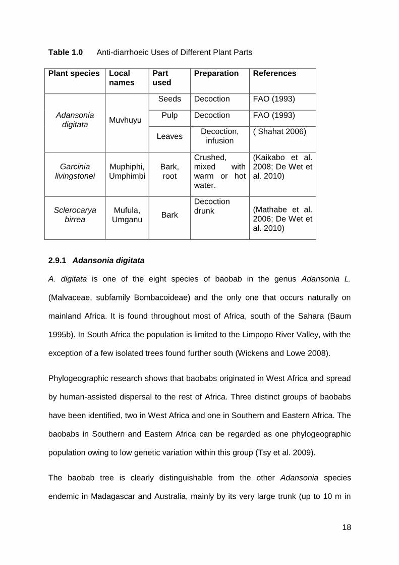
The following plants have been reported to be used traditionally in treating diarrhoea;
seeds, pulp and leaves of A. digitata, bark of G. livingstonei, and S. birrea (FAO
1993; De Caluweet al. 2009; Masola et al. 2009; De Wet et al. 2010; Gouwakinnou
et al. 2011), as seen in Table 1.0. Determination of antibacterial activity and the
active components of these plants will provide baseline information on potential
usage of extracts from these plants for the treatment of infectious diarrhoea caused
by the following bacteria: Escherichia coli, Staphylococcus aureus, Klebsiella
oxytoca, Salmonella enterica and Shigella sonnei. The information obtained from this
research could then be used further in developing drugs from these plants or
synthesising drugs that mimic the active components in these plants.
18
Table 1.0 Anti-diarrhoeic Uses of Different Plant Parts
Plant species Local names
Part used
Preparation References
Adansonia digitata
Muvhuyu
Seeds Decoction FAO (1993)
Pulp Decoction FAO (1993)
Leaves Decoction,
infusion ( Shahat 2006)
Garcinia livingstonei
Muphiphi, Umphimbi
Bark, root
Crushed, mixed with warm or hot water.
(Kaikabo et al. 2008; De Wet et al. 2010)
Sclerocarya birrea
Mufula, Umganu
Bark
Decoction drunk (Mathabe et al.
2006; De Wet et al. 2010)
2.9.1 Adansonia digitata
A. digitata is one of the eight species of baobab in the genus Adansonia L.
(Malvaceae, subfamily Bombacoideae) and the only one that occurs naturally on
mainland Africa. It is found throughout most of Africa, south of the Sahara (Baum
1995b). In South Africa the population is limited to the Limpopo River Valley, with the
exception of a few isolated trees found further south (Wickens and Lowe 2008).
Phylogeographic research shows that baobabs originated in West Africa and spread
by human-assisted dispersal to the rest of Africa. Three distinct groups of baobabs
have been identified, two in West Africa and one in Southern and Eastern Africa. The
baobabs in Southern and Eastern Africa can be regarded as one phylogeographic
population owing to low genetic variation within this group (Tsy et al. 2009).
The baobab tree is clearly distinguishable from the other Adansonia species
endemic in Madagascar and Australia, mainly by its very large trunk (up to 10 m in
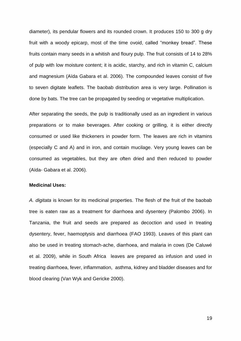
19
diameter), its pendular flowers and its rounded crown. It produces 150 to 300 g dry
fruit with a woody epicarp, most of the time ovoid, called “monkey bread”. These
fruits contain many seeds in a whitish and floury pulp. The fruit consists of 14 to 28%
of pulp with low moisture content; it is acidic, starchy, and rich in vitamin C, calcium
and magnesium (Aïda Gabara et al. 2006). The compounded leaves consist of five
to seven digitate leaflets. The baobab distribution area is very large. Pollination is
done by bats. The tree can be propagated by seeding or vegetative multiplication.
After separating the seeds, the pulp is traditionally used as an ingredient in various
preparations or to make beverages. After cooking or grilling, it is either directly
consumed or used like thickeners in powder form. The leaves are rich in vitamins
(especially C and A) and in iron, and contain mucilage. Very young leaves can be
consumed as vegetables, but they are often dried and then reduced to powder
(Aïda- Gabara et al. 2006).
Medicinal Uses:
A. digitata is known for its medicinal properties. The flesh of the fruit of the baobab
tree is eaten raw as a treatment for diarrhoea and dysentery (Palombo 2006). In
Tanzania, the fruit and seeds are prepared as decoction and used in treating
dysentery, fever, haemoptysis and diarrhoea (FAO 1993). Leaves of this plant can
also be used in treating stomach-ache, diarrhoea, and malaria in cows (De Caluwé
et al. 2009), while in South Africa leaves are prepared as infusion and used in
treating diarrhoea, fever, inflammation, asthma, kidney and bladder diseases and for
blood clearing (Van Wyk and Gericke 2000).

20
Previous Studies:
Previous studies on the methanolic extract of A. digitata roots have reported that it
has anti-trypanosomal activity against Trypanosoma congolense and T. brucei
(Atawodi et al. 2003). Stem and root barks of A. digitata also contain bioactive
constituents such as tannins, phlobatannins, terpenoids, cardiac glycosides and
saponins in the stem bark, as well as terpenoids in the aqueous extract of root bark,
which are responsible for significant antibacterial activity of the crude extracts of this
plant (Masola et al. 2009).
21
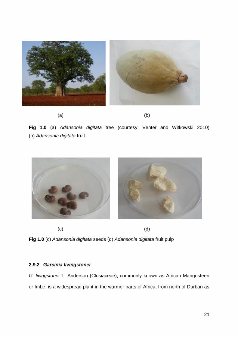
(a) (b)
Fig 1.0 (a) Adansonia digitata tree (courtesy: Venter and Witkowski 2010)
(b) Adansonia digitata fruit
(c) (d)
Fig 1.0 (c) Adansonia digitata seeds (d) Adansonia digitata fruit pulp
2.9.2 Garcinia livingstonei
G. livingstonei T. Anderson (Clusiaceae), commonly known as African Mangosteen
or Imbe, is a widespread plant in the warmer parts of Africa, from north of Durban as
22
far as Somalia and Guinea. In southern Africa, it is distributed widely in the Limpopo
and Zambezi Valleys (Johns et al. 1996; National Research Council 2008).
In South Africa, it is found in scrub, open woodland and forest; in Zimbabwe, usually
along rivers in the lowveld and frequently in riparian and Munga areas, Mopane
woodland and termite mounds in Zambia. It is also found on rocky soil away from
water and in open coastal forest (Orwa et al. 2009).
G. livingstonei is a small tree that grows to 18 m and bears small (10−40 mm
diameter) yellowish-orange fruits containing a sticky juice. The fruit is edible and has
a pleasant flavour. The pulp can be eaten fresh, made into a jam or jellies, or used to
prepare ice cream or alcoholic beverages (National Research Council 2008; Gene
2004; Glen 2007).
Medicinal Uses:
G. livingstonei is used for the treatment of diarrhoea by a rural community in northern
Maputaland located in KwaZulu-Natal Province, South Africa. Roots and bark are
crushed and mixed with warm or hot water, which is administered orally or anally. An
infusion (125 ml) is drunk three times a day, until the diarrhoea subsides (De Wet et
al. 2010).
Previous Studies:
Acetone extracts of leaves of G. livingstonei have been studied for antibacterial
activity. Bioautographs showed that two compounds were mainly responsible for the
antibacterial activity and these were tested against nosocomial pathogens, namely
Escherichia coli, Staphylococcus aureus, Enterococcus faecalis and Pseudomonas
23
aeruginosa. Three of the tested organisms were sensitive to both compounds,
except P. aeruginosa, which was resistant (Kaikabo et al. 2009).
Methanol extracts of root bark of G. livingstonei have also shown antiparasitic activity
against some selected parasites (Mbwambo et al. 2006).
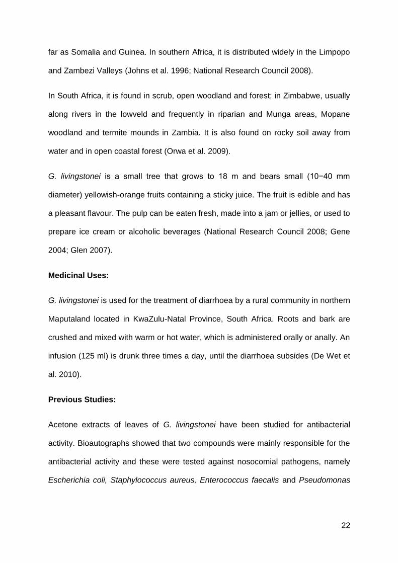
(a) (b)
Fig 2.0 (a) Garcinialivingstonei tree (b) Garcinialivingstonei tree bearing fruits
(courtesy: Glen 2007)
2.9.3 Sclerocarya birrea
S. birrea (Anacardiaceae) is a fast-growing tree. Three subspecies of S. birrea are
known. The subspecies caffra occurs mainly in the southern part of Africa and is
known as marula. The subspecies multifoliolata is restricted to Tanzania and
possibly the neighbouring part of Kenya and the subspecies birrea is present in
Western and Central Africa (Nghitoolwa et al. 2003). Flowering takes place in the dry
24
season when trees are leafless. The major pollinators (or flower visitors) of S. birrea
are honey bees. Secondary pollinators include flies and wasps (Chirwa and
Akinnifesi 2008). S. birrea bears plum-sized stone fruit with a thick yellow peel and
translucent white flesh. Many are eaten fresh, but most are processed into products
such as beverages, jams and jellies. Regardless of taste (sweet-and-sour or tart),
the juice is reported to be nutritionally important, containing as much as four times
the vitamin C of orange juice (National Research Council 2008).
Medicinal Uses:
In traditional medicine, the bark of S. birrea is the part most frequently used to treat
ailments that are mostly bacteria-related (stomach-aches, diarrhoea, wounds and
coughs) (Gouwakinnou et al. 2011). The bark of S. birrea subsp. caffra is crushed
and mixed with hot, warm or cold water. The mixture is administered anally or orally.
The mixture (125 ml) is drunk three times a day, until diarrhoea subsides. If
administered anally, the dosage depends on the person's weight (Mathabe et al.
2006; De Wet et al. 2008).
Previous Studies:
Previous studies have shown that extracts from the stem bark and leaves of S. birrea
possess a catalogue of pharmacological activities, including analgesic, anti-
inflammatory, anti-diabetic and hypoglycaemic (Ojewole 2004), antidiarrhoeal
(Galvez 1991;Galvez 1993), antibacterial (Eloff 2001) and insecticidal properties
(Fatope et al. 1993).
Galvez’s (1991) investigation of antidiarrhoeic activity of the bark of Sclerocarya
birrea in rats revealed that the antidiarrhoeic activity was related to an inhibition of
25
intestinal transit rather than to inhibition of net secretion of fluid and electrolytes
provoked by the laxative agents used. A condensed tannin was isolated from the
crude drug which produced inhibition in intestinal motility, and the monomer was
identified as procyanidin.
Eloff (2001) reported antibacterial activities against Staphylococcus aureus,
Pseudomonas aeruginosa, Escherichia coli and Enterococcus faecalis using acetone
extracts of bark and leaves of Sclerocarya birrea with MIC values from 0.15 to 3
mg/ml.
26
(a) (b)
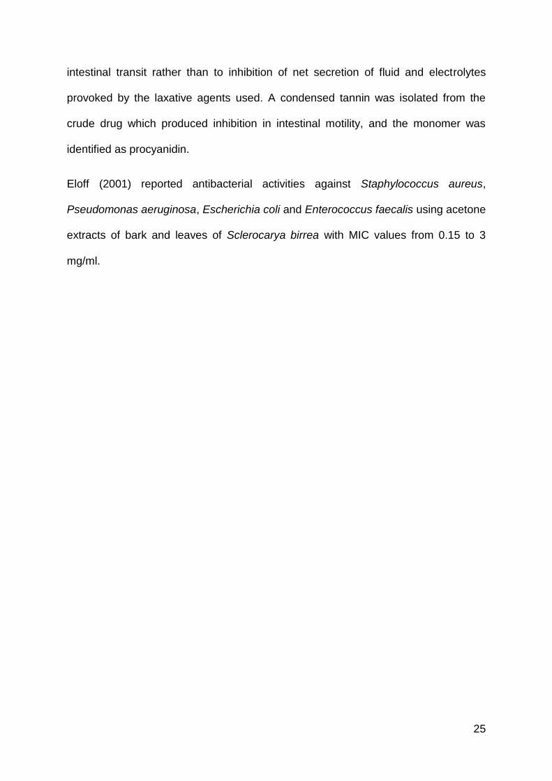
Fig 3.0 (a) Sclerocarya birrea tree (b) Sclerocarya birrea treebearing fruits (courtesy:
Mutshinyalo and Tshisevhe 2003)
(c)
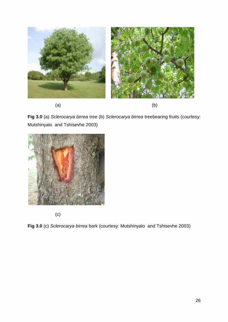
Fig 3.0 (c) Sclerocarya birrea bark (courtesy: Mutshinyalo and Tshisevhe 2003)
27
2.10 HYPOTHESIS AND OBJECTIVES
HYPOTHESIS
1. Crude extracts of A. digitata seeds, pulp and leaves, G. livingstonei and S.
birrea barks will show antibacterial activities against bacteria causing
diarrhoea, such as Escherichia coli, Staphylococcus aureus, Klebsiella
oxytoca, Salmonella enterica and Shigella sonnei.
2. The crude extract of the plants that show antimicrobial activity will have active
components with antibacterial properties.
OBJECTIVES
1. To determine the susceptibility of Escherichia coli, Staphylococcus aureus,
Klebsiella oxytoca, Salmonella enterica, and Shigella sonnei to the crude
extracts of A. digitata, G. livingstonei and S. birrea.
2. To determine the MIC and minimum bactericidal concentration (MBC) of A.
digitata, G. livingstonei, and S. birrea plant extracts on the test bacteria.
3. To carry out cytotoxicity tests on plant extracts with most antimicrobial
properties.
4. To determine the antioxidant properties and phytochemical analysis of the
plant extracts.
28
CHAPTER 3: ANTIMICROBIAL AND CYTOTOXICITY ASSAYS
3.1 MATERIALS AND METHOD
3.1.1 PLANT MATERIALS AND SAMPLE COLLECTION
The leaves, fruit pulps and seeds of A. digitata, bark of G. livingstonei and S. birrea
were collected from Venda, Limpopo Province of South Africa, and voucher
specimens were prepared and identified at the H.G.W.J Schweikerdt Herbarium,
University of Pretoria.
3.1.2 INSTRUMENTS AND REAGENTS USED
Grinding machine, shaker, rotavapor, freeze drier, beakers, conical flasks, round-
bottom flasks, vacuum filter, filter papers, methanol and water.
3.1.3 PREPARATION OF PLANT EXTRACT
The method described by Ndip et al. (2007) was employed; different plant parts were
harvested, air-dried for about two weeks and ground to a fine powder. Methanol
(100%) and water were used as solvents for extraction of crude extract. Water was
chosen as a solvent so as to mimic the traditional style, since most of these plant
parts were administered as either infusions or decoctions. Twenty grams of each
powdered plant material was macerated in 200 ml of each solvent in extraction pots
such that the level of the solvent was above that of the plant material. The
macerated mixtures were then left on the shaker for 72 hours at room temperature.
The extracts were filtered out from the macerated mixture by vacuum filtration. The
methanol extracts were concentrated in a Buchi Rotavapor R-200, transferred to
appropriately labelled vials and allowed to stand at room temperature to permit
evaporation of residual solvents. The water extracts were concentrated using a
freeze dryer.
29
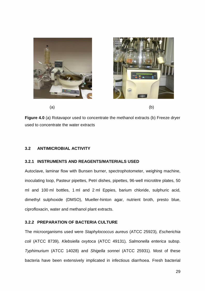
(a) (b)
Figure 4.0 (a) Rotavapor used to concentrate the methanol extracts (b) Freeze dryer
used to concentrate the water extracts
3.2 ANTIMICROBIAL ACTIVITY
3.2.1 INSTRUMENTS AND REAGENTS/MATERIALS USED
Autoclave, laminar flow with Bunsen burner, spectrophotometer, weighing machine,
inoculating loop, Pasteur pipettes, Petri dishes, pipettes, 96-well microtitre plates, 50
ml and 100 ml bottles, 1 ml and 2 ml Eppies, barium chloride, sulphuric acid,
dimethyl sulphoxide (DMSO), Mueller-hinton agar, nutrient broth, presto blue,
ciprofloxacin, water and methanol plant extracts.
3.2.2 PREPARATION OF BACTERIA CULTURE
The microorganisms used were Staphylococcus aureus (ATCC 25923), Escherichia
coli (ATCC 8739), Klebsiella oxytoca (ATCC 49131), Salmonella enterica subsp.
Typhimurium (ATCC 14028) and Shigella sonnei (ATCC 25931). Most of these
bacteria have been extensively implicated in infectious diarrhoea. Fresh bacterial
30
cultures were prepared by sub-culturing stock bacterial cultures into freshly prepared
nutrient agar and incubating at 370C for 24 hours. These 24-hour-old bacterial
cultures were transferred into freshly prepared nutrient broth and standardised to 0.5
McFarland turbidity standards using the spectrophotometer to obtain the desired cell
density of 1.5 X 10⁸ (cells/ml).
The 0.5 McFarland turbidity standard was prepared by adding 0.05 ml of 1.175% of
barium chloride dihydrate (BaCl2•2H2O), with 9.95 mL of 1% sulphuric acid (H2SO4).
3.2.3 AGAR WELL DIFFUSION ASSAY
The Agar Well Diffusion Assay was employed with modifications as described by
Irshad et al. (2012). The Agar Well Diffusion medium was prepared by pouring
molten Mueller-Hinton agar on petri dishes and allowing it to solidify. Afterwards,
100µl of inoculums, approximately 1.5 X 10⁸ cells /ml was seeded into warm molten
Mueller-Hinton agar and poured on the surface of the solidified agar. This was
allowed to solidify and holes of 5 mm width were made into the agar using sterile
Pasteur pipettes.
An amount of 100 mg/ml stock of crude plant extract was prepared for each plant by
dissolving 100 mg of dried plant extract in 1 ml of 10% DMSO. 100 µL of the stock
extract was pipetted onto the holes to give a concentration of 10 mg per hole. 100 µL
of 0.5 mg/ml ciprofloxacin was also pipetted into one of the holes to give a final
concentration of 0.05 mg. This served as the positive control, while 100 µL of 10%
DMSO was pipetted into one of the holes, which served as the negative control.
3.2.4 BROTH MICRODILUTION ASSAY FOR MIC AND MBC
The MIC of the plant extracts was determined with some modification, as described
by Eloff (1998). 100 µL of nutrient broth was added to all the wells of a 96-well
31
microtitre plate. 100 µL of each dissolved plant extract (50mg/ml) was then added in
triplicate for each bacterial plate on the first rows (row A). These were serially diluted
row by row and 100 µL of the mixture was discarded from the last row, thus leaving
each diluted well with a volume of 100 µL. The same procedure was carried out for
ciprofloxacin (positive control), during which 100 µL of 2.5 mg/ml of dissolved
ciprofloxacin was added in triplicate to row A. 100 µL of each bacterial suspension in
suitable growth medium (nutrient broth) was then added to all the wells except the
last column, which served as the sterile control (containing 200 µL of nutrient broth).
Wells containing bacterial suspensions and growth medium, as well as wells
containing 10% DMSO, bacteria suspensions and growth medium, were used as
negative control. The microtitre plates were incubated at 37°C for 24 hours and the
MICs were the lowest concentration where no viability was observed after 24 hours
on the basis of metabolic activity. To indicate respiratory activity, a change in colour
from blue to pink would be determined after adding 20 µL of Presto blue per well in
duplicate and incubating it at 37°C for 30 min.
In order to determine the MBC, the method described by Eloff (1998) was also
employed: 150 µL of nutrient broth was pipetted into all the wells of a 96-well
microtitre plate, and 50 µL was taken from the 24-hour-old suspension of the
undisturbed column (the column without Presto blue) in the MIC plates and added to
the freshly prepared microtitre plates to make up a final volume of 200 µL. These
were incubated at 370C for 24 hours.
After 24 hours, the plates were taken out of the incubator and 20 µL of Presto blue
was added to observe for colour change. The MBC would be the concentration
where no change in colour was observed.
32
3.3 CYTOTOXICITY EXPERIMENT
3.3.1 INSTRUMENTS AND REAGENTS/MATERIALS USED
Weighing machine, microscope, laminar flow, Pasteur pipettes, 96-well microtitre
plates, 24-well microtitre plates, 50 ml and 100 ml bottles, 1 ml and 2 ml Eppies,
pipettes, ELISA plate reader, Dulbecco’s Modified Eagle’s Medium (DMEM), Foetal
bovine serum (FBS), penicillin/streptomycin (PS), DMSO, XTT reagent, actinomycin
D, human embryonic kidney (HEK) 293 cells, water and methanol plant extracts.
3.3.2 XTT CYTOTOXICITY ASSAY
The cytotoxicity of water and methanol extracts of G. livingstonei and S. birrea were
tested on Human Embryonic Kidney (HEK) 293 cells. 100 µL of HEK cell suspension
(1 x 10⁵ cells/ml) was added to the inner wells of a 96-well plate and 200 µL of
incomplete medium (Dulbecco’s modified eagle’s medium(DMEM)) to the outer
wells. The plates were incubated for 24 hours to allow the cells to attach to the base
of the plate. Serial dilution of the plant extracts (already dissolved in DMSO), positive
control (Actinomycin D) and negative control (DMSO) with the complete medium
(10% foetal bovine serum + DMEM + Penicillin/Streptomyosin) were carried out in a
24-well plate to give eight different concentrations of each sample and a final
volume of 1ml per well.
100µL of each concentration from the 24-well plate was added to the 96-well plate in
triplicate and there was a triplicate medium control and a DMSO control for each
extract. A reference plate was prepared to account for the colour of the plant
extracts. This plate contained the plant extracts and medium in duplicate but no
cells. The plates were incubated for 72 hours, after which 50 µL of XTT reagent from
ROCHE was added to all the wells and they were incubated for about 2 h 30 min.
33
After incubation the plates were read on an ELISA plate with KC Junior software to
read the absorbance at 450 nm and 690 nm as the reference wavelength. Graph pad
prism software was used to analyse the data.
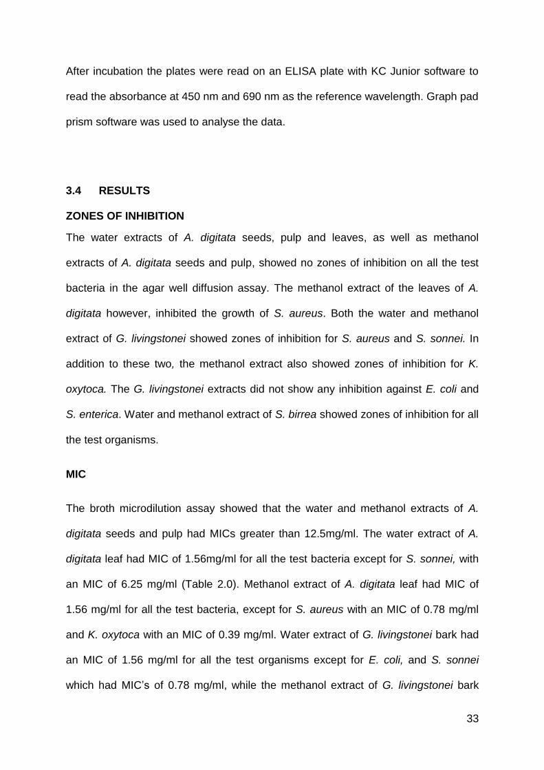
3.4 RESULTS
ZONES OF INHIBITION
The water extracts of A. digitata seeds, pulp and leaves, as well as methanol
extracts of A. digitata seeds and pulp, showed no zones of inhibition on all the test
bacteria in the agar well diffusion assay. The methanol extract of the leaves of A.
digitata however, inhibited the growth of S. aureus. Both the water and methanol
extract of G. livingstonei showed zones of inhibition for S. aureus and S. sonnei. In
addition to these two, the methanol extract also showed zones of inhibition for K.
oxytoca. The G. livingstonei extracts did not show any inhibition against E. coli and
S. enterica. Water and methanol extract of S. birrea showed zones of inhibition for all
the test organisms.
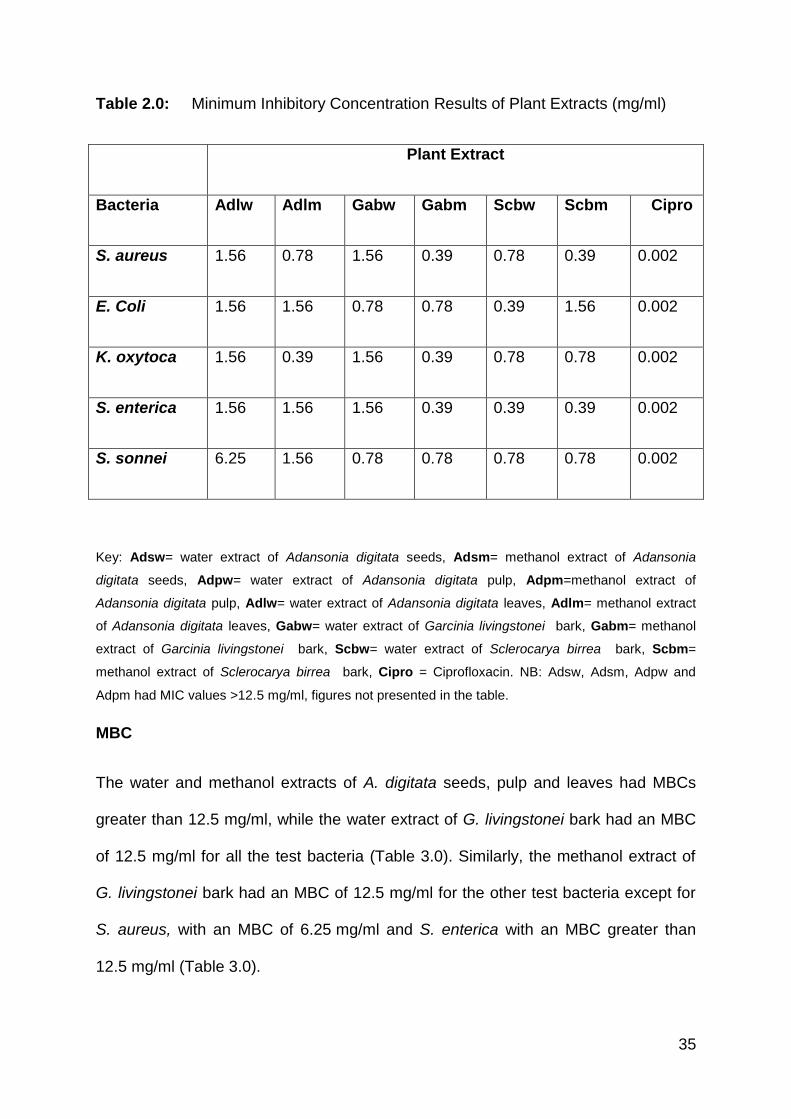
MIC
The broth microdilution assay showed that the water and methanol extracts of A.
digitata seeds and pulp had MICs greater than 12.5mg/ml. The water extract of A.
digitata leaf had MIC of 1.56mg/ml for all the test bacteria except for S. sonnei, with
an MIC of 6.25 mg/ml (Table 2.0). Methanol extract of A. digitata leaf had MIC of
1.56 mg/ml for all the test bacteria, except for S. aureus with an MIC of 0.78 mg/ml
and K. oxytoca with an MIC of 0.39 mg/ml. Water extract of G. livingstonei bark had
an MIC of 1.56 mg/ml for all the test organisms except for E. coli, and S. sonnei
which had MIC’s of 0.78 mg/ml, while the methanol extract of G. livingstonei bark
34
had an MIC of 0.39 mg/ml for all the test organisms with the exception of E. coli, and
S. sonnei which maintained MIC’s of 0.78 mg/ml. Water extract of S. birrea bark had
an MIC of 0.78 mg/ml for all the test bacteria except E. coli and S. enterica which
had lower MIC’s of 0.39 mg/ml, while the methanol extract of S. birrea bark had
varied MIC values ranging from 0.39 to 1.56 mg/ml (Table 2.0).
(a) (b) (c)
Figure 5.0 Images of micro titre plates with plant extracts and test organism, as
well as the positive and negative controls before incubation
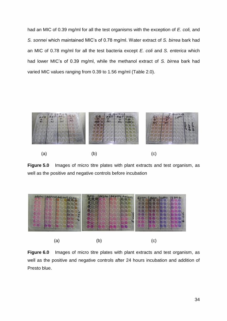
(a) (b) (c)
Figure 6.0 Images of micro titre plates with plant extracts and test organism, as
well as the positive and negative controls after 24 hours incubation and addition of
Presto blue.
35
Table 2.0: Minimum Inhibitory Concentration Results of Plant Extracts (mg/ml)
Plant Extract
Bacteria Adlw Adlm Gabw Gabm Scbw Scbm Cipro
S. aureus 1.56 0.78 1.56 0.39 0.78 0.39 0.002
E. Coli 1.56 1.56 0.78 0.78 0.39 1.56 0.002
K. oxytoca 1.56 0.39 1.56 0.39 0.78 0.78 0.002
S. enterica 1.56 1.56 1.56 0.39 0.39 0.39 0.002
S. sonnei 6.25 1.56 0.78 0.78 0.78 0.78 0.002
Key: Adsw= water extract of Adansonia digitata seeds, Adsm= methanol extract of Adansonia
digitata seeds, Adpw= water extract of Adansonia digitata pulp, Adpm=methanol extract of
Adansonia digitata pulp, Adlw= water extract of Adansonia digitata leaves, Adlm= methanol extract
of Adansonia digitata leaves, Gabw= water extract of Garcinia livingstonei bark, Gabm= methanol
extract of Garcinia livingstonei bark, Scbw= water extract of Sclerocarya birrea bark, Scbm=
methanol extract of Sclerocarya birrea bark, Cipro = Ciprofloxacin. NB: Adsw, Adsm, Adpw and
Adpm had MIC values >12.5 mg/ml, figures not presented in the table.
MBC
The water and methanol extracts of A. digitata seeds, pulp and leaves had MBCs
greater than 12.5 mg/ml, while the water extract of G. livingstonei bark had an MBC
of 12.5 mg/ml for all the test bacteria (Table 3.0). Similarly, the methanol extract of
G. livingstonei bark had an MBC of 12.5 mg/ml for the other test bacteria except for
S. aureus, with an MBC of 6.25 mg/ml and S. enterica with an MBC greater than
12.5 mg/ml (Table 3.0).
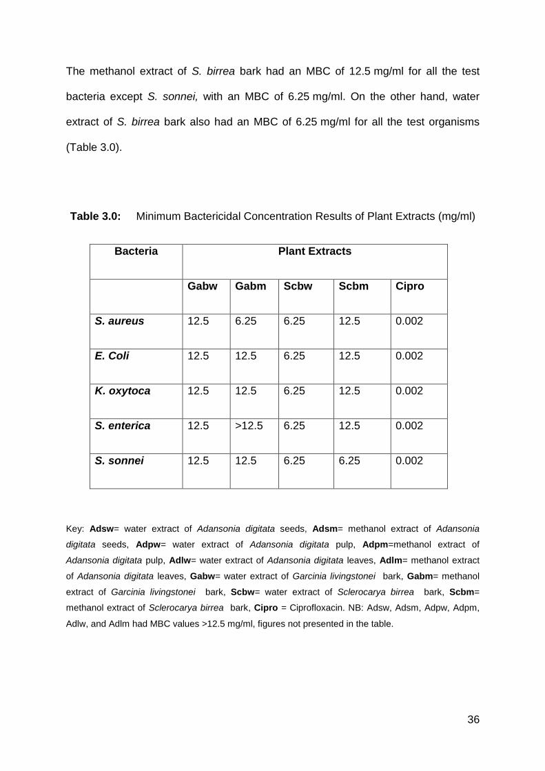
36
The methanol extract of S. birrea bark had an MBC of 12.5 mg/ml for all the test
bacteria except S. sonnei, with an MBC of 6.25 mg/ml. On the other hand, water
extract of S. birrea bark also had an MBC of 6.25 mg/ml for all the test organisms
(Table 3.0).
Table 3.0: Minimum Bactericidal Concentration Results of Plant Extracts (mg/ml)
Bacteria Plant Extracts
Gabw Gabm Scbw Scbm Cipro
S. aureus 12.5 6.25 6.25 12.5 0.002
E. Coli 12.5 12.5 6.25 12.5 0.002
K. oxytoca 12.5 12.5 6.25 12.5 0.002
S. enterica 12.5 >12.5 6.25 12.5 0.002
S. sonnei 12.5 12.5 6.25 6.25 0.002
Key: Adsw= water extract of Adansonia digitata seeds, Adsm= methanol extract of Adansonia
digitata seeds, Adpw= water extract of Adansonia digitata pulp, Adpm=methanol extract of
Adansonia digitata pulp, Adlw= water extract of Adansonia digitata leaves, Adlm= methanol extract
of Adansonia digitata leaves, Gabw= water extract of Garcinia livingstonei bark, Gabm= methanol
extract of Garcinia livingstonei bark, Scbw= water extract of Sclerocarya birrea bark, Scbm=
methanol extract of Sclerocarya birrea bark, Cipro = Ciprofloxacin. NB: Adsw, Adsm, Adpw, Adpm,
Adlw, and Adlm had MBC values >12.5 mg/ml, figures not presented in the table.
37
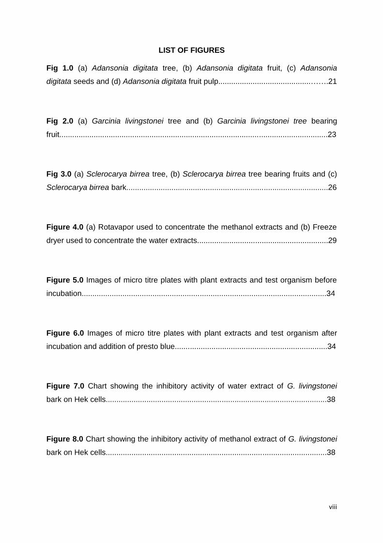
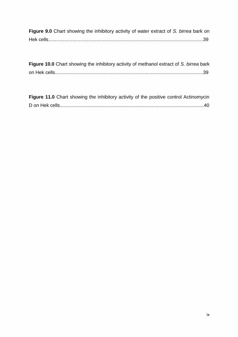
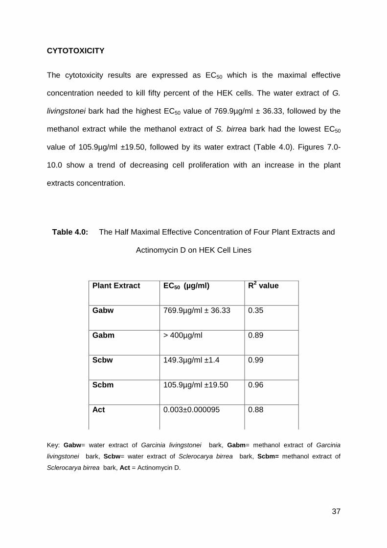
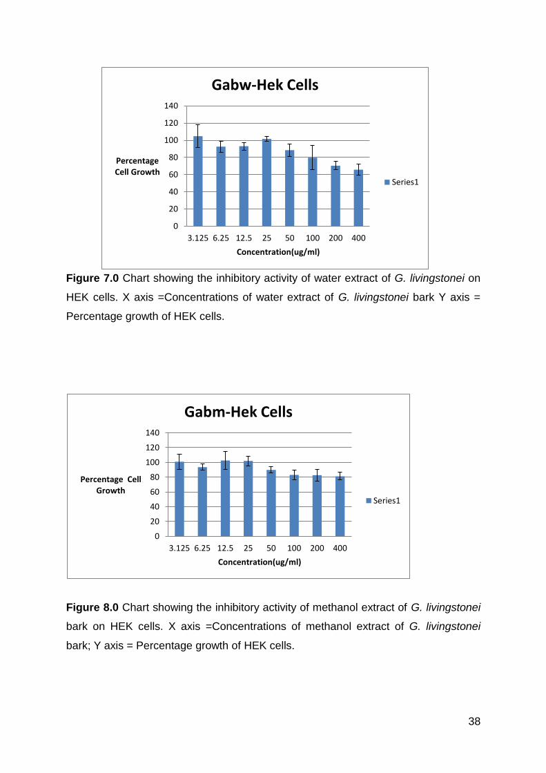
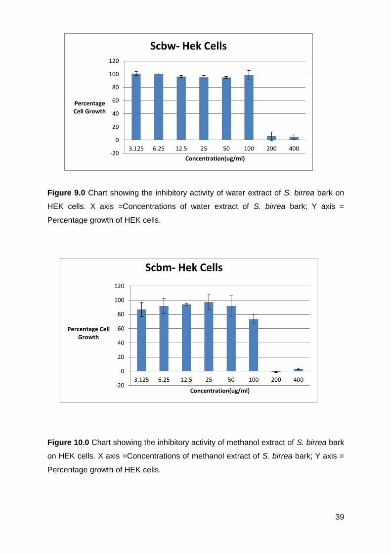
CYTOTOXICITY
The cytotoxicity results are expressed as EC50 which is the maximal effective
concentration needed to kill fifty percent of the HEK cells. The water extract of G.
livingstonei bark had the highest EC50 value of 769.9µg/ml ± 36.33, followed by the
methanol extract while the methanol extract of S. birrea bark had the lowest EC50
value of 105.9µg/ml ±19.50, followed by its water extract (Table 4.0). Figures 7.0-
10.0 show a trend of decreasing cell proliferation with an increase in the plant
extracts concentration.
Table 4.0: The Half Maximal Effective Concentration of Four Plant Extracts and
Actinomycin D on HEK Cell Lines
Key: Gabw= water extract of Garcinia livingstonei bark, Gabm= methanol extract of Garcinia
livingstonei bark, Scbw= water extract of Sclerocarya birrea bark, Scbm= methanol extract of
Sclerocarya birrea bark, Act = Actinomycin D.
Plant Extract EC50 (µg/ml) R2 value
Gabw 769.9µg/ml ± 36.33 0.35
Gabm > 400µg/ml 0.89
Scbw 149.3µg/ml ±1.4 0.99
Scbm 105.9µg/ml ±19.50 0.96
Act 0.003±0.000095 0.88
38
Figure 7.0 Chart showing the inhibitory activity of water extract of G. livingstonei on
HEK cells. X axis =Concentrations of water extract of G. livingstonei bark Y axis =
Percentage growth of HEK cells.
Figure 8.0 Chart showing the inhibitory activity of methanol extract of G. livingstonei
bark on HEK cells. X axis =Concentrations of methanol extract of G. livingstonei
bark; Y axis = Percentage growth of HEK cells.
0
20
40
60
80
100
120
140
3.125 6.25 12.5 25 50 100 200 400
Percentage Cell Growth
Concentration(ug/ml)
Gabm-Hek Cells
Series1
0
20
40
60
80
100
120
140
3.125 6.25 12.5 25 50 100 200 400
Percentage Cell Growth
Concentration(ug/ml)
Gabw-Hek Cells
Series1
39
Figure 9.0 Chart showing the inhibitory activity of water extract of S. birrea bark on
HEK cells. X axis =Concentrations of water extract of S. birrea bark; Y axis =
Percentage growth of HEK cells.
Figure 10.0 Chart showing the inhibitory activity of methanol extract of S. birrea bark
on HEK cells. X axis =Concentrations of methanol extract of S. birrea bark; Y axis =
Percentage growth of HEK cells.
-20
0
20
40
60
80
100
120
3.125 6.25 12.5 25 50 100 200 400
Percentage Cell Growth
Concentration(ug/ml)
Scbw- Hek Cells
-20
0
20
40
60
80
100
120
3.125 6.25 12.5 25 50 100 200 400
Percentage Cell Growth
Concentration(ug/ml)
Scbm- Hek Cells
40
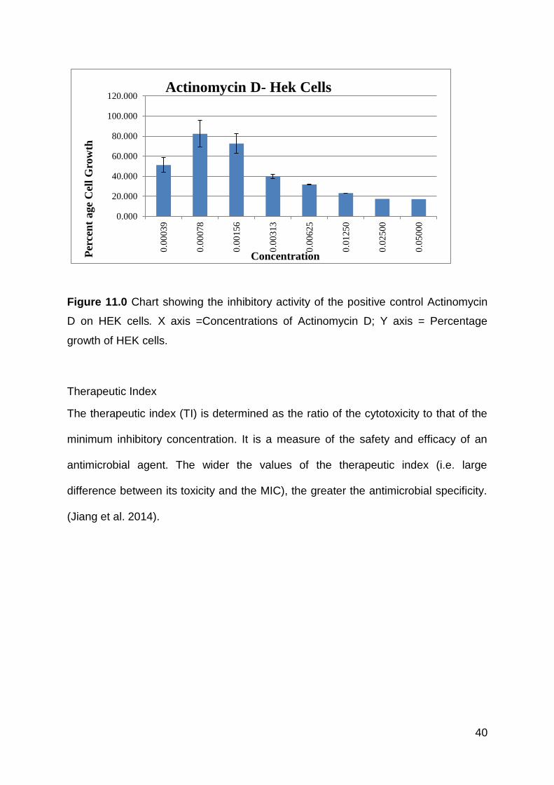
Figure 11.0 Chart showing the inhibitory activity of the positive control Actinomycin
D on HEK cells. X axis =Concentrations of Actinomycin D; Y axis = Percentage
growth of HEK cells.
Therapeutic Index
The therapeutic index (TI) is determined as the ratio of the cytotoxicity to that of the
minimum inhibitory concentration. It is a measure of the safety and efficacy of an
antimicrobial agent. The wider the values of the therapeutic index (i.e. large
difference between its toxicity and the MIC), the greater the antimicrobial specificity.
(Jiang et al. 2014).
0.000
20.000
40.000
60.000
80.000
100.000
120.000
0.0
0039
0.0
0078
0.0
0156
0.0
0313
0.0
0625
0.0
1250
0.0
2500
0.0
5000
Per
cen
t age
Cel
l G
row
th
Concentration
Actinomycin D- Hek Cells
41
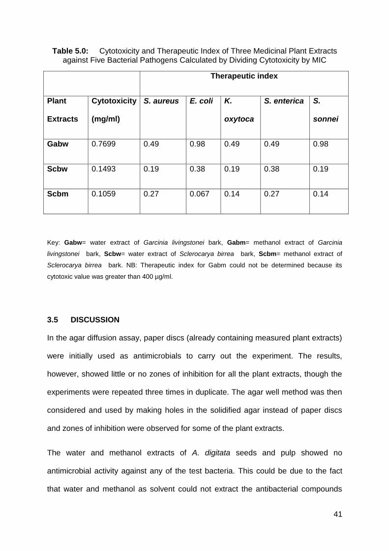
Table 5.0: Cytotoxicity and Therapeutic Index of Three Medicinal Plant Extracts against Five Bacterial Pathogens Calculated by Dividing Cytotoxicity by MIC
Therapeutic index
Plant
Extracts
Cytotoxicity
(mg/ml)
S. aureus E. coli K.
oxytoca
S. enterica S.
sonnei
Gabw 0.7699 0.49 0.98 0.49 0.49 0.98
Scbw 0.1493 0.19 0.38 0.19 0.38 0.19
Scbm 0.1059 0.27 0.067 0.14 0.27 0.14
Key: Gabw= water extract of Garcinia livingstonei bark, Gabm= methanol extract of Garcinia
livingstonei bark, Scbw= water extract of Sclerocarya birrea bark, Scbm= methanol extract of
Sclerocarya birrea bark. NB: Therapeutic index for Gabm could not be determined because its
cytotoxic value was greater than 400 µg/ml.
3.5 DISCUSSION
In the agar diffusion assay, paper discs (already containing measured plant extracts)
were initially used as antimicrobials to carry out the experiment. The results,
however, showed little or no zones of inhibition for all the plant extracts, though the
experiments were repeated three times in duplicate. The agar well method was then
considered and used by making holes in the solidified agar instead of paper discs
and zones of inhibition were observed for some of the plant extracts.
The water and methanol extracts of A. digitata seeds and pulp showed no
antimicrobial activity against any of the test bacteria. This could be due to the fact
that water and methanol as solvent could not extract the antibacterial compounds
42
present in these plant parts, considering that inhibition of Staphylococcus aureus,
Staphylococcus epidermidis, Streptococcus mutans and Pseudomonas aeruginosa
by the ethyl acetate and n-butanol extracts of the pericarp, pulp and seed portion of
A. digitata has been reported (Shukla et al. 2003).
Contrary to the agar well diffusion assay in which methanol and water extract of A.
digitata leaves showed little and no inhibition respectively, the broth microdilution
method indicated that both the water and methanol extracts of A. digitata leaves
exhibited inhibitory activities against all of the test bacteria at concentrations of 0.39
mg/ml to 6.25 mg/ml. Their bactericidal effect, however, was at concentrations
greater than 12.5 mg/ml. The methanol extracts of A. digitata root bark and leaves
have been shown to exhibit antibacterial activity against Staphylococcus aureus,
Streptococcus faecalis, Bacillus subtilis, Escherichia coli and Mycobacterium phlei
(Anani et al. 2000).
The disadvantage of using the agar diffusion method to determine antimicrobial
activity is that the antimicrobial effect may be affected by the agar type, salt
concentration, incubation temperature and molecular size of the antimicrobial
component. Furthermore, it does not distinguish between bactericidal and
bacteriostatic effects (Eloff 1998).
The MIC results (0.39-1.56mg/ml) showed that the G. livingstonei plant extracts
displayed antimicrobial activity against all the test bacteria. Elsewhere, two
compounds isolated from acetone extracts of G. livingstonei leaves were shown to
exhibit antimicrobial activity against Escherichia coli, Staphylococcus aureus and
Enterococcus faecalis with MIC’s ranging from 8-100 µg/ml, while the methanol
43
extract of its root bark showed antiparasitic activity against some selected parasites
(Kaikabo et al. 2009; Kaikabo and Eloff 2011; Mbwambo et al. 2006).
Water and methanol extracts of S. birrea bark showed both significant inhibitory and
bactericidal effect on all the test organisms with MIC which ranged from 0.39 to 1.56
mg/ml. This is in line with the findings of Eloff (2001), who reported that acetone
extracts of S. birrea bark and leaves showed significant antimicrobial activities with
MIC values ranging from 0.15 to 3 mg/ml against Staphylococcus aureus,
Pseudomonas aeruginosa, Escherichia coli and Enterococcus faecalis.
The EC50 of all the plant extracts, which ranged from 105.9µg/ml to 769.9µg/ml, was
much higher than that of Actinomycin D, the positive control. At this level, these
extracts are considered safe, considering that the values are greater than 100 µg/ml,
which has been considered safe for extracts of Rubia cordifolia roots (Patel et
al.2010).
Of all the extracts studied, water extract of G. livingstonei bark had the highest
therapeutic index of 0.98 for E. coli and S. sonnei, which is similar to the findings of
Adamu et al. (2014) who reported that Strychnos mitis extracts had a therapeutic
index of 1.1 for E. coli.
44
CHAPTER 4: PHYTOCHEMICAL AND ANTIOXIDANT ASSAYS
4.1 MATERIALS AND METHODS
4.1.1 PREPARATION OF PLANT EXTRACT
The method described in 3.1.3 above was employed in preparing the plant extracts.
4.1.2 INSTRUMENTS AND REAGENTS/MATERIALS USED
Weighing machine, test tubes, spray bottle, 1 ml and 2 ml Eppies, pipettes, filter
papers, 10% DMSO, methanol, Wagner’s reagent, Dragendroff’s reagent, sodium
hydroxide, hydrochloric acid, ethyl acetate, aluminium chloride, phoshomolybdic acid
reagent, ammonia vapours, alcoholic ferric chloride, chloroform, sulphuric acid,
distilled water, water and methanol plant extracts, ELISA plate reader, 96-well
microtitre plates, foil paper, Vitamin C, ethanol, 1,1-diphenyl-2-picrylhydrazyl
(DPPH).
4.2 PHYTOCHEMICAL SCREENING
The methods described below were used to assess the phytochemical constituents
of the plant extracts.
4.2.1 TEST FOR ALKALOIDS
WAGNER’S TEST: A fraction of extract was treated with Wagner’s test reagent
[1.27 g of iodine and 2 g of potassium iodide in 100 ml of water] and observed for the
formation of a reddish brown colour precipitate (Lalitha and Jayanthi 2012).
DRAGENDROFF’S TEST: A few drops of Dragendroff’s reagent were added to a
test tube containing 1 ml of extract and a colour change was observed. The
45
appearance of an orange colour was an indication of the presence of alkaloids
(Firdouse and Alam 2011).
4.2.2 TEST FOR FLAVONOIDS
SODIUM HYDROXIDE TEST: Plant extract is treated with dilute NaOH, followed by
addition of dilute HCl. A yellow solution with NaOH turns colorless with dilute HCl,
which shows the presence of flavonoids (Onwukaeme et al. 2007). A conclusive test
was carried out using the aluminum chloride Test.
ALUMINUM CHLORIDE TEST: A quantity (0.2 g) of each of the extracts was heated
with 10 ml of ethyl acetate in boiling water for three minutes, after which 4 ml of the
filtrate was shaken with 1 ml of 1% aluminum chloride solution and observed for light
yellow colouration. A yellow precipitate indicated the presence of flavonoids (Subash
et al. 2013).
4.2.3 TEST FOR PHENOLS
In this test, one spots the extract on a filter paper, adds a drop of phoshomolybdic
acid reagent and exposes it to ammonia vapours. Blue colouration of the spot
indicates the presence of phenols (Kumar et al. 2007).
4.2.4 TEST FOR TANNINS
In this test 10% alcoholic ferric chloride is added to 2–3 ml of methanolic extract
(1:1). Dark blue or greenish grey colouration of the solution reveals the presence of
tannins (Kumar et al. 2007; Parekh and Chanda 2007).
46
4.2.5 TEST FOR TERPENOIDS
To conduct this test, 5 ml of plant extract is added to 2 ml of chloroform and 3 ml of
concentrated sulphuric acid. The presence of terpenoids gives a reddish brown
colour of interface (Edeoga et al. 2005).
4.2.6 TEST FOR SAPONINS
One adds 0.5 ml of plant extract filtrate to 5 ml of distilled water and shakes it well.
Persistence of frothing is an indication of the presence of saponins (Parekh and
Chanda 2007).
4.3 DETERMINATION OF ANTIOXIDANT ACTIVITY
The method described by Du Toit et al. (2001) was employed to determine the
antioxidant content of the plant extracts as well as that of vitamin C so as to have a
basis for comparison. 2mg of plant extract was dissolved in 200µl of ethanol to give a
stock concentration of 10mg/ml while 2mg of vitamin C was dissolved in 1ml of
ethanol to give a stock concentration of 2 mg/ml which served as the positive control.
1,1-diphenyl-2-picrylhydrazyl (DPPH) which is a free radical that can be reduced by
an antioxidant, was prepared by dissolving 20 mg in 500 ml ethanol to give a stock
concentration of 0.04 mg/ml. All the samples were tested in triplicate on 96-well
microtitre plates. The start-up concentration for the test samples in the first row was
500 µg/ml, which is a composition of distilled water and the extract, while the positive
control (vitamin C) was a mixture of distilled water and vitamin C with a final
concentration of 100 µg/ml in the first row. Blank controls were prepared by adding
ethanol to distilled water and negative controls also had test samples and distilled
water. These were serially diluted from the first row to the last row, and DPPH was
47
added into all the wells except the negative controls. The plates were covered in foil
and incubated for 30 minutes. The absorbency were read with an ELISA plate reader
(KC junior) at 515 nm and the absorbency values were analysed with the graph pad
prism software.
4.4 RESULTS
PHYTOCHEMICALS
The water and methanol extracts of each plant part had the same test result, except
for extracts of S. birrea, as seen in Table 6.0. Alkaloids were absent in both the
water and methanol extracts of A. digitata seeds, pulps and leaves. Flavanoids and
phenols were present in both the water and methanol extracts of A. digitata seeds,
pulp and leaves. Tannins were only detected in the leaf extract of A. digitata, but
were absent in the seed and pulp extracts. Terpenoids were only detected in the
pulp extract of A. digitata but not in its seed or leaf extracts. Saponins were present
in both the pulp and leaf extracts of A. digitata, but were not detected in its seed
extracts. Alkaloids, flavonoids, phenols, tannins, terpenoids and saponins were
present in the bark extracts of G. livingstonei and S. birrea, with the exception of the
water extract of S. birrea, which did not contain any flavonoids.
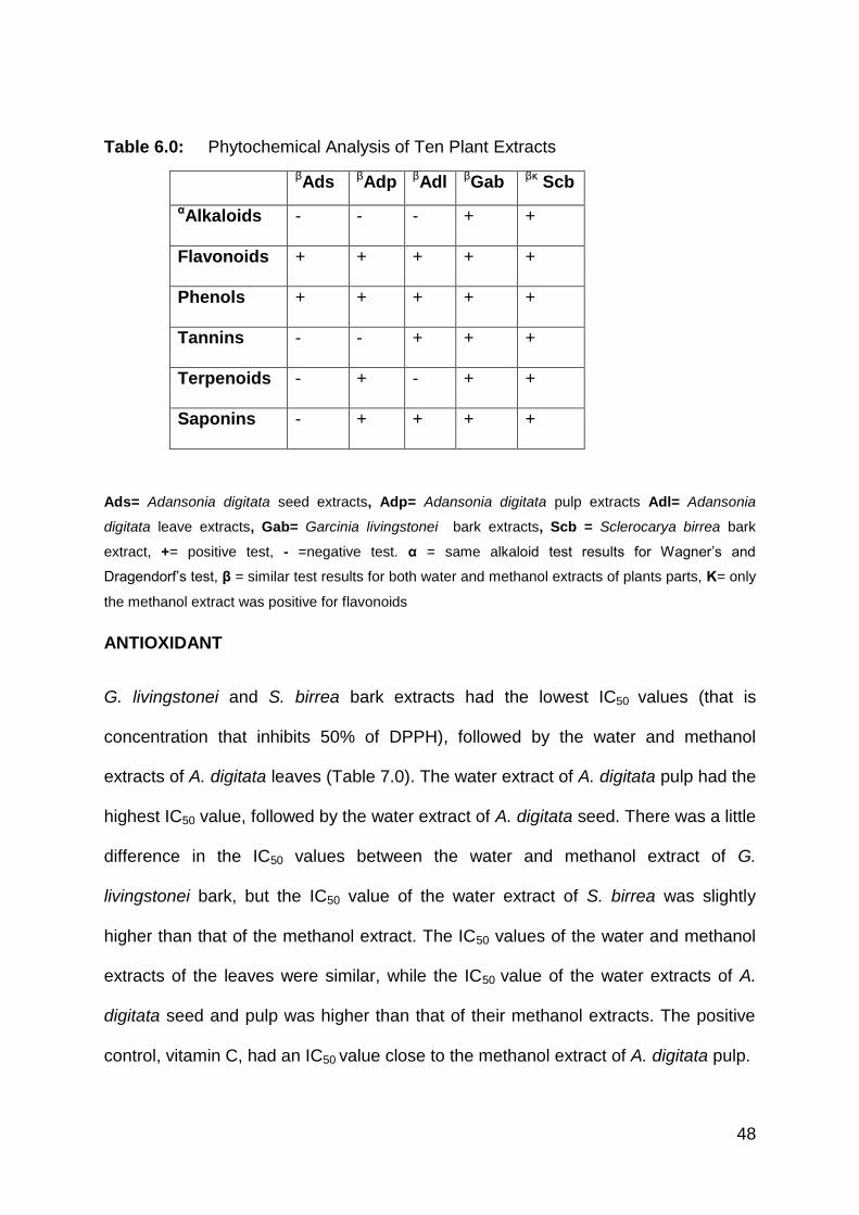
48
Table 6.0: Phytochemical Analysis of Ten Plant Extracts
βAds βAdp βAdl βGab βκ Scb
αAlkaloids
- - - + +
Flavonoids
+ + + + +
Phenols
+ + + + +
Tannins
- - + + +
Terpenoids
- + - + +
Saponins
- + + + +
Ads= Adansonia digitata seed extracts, Adp= Adansonia digitata pulp extracts Adl= Adansonia
digitata leave extracts, Gab= Garcinia livingstonei bark extracts, Scb = Sclerocarya birrea bark
extract, += positive test, - =negative test. α = same alkaloid test results for Wagner’s and
Dragendorf’s test, β = similar test results for both water and methanol extracts of plants parts, Κ= only
the methanol extract was positive for flavonoids
ANTIOXIDANT
G. livingstonei and S. birrea bark extracts had the lowest IC50 values (that is
concentration that inhibits 50% of DPPH), followed by the water and methanol
extracts of A. digitata leaves (Table 7.0). The water extract of A. digitata pulp had the
highest IC50 value, followed by the water extract of A. digitata seed. There was a little
difference in the IC50 values between the water and methanol extract of G.
livingstonei bark, but the IC50 value of the water extract of S. birrea was slightly
higher than that of the methanol extract. The IC50 values of the water and methanol
extracts of the leaves were similar, while the IC50 value of the water extracts of A.
digitata seed and pulp was higher than that of their methanol extracts. The positive
control, vitamin C, had an IC50 value close to the methanol extract of A. digitata pulp.
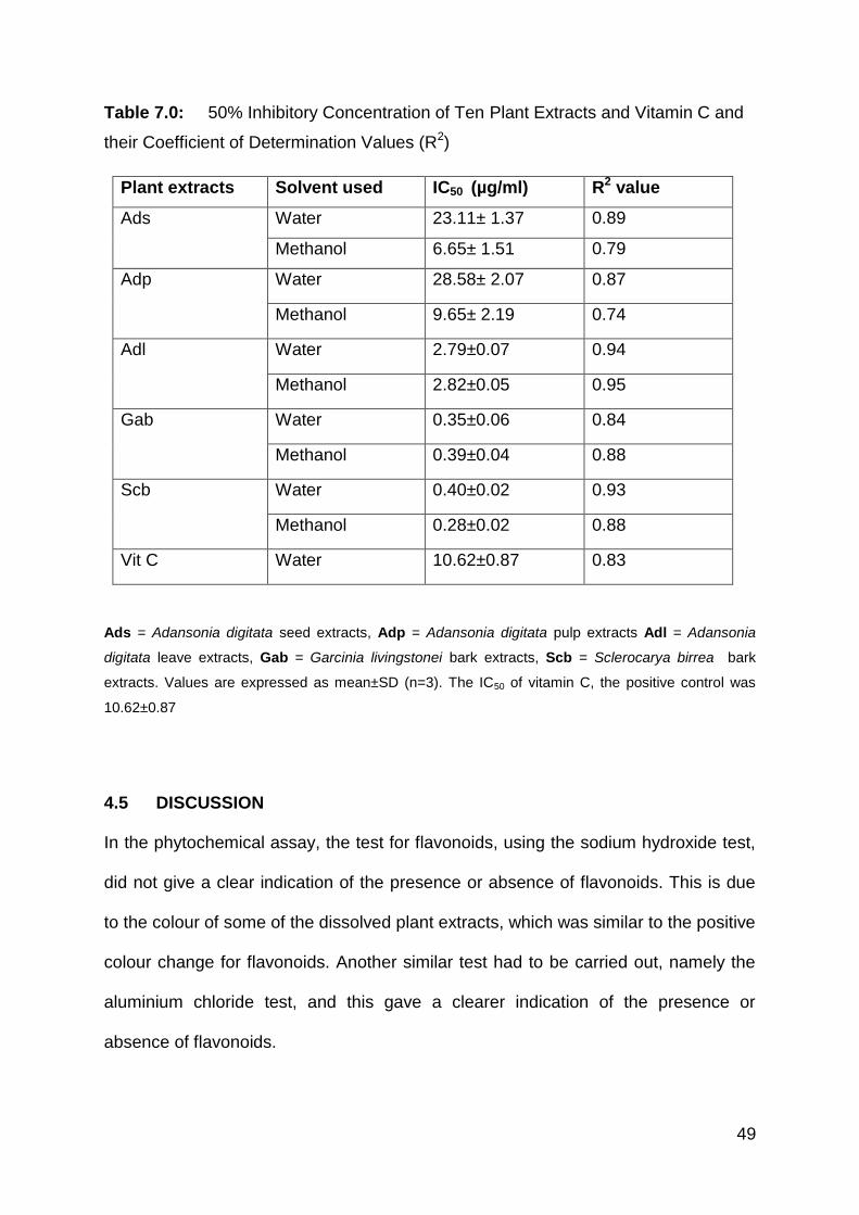
49
Table 7.0: 50% Inhibitory Concentration of Ten Plant Extracts and Vitamin C and
their Coefficient of Determination Values (R2)
Plant extracts Solvent used IC50 (µg/ml) R2 value
Ads Water 23.11± 1.37 0.89
Methanol 6.65± 1.51 0.79
Adp Water 28.58± 2.07 0.87
Methanol 9.65± 2.19 0.74
Adl Water 2.79±0.07 0.94
Methanol 2.82±0.05 0.95
Gab Water 0.35±0.06 0.84
Methanol 0.39±0.04 0.88
Scb Water 0.40±0.02 0.93
Methanol 0.28±0.02 0.88
Vit C Water 10.62±0.87 0.83
Ads = Adansonia digitata seed extracts, Adp = Adansonia digitata pulp extracts Adl = Adansonia
digitata leave extracts, Gab = Garcinia livingstonei bark extracts, Scb = Sclerocarya birrea bark
extracts. Values are expressed as mean±SD (n=3). The IC50 of vitamin C, the positive control was
10.62±0.87
4.5 DISCUSSION
In the phytochemical assay, the test for flavonoids, using the sodium hydroxide test,
did not give a clear indication of the presence or absence of flavonoids. This is due
to the colour of some of the dissolved plant extracts, which was similar to the positive
colour change for flavonoids. Another similar test had to be carried out, namely the
aluminium chloride test, and this gave a clearer indication of the presence or
absence of flavonoids.
50
The fact that water and methanol extracts of all samples yielded the same test
results, with the exception of the flavonoid test result of S. birrea, is because water
and methanol are polar solvents and therefore extract similar compounds. Methanol,
on the other hand, could have extracted more polyphenols such as flavonoids better
than water (Medini et al. 2014), which explains why the methanol extract of S. birrea
tested positive for flavonoid. Looking at this test result, the water extract of S. birrea
is probably not a good source of flavonoids.
Alkaloids were absent in both the water and methanol extracts of A. digitata seeds,
pulp and leaves. Chemicals that have been isolated and characterised from the A.
digitata plant usually belong to the classes of terpenoids, flavonoids, steroids,
vitamins, amino acids, carbohydrates and lipids (Shukla et al. 2001). This explains
why A. digitata seeds, pulp and leaves contain flavonoids and phenols, as most
phenolic compounds belong to the flavonoid group.
Tannin was only detected in the leaf extract of A. digitata but was absent in the seed
and pulp extracts. This agrees with the findings of Ayele et al. (2013), who
determined the total polyphenol content of A. digitata leaves and expressed it as
tannic acid equivalents. Previous work has also proven tannins to be present in A.
digitata bark (Tanko et al. 2008). The stem bark of A. digitata is known to contain
both soluble and insoluble tannins (Yusha’u et al. 2010).
Saponin was present in both the pulp and leaf extract of A. digitata, but was not
detected in its seed extracts. Warra et al. (2012), however, reported slight presence
of saponin in A.digitata seed oil, but this could be attributed to the solvent used in
extraction, which was hexane. The fruit pulp of A. digitata has been found to have
51
anti-inflammatory properties; this is often attributed to the presence of sterols,
saponins and triterpenes in its aqueous extract (Ramadan et al. 1994).
Terpenoid was present only in the pulp extract of A. digitata but not in its seed and
leaf extracts. This is also consistent with the findings of Al-Qarawi et al. (2003), who
reported isolating triterpene from A. digitata fruit. In research carried out by Warra et
al. (2012) though, terpenoids were found to be present in an extract of A. digitata
seeds. This might be because the seeds were extracted with a different solvent,
namely hexane.
Alkaloids, flavonoids, phenol, tannins, terpenoids and saponins were present in the
bark extracts of G. livingstonei and S. birrea, with the exception of the water extract
of S. birrea, which did not contain any flavonoid. Phytochemistry of the stem bark of
S. birrea in previous work has been reported to contain tannins, flavonoids, alkaloids,
and steroids (Ojewole et al. 2010). Previous work also showed that xanthones and
biflavonoids had been isolated from the root bark of G. livingstonei (Mbwambo et al.
2006), while benzophenones and biflavonoids were isolated from its fruit (Yang et al.
2010). The significance of this finding is that bark extracts of G. livingstonei and S.
birrea are a good source of alkaloids, flavonoids, phenol, tannins, terpenoids and
saponins.
Tannins and flavonoids are known for their laxative effect and are thought to be
responsible for antidiarrhoeal activity by increasing colonic water and electrolyte
reabsorption (Palombo 2006; Misra et al. 2014), which explains why the bark of
these plants are used traditionally in treating diarrhoea.
Terpenoids have been shown to be active against bacteria, fungi, viruses and
protozoa and their mechanism of action is speculated to involve membrane
52
disruption by the lipophilic compounds (Cowan 1999). Alkaloids have been found to
have antimicrobial properties with microbicide effects against Giardia and
Entamoeba species, as well as antidiarrheal effects which are probably due to their
effects on transit time in the small intestine (Cowan 1999). Saponins have several
biological effects, some of which are antibacterial, antifungal, antiparasitic,
antitumours/cytotoxicity, antiviral and antioxidant activities (Sparg et al. 2004).
The serial dilution done on a 96-well microtitre plate for the antioxidant assay did not
give a 50% inhibition for DPPH when the absorbencies were read on the ELISA plate
reader. Further dilution from the last well of the initial plate had to be done in a new
microtitre plate. The absorbencies of the second plate were read on the ELISA
machine and 50% inhibition was observed.
Water and methanol extracts of bark of S. birrea and G. livingstonei exhibited
excellent antioxidant activities with their 50% inhibitory concentration of DPPH
radical ranging from 0.28±0.02 µg/ml to 0.40±0.02 µg/ml. This is quite impressive
when compared to the positive control, vitamin C, which had a 50% inhibitory
concentration of 10.62±0.87 µg/ml. These results can be attributed to the presence
of phenols, flavonoids, tannins, alkaloids, saponins and terpenoids and this is in
agreement with studies in which these compounds have been associated with high
antioxidant activities (Dangles et al. 2000; Moura et al. 2007; Gulcin et al. 2004;
Grabmann 2005).
Extracts of A. digitata leaves also exhibited good antioxidant properties, with their
IC50 values being 2.79±0.07 µg/ml and 2.82±0.05 µg/ml. This can also be attributed
to the presence of phenols, flavonoids, tannins and saponins.
53
CHAPTER 5: GENERAL DISCUSSION AND CONCLUSION
5.1 GENERAL DISCUSSION
Water and methanol extracts of A. digitata seeds and pulp showed no antimicrobial
activity with both the agar well diffusion method and the broth microdilution method.
Their phytochemical analysis, however, revealed that both plant parts contain
flavonoids and phenols, while the extracts of A. digitata pulp also contain terpenoids
and saponins. These phytochemicals might have contributed to the antioxidant
properties exhibited by both water and methanol extracts of A. digitata seeds and
pulp, though their methanol extracts exhibited better antioxidant properties than their
water extracts. This is because methanol extracts more polyphenols such as
flavonoids, which are thought to have antioxidant properties, better than water
(Medini et al. 2014; Pietta 2000).
Water extract of A. digitata leaves showed lower antimicrobial activity when
compared to its methanol extract. The MIC of the methanol extract for A. digitata
leaves was lower than its water extract. This is because the methanol extract
dissolved better in 10% DMSO than the water extract. Phytochemistry of the extracts
of A. digitata leaves revealed that they contained flavonoid, phenol, tannin and
saponin, which explains why the extracts of A. digitata leaves also had good
antioxidant properties, as their IC50 ranged between 2.79±0.07 to 2.82±0.05 µg/ml.
The presence of these phytochemicals also explains why the methanol extract in
particular was effective against bacteria causing diarrhoea, as flavonoids, phenols,
saponins and tannins have been implicated as antimicrobials (Cowan 1999; Sparg et
al. 2004; Palombo 2006).
54
The result of the agar well diffusion method showed that the water and methanol
extracts of G. livingstonei and S. birrea had antimicrobial potential, as they showed
zones of inhibitions similar to the positive control ciprofloxacin. The broth
microdilution method however, was more conclusive, as it indicated the
concentrations at which these plant extracts showed inhibition of the test organisms.
Similar values are seen amongst the MICs of extracts of G. livingstonei and S.
birrea, ranging between 0.39 mg/ml to 1.56mg/ml. This may be attributed to the fact
that the solvents used are both polar solvents and at such they extract similar
antibacterial compounds and this justifies their traditional use in the treatment of
diarrhoea and the method of preparation, which is usually infusion or decoction.
The phytochemical analysis of G. livingstonei and S. birrea extracts reveal that they
all contain alkaloids, phenols, flavonoids, tannins, terpenoids and saponins, with the
exception of the water extract of S. birrea, which did not contain any flavonoid. The
IC50 of G. livingstonei and S. birrea extracts, which ranged from 0.28±0.02 to
0.40±0.02 µg/ml, is an indication that they were very good antioxidants, as vitamin C,
which was the positive control, only had an IC50 of 10.62±0.87 µg/ml. Cytotoxicity
assay also revealed the safety profile of G. livingstonei and S. birrea extracts. Their
EC50 ranged from 105.9 µg/ml ±19.50 to 769.9 µg/ml ± 36.33 in comparison to
Actinomycin D, which had an EC50 of 0.003 µg/ml ±0.000095. This shows that these
plants extracts are tolerated by the human kidney cells even at high concentrations.
55
5.2 CONCLUSION
This study investigated the antimicrobial, antioxidant and phytochemical content of
all the plant extracts, as well as the cytotoxicity of extracts of bark of G. livingstonei
and S. birrea.
This study demonstrates that the water and methanol extract of S. birrea, and G.
livingstonei were active against all the test organisms. They exhibited antioxidant
activities and six phytochemical compounds were detected in them with the
exception of the water extract of S. birrea, which contained five, as well as having a
safe cytotoxicity profile.
The water and methanol extract of A. digitata leaves also exhibited inhibitory
activities against the entire test organisms, had good antioxidant activities and four
phytochemical compounds were detected in them.
The water and methanol extracts of A. digitata seeds and pulp, however, showed no
antimicrobial activities but exhibited antioxidant properties and two phytochemical
compounds were detected in A. digitata seed extracts, while four phytochemical
compounds were found in the pulp extracts.
5.3 RECOMMENDATIONS
The agar well diffusion assay was not a conclusive method to determine the
antimicrobial content of the plant extracts. A further conclusive test was therefore
carried out using the broth microdilution method to determine the MICs and
bactericidal concentrations of the plant extracts.
56
Further research needs to be carried out on the bark extracts of G. livingstonei and
S. birrea to isolate and identify the active compound(s) responsible for their
antimicrobial, antioxidant and low cytotoxic properties.
57
REFERENCES
ABOUTALEB, N., E.J. KUIJPER and J.T. VAN DISSEL. (2014). Emerging infectious
colitis. Current Opinion in Gastroenterology, vol. 30, no. 1, pp. 106–115.
ABREU, A.C., A.J. MCBAIN and M. SIMÕES.(2012). Plants as sources of new
antimicrobials and resistance-modifying agents.Natural Product Report, July, no.29,
pp.1007-1021.
ADAMU, M., V. NAIDOO and J.N. ELOFF. (2014). The antibacterial activity,
antioxidant activity and selectivity index of leaf extracts of thirteen South African tree
species used in ethnoveterinary medicine to treat helminth infections. BMC
Veterinary Research, Vol. 10, No. 1, p. 1-15
AHMED, A.S., L.J. MCGAW and J.N. ELOFF.(2013). Evaluation of pharmacological
activities, cytotoxicity and phenolic composition of four Maytenus species used in
southern African traditional medicine to treat intestinal infections and diarrhoeal
diseases.BMC Complementary and Alternative Medicine, vol.13, pp.100.
AIDA-GABARA, D., S. MAMAA, D. MANUELA, C. MADYA, and R. MAXA. (2006).
The African baobab tree (Adansonia digitata L.): principal characteristics and uses.
Fruits, vol. 61, pp. 55-69.
58
AL-QARAWI, A.A., M.A. AL-DAMEGH and S.A. EL-MOUGY.(2003).
Hepatoprotective influence of Adansonia digitata pulp.Journal of Herbs Spices and
Medicinal Plants, vol. 10, pp. 1–6.
ANANI, K., J.B. HUDSON, C. DE SOUZA, K. AKPAGANA, G.H.N. TOWER, J.T.
ARNASON and M. GBEASSOR.(2000). Investigation of medicinal plants of Togo for
antiviral and antimicrobial activities. Pharmaceutical Biology, vol. 39, no. 1, pp. 40–
45.
ANDRADE R.J. and P.M. TULKENS.(2011). Hepatic safety of antibiotics used in
primary care.Journal of Antimicrobial Chemotherapy, vol. 66, no. 7, pp. 1431-1446.
APPIDI, J.R., D.S. GRIERSON and A.J. AFOLAYAN.(2008). Ethnobotanical study of
plants used for the treatment of diarrhoea in the Eastern Cape, South Africa.Pakistan
Journal of Biological Sciences, vol. 11, no. 15, pp. 1961–1963.
ATAWODI, S.E.,T. BULUS, S. IBRAHIM, D.A. AMEH, A.J. NOK, M. MAMMAN and
M.GALADIMA.(2003). In vitro trypanocidal effect of methanolic extracts of some
Nigerian savannah plants.African Journal of Biotechnology, vol. 2, no. 9, pp. 317-
321.
59
AYELE, Y., J. KIM, E. PARK, Y. KIM, N. RETTA, G. DESSIE, S. RHEE, K. KOH, K.
NAM and H.S. KIM. (2013). A methanol extract of Adansonia digitata L. leaves
inhibits pro-inflammatory iNOS possibly via the inhibition of NF-κB activation.
Biomolecules and Therapeutics, vol. 21, no. 2, pp. 146–152.
BALICK, M.J. and P.A. COX. (1996). Plants, people, and culture: the science of
ethnobotany. New York: Scientific American Library.
BALUNASA, M.J and A.D. KINGHORN.(2005). Drug discovery from medicinal
plants.Life Sciences, vol. 78, no. 5, pp. 431–441.
BARRETT, B. and D. KIEFFER.(2001). Medicinal plants, science, and health
care.Journal of Herbs, Spices and Medicinal Plants, vol. 8 no. 2-3, pp. 1-36.
BAUM, D.A. (1995b). A systematic revision of Adansonia (Bombacacea).Annals of
the Missouri Botanical Garden vol. 82, no. 3, pp. 440-470.
BHATNAGAR, S., N. BHANDARI, U.C. MOULI and M.K. BHAN. (2004). Consensus
statement of IAP national task force: status report on management of acute
diarrhoea. Indian Pediatrics, vol. 41, no. 4, pp. 335–348.
60
BIELASZEWSKA, M., A. MELLMANN, W. ZHANG, R. KOCK,A. FRUTH, A.
BAUWENS, G. PETERS and H. KARCH. (2011). Characterisation of the Escherichia
coli strain associated with an outbreak of haemolytic uraemic syndrome in Germany,
2011: a microbiological study. The Lancet Infectious Diseases, vol. 11, no. 9, pp.
671–676.
BLACK, R.E.,S. COUSENS, H.L. JOHNSON,J.E. LAWN,I. RUDAN,D.G. BASSANI,
P. JHA, H. CAMPBELL, C.F. WALKER, R. CIBULSKIS, T. EISELE, L. LIU and C.
MATHERS. (2010). Global, regional, and national causes of child mortality in 2008: a
systematic analysis. The Lancet, vol. 375 no. 9730, pp 1969-1987.
BOURGAUD, F., A. GRAVOT, S. MILESI, and E.GONTIER. (2001). Production of
plant secondary metabolites: a historical perspective. Plant Science, vol. 161,
pp.839–851.
BUWA, L.V. and J. VAN STADEN. (2006). Antibacterial and antifungal activity of
traditional medicinal plants used against venereal diseases in South Africa. Journal
of Ethnopharmacology, vol. 103, no. 1, pp 139–142.
CHAN, C., G. NGOH and R. YUSOFF. (2012). A brief review on anti diabetic plants:
global distribution, active ingredients, extraction techniques and acting mechanisms.
Pharmacognosy Review, vol. 6, no. 11, pp. 22–28.
61
CHEN, H.L., C.H. WANG, C.T. CHANG and T.C. WANG.(2003). Effects of
Taiwanese yam (Dioscorea japonica Thunb var.pseudojaponica Yamamoto) on
upper gut function and lipid metabolism in Balb/c mice.Nutrition, vol. 19, pp. 646–
651.
CHEW, Y.L., J.K. GOH and Y.Y. LIM. (2009). Assessment of in vitro antioxidant
capacity and polyphenolic composition of selected medicinal herbs from
Leguminosae family in Peninsular Malaysia.Food Chemistry, vol. 119, pp. 373–378.
CHHAGAN, M.K. and S. KAUCHALI. (2006). Comorbidities and mortality among
children hospitalized with diarrheal disease in an area of high prevalence of human
immunodeficiency virus infection. Pediatric Infectious Disease Journal, vol. 25, pp.
333–338.
CHIRWA, P.W. and F.K. AKINNIFESI.(2008). Ecology and biology of Uapaca
kirkiana, Strychnos cocculoides and Sclerocarya birrea in Southern Africa.
Indigenous fruit trees in the tropics: domestication, utilization and commercialization.
Wallingford: CAB International, pp. 322–340.
CHUKWUJEKWU, J.C., C.A. LATEGAN, P.J. SMITH, F.R. VAN HEERDEN and V.J.
VAN STADEN.(2009). Antiplasmodial and cytotoxic activity of isolated sesquiterpene
lactones from the acetone leaf extract of Vernonia colorata.South African Journal of
Botany, vol. 75, pp. 176–179.
62
CLSI (NCCLS). (2003). Methods for dilution antimicrobial susceptibility tests for
bacteria that grow aerobically; approved standard M7-A6. 6th ed. Wayne, Pa, USA:
National Committee for Clinical Laboratory Standards.
COWAN, M.M. (1999). Plant products as antimicrobial agents.Clinical Microbiology
Reviews, vol. 12, no. 4, pp. 564–582.
CROZIER, A., M.N. CLIFFORD and H. ASHIHARA.(2006). Plant secondary
metabolites occurrence, structure and role in the human diet. Oxford: Blackwell
Publishing.
DANGLES, O., G. FARGEIXA and C. DUFOURB. (2000). Antioxidant properties of
anthocyanins and tannins: a mechanistic investigation with catechin and the 3′,4′,7-
trihydroxyflavylium ion. Journal of the Chemical Society, Perkin Transactions, vol. 2,
pp. 1653-1663.
DAS, K., R.K.S. TIWARI and D.K. SHRIVASTAVA. (2010). Techniques for
evaluation of medicinal plant products as antimicrobial agent: current methods and
future trends. Journal of Medicinal Plants Research, vol. 4, no. 2, pp. 104-111.
DE CALUWE, E., S. DE SMEDT, A.E. ASSOGBADJO, R. SAMSON, B. SINSIN and
P. VAN DAMME.(2009). Ethnic differences in use value and use patterns of baobab
63
(Adansonia digitata L.) in northern Benin.African Journal of Ecology, vol. 47, pp.
433–440.
DEEPALI, P., P. ASHISH, M. GAETANO, D. VISHAL and C.S. LUNDBORG.
(2011). Adherence to treatment guidelines for acute diarrhoea in children up to 12
years in Ujjain, India - a cross-sectional prescription analysis.Biomed Central
Infectious Disease, vol. 11, no. 32.
DE WET, H., P.M. DLUDLA, S.E. MKHALIPHI, and S.N.M. SHABALALA. (2008). An
ethnopharmacological investigation of home grown plants used for treating diarrhoea
and wound infections in the Mbazwana area, Maputaland. South African Journal of
Botany, vol. 74, pp. 365–366.
DE WET, H., M.N. NKWANYANAA and S.F. VAN VUUREN. (2010). Medicinal plants
used for the treatment of diarrhoea in northern Maputaland, KwaZulu-Natal Province.
South Africa Journal of Ethnopharmacology, vol. 130 no. 2, pp. 284-289.
DU TOIT, R., Y. VOLSTEEDT and Z. APOSTOLIDES. (2001). Comparison of the
antioxidant content of fruits, vegetables and teas measured as Vitamin c equivalents.
Toxicology, vol.166, pp. 63-69.
64
EDEOGA, H.O., D.E. OKWU and B.O. MBAEBIE.(2005). Phytochemical constituents
of some Nigerian medicinal plants.African Journal of Biotechnology, vol. 4, pp. 685–
688.
ELOFF, J.N. (1998). A sensitive and quick microplate method to determine the
minimal inhibitory concentration of plant extracts for bacteria. Planta Medica, vol. 64,
pp. 711–713.
ELOFF, J.N. (2001). Antibacterial activity of marula (Sclerocarya birrea) (A. Rich.)
Hochst.subsp. caffra (Sond.) Kokwaro (Anacardiaceae) bark and leaves. Journal of
Ethnopharmacology, vol. 76, pp. 305–308.
FABRICANT D.S. and N.R. FARNSWORTH. (2001). Thevalue of plants used in
traditional medicine for drug discovery. Environmental Health Perspectives, vol. 109,
no. 1, pp. 69-75.
FATOPE, M.O., H. IBRAHIM, and Y. TAKEDA. (1993). Screening of higher plants
reputed as pesticides using the brine shrimp lethality assay. International Journal
Pharmacognosy, vol. 31, no. 4, pp. 250–254.
65
FAO (1993). Indigenous multipurpose trees of Tanzania: uses and economic
benefits for people. Available at:
http://www.fao.org/docrep/x5327e/x5327e0g.htm1993 [Accessed 9 May 2011].
FARTHING, M.J.G andP. KELLY. (2007). Infectious diarrhoea.Medicine, vol. 35, no.
5, pp. 251-256.
FHOGARTAIGH, C.N. and J.D. EDGEWORTH.(2009). Bacterial
gastroenteritis.Medicine, vol. 37, no. 11, pp. 586-593.
FIRDOUSE, S. and P. ALAM.(2011). Phytochemical investigation of extract of
Amorphophallus campanulatus tubers.International Journal of Phytomedicine, vol. 3,
pp. 32-35.
FISCHER, C.L., O.F. WALKER, M.W. YOUNG and R.E. BLACK. (2009). Zinc and
low osmolarity oral rehydration salts for diarrhoea: a renewed call to action. Bulletin
WHO, vol. 87, no. 10, pp. 733–804.
GAGINELLA, T.S., J.F. KACHUR, H. TAMAI and A. KESHAVARZIAN.(1995).
Reactive oxygen and nitrogen metabolites as mediators of secretory
diarrhea.Gastroenterology, vol. 109, pp. 2019-2028.
66
GALVEZ, J., A. ZARZUELO, M.E. CRESPO, M.P. UTRILLA, J. JIMÉNEZ, C.
SPIESSENS and P. DE WITTE. (1991). Antidiarrhoeic activity of Sclerocarya birrea
extract and its active tannin constituents in rats. Phytotherapy Research, vol. 5, pp.
276–278.
GALVEZ, J., M.E. CRESPO, A. ZARZUELO, P. DE WITTE and C. SPIESSENS.
(1993). Pharmacological activity of a procyanidin isolated from Sclerocarya birrea
bark: antidiarrhoeal activity on isolated guinea-pig ileum. Phytotherapy Research,
vol. 7, pp. 25–28.
GAULT, G., F.X. WEILL, P. MARIANI-KURKDJIAN, N. JOURDAN-DA SILVA, L.
KING, B. ALDABE, M. CHARRON, N. ONG, C. CASTOR, M. MACE, E. BINGEN, H.
NOE, V. VAILLANT, A. BONE, B. VENDRELY, Y. DELMAS, C. COMBE, R.
BERCION, E. D’ANDIGNE, M. DESJARDIN, H. DE VALK and P. ROLLAND. (2011).
Outbreak of haemolytic uraemic syndrome and bloody diarrhoea due to Escherichia
coli O104:H4, South-West France. Eurosurveillance, vol. 16, no. 26, 30 June
Available online: http://www.eurosurveillance.org/ViewArticle.aspx?ArticleId=19905
[Accessed September 2011].
GENE, J. (2004). The Imbe.[IFAS Palm Beach County Cooperative Extension
Service]. Available at: http://www.quisqualis.com/19imbejoy.html [Accessed January,
2010].
67
GLEN, H.F. (2007). Garcinia livingstonei T. Anderson. [South African National
Biodiversity Institute]. Available at:
http://www.plantzafrica.com/plantefg/garcinliving.htm [Accessed January, 2010].
GOUWAKINNOU, G.N., A.M. LYKKE, A.E. ASSOGBADJO, and B.
SINSIN.(2011).Local knowledge, pattern and diversity of use of Sclerocarya
birrea.Journal of Ethnobiology and Ethnomedicine, vol. 7, pp. 8.
GRABMANN, J. 2005. Terpenoids as plant antioxidants. Vitamins & Hormones, vol.
72, pp. 505–535.
GRARE, M., S. FONTANAY, C. CORNIL, C. FINANCE and R.E. DUVAL. (2008).
Tetrazolium salts for MIC determination in microplates: Why? Which salt to select?
How? Journal of Microbiological Methods, vol. 75, no. 1, pp.156–159.
GROVER, J.K., V. VATS and S.S. RATHI.(2000). Medicinal plants of India with anti-
diabetic potential.Journal of Ethnopharmacology, vol. 73, pp. 461–470.
GULCIN I. 2010. Antioxidant properties of resveratrol: A structure-activity insight.
Innovative Food Science Emerging Techniques, vol. 11, pp. 210–218.
68
GULCIN, I., V. MSHVILDADZE, A. GEPDIREMEN and R. ELIAS. (2004). Antioxidant
activity of saponins isolated from Ivy: α-Hederin, Hederasaponin-C,
Hederacolchiside-E and Hederacolchiside-F. Planta Medica, vol. 70, no. 6, pp. 561-
563.
GULCIN, I., E. BURSAL, M.H. SEHITOGLU, M. BILSEL and A.C. GOREN. (2010).
Polyphenol contents and antioxidant activity of lyophilized aqueous extract of
propolis from Erzurum, Turkey. Food and Chemical Toxicology, vol. 48, pp. 2227–
2238.
GURIB-FAKIM A. (2006). Medicinal plants: Traditions of yesterday and drugs of
tomorrow. Molecular Aspects of Medicine, vol. 27, no. 1, pp. 1–93
HARBORNE, J.B. (1999). Classes and functions of secondary products from plants.
In N.J. WALTON and D.E. BROWN (Eds). Chemicals from Plants. London: Imperial
College Press. pp. 1-25.
HEINRICH, M., J. BARNES, S. GIBBONS and E.M. WILLIAMSON. (2004).
Fundamentals of pharmacognosy and phytotherapy. 1st ed. Edinbrugh: Churchill
Livingstone. pp. 245–252.
69
IRSHAD, S., M. MAHMOOD, and F. S. PERVEEN. (2012). In-Vitro anti-bacterial
activities of three medicinal plants using agar well diffusion method. Research
Journal of Biology, vol. 02, no. 01, pp. 1-8.
ISENBARGER, D.W., C.W. HOGE, A. SRIJAN, C. PITARANGSI, N. VITHAYASAI,
L. BODHIDATTA, K.W.HICKEY and P.D. CAM. (2002). Comparative antibiotic
resistance of diarrheal pathogens from Vietnam and Thailand, 1996–1999.Emerging
Infectious Diseases, vol. 8, pp. 175–180.
JAGER, A.K., A. HUTCHINGS and J. VAN STADEN.(1996). Screening of Zulu
medicinal plants for prostaglandin-synthesis inhibitors.Journal of
Ethnopharmacology, vol. 52, pp. 95–100.
JEONG, E., S.Y. LEE, S.M. YU, N.H. PARK, H. LEE, Y. YIM, G. HWANG, C.
CHEONG, J.H. JUNG and J. HONG. (2012). Identification of structurally diverse
alkaloids in Corydalis species by liquid chromatography/electrospray ionization
tandem mass spectrometry.Rapid Communications in Mass Spectrometry, vol.26,
no. 15, pp.1661–1674.
JOHNS, T., E.B. MHORO and P. SANAYA.(1996). Food plants and masticants of
the Batemi of Ngorongoro district.Tanzania Economic Botany, vol. 50, no. 1, pp.
115– 121.
70
JOSEPH, B. and S.J. RAJ. (2010). Pharmacognostic and phytochemical properties
of Aleo vera Linn: an overview. International Journal of Pharmaceutical Sciences
Review and Research, vol. 4, no. 2, pp 106-110.
JENSSEN, H., P. HAMILL, and R.E.W. HANCOCK.(2006). Peptide antimicrobial
agents. Clinical Microbiology Review, vol. 19, no. 3, pp. 491-511.
JIANG, Z., A.I. VASIL, M.L. VASIL and R.S. HODGES. (2014). “Specificity
Determinants” Improve therapeutic indices of two antimicrobial peptides Piscidin 1
and Dermaseptin S4 against the gram-negative pathogens Acinetobacter baumannii
and Pseudomonas aeruginosa. Pharmaceuticals, vol. 7, no.4, pp. 366-391.
KAIKABO, A.A., M.M. SULEIMAN, B.B. SAMUEL and J.N. ELOFF. (2008).
Antibacterial activity of eleven South African plants use in treatment of diarrhoea in
folkloric medicine. African Journal of Traditional, Complementary and Alternative
Medicine, vol. 5, pp. 315–316.
KAIKABO, A.A., B.B. SAMUEL and ELOFF, J.N. (2009). Isolation and activity of two
antibacterial biflavonoids from leaf extracts of Garcinia livingstonei (Clusiaceae).
Natural Product Communications, vol. 4, no. 10, pp.1363-1366.
71
KAIKABO, A.A. and J.N. ELOFF. (2011). Antibacterial activity of two biflavonoids
from Garcinia livingstonei leaves against Mycobacterium smegmatis. Journal of
Ethnopharmacology, vol. 138, no. 1, pp. 253–255.
KARUNA, S.B., C.S. RAJENDRAKUMAR and A.R. REDDY. (2000). Aldose
reductase in rice (Oryza sativa L.): stress response and developmental specificity.
Plant Science, vol.160, no. 1, pp. 149-157.
KUMAR, G.S., K.N. JAYAVEERA, C.K. KUMAR, U.P. SANJAY, B.M. SWAMY and
D.V. KUMAR.(2007). Antimicrobial effects of Indian medicinal plants against acne-
inducing bacteria.Tropical Journal of Pharmaceutical Research, vol. 6, no.2, pp.717–
723.
LALITHA, T.P. and P. JAYANTHI. (2012). Preliminary studies on phytochemicals
and antimicrobial activity of solvent extracts of Eichhornia crassipes (Mart.)Solms.
Asian Journal of Plant Science and Research, vol. 2, no. 2, pp. 115-122.
LEI, Z., D.V. HUHMAN and L.W. SUMNER. (2011). Mass spectrometry strategies in
metabolomics. The Journal of Biological Chemistry, vol. 286, pp. 25435-25442.
Lewis, N. (2014). African traditional herbal research clinic.Respiratory Diseases
2013, Newsletter, vol. 9, no. 1, pp. 1.
72
LI, J., M. ZHANG and T. ZHENG. (2009). Thein vitro antioxidant activity of lotus
germ oil from supercritical fluid carbon dioxide extraction. Food Chemistry, vol. 115,
pp. 939–44.
LIGHT, M.E., S.G. SPARG, G.I. STAFFORD and J. VAN STADEN. (2005). Riding
the wave: South Africa's contribution to ethnopharmacological research over the last
25 years. Journal of Ethnopharmacology, vol. 100, pp. 127–130.
MACINTYRE, U.E. and F.P.DE VILLIERS. (2010). The economic burden of diarrheal
disease in a tertiary level hospital, Gauteng, South Africa. Journal of Infectious
Diseases, vol. 202, no. 1, pp. S116-S125.
MARTINEZ, M.J.A., L.M.B. DEL OLMO, L.A. TICONA, and P.B. BENITO. (2012).
The Artemisia L. genus: a review of bioactive sesquiterpene lactones. In Atta-ur-
Rahman (Eds). Studies in Natural Products Chemistry, vol. 37, chap. 2. Britain:
Elsevier.
MASOLA, S.N., R.D. MOSHA, and P.N. WAMBURA. (2009). Assessment of
antimicrobial activity of crude extracts of stem and root barks from Adansonia
digitata (Bombacaceae) (African baobab). African Journal of Biotechnology, vol. 8,
no. 19, pp. 5076-5083.
73
MATHABE, M.C., R.V. NIKOLOVA, N. LALL and N.Z. NYAZEMA.(2006).
Antibacterial activities of medicinal plants used for the treatment of diarrhoea in
Limpopo Province, South Africa. Journal of Ethnopharmacology, vol. 105, pp. 286–
293.
MBWAMBO, Z.H., M.C. KAPINGU,M.J. MOSHI,F. MACHUMI,S. APERS,P. COS,D.
FERREIRA, J.P.J. MARAIS,D.V. BERGHE,L. MAES,A. VLIETINCK and L.
PIETERS.(2006). Antiparasitic activity of some xanthones and biflavonoids from the
root bark of Garcinia livingstonei. Journal of Natural Productions, vol. 69, no. 3, pp.
369–372.
MEDINI, F., H. FELLAH, R. KSOURI and C. ABDELLY.(2014). Total phenolic,
flavonoid and tannin contents and antioxidant and antimicrobial activities of organic
extracts of shoots of the plant Limonium delicatulum.Journal of Taibah University for
Science,Available at: http://dx.doi.org/10.1016/j.jtusci.2014.01.003
MISRA, A., S. SRIVASTAVA and M. SRIVASTAVA. (2014). Evaluation of anti
diarrheal potential of Moringa oleifera (Lam.) leaves. Journal of Pharmacognosy and
Phytochemistry, vol. 2, no. 5, pp. 43-46.
MOURA, D.J., M.F. RICHTER, J.M. BOEIRA, J.A.P. HENRIQUES and J. SAFFI.
(2007). Antioxidant properties of β-carboline alkaloids are related to their
antimutagenic and antigenotoxic activities. Mutagenesis, vol. 22, no. 4, pp. 293-302.
74
MUTSHINYALO, T. and J. TSHISEVHE. (2003). Available at:
http://www.plantzafrica.com/plantqrs/sclerobirr.htm [Accessed March 2013]
National Research Council.(2008). Lost Crops of Africa.Volume III: Fruits.
Washington, D.C.: The National Academies Press. pp. 209− 297.
NDIP, R.N., A.E. MALANGE, A.E. TARKANG, S.M. MBULLAH, H.N. LUMA, A.
MALONGUE, L.M. NDIP, K. NYONGBELA, C. WIRMUM and S.M.EFANGE.
(2007). In vitro anti-Helicobacter pylori activity of extracts of selected medicinal
plants from North West Cameroon.Journal of Ethnopharmacology, vol. 114, pp. 452–
457.
NGHITOOLWA, E., J.B. HALL, and F.L. SINCLAIR.(2003). Population status and
gender imbalance of the Marula tree, Sclerocarya birrea subsp. caffra in Northern
Namibia.Agroforestry Systems, vol. 59, no. 3, pp. 289–294.
NITTA, T., T. ARAI, H. TAKAMATSU, Y. INATOMI, H. MURATA, M. IINUMA, T.
TANAKA, T. ITO, F. ASAI, I. IBRAHIM, T. NAKANISHI and K. WATABE.(2002).
Antibacterial activity of extracts prepared from tropical and subtropical plants on
methicillin-resistant Staphylococcus areus.Journal of Health Science, vol. 48, no. 3,
pp. 273-276.
75
NOUMI, E. and A. YOMI.(2001). Medicinal plants used for intestinal diseases in
Mbalmayo Region, Centre Province, Cameroon.Fitoterapia, vol. 72, pp. 246-254.
ODIIT, A., A. MULINDWA, E. NALUMANSI, M.J. MPHAHLELE, L. SEHERI, J.M.
MWENDA and A. KISAKYE. (2014). Rotavirus prevalence and genotypes among
children younger than 5 years with acute diarrhoea at Mulago National Referral
Hospital, Kampala, Uganda.Pediatric Infectious Disease Journal, vol. 33, pp. S41-
S44.
OJEWOLE, J.A.O. (2004). Evaluation of the analgesic, anti-inflammatory and
antidiabetic properties of Sclerocarya birrea (A. Rich.)Hochst.stem-bark aqueous
extract in mice and rats. Phytotherapy Research, vol. 18, pp. 601–608.
OJEWOLE, J.A.O., T. MAWOZA, W.D.H. CHIWORORO and P.M.O. OWIRA.(2010).
Sclerocarya birrea (A. Rich) Hochst. [‘Marula’] (Anacardiaceae): a review of its
phytochemistry, pharmacology and toxicology and its ethnomedicinal uses.
Phytotherapy Research, vol. 24, pp. 633–639.
OKPEKON, T., S. YOLOU, C. GLEYE, F. ROBLOT, P. LOISEAU, C. BORIES, F.
GRELLIER, F. FRAPPIER, A. LAURENS and R. HOCQUEMILLER. (2004).
Antiparasitic activities of medicinal plants used in Ivory Coast.Journal of
Ethnopharmacology, vol. 90, pp. 91-97.
76
OKSMAN-CALDENTEYA, K. and D. INZÉ. (2004). Plant cell factories in the post-
genomic era: new ways to produce designer secondary metabolites. Trends in Plant
Science, vol. 9, no. 9, pp. 433–440.
ONWUKAEME, D.N., T.B. IKUEGBVWEHA and C.C. ASONYE.(2007). Evaluation of
phytochemical constituents, antibacterial activities and effect of exudates of
Pycanthus angolensis Weld Warb (Myristicaceae) on corneal ulcers in
rabbits.Tropical Journal of Pharmaceutical Research, vol. 6, pp.725–730.
ORWA, C., A. MUTUA, R. KINDT, R. JAMNADASS and S. ANTHONY. (2009).
Agroforestree Database: a tree reference and selection guide version 4.0.World
Agroforestry Centre, Kenya. Available
at:http://www.worldagroforestry.org/sites/treedbs/treedatabases.asp [Accessed
January, 2010].
PALOMBO, E.A. (2006). Phytochemicals from traditional medicinal plants used in
the treatment of diarrhoea: modes of action and effects on intestinal function.
Phytotherapy Research, vol. 20, pp. 717–724.
PAREKH, J. and S.V. CHANDA.(2007). In vitro antimicrobial activity and
phytochemical analysis of some Indian medicinal plants.Turkish Journal of Biology,
vol. 31, pp. 53–58.
77
PATEL, P.R., B.P. RAVAL, H.A. KARANTH and V.R. PATEL.(2010). Potent
antitumor activity of Rubia cordifolia.International Journal of Phytomedicine, vol. 2
pp. 44-46.
PETRI, W.A., M. MILLER, H.J. BINDER, M.M. LEVINE, R. DILLINGHAM, and R.L.
GUERRANT.(2008). Enteric infections, diarrhoea, and their impact on function and
development.Journal of Clinical Investigation, vol. 118, no. 4, pp. 1277–1290.
PHILLIPSON, J.D. (2001). Phytochemistry and medicinal plants.Phytochemistry,
vol. 56, no. 3, pp. 237–243.
PICHERSKY, E. and J. GERSHENZON. (2002). The formation and function of plant
volatiles: perfumes for pollinator attraction and defense. Current Opinion in Plant
Biology, vol. 5, pp. 237–243.
PIETTA, P. (2000). Flavonoids as antioxidants.Journal of Natural Products, vol. 63,
no. 7, pp. 1035–1042.
78
PILLAY, D.G., A.J. KARAS and A.W. STURM.(1997).An outbreak of Shiga bacillus
dysentery in KwaZulu/Natal, South Africa. Journal of Infection, vol. 34, no. 2, pp.107-
111.
RAMADAN, A., F.M. HARRAZ and S.A. EL-MOUGY.(1994). Anti-inflammatory,
analgesic and antipyretic effects of the fruit pulp of Adansonia digitata.Fitoterapia,
vol. 65, no. 5, pp. 418–422.
RAO, V.R. and R.K. ARORA.(2004). Rationale for conservation of medicinal plants
Medicinal Plants Research in Asia, vol. 1, pp. 7-22.
SAMIE, A.,J. RAMALIVHANA,E.O. IGUMBOR andC.L. OBI.(2007). Prevalence,
haemolytic and haemagglutination activities and antibiotic susceptibility profiles of
campylobacter spp. isolated from human diarrhoeal stools in Vhembe district, South
Africa. Journal of Health Population and Nutrition, vol. 25, no. 4, pp. 406–413.
SAMIE, A., M.B. MASHAO, P.O. BESSONG, T.F. NKGAU, M.N.B. MOMBA and C.L.
OBI. (2012). Diversity and antibiograms of bacterial organisms isolated from samples
of household drinking-water consumed by HIV-positive individuals in rural settings,
South Africa. Journal of Health Population and Nutrition, vol. 30, no. 3, pp. 241–249.
SAMUELSSON, G. (2004). Drugs of natural origin: a textbook of pharmacognosy. 5th
ed. Stockholm: Swedish Pharmaceutical Press.
79
SASIDHARAN, S., Y. CHEN, D. SARAVANAN, K.M. SUNDRAM and L.Y.
LATHA.(2011). Extraction, isolation and characterization of bioactive compounds
from plants' extracts.African Journal of Traditional Complementary Alternative
Medicines, vol. 8, no. 1, pp. 1–10.
SHAHAT, A.A. (2006). Procyanidins from Adansonia digitata. Pharmaceutical
Biology, vol. 44, no. 6, pp. 445–450.
SHUKLA, Y.N., S. DUBEY, S.P. JAIN, and S. KUMAR.(2001). Chemistry, biology
and uses of Adansonia digitata — a review.Journal of Medicinal and Aromatic Plant
Sciences, vol. 23, pp. 429–434.
SHUKLA, Y.N., S. DUBEY, A. SRIVASTAVA, S.P. JAIN and S. KUMAR.(2003).
Antibacterial activity and some chemical constituents of Adansonia digitata
Linn.Indian drugs, vol. 40, no. 3, pp. 186-187.
SINGH, S.B. and J.F. BARRETT. (2006). Empirical antibacterial drug discovery—
foundation in natural products.Biochemical Pharmacology, vol. 71, no. 7, pp. 1006–
1015.
80
SPARG, S.G., M.E. LIGHT and J. VAN STADEN. (2004). Biological activities and
distribution of plant saponins.Journal of Ethnopharmacology, vol. 94, no. 2–3, pp.
219–243.
SUBASH, K.R., B.G. MUTHULAKSHMI R.N. JAGAN and V.C. BINOY (2013).
Phytochemical screening and acute toxicity study of ethanolic extract of Alpinia
galanga in rodents. International Journal of Medical Research & Health Sciences,
vol. 2, no. 1, pp. 93-100.
SUMNER, L.W., P. MENDES, R.A. DIXON. (2003). Plant metabolomics: large-scale
phytochemistry in the functional genomics era. Phytochemistry, vol. 62, no. 6, pp.
817–836.
TANKO, Y., M. YERIMA, M.A. MAHDI, A.H. YARO, K.Y. MUSA and A.
MOHAMMED.(2008). Hypoglycemic activity of methanolic stem bark of adansonnia
digitata extract on blood glucose levels of streptozocin-induced diabetic wistar
rats.International Journal of Applied Research in Natural products, vol. 1, no. 2, pp.
32-36.
TSY, J.L.P., R. LUMARET, D. MAYNE, A.O.M. VALL, Y.I.M. ABUTABA, M. SAGNA,
S.O.R. RAOSETA and P. DANTHU. (2009). Chloroplast DNA phylogeography
suggests a West African centre of origin for baobab, Adansonia digitata L.
(Bombacoideae, Malvaceae). Molecular Ecology, vol. 18, pp. 1707–1715.
81
UNICEF (2010) Water Sanitation and Hygiene Annual Report 2009. New York:
UNICEF.
VAN WYK, B.E. and N. GERICKE. (2000). People's plants—a guide to useful plants
of Southern Africa. Pretoria, South Africa: Briza Publications.
VENTER, S.M., and E.T.F. WITKOWSKI.(2010). Baobab (Adansonia digitata L.)
density, size-class distribution and population trends between four land-use types in
northern Venda, South Africa.Forest Ecology and Management, vol. 259, no. 3, pp.
294–300.
WALKER, C.L.F., I. RUDAN, L. LIU, H. NAIR, E. THEODORATOU, Z.A. BHUTTA,
K.L. O'BRIEN, H.C. AMPBELL and R.E. BLACK. (2013). Global burden of childhood
pneumonia and diarrhoea.The Lancet, vol. 381, no. 9875, pp. 1405–1416.
WARRA, A.A., R.A. UMAR, F.A. ATIKU, A. NASIRU and M.K. GAFAR.(2012).
Physical and phytochemical characteristics of seed oils from selected cultivars grown
in northern Nigeria.Research and Reviews: Journal of Agriculture and Allied
Sciences, vol 1, no 1, pp. 4-8.
82
WHO. (2013). Ending preventable deaths from pneumonia and diarrhoea by 2025.
Available at:
http://www.who.int/maternal_child_adolescent/news_events/news/2013/gappd_launc
h/en/. [Accessed 9 May 2011].
WEI, X., M. CHEN, J. XIAO, Y. LIU, L. YU, H. ZHANG and Y. WANG. (2010).
Composition and bioacitivity of tea flower polysaccharides obtained by different
methods. Carbohydrate Polymers, vol. 79, pp. 418–422.
WICKENS, G.E. and P. LOWE. (2008). The baobabs: Pachycauls of Africa,
Madagascar and Australia. United kingdom: Springer.
YANG, B., M. ZHAO and Y. JIANG. (2009). Anti-glycated activity of polysaccharides
of longan (Dimocarpus longan Lour.) fruit pericarp treated by ultrasonic wave. Food
Chemistry, vol. 114, pp. 629–633.
YANG, H., M. FIGUEROA, S. TO, S. BAGGETT, B. JIANG, M.J. BASILE,
I.B.WEINSTEIN, and E.J. KENNELLY. (2010). Benzophenones and biflavonoids
from Garcinia livingstonei Fruits.Journal of Agricultural and Food Chemistry, vol. 58,
no. 8, pp. 4749–4755.
83
YUSHA’U, M., M.M. HAMZA and N. ABDULLAHI. (2010). Antibacterial activity of
Adansonia digitata stem bark extracts on some clinical bacterial isolates.International
Journal of Biomedical and Health Sciences, vol. 6, no. 3, pp. 129-135.