Embed Size (px)
Citation preview

Research ArticleAntifungal Activity and Major Bioactive Compounds of WaterExtract of Pangium edule Seed against Aspergillus flavus
Kharisma I. Listyorini,1 Harsi D. Kusumaningrum ,2 and Hanifah N. Lioe 2
1Food Science Study Program, Graduate School, IPB University, Bogor, Indonesia2Department of Food Science and Technology, Faculty of Agricultural Engineering and Technology, IPB University, Bogor, Indonesia
Correspondence should be addressed to Harsi D. Kusumaningrum; [email protected]
Received 7 May 2021; Revised 7 September 2021; Accepted 13 September 2021; Published 4 October 2021
Academic Editor: Zheng-Fei Yan
Copyright © 2021 Kharisma I. Listyorini et al. This is an open access article distributed under the Creative Commons AttributionLicense, which permits unrestricted use, distribution, and reproduction in any medium, provided the original work isproperly cited.
Pangium edule seeds are widely used as spices in Southeast Asia in a fresh and fermented form and are reported to have activecompounds for food preservation. However, scientific data on the active compounds of P. edule seed that can prevent thegrowth of toxigenic Aspergillus flavus have not been widely reported. This research subjected to determine the antifungalactivity and identify the active compounds of water extract of old and fermented seed of P. edule against A. flavus. The waterextract was compared to the extracts obtained by multilevel maceration using 50% ethanol, ethyl acetate, and n-hexane assolvents. Alkaloid, saponin, phenolic compound, flavonoid, triterpenoid, and glycoside were detected qualitatively in the crudeextracts. The water extract showed the best activity to suppress the growth of A. flavus, determined by the agar dilutionmethod, with the minimum inhibitory concentration (MIC) of 12.5 and 25mg/mL for old and fermented seed, respectively.The water extracts showed a moderate toxicity with LC50 of 100-500 μg/mL, determined by the brine-shrimp toxicity test.After fractionation using 3 kDa molecular-weight (MW) cut-off ultrafiltration membrane, two fractions, i.e., fraction with MW< 3 kDa and >3 kDa, were obtained. The fraction with MW< 3 kDa showed the best antifungal activity with the MIC of 6.25and 12.5mg/mL for old and fermented seed, respectively. LC-MS/MS profile showed that different compounds belong to fattyacid, amino acid, glycoside, and peptide were found as major active compounds in the fractionated water extract. The principalcompounds and partial least-square analysis, however, suggested that fatty acid and glycoside are responsible for the antifungalactivity. Hence, this study concluded that the water extract of P. edule seed had promising antifungal activity against A. flavuswhich was due to presence of particular compounds belong to fatty acid and glycoside.
1. Introduction
Pangium edule is a traditional medicinal plant which its fruit-seed is often used as spices in Southeast Asian countries,including Indonesia. The fresh, old, and fermented seedsare widely used in different culinary recipes. The fermenta-tion process of P. edule seed usually done by buried the boiledP. edule seeds in the ground for 40 days covered with huskashes [1]. After fermentation, the seeds turn to a dark-brown product with slightly mushy texture.
Some studies have shown that P. edule seeds containglycoside, unsaturated fatty acids, tannins, phenolic com-pounds, and alkaloid which have an inhibitory effect onthe microbial growth [2–5]. Traditionally, the Indonesian
people use P. edule seed to prolong the shelf-life of fisheryproducts and using it as a fermentation medium [6]. Freshand fermented P. edule seeds that have also been reportedcan be used as potential preservative in fresh fish by inhibit-ing the growth of pathogenic and histamine-producingmicroorganisms in fish [2, 7, 8]. The preservation of fisheryproducts with P. edule seed suggested that P. edule seed haspotential antifungal activity.
Previous studies have been conducted to assess the anti-fungal activity of P. edule seed against some fungi [9, 10].Study using fresh seed showed that the water extract of freshP. edule seed with concentrations of 20, 40, 60, 80, and 100%(v/v) could inhibit the growth of Rhizoctonia sp. The opti-mum inhibition was showed by 100% (v/v) water extract,
HindawiInternational Journal of Food ScienceVolume 2021, Article ID 3028067, 11 pageshttps://doi.org/10.1155/2021/3028067

with inhibition of 39.34%. This study also indicated that 30%(v/v) water extract of fresh P. edule seed could inhibit thegrowth of Cylindrocladium sp. [9]. Furthermore, anotherstudy reported that methanolic-extract of fresh P. edule seedwith a concentration of 15% could optimally inhibit thegrowth of Aspergillus flavus [10]. However, the data on theantifungal activity and the identified active compounds ofwater extract of P. edule against A. flavus are still limited.A. flavus has been known as aflatoxin producer, a toxin thatcan affect the mechanism of the liver in humans, mammals,and poultry, thus becoming a factor causing liver cancer [11,12]. A. flavus that has been reported is also found in thepreserved fishery products, such as semidried and salted fishproducts [13, 14]. Hence, this research is aimed at determin-ing the antifungal activity of water extract of old andfermented seed of P. edule against toxigenic A. flavus. Thefractionated extracts by ultrafiltration membrane were alsostudied, and the major active compounds were identifiedby LC-MS/MS.
2. Materials and Methods
2.1. Sample Collection and Preparation. Old (dark-browncolor) P. edule seeds were collected from Padang Pariaman,Padang, West Sumatra. Fermented P. edule seeds were col-lected from Nganjuk, East Java. Both samples were collectedin 2019. The collected seeds were peeled manually and air-dried for 2 days. The dried seeds were then crushed to obtainpowdered P. edule seed. The powdered sample were kept inplastic bags and transported to the laboratory for analysis.
2.2. Physico-Chemical Property Assay. Proximate analysiswas carried out using the standard method, including mois-ture content (AOAC, 2012; method 935.29), ash content(AOAC, 2012; method 942.05), protein content (AOAC,2012; method 960.52) [15], fat content (AOAC, 2005;method 2003.06) [16], and carbohydrate content (by differ-ence). Color analysis was performed using a Chromameter(Konica Minolta, CR-300).
2.3. Sample Extraction. The extraction process was carriedout by multilevel maceration methods using water, 50% etha-nol, ethyl acetate, and n-hexane as solvents. Seventy-fivegrams of both P. edule seed powders, i.e., old and fermentedseed, was macerated using orbital shaker (New Brunswick,Germany) for 72 hours at a weight ratio of the sample, andthe volume of solvent was 1 : 4. The extracts obtained frommaceration processes were filtered using Whatman grade 1filter paper and concentrated using vacuum rotary evaporator(Buchi, Switzerland) at high pressure at 50-60°C. The extractsthat have been obtained then be stored in a freezer with atemperature of -15 to -20°C until it was used for analysis.
2.4. Qualitative Phytochemical Screening. The P. edule crudeseed extracts were used to perform the phytochemical anal-ysis. The various natural compounds present in the extractswere determined qualitatively using the methods for thedetection of alkaloid, saponin, tannin, phenolic, flavonoid,triterpenoid, steroid, and glycoside [17, 18].
2.5. Determination of Antifungal Activity. Pure culture ofAspergillus flavus was maintained on slant of potato dextroseagar (PDA, Oxoid, UK) at 4°C. Prior to the testing, A. flavuswas subcultured by transferring a loop of cells onto slant ofPDA medium and incubated for five days at 28°C, and thespores were harvested in sterilized saline solution (NaCl0.85%) [19]. The crude extracts that have been obtainedwas dissolved in 100% dimethyl sulfoxide (DMSO, Merck,Germany) and prepared as stock solutions, and serial two-fold dilutions were performed. The final concentrations ofthe extracts ranged from 8 to 250mg/mL. The antifungalactivity of the extracts against A. flavus was measured usingthe agar dilution method [20, 21]. These extracts werediluted 1 : 10 in plates containing melted PDA medium toobtain the test concentrations from 0.8 to 25mg/mL and afinal DMSO concentration 10%. The harvested spores of A.flavus were then inoculated into these plates, after themedium had solidified, in 32 spots. The plates were incu-bated at 28°C for three days. Dimethyl sulfoxide (DMSO)100% was tested as a negative control. The MIC value wasread as the lowest concentration showing 100% growth inhi-bition. Each assay was conducted in three replicates.
2.6. Fractionation of Water Extract of P. edule Seed. Frac-tionation was performed according to Andayani et al. [22],with slight modification. The water extract was dissolved inDMSO to obtain an extract concentration of 500mg/mL.The extract solution was then centrifuged (Hermle Z383-K,Germany) at 1600 × g for 30 minutes at room temperatureand filtered using a glass microfiber filter (Whatman GF/AGlass Circles, 1.6μm pore size, 110mm, UK). Then, theextract solution was filtered again using nylon membrane fil-ter (nylon 25mm; 0.22μm pore size, Aijiren, China). Each of20mL of extract solution obtained was fractionated usingthe 3 kDa MWCO (molecular weight cut off) ultrafiltrationmembrane (Amicon Ultra-15 3K Centrifugal Filter Unit,Ultracel-3 regenerated cellulose membrane, Merck Milli-pore, USA) for 30 minutes at 3900 × g, 28°C. Two fractionswere obtained, the fraction with molecular weight (MW) of<3 kDa and >3 kDa. Each fraction was adjusted to the finalvolume of 20mL using DMSO. Antifungal activity wastested for the both fractions by agar dilution method. Thefraction which had the best inhibitory activity was definedas the fraction that showed the lowest MIC [20, 21].
2.7. LC-MS/MS Analysis. The fraction which showed thebest inhibitory activity was analyzed using LC-MS/MS. Thesamples (10mg) were dissolved in 5mL methanol and fil-tered using PTFE membrane filter (PTFE 25mm; 0.22μm;pore size, Anpel, China). An amount of 2μL sample wasinjected into LC-MS/MS with a flow rate of 0.2mL/min.LC-MS/MS analysis was performed on a Thermo ScientificVanquish Flex Binary UHPLC (Thermo Fisher Scientific,USA) connected to a Thermo Scientific Q Exactive PlusOrbitrap High Resolution Mass Spectrometer (ThermoFisher Scientific, USA). The mass spectrometer wasequipped with electrospray ionization (ESI) in the positivemode. The MS/MS spectra were obtained with a mass rangeof m/z 100-1500. The chromatographic separation of the
2 International Journal of Food Science

sample was performed on a C18 column (100mm × 2:1mm;1.5μm, Accucore C18, Thermo Fisher Scientific, USA).The mobile phase consisted of water containing 0.1% for-mic acid for solvent A and acetonitrile containing 0.1%formic acid for solvent B. The gradient elution was 0-1min (5% B), 1-25min (5-95% B), 25-28min (95% B),and 28-30min (5% B).
2.8. Brine Shrimp Lethality Assay. Artemia salina Leach eggswere hatched in artificial sea water by dissolving 27 g com-mercial sea-salt in 3 L of water. After 48 hours, the shrimpsmatured as nauplii and were ready for the assay. The brineshrimp lethality test was carried out on the water extract ofP. edule seed using the standard procedure [23, 24]. Ten mil-ligrams of the extract was dissolved in 1mL of water to givean extract concentration of 10mg/mL as stock solution.Concentrations of 1mg/mL, 500μg/mL, 100μg/mL, and10μg/mL were prepared by making a serial dilution fromthe stock solution. A suspension of nauplii, containing 10nauplii, was added into each test tube, and the number of deadnauplii was counted after 24 hours. Each assay was conductedin three replicates. Lethal concentration (LC50) was deter-mined using the probit analysis method by SPSS 24.
2.9. Statistical Analysis. Data generated from the antifungalactivity analysis were processed with SPSS 24 to performone-way analysis of variance (ANOVA) using Tukey’s mul-tiple comparisons. Differences were considered statisticallysignificant when p < 0:05. The data were expressed bymeans ± standard deviation. The categorization and contri-bution of compounds identified by LC-MS/MS analysis wereanalyzed by principal component analysis (PCA) and partialleast-square regression (PLSR) using XLSTAT.
3. Results and Discussion
3.1. Proximate and Color of Seed Powders and the Yield ofExtraction. The fermented P. edule seed showed higher mois-ture and protein content but lower in carbohydrate content(p < 0:05) than the old P. edule seed (Table 1). The differ-ences were most likely attributed to the fermentation process,where the breakdown of chemical components occurs natu-rally. Microorganisms that are involved in the spontaneousfermentation process of P. edule seed thought to have amylo-lytic properties, indicated by the decrease in carbohydratecontent in the fermented seeds. A previous study reportedthat fresh P. edule seed contains about 52% water (wet basis),16% fat, 18% carbohydrate, and 13% protein [8].
The L∗ value ranges between 0 and 100, with 0 pointingblack/dark color and 100 pointing to white/light. The morepositive the a∗ value, the sample tends to be red, while themore negative the a∗ value, the sample tends to be green.The more positive the b∗ value, then the sample tends tobe yellow, while the more negative the b∗ value, the sampletends to be blue. As shown in Table 1, the color of P. eduleseed powders had a low brightness intensity and was amixture of red and blue since it had positive a∗ value andnegative b∗ value. Brightness intensity in old P. edule seed
(23.70) was significantly higher than that of fermented P.edule seed (18.92) (p < 0:05).
The P. edule seeds were peeled manually and air-driedfor 2 days before the extraction which yielded 24% dry mate-rial. The dried seeds were then crushed to obtain powderedP. edule seed, and 75 g of both powdered seeds was extractedusing the maceration method. The multilevel extraction pro-cess using the maceration method was chosen because thismethod can produce large amounts of extract and avoidchemical changes due to heating [25]. The extraction ofold and fermented P. edule seed with water solvent resultedin the highest yield extract value, i.e., 16.71% and 19.67%,respectively (Figure 1). However, the yield of ethyl acetateextract was relatively high due to the oil in P. edule seedwas also extracted. Proximate analysis showed that the fator oil content of old and fermented P. edule seed was rela-tively high, i.e., 42.38% and 41.83%, respectively. The oil inP. edule seed could be extracted when the seeds were macer-ated using ethyl acetate solvent since oil can dissolve in ethylacetate. It causes the ethyl acetate extract not be able to fullydried by evaporative process.
Heruwati et al. [8] reported that the extract-yield of freshP. edule seed using water, 50% ethanol, and n-hexane was2.46%, 2.72%, and 0.54%, respectively. On the other hand,extraction of fermented P. edule seed using water, 50% etha-nol, and n-hexane gave the yield of 7.72, 10.26, and 0.56%,respectively. It shows that the results of both studies showedsame trend, i.e., the polar solvent gave high yield.
3.2. Qualitative Phytochemicals of Seed Extracts. Alkaloidwas present in water and ethanolic seed extracts, whiletannin and steroid were absent in all P. edule crude seedextracts (Table 2). All extracts contain saponin, phenoliccompounds, flavonoid, and triterpenoid as well as glyco-sides. Phytochemicals are nonnutritive chemical compoundsin plants that have protective or preventive propertiesagainst disease which are beneficial to human health [26].
These results have some similarities with previous study.Alkaloid was also found in crude extract of fermented P.edule seeds using water, 70% ethanolic, acetone, and n-hexane as solvents, while saponin, flavonoid, and tannin
Table 1: Proximate and color of P. edule seed powders (dryweight basis).
Parameter Fermented seed Old seed
Moisture (%)a 9:09 ± 0:72 7:41 ± 0:56Ash (%) 3:56 ± 0:15 3:36 ± 0:29Protein (%)a 24:96 ± 0:09 20:17 ± 0:15Fat (%) 41:83 ± 0:42 42:38 ± 0:26Carbohydrate (%)a 29:65 ± 0:56 34:08 ± 0:41L∗a 18:92 ± 3:12 23:71 ± 1:23a∗a 3:30 ± 0:28 5:87 ± 0:31b∗ −5:49 ± 0:31 −5:49 ± 1:20L∗ indicates the level of brightness; a∗ and b∗ values indicate the color trend.Super indexes of letter a mean significantly different between the extracts(p < 0:05).
3International Journal of Food Science
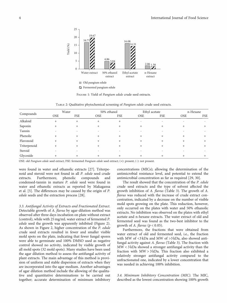
were found in water and ethanolic extracts [27]. Triterpe-noid and steroid were not found in all P. edule seed crudeextracts. Furthermore, phenolic compounds andcondensed-tannin in mature P. edule seed were found inwater and ethanolic extracts as reported by Makagansaet al. [5]. The differences may be caused by the origin of P.edule seeds and the extraction process [28].
3.3. Antifungal Activity of Extracts and Fractionated Extract.Detectable growth of A. flavus by agar-dilution method wasobserved after three days incubation on plate without extract(control), while with 25mg/mL water extract of fermented P.edule seed the growth was apparently inhibited (Figure 2).As shown in Figure 2, higher concentration of the P. edulecrude seed extracts resulted in fewer and smaller visiblemold spots on the plate, indicating that fewer fungal sporeswere able to germinate and 100% DMSO used as negativecontrol showed no activity, indicated by visible growth ofall mold spots (32 mold spots). Many studies have been usedthe agar dilution method to assess the antifungal activity ofplant extracts. The main advantage of this method is provi-sion of uniform and stable dispersion of extracts when theyare incorporated into the agar medium. Another advantagesof agar dilution method include the allowing of the qualita-tive and quantitative determinations to be carried outtogether; accurate determination of minimum inhibitory
concentrations (MICs); allowing the determination of theantimicrobial resistance level, and potential to extend theantimicrobial concentration as far as required [29, 30].
The result showed that the concentration of the P. edulecrude seed extracts and the type of solvent affected thegrowth inhibition of A. flavus (Table 3). The growth of A.flavus was reduced with the increase of crude extract con-centration, indicated by a decrease on the number of visiblemold spots growing on the plate. This reduction, however,only occurred on the plates with water and 50% ethanolicextracts. No inhibition was observed on the plates with ethylacetate and n-hexane extracts. The water extract of old andfermented seed was found as the two-best inhibitor to thegrowth of A. flavus (p < 0:05).
Furthermore, the fractions that were obtained fromwater extract of old and fermented seed, i.e., the fractionwith MW of <3 kDa and MW of >3 kDa, also showed anti-fungal activity against A. flavus (Table 3). The fraction withMW< 3 kDa showed a stronger antifungal activity than thefraction with MW> 3 kDa. This fraction also exhibited arelatively stronger antifungal activity compared to theunfractionated one, indicated by a lower concentration thatwas needed in achieving 100% inhibition.
3.4. Minimum Inhibitory Concentration (MIC). The MIC,described as the lowest concentration showing 100% growth
16.71
4.86
16.08
2.06
19.67
3.84
14.31
1.580
5
10
15
20
25
Water extract 50% ethanolextract
Ethyl acetateextract
n-Hexaneextract
Yiel
d (%
)
Old pangium eduleFermented pangium edule
Figure 1: Yield of Pangium edule crude seed extracts.
Table 2: Qualitative phytochemical screening of Pangium edule crude seed extracts.
CompoundsWater 50% ethanol Ethyl acetate n-Hexane
OSE FSE OSE FSE OSE FSE OSE FSE
Alkaloid + + + + - - - -
Saponin + + + + + + + +
Tannin - - - - - - - -
Phenolic + + + + + + + +
Flavonoid + + + + + + + +
Triterpenoid + + + + + + + +
Steroid - - - - - - - -
Glycoside + + + + + + + +
OSE: old Pangium edule seed extract; FSE: fermented Pangium edule seed extract; (+): present; (-): not present.
4 International Journal of Food Science

inhibition, was only determined for water extract and itsfraction with MW< 3 kDa (Table 4). As shown in Table 3,apparent mold spots were still found on all plates of theother extracts in a concentration between 0.8mg/mL and25mg/mL. The MIC50 and MIC90, that indicated, respec-tively, ≥50% and ≥90% inhibition, were also determined tofacilitate the comparison between antifungal activity levelsof P. edule seed extracts [20, 31].
As described in Table 4, the MIC shown by the waterextract of old seed was lower than that of fermented seed.The fraction with MW of <3 kDa of old seed, however,showed the best inhibitory activity than the other samples(p < 0:05), indicated by the lowest MIC. The results are inline with the previous studies which indicated that P. eduleseed extracted using polar solvents could inhibit thegrowth of A. flavus [10]. Some studies have shown thatlow MW compounds have a contribution to antifungalactivity [32–35].
3.5. Identified Compounds in Fractionated Extract. Theantifungal compound profiles of fraction < 3 kDa of oldand fermented seeds which found as the two-best inhibitorfor A. flavus were analyzed using LC-MS/MS instrument(Figure 3). Thirteen major compounds were detected onthe chromatogram and identified based on the retentiontime and MS data (molecular mass, m/z value of the MS/MSfragments) (Table 5). Based on the results of this analysis,the chemical profiles of fraction < 3 kDa of water extract ofold and fermented P. edule seeds were identified as fattyacids (peaks 2, 3, and 6), amino acids (peaks 1, 4, 7, 8, and9), glycoside (peak 5), and peptides (peaks 10-13). Further-more, the description of the fraction < 3 kDa of water extractof old and fermented P. edule seeds towards the chromato-gram peaks of the LC-MS/MS and its antifungal activitywas visualized using principal component analysis (PCA)(Figure 4). F1 and F2 represented 99.72% of total datavariance, with F1 covered for 78.73% of the data.
(a) (b) (c)
Figure 2: Growth of A. flavus on plates exposed to crude water extract of fermented P. edule seed, after three days incubation at 28°C: (a)negative control (100% DMSO); (b) 3.12mg/mL; (c) 25mg/mL.
Table 3: Antifungal activity of extracts and fractionated water extract of P. edule seed against A. flavus as determined by agar dilution.
Seed Extraction solventNumber of detected mold spot at different concentrations
25mg/mL 12.5mg/mL 6.25mg/mL 3.12mg/mL 1.6mg/mL 0.8mg/mL100% DMSO(control)
Old seed
Water 0 ± 0:0a 0 ± 0:0a 4 ± 3:2a 21 ± 5:5c 29 ± 2:7ef 31 ± 1:3f
32 ± 0:0f50% ethanol 3 ± 2:9a 4 ± 3:9a 13 ± 2:5b 19 ± 2:6c 22 ± 2:7cd 26 ± 4:5de
Ethyl acetate 32 ± 0:8f 32 ± 0:8f 31 ± 2:4f 32 ± 0:4f 32 ± 0:5f 32 ± 0:5f
n-Hexane 32 ± 0:0f 32 ± 0:8f 32 ± 0:0f 32 ± 0:0f 32 ± 0:0f 32 ± 0:4f
Fermented seed
Water 0 ± 0:0a 3 ± 3:6ab 8 ± 2:0bc 16 ± 2:9de 23 ± 2:5fgh 30 ± 1:6i
32 ± 0:0i50% ethanol 3 ± 2:3ab 4 ± 4:0ab 11 ± 2:0cd 19 ± 4:9ef 21 ± 5:6efg 27 ± 4:4ghi
Ethyl acetate 29 ± 4:0i 29 ± 4:8hi 31 ± 1:7i 31 ± 2:1i 31 ± 1:5i 31 ± 1:1i
n-Hexane 32 ± 0:0i 32 ± 0:8i 32 ± 0:0i 32 ± 0:4i 32 ± 0:0i 32 ± 0:0i
Old seedWater, <3 kDa 0 ± 0:0a 0 ± 0:0a 0 ± 0:0a 14 ± 3:2b 18 ± 1:8bcd 18 ± 1:6bcd 32 ± 0:0eWater, >3 kDa 14 ± 3:0b 15 ± 3:1bc 16 ± 4:7bc 17 ± 4:1bcd 19 ± 3:2cd 21 ± 2:0d
Fermented seedWater, <3 kDa 0 ± 0:4a 0 ± 0:0a 15 ± 2:8b 17 ± 2:8bcde 18 ± 2:1bcde 20 ± 2:6de 32 ± 0:0fWater, >3 kDa 15 ± 1:6b 16 ± 1:6bc 17 ± 1:9bcd 18 ± 1:2bcde 19 ± 2:5cde 21 ± 2:8e
Numbers are means of three replications. Numbers followed by different letters in each row of P. edule seed show a significant difference (p < 0:05) withindifferent extract concentration and also between control.
5International Journal of Food Science

The PCA score plot discriminated OSE1 and OSE2 fromother samples by separate cluster on the positive score valueof F1, which showed the similar characteristics as antifungal
activity (Figure 4(a)). The loading plot (Figure 4(b)) of PCAresult showed that the most discriminatory constituents inOSE1 and OSE2 were peaks 2, 5, and 6 which presented in
Table 4: The MIC, MIC90, and MIC50 that were determined using the agar dilution method.
Seed Sample MIC MIC90 MIC50
Old seed
Water extract 12.5mg/mL 12.5mg/mL 6.25mg/mL
50% ethanolic extract — 25mg/mL 6.25mg/mL
Water extract fraction < 3 kDa 6.25mg/mL 6.25mg/mL 3.12mg/mL
Fermented seed
Water extract 25mg/mL 12.5mg/mL 3.12mg/mL
50% ethanolic extract — 25mg/mL 6.25mg/mL
Water extract fraction < 3 kDa 12.5mg/mL 12.5mg/mL 6.25mg/mL
MIC: minimum inhibitory concentration, the lowest concentration showing 100% growth inhibition; MIC50 and MIC90 indicated ≥50% and ≥90% inhibition,respectively.
0 5 20 30
Rela
tive a
bund
ance
OSE1
OSE2
FSE1
FSE2
2, 5
6
8 9
2, 5
6
8 9
1, 3, 4
5
8
67
9
12 13
6
8
7
9
251510
Retention time (min)
1110
1, 3, 4, 5
Figure 3: LC-MS/MS chromatogram of fraction < 3 kDa of water extract of old (OSE) and fermented (FSE) of P. edule seed (positiveion mode).
6 International Journal of Food Science

Table 5: Compounds identified in the water extract of old and fermented Pangium edule seed by LC-MS/MS analysis.
Peakno.
Identified compoundsRt
(mins)Precursorion (m/z)
Productions (m/z)
Formula[M-H]+
MW (g/mol)
Detectedsamples
11-[(3-Carboxypropyl)amino]-1-deoxy-
beta-D-fructofuranose0.99 266.1228 248 C10H19NO7 265.1156 FSE1, FSE2
2Methyl (4Z)-5-(1,3-dioxolan-2-yl)
-2-hydroxy-4-(hydroxyimino)pentanoate1.00 234.0970
2161881029790
C9H15NO6 233.0900 OSE1, OSE2
3Ethylenediamine-N,N′
-diacetic-N,N′-dipropionic acid 1.02 321.128516012484
C12H20N2O8 320.1213 FSE1, FSE2
4 2′-Deoxymugineic acid 1.06 305.133213084
C12H20N2O7 304.1260 FSE1, FSE2
5Methyl 4,6-dideoxy-4-[(2,4-
dihydroxybutanoyl)amino]-2-O-methylhexopyranoside
1.09 294.154327623088
C12H23NO7 293.1465OSE1, OSE2,FSE1, FSE2
6Methyl (5-acetamido-2,2-dimethyl-4,6-dioxo-1,
3-dioxan-5-yl)acetate1.22 274.0916
2562381309784
C11H15NO7 273.0843OSE1, OSE2,FSE1, FSE2
7 6-Hydroxypicolinic acid 1.40 140.0340 112 C6H5NO3 139.0267 FSE1, FSE2
8 Isoleucine 1.56 132.1017 86 C6H13NO2 131.0944OSE1, OSE2,FSE1, FSE2
9 Phenylalanine 2.27 166.0860 120 C9H11NO2 165.0787OSE1, OSE2,FSE1, FSE2
10 Leucyl-valine 4.83 231.1696 72 C11H22N2O3 230.1624 FSE2
11 Leucyl-alanyl-proline 5.67 300.190911686
C14H25N3O4 299.1836 FSE2
12 Leucyl-aspartyl-valine 6.40 346.196321518772
C15H27N3O6 345.1890 FSE1
13 Phenylalanyl-valyl-aspartic acid 7.37 380.1805
21518712072
C18H25N3O6 379.1732 FSE1
OSE1OSE2
FSE1
FSE2
–3
–2
–1
0
1
2
3
–4 –3 –2 –1 0 1 2 3 4
F2 (2
0.99
%)
F1 (78.73%)
(a)
Peak 1
Peak 2
Peak 3
Peak 4
Peak 5
Peak 6
Peak 7
Peak 8Peak 9
Peak 10 Peak 11
Peak 12 Peak 13–1
–0.5
0
0.5
1
–1 –0.5 0 0.5 1
F2 (2
0.99
%)
F1 (78.73 %)
(b)
Figure 4: PCA results of the fraction < 3 kDa of water extract of old and fermented P. edule seeds: (a) score plot; (b) loading plot.
7International Journal of Food Science

high amounts in OSE1 and OSE2 compared to in FSE1 andFSE2. Loading plot also showed that peaks 12 and 13suggested as discriminant compounds in FSE1, while FSE2formed a separate cluster with peaks 10 and 11 as discrimi-nant compounds.
Contribution and correlation of each peak to the anti-fungal activity were described by variable importance in pro-jection (VIP) scores and correlation coefficient (r) usingpartial least squares regression (PLSR) analysis. The resultof PSLR indicated that peaks 2, 5, and 6 were contributedto the antifungal activity of water extract of old P. edule seed(VIP score > 1) and showed a positive correlation to antifun-gal activity (r > 0:90) (Figure 4(b)). Meanwhile, peaks 1, 3, 4,7, 8, and 9 were contributed to the antifungal activity of waterextract of fermented P. edule seed (VIP score > 1) but showeda negative correlation to antifungal activity (r < 0). Moreover,the result of PSLR also indicated that peaks 10, 11, 12, and 13were not contributed to the antifungal activity of waterextract of old and fermented P. edule seed. The different com-pounds present in the different extracts cannot be describedby the result as shown in Table 2.
Based on the LC-MS/MS chromatogram in Figure 3,from the three peaks that contributed and are positively cor-related on antifungal activity (peaks 2, 5, and 6), there weretwo peaks that were detected and identified in both waterextract of old P. edule seed (OSE) and water extract of fer-mented P. edule seed (FSE), i.e., peaks 5 and 6 which wereclassified as glycoside and fatty acid. Peak 2 which classifiedas fatty acid was only detected and identified in waterextract of old P. edule seed. This indicated that the peaks(compounds) responsible for the antifungal activity of thewater extract of old P. edule seed are peaks 2, 5, and 6,while the peaks (compounds) responsible for the antifun-gal activity of the water extract of fermented P. edule seedare only peaks 5 and 6.
Overall, the PCA and PSLR analysis revealed that pep-tides were not contributed to the antifungal activity of thefraction < 3 kDa of water extract of old and fermented seeds.All of contributed peaks in this study classified as fatty acid,amino acid, and glycoside compounds (Table 5). Aminoacids showed a negative correlation to the antifungal activityof the fraction < 3 kDa of water extract of old and fermentedseeds, while fatty acids and glycoside showed a positive cor-relation to its antifungal activity. Therefore, both fatty acidsand glycoside were compounds that are responsible for theantifungal activity of the fraction < 3 kDa of water extractof old and fermented P. edule seeds. Previous studies havebeen reported that fatty acids showed antifungal activityagainst pathogenic fungi, such as Candida albicans, Aspergil-lus flavus, and Rhizopus nigricans [36, 37]. Fatty acids inducefungal cell inhibition through cell membrane which is amajor target for these compounds. Fatty acids can increasethe membrane fluidity and cause a generalized disorganiza-tion of the cell membrane. It leads to conformationalchanges in membrane proteins which will result in leakageof the intracellular components and cell death [38, 39].
Moreover, the PCA and PSLR results also showed thatglycosides contributed to antifungal activity of water extractof old and fermented P. edule seed. Glycosides have been
reported to have antifungal activity against T. mentagro-phytes, C. albicans, B. cinerea, A. alternata, A. flavus, Asper-gillus niger, Aspergillus ochraseus, Aspergillus versicolor,Penicillium funiculosum, Rhizoctonia cerealis, and Tricho-derma viride [40–43]. Zhang et al. [44] revealed that mecha-nism of antifungal properties of glycosides was damaging theplasma membrane and cause the leakage of cytoplasmicmaterials which leads to cell death.
3.6. The Median Lethal Concentration (LC50). The LC50determined by the brine-shrimp lethality test (BSLT) forwater extract of P. edule seed is given in Table 6. The resultshows that the water extracts are moderately toxic toA. salina(LC50: 468μg/mL for old seed and 244μg/mL for fermentedseed). The toxicity based on the BSLT results is classified asfollows: LC50 0-100μg/mL, highly toxic; LC50 100-500μg/mL,moderately toxic; LC50 500-1000μg/mL, low toxic; and LC50> 1000μg/mL, nontoxic [45]. According toMeyer’s research,the plant extracts with LC50 of 30-1000μg/mLhad potential asantimicrobial agent and pesticide [23].
The toxicity test of the seed water extracts against A.salina larvae has not been reported before, though Sudjanaet al. [46] and Simanjuntak et al. [47] have described the tox-icity of P. edule seed extract with other solvents. The meth-anolic and chloroform extracts of P. edule seed had LC50 of274.26μg/mL and 916.13μg/mL, respectively, while the n-hexane extract was nontoxic since it had LC50 > 1000μg/mL [46]. Furthermore, the ethanolic extract of fresh P.edule seed was also considered as nontoxic against A. salinalarvae (LC50 > 1000μg/mL) as reported by Simanjuntaket al. [47]. The differences in the LC50 value can be due todifferent type of P. edule used which cause differences inthe secondary metabolites produced. In addition, differentextraction methods and solvents also affect the dissolvedactive compounds, causing differences in biological activitiesof same plant species [48, 49].
Previous studies reported that LC50 of the BSLT and theLD50 of the acute oral toxicity assay in animal models has apositive correlation [45, 50]. According to Parra’s research,the brine shrimp LC50 < 10μg/mL has LD50 100-1000mg/kg; LC50 < 20μg/mL has LD50 1000-2500mg/kg; LC50 > 25μg/mL has LD50 2500-8000mg/kg [50]. We canassume that the LD50 of oral acute toxicity for water extract
Table 6: LC50 by the brine-shrimp lethality test of water extract ofP. edule seed.
ExtractsConcentration (μg/
mL)Mortality
(%)LC50 (μg/
mL)
Old seed extract
1000 80.00
468500 53.33
100 26.67
10 23.33
Fermented seedextract
1000 90.00
244500 60.00
100 43.33
10 36.67
8 International Journal of Food Science

of old and fermented of P. edule seed also will be more than2500mg/kg, because the LC50 of BSLT is 468 and 244μg/mL,respectively. The chemical labeling and classification ofacute systemic toxicity based on oral LD50 are recom-mended by the Organization for Economic Co-operationand Development (OECD) as follows: ≤ 5mg/kg, verytoxic; 5-50mg/kg, toxic; 50-500mg/kg, harmful; and 500-2000mg/kg, no label [51]. Hence, this finding showed thatthe water extract of P. edule seed could be developed as anantifungal or preservative agent since the oral LD50suggests that the extract is nontoxic.
4. Conclusions
This study found that the water extract fraction with MW< 3 kDa of old P. edule seed showed the best inhibitoryactivity against A. flavus, followed by the fraction withMW< 3 kDa of fermented seed. These fractions showed astronger antifungal activity than the unfractionated waterextract, indicated by the lower MIC. The responsible com-pounds for the antifungal activity of water extract of oldand fermented seed were classified into the same group,i.e., fatty acid and glycoside. Purification and confirmationof the antifungal activity of the purified compound wereincluded in the further study.
Data Availability
All datasets used to support the findings of this study areavailable upon reasonable request from the correspondingauthor.
Disclosure
The main results of this article have been published as amaster thesis in IPB repository and have been approved torepublish the data.
Conflicts of Interest
The authors declare that there is no conflict of interestregarding the publication of this paper.
Acknowledgments
This work was supported by the Ministry of Research, Tech-nology and Higher Education of the Republic of Indonesiaunder grant No: 2108/IT3.L1/PN/2020.
References
[1] E. G. Samudry, A. Sukainah, and A. Mustarin, “Analisis kuali-tas kluwek (Pangium edule Reinw) hasil fermentasi menggu-nakan media tanah dan abu sekam,” Jurnal PendidikanTeknologi Pertanian, vol. 3, no. 1, pp. 25–33, 2018.
[2] E. S. Heruwati, H. E. Widyasari, and J. Haluan, “Fresh fishpreservation using picung (Pangium edule Reinw) kernel,”Jurnal Pascapanen dan Bioteknologi Kelautan dan Perikanan,vol. 2, no. 1, pp. 9–18, 2014.
[3] W. Mangunwardoyo, L. Ismaini, and E. S. Heruwati, “Anal-ysis of bioactive compounds in fresh seed extract of picung(Pangium edule Reinw.),” Berita Biologi, vol. 9, no. 3,pp. 259–264, 2008.
[4] F. Y. Chye and K. Y. Sim, “Antioxidative and antibacterialactivities of Pangium edule seed extracts,” International Jour-nal of Pharmacology, vol. 5, no. 5, pp. 285–297, 2009.
[5] C. Makagansa, C. F. Mamuaja, and L. C. Mandey, “The anti-bacterial activity of pangi kernel extract (Pangium eduleReinw) towards Staphylococcus aureus, Bacillus cereus, Pseu-domonas aeruginosa, and Escherichia coli in vitro,” JurnalIlmu dan Teknologi Pangan, vol. 3, no. 1, pp. 16–25, 2015.
[6] A. K. Hasbullah and H. N. Novelina, “Characterization tradi-tional food pado from West Sumatra,” Der Pharmacia Lettre,vol. 8, no. 15, pp. 202–205, 2016.
[7] A. Kusmarwati and N. Indriati, “The inhibition rate of theactive compound of Pangium edule Reinw. seeds extract onthe growth of histamine producing bacteria,” Jurnal Pascapa-nen dan Bioteknologi Kelautan dan Perikanan, vol. 3, no. 1,pp. 29–36, 2008.
[8] E. S. Heruwati, L. Ismaini, andW.Mangunwardoyo, “Antibac-terial test of pangium (Pangium edule Reinw) extract againstthe growth of fish spoilage bacteria,” Indonesian FisheriesResearch Journal, vol. 15, no. 2, pp. 65–73, 2017.
[9] I. Achmad, E. Anggraeni, N. Herliyana, A. Asrori, and S. Rijal,“Effectiveness of Pangium edule Reinw. flesh seed extract inhi-bition on in vitro growth of Rhizoctonia sp. and Cylindrocla-dium sp,” Journal of Horticulture, vol. 22, no. 3, pp. 268–275,2012.
[10] V. Membalik, Y. W. George, A. Rahman, and A. Asman,“Antifungal activities of Pangium edule Reinw. seed extractsinhibit the growth of Aspergillus flavus, producer of aflatoxins,through the in-vitro test,” International Journal of Pharmaceu-tical Sciences and Research, vol. 10, no. 6, pp. 2718–2722, 2019.
[11] IARC (International Agency for Research on Cancer), “Afla-toxin in traditional herbal medicines, some mycotoxins, naph-thalene, and styrene,” International Agency for Research onCancer, Lyon, 2002.
[12] M. T. Hedayati, A. C. Pasqualotto, P. A. Warn, P. Bowyer, andD. W. Denning, “Aspergillus flavus: human pathogen, allergenand mycotoxin producer,” Microbiology, vol. 153, no. 6,pp. 1677–1692, 2007.
[13] N. Indriati, I. Hermana, I. Hidayah, and E. S. Rahayu, “Preva-lence of aflatoxin B1 in commercial dried fish from someregions of Java,” Squalen Bulletin of Marine and Fisheries Post-harvest and Biotechnology, vol. 12, no. 3, pp. 107–115, 2018.
[14] I. Hermana, A. Kusmarwati, and Y. Yennie, “Isolasi dan iden-tifikasi kapang dari ikan pindang,” Jurnal Pascapanen dan Bio-teknologi Kelautan dan Perikanan, vol. 13, no. 1, pp. 79–92,2018.
[15] AOAC (Association of Official Analytical Chemists), OfficialMethod of Analysis, Association of Official Analytical Chem-ists, Washington DC, 19th Ed edition, 2012.
[16] AOAC (Association of Official Analytical Chemists), OfficialMethod of Analysis, Association of Official Analytical Chem-ists, Washington DC, 18th Ed edition, 2005.
[17] R. Gul, S. U. Jan, S. Faridullah, S. Sherani, and N. Jahan, “Pre-liminary phytochemical screening, quantitative analysis ofalkaloids, and antioxidant activity of crude plant extracts fromEphedra intermedia indigenous to Balochistan,” The ScientificWorld Journal, vol. 2017, Article ID 5873648, 7 pages, 2017.
9International Journal of Food Science

[18] T. M. B. Bandiola, “Extraction and qualitative phytochemicalscreening of medicinal plants: a brief summary,” InternationalJournal of Pharmacy, vol. 8, no. 1, pp. 137–143, 2018.
[19] V. Singh, R. S. Upadhyay, B. K. Sarma, and H. B. Singh, “Tri-choderma asperellum spore dose depended modulation ofplant growth in vegetable crops,” Microbiological Research,vol. 193, pp. 74–86, 2016.
[20] C. R. A. Mota, K. C. Miranda, J. A. Lemos et al., “Comparisonof in vitro activity of five antifungal agents against dermato-phytes, using the agar dilution and broth microdilutionmethods,” Revista da Sociedade Brasileira de Medicina Tropi-cal, vol. 42, no. 3, pp. 250–254, 2009.
[21] M. Balouiri, M. Sadiki, and S. K. Ibnsouda, “Methods forin vitro evaluating antimicrobial activity: a review,” Journalof Pharmaceutical Analysis, vol. 6, no. 2, pp. 71–79, 2016.
[22] S. N. Andayani, H. N. Lioe, C. H. Wijaya, and M. Ogawa,“Umami fractions obtained from water-soluble extracts ofred oncom and black oncom−Indonesian fermented soybeanand peanut products,” Journal of Food Science, vol. 85, no. 3,pp. 657–665, 2020.
[23] B. N. Meyer, N. R. Ferrigni, J. E. Putnam, L. B. Jacobsen, D. E.Nichols, and J. McLaughlin, “Brine shrimp: a convenient gen-eral bioassay for active plant constituents,” Journal of Medici-nal Plant Research, vol. 45, no. 5, pp. 31–34, 1982.
[24] Q. S. Sarah, F. C. Anny, and M. Misbahuddin, “Brine shrimplethality assay,” Bangladesh Journal of Pharmacology, vol. 12,no. 2, pp. 186–189, 2017.
[25] M. Heinrich, J. Barnes, J. Prieto-Garcia, S. Gibbons, andE. Williamson, Fundamentals of Pharmacognosy and Phy-totherapy, Elsevier, Hongaria, 3rd Edition edition, 2018.
[26] M. G. Ajuru, L. F. Williams, and G. Ajuru, “Qualitative andquantitative phytochemical screening of some plants used inethnomedicine in the Niger Delta region of Nigeria,” Journalof Food and Nutrition Sciences, vol. 5, no. 5, pp. 198–205, 2017.
[27] S. Warnasih and U. Hasanah, “Phytochemical characterizationand tannin stability test from kluwek (Pangium edule Reinw),”Ekologia, vol. 1, no. 2, pp. 44–49, 2018.
[28] B. S. Hartati, S. B, and H. Karim, “Pengaruh jenis pelarut ter-hadap kandungan senyawametabolit sekunder klika kayu jawa(Lannea coromendelica),” Sainsmat, vol. 8, no. 2, pp. 19–27,2019.
[29] M. T. G. Silva, S. M. Simas, T. G. F. M. Batista, P. Cardarelli,and T. C. B. Tomassini, “Studies on antimicrobial activity,in vitro, of Physalis angulata L. (Solanaceae) fraction and phy-salin B bringing out the importance of assay determination,”Memórias do Instituto Oswaldo Cruz, vol. 100, no. 7,pp. 779–782, 2005.
[30] J. B. L. Tan and Y. Y. Lim, “Critical analysis of current methodsfor assessing the in vitro antioxidant and antibacterial activityof plant extracts,” Food Chemistry, vol. 172, pp. 814–822, 2015.
[31] S. Schwarz, P. Silley, S. Simjee et al., “Editorial: assessing theantimicrobial susceptibility of bacteria obtained from ani-mals,” Journal of Antimicrobial Chemotherapy, vol. 65, no. 4,pp. 601–604, 2010.
[32] F. P. Hu and J. M. Young, “Biocidal activity in plant patho-genic Acidovorax, Burkholderia, Herbaspirillum, Ralstoniaand Xanthomonas spp,” Journal of Applied Microbiology,vol. 84, no. 2, pp. 263–271, 1998.
[33] R. González-Lamothe, G. Mitchell, M. Gattuso, M. Diarra,F. Malouin, and K. Bouarab, “Plant antimicrobial agents andtheir effects on plant and human pathogens,” International
Journal of Molecular Sciences, vol. 10, no. 8, pp. 3400–3419,2009.
[34] S. S. Rathore, M. Isravel, S. Vellaisamy et al., “Exploration ofantifungal and immunomodulatory potentials of a furanonederivative to rescue disseminated cryptococosis in mice,” Sci-entific Reports, vol. 7, no. 1, article 15400, pp. 1–14, 2017.
[35] A. Matuszewska, M. Jaszek, D. Stefaniuk, T. Ciszewski, andL. Matuszewski, “Anticancer, antioxidant, and antibacterialactivities of low molecular weight bioactive subfractions iso-lated from cultures of wood degrading fungus Cerrena unico-lor,” PLoS One, vol. 13, no. 6, article e0197044, pp. 1–14, 2018.
[36] R. Hassan, S. El-Kadi, and M. Sand, “Effect of some organicacids on some fungal growth and their toxin production,”International Journal of Advances in Biology, vol. 2, no. 1,pp. 1–11, 2015.
[37] J. Yun and D. G. Lee, “A novel fungal killing mechanism ofpropionic acid,” FEMS Yeast Research, vol. 16, no. 7, pp. -fow089–fow088, 2016.
[38] T. J. Avis and R. R. Bélanger, “Specificity and mode of action ofthe antifungal fatty acid cis-9-heptadecenoic acid produced byPseudozyma flocculosa,” Applied and Environmental Microbi-ology, vol. 67, no. 2, pp. 956–960, 2001.
[39] T. J. Avis, “Antifungal compounds that target fungal mem-branes: applications in plant disease control,” Canadian Jour-nal of Plant Pathology, vol. 29, no. 4, pp. 323–329, 2007.
[40] I. A. Khan, A. M. Clark, and J. D. McChesney, “Antifungalactivity of a new triterpenoid glycoside from Pithecellobiumracemosum (M.),” Pharmaceutical Research, vol. 14, no. 3,pp. 358–361, 1997.
[41] K. S. Rao, G. V. Babu, and Y. V. Ramnareddy, “Acylated fla-vone glycosides from the roots of Saussurea lappa and theirantifungal activity,” Molecules, vol. 12, no. 3, pp. 328–344,2007.
[42] D. M. Wang, W. J. Pu, Y. H. Wang, Y. J. Zhang, and S. S.Wang, “A new isorhamnetin glycoside and other phenoliccompounds from Callianthemum taipaicum,” Molecules,vol. 17, no. 4, pp. 4595–4603, 2012.
[43] A. M. O. Amoussa, M. Bourjot, L. Lagnika, C. Vonthron-Séné-cheau, and A. Sanni, “Acthaside: a new chromone derivativefrom Acacia ataxacantha and its biological activities,” BMCComplementary and Alternative Medicine, vol. 16, no. 1,pp. 506–513, 2016.
[44] J. D. Zhang, Z. Xu, Y. B. Cao et al., “Antifungal activities andaction mechanisms of compounds from Tribulus terrestrisL.,” Journal of Ethnopharmacology, vol. 103, no. 1, pp. 76–84,2006.
[45] M. Hamidi, B. Jovanova, and T. Kadifkova Panovska, “Toxico-logical evaluation of the plant products using brine shrimp(Artemia salina L.) model,” Macedonian Pharmaceutical Bul-letin, vol. 60, no. 1, pp. 9–18, 2014.
[46] T. S. Sudjana, E. Rohaeti, and F. C. Yunita, “Comparison of theExtraction Methods towards Picung Seed (Pangium eduleReinw) and Toxicity Assay to Artemia salina Leach,” in Roleof Chemistry in Industrial Progress, pp. 330–333, Proceedingsof the National Seminar of Indonesian Chemical Society,Bogor: Indonesian Chemical Society, 2006.
[47] I. N. Simanjuntak, R. A. Repi, E. M. Moko, M. N. Tanor, andD. J. J. Rayer, “Potensi ekstrak biji pangi (Pangium eduleReinw) sebagai pengawet alami pada ikan mujair (Oreochro-mis mossambicus),” Fullerene Journal of Chemistry, vol. 5,no. 2, pp. 117–123, 2020.
10 International Journal of Food Science

[48] J. Azmir, I. S. M. Zaidul, M. M. Rahman et al., “Techniques forextraction of bioactive compounds from plant materials: areview,” Journal of Food Engineering, vol. 117, no. 4,pp. 426–436, 2013.
[49] M. Saxena, J. Saxena, R. Nema, D. Singh, and A. Gupta, “Phy-tochemistry of medicinal plants,” Journal of Pharmacognosyand Phytochemistry, vol. 1, no. 6, pp. 168–182, 2013.
[50] A. L. Parra, R. S. Yhebra, I. G. Sardiñas, and L. I. Buela, “Com-parative study of the assay of Artemia salina L. and the esti-mate of the medium lethal dose (LD50 value) in mice, todetermine oral acute toxicity of plant extracts,” Phytomedicine,vol. 8, no. 5, pp. 395–400, 2001.
[51] E. Walum, “Acute oral toxicity,” Environmental Health Per-spectives, vol. 106, supplement 2, pp. 497–503, 1998.
11International Journal of Food Science