Embed Size (px)
Citation preview
Antibody mediated fluorescence enhancement of nucleoside analogue1,3-diaza-2-oxophenoxazine (tC1)
Frank Sellrie a,n, Christine Lenz a, Anika Andersson a, L. Marcus Wilhelmsson b,Jörg A. Schenk a,c
a UP Transfer GmbH, Am Neuen Palais 10, Potsdam, Germanyb Department of Chemical and Biological Engineering, Chalmers University of Technology, Gothenburg, Swedenc Hybrotec GmbH, Am Mühlenberg 11, Potsdam, Germany
a r t i c l e i n f o
Article history:Received 4 November 2013Received in revised form14 February 2014Accepted 20 February 2014Available online 28 February 2014
Keywords:Fluorescent nucleoside analogue1,3-Diaza-2-oxophenoxazine (tC1)Monoclonal antibodyFluorescence enhancementHomogeneous immunoassayHypsochromic shift
a b s t r a c t
We report on the generation and analytical application of the monoclonal antibody G93-ED2 raisedagainst the tricyclic fluorescent nucleoside analogue 1,3-diaza-2-oxophenoxazine (tC1). G93-ED2 isspecifically binding this deoxycytidine analogue and was found to raise its fluorescence intensity by afactor of 5. This unique feature makes it a valuable tool in fluorescence dependent immunoassays. G93-ED2 was successfully applied in a homogeneous fluorescence quenching immunoassay (DNA-Q) for thesequence specific determination of DNA.
& 2014 Elsevier B.V. All rights reserved.
1. Introduction
The binding of monoclonal antibodies directed against fluor-ophores, often leads to dramatic changes in the fluorescenceproperties of the bound fluorophore. Decades ago, Voss et al.found quenching effects and bathochromic shifts for polyclonaland monoclonal antibodies to fluorescein [1–3]. In our group, wealso found such effects for another fluorescein antibody [4] and itsrecombinant scFv fragment [5,6]. Fluorescence quenching was alsodescribed for an anti-TAMRA antibody [7] and even for time-resolved fluorescence of Eu-kryptate antibodies [8].
Antibody mediated fluorescence quenching is caused by staticquenching. Amino acid side chains of antigen binding site and fluoro-phore form a nonfluorescent complex. This is a unique property of anindividual monoclonal antibody, meaning not all antibodies bindingthe fluorophore necessarily alter its fluorescence.
Surprisingly, the opposite effect (fluorescence enhancementand hypsochromic shift) is rarely described, mostly for fluorophoreconjugates [9,10].
One group of fluorophores are fluorescent nucleoside analo-gues, which replace normal nucleobases inside the DNA/RNA
duplex and aims at minimal perturbation of the natural propertiesof the nucleic acid system under investigation [11–13]. These areapplied in photo-physical studies of DNA interacting with macro-molecules (proteins) or for determining size and shape of DNAtertiary structure. Usually, fluorescent base analogues, such as 2-aminopurine and pyrrolo-C are highly sensitive to their micro-environment [12,14,15].
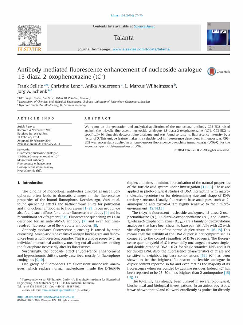
The tricyclic fluorescent nucleoside analogues, 1,3-diaza-2-oxo-phenothiazine (tC), 1,3-diaza-2-oxophenoxazine (tC1) and 7-nitro-1,3-diaza-2-oxophenothiazine (tCnitro) are a family of deoxycytidineanalogues that have been shown to base pair faithfully with dGwithvirtually no disruption of the normal duplex structure [16–18]. Thismeans that the stability of the DNA duplex is not compromised ascompared to the control regardless of DNA sequence. The fluores-cence quantum yield of tC is essentially unchanged between single-and double-stranded DNA - 0.21 for single stranded DNA and 0.19for duplex DNA. Also, the fluorescence characteristics of tC are notsensitive to neighbouring base combinations [19]. tC1 has beenshown to be the brightest fluorescent nucleoside analogue induplex context reported so far and even retains the majority of itsfluorescence when surrounded by guanine residues. Indeed, tC1 hasbeen reported to be 25–50 times brighter than 2-aminopurine [16](Fig. 1).
The tC-family has already been utilized in several biophysical,biochemical and biological investigations. In an anisotropy study,it was shown that tC and tC1work excellently as probes for directly
Contents lists available at ScienceDirect
journal homepage: www.elsevier.com/locate/talanta
Talanta
http://dx.doi.org/10.1016/j.talanta.2014.02.0460039-9140 & 2014 Elsevier B.V. All rights reserved.
n Correspondence to: UP Transfer GmbH c/o Fraunhofer Institute for BiomedicalEngineering, Am Mühlenberg 13, D-14476 Potsdam, Germany.Tel.: þ49 331 58187 231; fax: þ49 331 58187 299.
E-mail address: [email protected] (F. Sellrie).
Talanta 124 (2014) 67–70
monitoring motion of nucleic acid helical structures, rather thana combination of motion of the overall structure and the probeitself, as is the case for most of the currently available fluorescentbase analogues [16]. Furthermore, tC has been utilized as a FRET-donor in pair with rhodamine in a PNA-DNA-hybrid [20] and inpair with Alexa-555 in a study of conformational dynamics of DNApolymerase [21]. Importantly, tC1 and tCnitro were recently devel-oped to be the first nucleic acid base analogue FRET-pair andshown to successfully monitor both distance and geometrychanges within natural duplex systems [18].
The 5'-triphosphates of tC and tC1 have also been synthesizedand been shown in several polymerase studies to be efficientlyincorporated into DNA although with increased frequency ofmutations [22,23].
Here we present the generation of a novel monoclonal antibodybinding the fluorescent nucleoside analogue 1,3-diaza-2-oxophe-noxazine (tC1). This antibody dramatically increased the fluores-cence of its antigen and induced a hypsochromic shift. We wereable to apply this monoclonal antibody G93-ED2 successfully in aDNA-Quenching immunoassay (DNA-Q) for the sequence specificdetermination of oligonucleotide DNA.
This assay format was recently published (Sellrie et al. [7]). Inthis work we applied 5-TAMRA as fluorophore and the 5-TAMRAbinding and quenching monoclonal antibody G71-DC7. DNA-Quenching assay (DNA-Q) bases on our finding that an antibodysensitive fluorophore gets protected from fluorescence quenchingcaused by an anti-fluorophore antibody, if the fluorophore replacesone of the internal nucleotides of one of the strands of the DNAduplex. In the DNA-Q assay the fluorescence probe is annealed tosingle stranded analyte DNA in the sample (for example denaturedor asymmetric PCR amplicon). Then fluorescence quenching anti-body is added and the change in fluorescence is detected. If thesample contains the probe target sequence, the probe hybridizeswith the target. That escapes the fluorophore from antibodyquenching and results in a positive fluorescence signal.
2. Experimental
2.1. Generation of fluorescence enhancing antibodies
Monoclonal tC1 specific antibodies were generated by hybridomatechnology [24]. For this purpose Balb/c mice were immunized threetimes with a BSA-tC1 conjugate (1,3-diaza-2-oxophenoxazine–bovine
serum albumin). Fluorophore conjugates were synthesized by empBiotech GmbH (Germany). Immunization started with 100 mg con-jugate using Freund's complete adjuvant. Booster immunizationswere carried out six and eight weeks after the first immunizationusing 50 mg conjugate without adjuvant. Four days after the finalbooster immunization electrofusion of spleen cells with myelomacells (P3�63Ag8.653, ATCC CRL-1580) in the presence of polyethy-lene glycol 8000 was performed as described [25]. Selected hybridswere cultivated in RPMI 1640 medium (containing 10% FCS, 2 mMglutamine and 50 mM β-mercaptoethanaol) and subcloned by limit-ing dilution on mouse peritoneal feeder cells. Culture supernatantsof clones and subclones were tested in an enzyme immunoassay(ELISA) for antigen binding to a Biotin (3'-terminal) and tC1 (internal)modified oligonucleotide, adsorbed to streptavidin coated microtiterplates. The class and subclass of monoclonal antibodies weredetermined as described [25]. Purification of antibody from culturesupernatant was performed by protein A affinity chromatography[26].
2.2. Competitive ELISA for tC1 and tC binding
Microtiter plates (Greiner Bio-One GmbH-Germany) werecoated with BSA-tC1 conjugate (incubation overnight with 50 mlper well containing 5 mg/ml in PBS), washed with tap water andblocked with 60 ml PBS-NCS (neonatal calf serum) per well for40 min at room temperature. The wells were then incubated for2 h with 50 ml of a mixture (incubated in advance for 30 min) ofantibody G93-ED2 (0.5 mg/ml) and different tC1 and tC concentra-tions. Microtiter plates were washed and incubated (50 ml perwell; 1 h) with goat anti-mouse IgG-HRP conjugate (Sigma Aldrich-Germany). Plates were washed, and substrate reaction using TMB(3,3',5,5'-tetramethylbenzidine) performed. Reaction was stoppedafter 10 min and absorption measured at 450 nm.
2.3. Fluorescence enhancement experiments
Excitation and emission spectra of fluorophores tC1 and tC wereexamined for the free fluorophores and in the presence of anti-body G93-ED2 at equimolar concentrations of 0.2 mM.
BSA-tC1 conjugate and tC1 binding antibody G93-ED2 or controlantibody G63-EC9 [27] were mixed (conjugate and antibody 0.2 mMin PBS) and fluorescence measured (Ex. 360 nm, Em. 432 nm).
Further fluorescence enhancement experiments were performedin the presence of 80% neonatal bovine serum (Biochrom GmbH,Germany) and 1 mg/ml DNA from salmon testis (Sigma-Aldrich,Germany). Concentration of fluorescence probe (see Section 2.4)and antibody G93-ED2 was 0.2 mM.
2.4. Oligonucleotide determination by DNA-quenching assay
tC1-CE phoshoramidite [5'-O-(4,4'-Dimethoxytrityl)-1'-(1,3-diaza-2-oxophenoxazin-1-yl)-2'-deoxy-Β-D-ribofuranosyl-3'-[(2-cyanoethyl)-(N,N-diisopropyl)]-phosphoramidite] was purchasedfrom Glen Research Corporation (USA). Fluorescence probe synth-esis was performed by IBA GmbH (Germany). Its sequence (5'-GGTTTT GTT GTX TTC TCT ATT-3'; X¼tC1) was chosen from Salmonellaenterica invA gene.
A template oligonucleotide complementary to fluorescence probewas synthesized by metabion GmbH (Germany) (5'-GGT GAC AATAGA GAA GAC AAC AAA ACC CAC-3'). The DNA-Quenching assay wasperformed in PBS. Fluorescence probe (40 ml of 0.3 mM solution) andtemplate oligonucleotide (40 ml, varying concentrations) were mixedand hybridized for 10 minutes at 40 1C. Antibody G93-ED2 (40 ml of0.3 mM) was added to get the final 0.1 mM concentration of antibodyand probe. Fluorescence measurement (Ex. 360 nm, Em. 432 nm)was carried out after 15 minutes of incubation.
Fig. 1. Tricyclic cytosine analogue structure. Guanine is shown base paired withfluorescent cytosine analogue tC1 and cytosine, respectively. The differencesbetween cytosine and tC1 were highlighted.
F. Sellrie et al. / Talanta 124 (2014) 67–7068
2.5. Fluorescence measurement
Fluorescence measurement was performed on an EdinburghInstruments (UK) F900 spectrometer. The excitation wavelengthfor tC1 was 360 nm. Fluorescence emission after antibody bindingwas detected at 432 nm.
3. Results and discussion
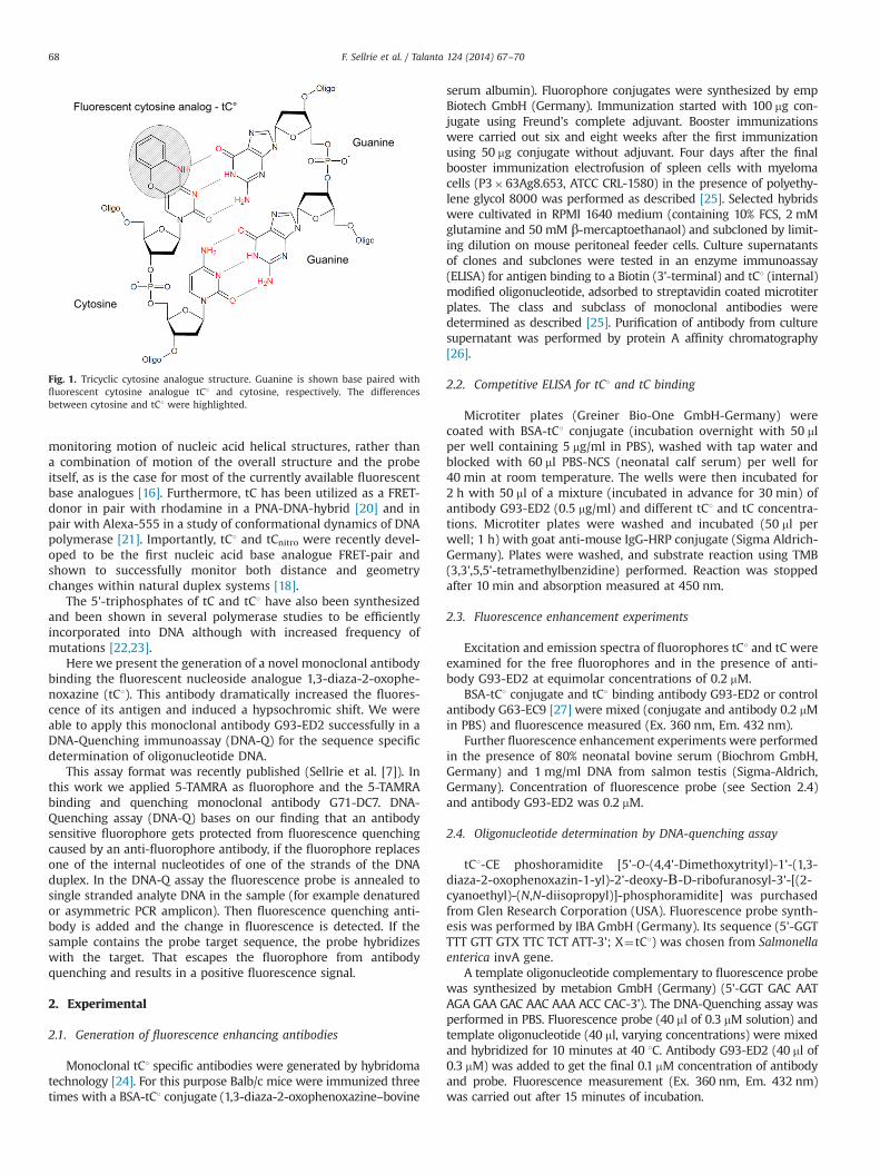
We performed hybridoma technology to raise monoclonalantibodies binding the tricyclic fluorescent deoxycytidine analo-gue tC1 (1,3-diaza-2-oxophenoxazine). These experiments yieldedthe monoclonal antibody G93-ED2. The isotype of the antibodywas determined as IgG1. G93-ED2 was tested to bind tC1 and tC incompetitive ELISA. The cytidine analogue tC1 was bound withhigher affinity then tC. The half maximal inhibitory concentration(IC50) of both antigen molecules differs by a factor of 6 with0.09 mM for tC1 and 0.56 mM for tC (Fig. 2). This fact is emphasisingthe high binding specificity of G93-ED2 as the molecule structuresof tC1 and tC vary in one single position (tC1—oxygen, tC—sulphur)(Fig. 1).
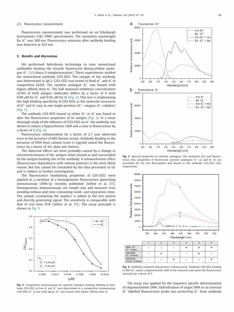
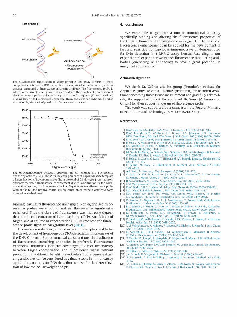
The antibody G93-ED2 bound to either tC1 or tC was found toalter the fluorescence properties of its antigen (Fig. 3). In a morethorough study of the influence of G93-ED2 on tC1 the antibody wasshown to induce a hypsochromic shift and a raise in fluorescence bya factor of 5 (Fig. 4).
Fluorescence enhancement by a factor of 2.7 was observedeven in the presence of 80% bovine serum. Antibody binding in thepresence of DNA from salmon testis [1 mg/ml] raised the fluores-cence by a factor of 4.6 (data not shown).
The observed effects are most probably caused by a change inmicroenvironment of the antigen when bound to and surroundedby the antigen binding site of the antibody. A solvatochromic effect(fluorescence dependence with solvent polarity) is the most likelyreason. But this cannot be concluded by the data presented so farand is subject to further investigation.
The fluorescence modulating properties of G93-ED2 wereapplied in a variation of a homogeneous fluorescence quenchingimmunoassay (DNA-Q) recently published (Sellrie et al. [7]).Homogeneous immunoassays are simple mix and measure testsavoiding tedious and time consuming wash- and separation-steps.The sample (containing the analyte) is added to the test systemand directly generating signal. The sensitivity is comparable withthat of real time PCR (Sellrie et al. [7]). The assay principle isshown in Fig. 5.
The assay was applied for the sequence specific determinationof oligonucleotide DNA. Hybridisation of target DNA to an internaltC1 labelled fluorescence probe was protecting tC1 from antibody
0.0001 0.0010 0.0100 0.1000 1.0000 10.00000.0
0.1
0.2
0.3
0.4
0.5
OD450nm
[µM]
Fig. 2. Competitive immunoassay for cytosine analogue binding. Binding of anti-body G93-ED2 to free tC and tC1 was determined in a competitive immunoassaywith BSA-tC1 at the solid phase. tC1 was bound with higher affinity then tC.
Fig. 3. Spectral properties of cytosine analogues. The excitation (Ex) and fluores-cence (Em) properties of fluorescent cytosine analogues tC1 (a) and tC (b) arepresented for the free fluorophore and bound to the antibody G93-ED2 (Ab),respectively.
Fig. 4. Antibody mediated fluorescence enhancement. Antibody G93-ED2 bindingto BSA-tC1 causes a hypsochromic shift of the emission and raises the fluorescenceintensity by a factor of 5.
F. Sellrie et al. / Talanta 124 (2014) 67–70 69
binding leaving its fluorescence unchanged. Non-hybridised fluor-escence probes were bound and its fluorescence significantlyenhanced. Thus the observed fluorescence was indirectly depen-dent on the concentration of hybridised target DNA. An addition oftarget DNA at equimolar concentration (0.1 mM) reduced the fluore-scence probe signal to background level (Fig. 6).
Fluorescence enhancing antibodies are in principle suitable forthe development of homogeneous DNA-detecting immunoassays ofthe DNA-Q format. But for practical considerations the applicationof fluorescence quenching antibodies is preferred. Fluorescenceenhancing antibodies lack the advantage of direct dependencybetween target concentration and fluorescence signal withoutproviding an additional benefit. Nevertheless fluorescence enhan-cing antibodies can be considered as valuable tools in immunoassayapplications not only for DNA detection but also for the determina-tion of low molecular weight analyts.
4. Conclusion
We were able to generate a murine monoclonal antibodyspecifically binding and altering the fluorescence properties ofthe tricyclic fluorescent deoxycytidine analogue tC1. The observedfluorescence enhancement can be applied for the development offast and sensitive homogeneous immunoassays as demonstratedfor DNA detection in a DNA-Q assay format. According to ourexperimental experience we expect fluorescence modulating anti-bodies (quenching or enhancing) to have a great potential inanalytical applications.
Acknowledgement
We thank Dr. Geßner and his group (Fraunhofer Institute forApplied Polymer Research - NanoPolyPhotonik) for technical assis-tance concerning fluorescence measurement and gratefully acknowl-edge the support of F. Ebert. We also thank Dr. Graser (AJ InnuscreenGmbH) for their support in design of fluorescence probe.
This work was supported by a grant from the Federal Ministryof Economics and Technology (ZIM KF2058407SK9).
References
[1] D.W. Ballard, R.M. Bates, E.W. Voss , J. Immunol. 135 (1985) 433–439.[2] D.W. Bedzyk, K.M. Weidner, L.K. Denzin, L.S. Johnson, K.D. Hardman,
M.W. Pantoliano, E.D. Asel, E.W. Voss , J. Biol. Chem. 265 (1990) 18615–18620.[3] E.W. Voss , J.C. Croney, D.M. Jameson, J. Protein Chem. 21 (2002) 231–241.[4] F. Sellrie, A. Warsinke, B. Micheel, Anal. Bioanal. Chem. 386 (2006) 206–210.[5] J.A. Schenk, F. Sellrie, V. Böttger, A. Menning, W.F. Stöcklein, B. Micheel,
Biochimie 89 (2007) 1304–1311.[6] M. Stech, H. Merk, J.A. Schenk, W.F. Stöcklein, D.A. Wüstenhagen, B. Micheel,
C. Duschl, F.F. Bier, S. Kubick, J. Biotechnol 164 (2012) 220–231.[7] F. Sellrie, E. Graser, C. Lenz, T. Hillebrand, J.A. Schenk, Biosens. Bioelectron 42
(2013) 512–515.[8] F. Sellrie, M. Beck, N. Hildebrandt, B. Micheel, Anal. Methods 2 (2010)
1298–1301.[9] A.P. Wei, J.N. Herron, J. Mol. Recognit 15 (2002) 311–320.[10] S. Inal, J.D. Kölsch, F. Sellrie, J.A. Schenk, E. Wischerhoff, A. Laschewsky,
D. Neher, J. Mater. Chem. B 1 (2013) 6373–6381.[11] R.W. Sinkeldam, N.J. Greco, Y. Tor, Chem. Rev. 110 (2010) 2579–2619.[12] L.M. Wilhelmsson, Q. Rev. Biophys 43 (2010) 159–183.[13] D.W. Dodd, R.H.E. Hudson, Mini-Rev. Org. Chem. 6 (2009) (2009) 378–391.[14] D.C. Ward, E. Reich, L. Stryer, J. Biol. Chem. 244 (1969) 1228–1237.[15] D.A. Berry, K.Y. Jung, D.S. Wise, A.D. Sercel, W.H. Pearson, H. Mackie,
J.B. Randolph, R.L. Somers, Tetrahedron Lett. 45 (2004) 2457–2461.[16] P. Sandin, K. Börjesson, H. Li, J. Mårtensson, T. Brown, L.M. Wilhelmsson,
B. Albinsson, Nucleic Acids Res. 36 (2008) 157–167.[17] K.C. Engman, P. Sandin, S. Osborne, T. Brown, M. Billeter, P. Lincoln, B. Nordén,
B. Albinsson, L.M. Wilhelmsson, Nucleic Acids Res. 32 (2004) 5057–5095.[18] K. Börjesson, S. Preus, A.H. El-Sagheer, T. Brown, B. Albinsson, L.
M. Wilhelmsson, J. Am. Chem. Soc. 131 (2009) 4288–4293.[19] P. Sandin, L.M. Wilhelmsson, P. Lincoln, V.E.C. Powers, T. Brown, B. Albinsson,
Nucleic Acids Res. 33 (2005) 5019–5025.[20] L.M. Wilhelmsson, A. Holmén, P. Lincoln, P.E. Nielsen, B. Nordén, J. Am. Chem.
Soc. 123 (2001) 2434–2435.[21] G. Stengel, J.P. Gill, P. Sandin, L.M. Wilhelmsson, B. Albinsson, B. Nordén,
D. Millar, Biochemistry 46 (2007) 12289–12297.[22] P. Sandin, G. Stengel, T. Ljungdahl, K. Börjesson, B. Macao, L.M. Wilhelmsson,
Nucleic Acids Res. 37 (2009) 3924–3933.[23] G. Stengel, B.W. Purse, L.M. Wilhelmsson, M. Urban, R.D. Kuchta, Biochemistry
48 (2009) 7547–7555.[24] G. Köhler, C. Milstein, Nature 256 (1975) 495–497.[25] J.A. Schenk, F. Matyssek, B. Micheel, In Vivo 18 (2004) 649–652.[26] R. Lindmark, K. Thorén-Tolling, J. Sjöquist, J. Immunol. Methods 62 (1983)
1–13.[27] J.A. Schenk, J. Fettke, C. Lenz, K. Albers, F. Mallwitz, N. Gajovic-Eichelmann,
E. Ehrentreich-Förster, E. Kusch, F. Sellrie, J. Biotechnol. 158 (2012) 34–35.
Fig. 5. Schematic presentation of assay principle. The assay consists of threecomponents: a template DNA molecule (single-stranded or denaturated), a fluor-escence probe and a fluorescence enhancing antibody. The fluorescence probe isadded to the sample and hybridized specifically to the template. Hybridization ofthe fluorescence probe and template protects the fluorophore (F) from antibodybinding leaving its fluorescence unaffected. Fluorophores of non-hybridized probesare bound by the antibody and their fluorescence enhanced.
Fig. 6. Oligonucleotide detection applying the tC1 binding and fluorescenceenhancing antibody G93-ED2. With increasing amount of oligonucleotide templatea larger fraction of fluorescent probe (from the total of 0.1 mM) gets protected fromantibody mediated fluorescence enhancement due to hybridization to the oligo-nucleotide resulting in a fluorescence decline. Negative control (fluorescence probewith antibody) and positive control (fluorescence probe without antibody) wereinserted as dashed lines.
F. Sellrie et al. / Talanta 124 (2014) 67–7070


![Synthesis and Photochemistry of Phenyl Substituted-1,2,4 ...The photochemically generated phenyl-1,3-diaza-5-thiabicyclo[2.1.0]pentenes are the key intermediates in this suggested](https://img.pdfslide.us/doc/110x75/5f938aac0564393e410bb222/synthesis-and-photochemistry-of-phenyl-substituted-124-the-photochemically.jpg)