Embed Size (px)
Citation preview
RSC Advances
REVIEW
Ope
n A
cces
s A
rtic
le. P
ublis
hed
on 1
4 M
ay 2
020.
Dow
nloa
ded
on 3
/19/
2022
12:
08:2
8 PM
. T
his
artic
le is
lice
nsed
und
er a
Cre
ativ
e C
omm
ons
Attr
ibut
ion-
Non
Com
mer
cial
3.0
Unp
orte
d L
icen
ce.
View Article OnlineView Journal | View Issue
Antibiotic resista
aCentre for Trans-Sahara Disease, Vaccine
Babangida University, Lapai, Niger State, NbDepartment of Microbiology, School of Agric
KwaZulu-Natal, Durban 4001, South AfricacFaculty of Pharmaceutical Sciences, Ahmad
NigeriadSchool of Laboratory Medicine and Medica
Durban 4001, South AfricaeMolecular Modeling and Drug Design Res
University of KwaZulu Natal, Westville Cam
Cite this: RSC Adv., 2020, 10, 18451
Received 15th February 2020Accepted 1st May 2020
DOI: 10.1039/d0ra01484b
rsc.li/rsc-advances
This journal is © The Royal Society o
nce: bioinformatics-basedunderstanding as a functional strategy for drugdesign
Umar Ndagi, *a Abubakar A. Falaki,b Maryam Abdullahi,c Monsurat M. Lawald
and Mahmoud E. Soliman e
The use of antibiotics to manage infectious diseases dates back to ancient civilization, but the lack of a clear
distinction between the therapeutic and toxic dose has been a major challenge. This precipitates the notion
that antibiotic resistancewas from time immemorial, principally because of a lack of adequate knowledge of
therapeutic doses and continuous exposure of these bacteria to suboptimal plasma concentration of
antibiotics. With the discovery of penicillin by Alexander Fleming in 1924, a milestone in bacterial
infections' treatment was achieved. This forms the foundation for the modern era of antibiotic drugs.
Antibiotics such as penicillins, cephalosporins, quinolones, tetracycline, macrolides, sulphonamides,
aminoglycosides and glycopeptides are the mainstay in managing severe bacterial infections, but
resistant strains of bacteria have emerged and hampered the progress of research in this field. Recently,
new approaches to research involving bacteria resistance to antibiotics have appeared; these involve
combining the molecular understanding of bacteria systems with the knowledge of bioinformatics.
Consequently, many molecules have been developed to curb resistance associated with different
bacterial infections. However, because of increased emphasis on the clinical relevance of antibiotics, the
synergy between in silico study and in vivo study is well cemented and this facilitates the discovery of
potent antibiotics. In this review, we seek to give an overview of earlier reviews and molecular and
structural understanding of bacteria resistance to antibiotics, while focusing on the recent bioinformatics
approach to antibacterial drug discovery.
1. Introduction
The use of antimicrobial entities dates back to ancient civili-zation1 during which a variety of naturally available healingmethods were used in curbing infections.1 Some of thesemethods require the use of moulds and plant extracts withcharacteristics antibiotic properties. This is true for compoundsthat were accidentally discovered (such as penicillin by Alex-ander Fleming in 1924) and even for those that are linked to theknowledge handed down by successive generations; Egyptiansapplied mouldy bread to infected wounds.2
However, until the 20th century, bacterial infections were thenumber one cause of human death in the developed world.2
and Drug Research, Ibrahim Badamasi
igeria. E-mail: [email protected]
ulture and Applied Sciences, University of
u Bello University Zaria, Kaduna State,
l Sciences, University of KwaZulu-Natal,
earch Group, School of Health Sciences,
pus, Durban 4001, South Africa
f Chemistry 2020
This is as a result of dose-related issues, the knowledge of theamount required to clear the organism from the system beinginadequate. Therefore, marked symptoms of bacterial infectionpersist until such time that a signicant amount is reached, andsymptoms begin to disappear. Exposure of bacteria to theinadequate dose of these agents is presumed as the number onecause of bacterial resistance, which could be a contributingfactor to bacterial evolution and perhaps the origin of bacteriaevolution.3
In the 1940s, antibiotics were major lifelines for treatingserious infections,4 this was re-established by the successful useof penicillin in controlling bacterial infections among militarypersonnel during the Second World War.5 Luria and Delbruckin the year 1943, conducted a uctuation test6 and acknowl-edged that bacterial resistance arises from heritable changes inthe bacterial cell (mutation),6 which is independent of factorssuch as co-habitation of the bacterial colony with the virus.Despite the advantages derived from the use of penicillin,resistance to these drugs emerged,7 such that in the 1950s, thegiant strides made in antibiotics development in the priordecade were threatened.8 In response to this, new beta (b)-lac-tam antibiotics were discovered and eventually upheld, therebyre-establishing the initial condence of “magic-use and magic
RSC Adv., 2020, 10, 18451–18468 | 18451
RSC Advances Review
Ope
n A
cces
s A
rtic
le. P
ublis
hed
on 1
4 M
ay 2
020.
Dow
nloa
ded
on 3
/19/
2022
12:
08:2
8 PM
. T
his
artic
le is
lice
nsed
und
er a
Cre
ativ
e C
omm
ons
Attr
ibut
ion-
Non
Com
mer
cial
3.0
Unp
orte
d L
icen
ce.
View Article Online
result” antibiotics are being portrayed.5,8 Within the samedecade, methicillin-resistant Staphylococcus aureus (MRSA) wasrst identied in the United Kingdom in 1962 and the UnitedStates in 1968.4,5 Unfortunately, resistance to most antibioticsemerged in time-course4 to the extent that even the most powerfulantibiotics developed in the early 1970s like vancomycin used inmanaging methicillin resistance in both Staphylococcus aureus (S.aureus) and coagulase-negative staphylococci had its share ofresistance4,5 in the late 1970s.
To understand the key aspect of bacterial survival, Baymet al. (2016) introduced microbial evolution and growth arena(MEGA) – plate,9 typically used to study microbial evolution.Their work suggests that evolution is not always led by the mostresistant mutants; highly resistant mutant can be trappedbehind more sensitive lineages.9 Baym concept of Petri dishoffers an unprecedented insight into the phenomenon of anti-biotic resistance.10
Resistance to antibiotics is eroding the scientic effort inkeeping up the pace of antibiotics' discovery in the 21stcentury.11 In the past (the golden era of antibiotics drugdiscovery), resistance to antibiotics' was not too challenging,11
this was because scientic research was coping with the chal-lenges of antibiotic resistance.11 However, recent events in theantibiotic resistance prole have pushed most pharmaceuticalindustries to rededicate their efforts in developing novel mole-cules used in managing chronic illnesses, this approachprovides more nancial benets that guaranty their survival inmonetary times. Thus, leading to drying up in the antibioticspipeline and leaving behind a few potent molecules that maylimit the choice of antibiotics in curing certain disease condi-tions. The emergence and widespread of antibiotic-resistantpathogens is an emerging challenge in the public healthdomain, this requires global action with a multi-dimensionalapproach that would address the fundamental challengesinherent in curbing drug-resistance and ne-tune the lead wayto minimise drug-resistance.12
The most recent World Economic Forum Global Risksreports have listed antibiotic resistance as one of the greatestthreats that put the health system under new pressure.13 Simi-larly, the United Nations (UN) ad hoc interagency coordinatinggroup on antimicrobial resistance in one of its recent reportwarned that drug-resistant diseases could account for up to 10million deaths each year by 2050 and damage to the economy ascatastrophic as the 2008–2009 global nancial crisis. By 2030, itis likely that antimicrobial resistance could drag up to 24million people into extreme poverty.14 Currently, at least700 000 people die each year due to drug-resistant diseases,including 230 000 people who die from multidrug-resistanttuberculosis.14 United State (U.S.) Centers for Disease, Controland Prevention in its 2019 report, states that more than 2.8million antibiotic-resistant infections occur in the U.S. eachyear,15 and more than 35 000 death result from this menace.15
Also, 223 900 cases of Clostridioides difficile occurred in 2017and at least 12 800 people died. Relative to this, there has beenan upsurge in antibiotic resistance prole and relative lack ofnew antibiotics in development.15
18452 | RSC Adv., 2020, 10, 18451–18468
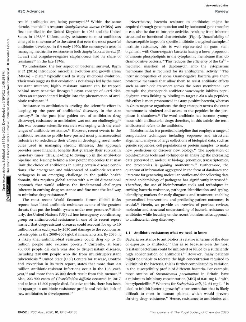
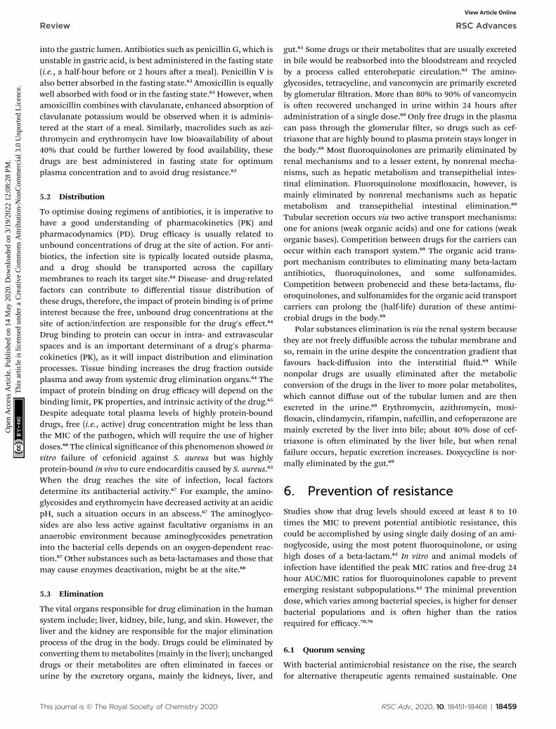
Nevertheless, bacteria resistant to antibiotics might beacquired through gene mutation and by horizontal gene transfer;it can also be due to intrinsic activities resulting from inherentstructural or functional characteristics (Fig. 1). Unavailability ofthe susceptible target of a specic antibiotic is a typical example ofintrinsic resistance, this is well represented in gram stainorganism, with Gram-negative bacteria having a lower proportionof anionic phospholipids in the cytoplasmic membrane than doGram-positive bacteria.16 This reduces the efficiency of the Ca2+ –mediated insertion of daptomycin into the cytoplasmicmembrane that is required for its antibacterial activity.17 Theintrinsic properties of some Gram-negative bacteria give themprotective measures that allow them to resist antibiotic effectssuch as antibiotic transport across the outer membrane. Forexample, the glycopeptide antibiotic vancomycin inhibits pepti-doglycan cross-linking by binding to target D-Ala-D-Ala peptides,this effect is more pronounced in Gram-positive bacteria, whereasin Gram-negative organisms, the drug transport across the outermembrane is hindered and access to the peptides in the peri-plasm is shutdown.18 The word antibiotic has become synony-mous with antibacterial drugs therefore, in this article; the termantibacterial refers to the antibiotic.
Bioinformatics is a practical discipline that employs a range ofcomputation techniques including sequence and structuralalignment, analyses of large collections of biological data, such asgenetic sequences, cell populations or protein samples, to makenew predictions or discover new biology.19 The application ofbioinformatics tools and techniques in analysing the increasingdata generated in molecular biology, genomics, transcriptomics,and proteomics is gaining momentum.20 Furthermore, thequantum of information aggregated in the form of databases andliterature for generating molecular proles and for collecting datarelated epidemiology of pathogens has signicantly increased.21
Therefore, the use of bioinformatics tools and techniques incurbing bacteria resistance, pathogen identication and typing,identifying markers for early diagnosis and treatment, enablingpersonalized interventions and predicting patient outcomes, iscrucial.22 Herein, we provide an overview of previous reviews,molecular and structural understanding of bacteria resistance toantibiotics while focusing on the recent bioinformatics approachto antibacterial drug discovery.
1.1 Antibiotic resistance; what we need to know
Bacteria resistance to antibiotics is relative in terms of the doseof exposure to antibiotic,23 this is so because even the mostresistant bacterium could be inhibited or killed by a sufficientlyhigh concentration of antibiotics.23 However, many patientsmight be unable to tolerate the high concentration required tokill/inhibit the bacteria, this is further complicated by variationin the susceptibility prole of different bacteria. For example,most strains of Streptococcus pneumoniae in Britain havea minimum inhibitory concentration (MIC) of 0.01 mg L�1 withbenzylpenicillin.23 Whereas for Escherichia coli, 32–64 mg L�1 isideal to inhibit bacteria growth;23 a concentration that is likelydifficult to meet in human plasma, which would preventthriving drug-resistance.23 Hence, resistance to antibiotics can
This journal is © The Royal Society of Chemistry 2020

Review RSC Advances
Ope
n A
cces
s A
rtic
le. P
ublis
hed
on 1
4 M
ay 2
020.
Dow
nloa
ded
on 3
/19/
2022
12:
08:2
8 PM
. T
his
artic
le is
lice
nsed
und
er a
Cre
ativ
e C
omm
ons
Attr
ibut
ion-
Non
Com
mer
cial
3.0
Unp
orte
d L
icen
ce.
View Article Online
be better considered as clinical resistance as opposed to drugresistance. Similarly, the rapid spread of the transposon(jumping DNA) drives bacterial resistance. This DNA is a vehiclefor spreading of bacteria resistance and characterise with theautonomous switch of locations in the genome.24
Antibiotics resistance is best understood using two conceptualmodels;25 rst, resistance to antibiotics could be considered asa result of microorganisms (bacteria) interaction with theirimmediate environment.25 The presence of thesemolecules, someof which are natural compounds, within the bacteria milieu exertthe survival pressure that allows the bacteria to adapt to a certainconcentration, hence, co-resident bacteria evolvedmechanisms toovercome these molecules' actions to survive. Therefore, theseorganisms are oen considered as “intrinsically” resistant to oneor more antibiotics.25 However, in clinical settings, the focus ismore on “acquired resistance” in a bacterial population that wasoriginally susceptible to the antibiotics.25 Usually, acquired resis-tance results from mutations in chromosomal genes or perhapsdue to acquired external genetic determinants of resistance,25
which is likely obtained from intrinsically resistant organismspresent in the environment.25
In the second model, antibiotics resistance/susceptibility inclinical practice could be considered as a relative phenomenonwith different strata.25 The establishment of clinical resistanceor susceptibility endpoints (susceptible, intermediate andresistant) depends on the in vitro activity of antibiotics againsta particular bacterial population, combined with some phar-macodynamics parameters.25 Thus, when treating antibiotic-resistant bacteria, susceptibility patterns interpretations mayvary according to the clinical scenario and treatment optionsavailability.25 For instance, the plasma concentration of cefur-oxime achieved in the lungs could be sufficiently high to treatupper respiratory tract infection caused by an organism re-ported as cefuroxime-resistant.25 However, the in vivo suscepti-bility of an organism to a particular antibiotic depends on thedose of bacterial inoculum.25 As noted from research, somecephalosporins (like cefazolin) may fail in the setting of high-inocula (deep-seated infections) caused by cephalosporin-susceptible S. aureus. This situation has been well docu-mented in S. aureus infections with some cephalosporins.26
2. Molecular basis of antibioticsresistance
Bacteria genome exibility allows them to respond to a range ofenvironmental challenges,25 including when antibiotic mole-cules that may distort the strength of their survival are present.Just like other organisms, bacteria share atomic (chemicalstructure) similarities with antibacterial agents and hence easeadaptability to the harmful effect of antibiotics.27 Two majorgenetic strategies dene bacteria adaptability to antibiotics: (1)gene mutation and (2) horizontal transfer of resistance gene.
2.1 Resistance resulting from a gene mutation
Mutation is one of the useful means of survival of bacterialamidst antibiotics threat; it usually results from exposure of
This journal is © The Royal Society of Chemistry 2020
bacteria to the sub-optimal concentration of antibiotics. Oncea resistant mutant emerges,27 the antibiotic eliminates thesusceptible population and the resistant bacterial predomi-nate.27 From a molecular perspective, mutation undermines thenetwork of interaction between the atoms of the drug and atomsof the amino acid from the bacterial enzyme. Consequently,drug-receptor resident-time would be shortened and potency ofthe drug molecule could be affected. In many instances,mutational changes leading to resistance affects cell homeo-stasis27 with an irreversible decrease in drug tness. Severalmechanisms of bacteria resistance are available in the litera-ture, the most common ones include; (1) changes in bacteriadrug target site (2) reduction in drug uptake (3) activation ofefflux mechanism to exclude drug molecule and (4) changes inessential metabolic pathways.
2.2 Horizontal gene transfer (HGT)
Transfer of genetic trait through DNA has been known as themostcommon method of bacteria resistance,25 it involves acquiringforeign DNAmaterial through horizontal gene transfer. It is a well-known fact that bacteria share the same environmental charac-teristic with most pharmaceuticals, thereby developing anintrinsic resistance trait required to facilitate the growth of anti-microbial resistance. This genetic exchange could be associatedwith the widespread resistance to many frequently used antibi-otics.27 Acquisition and transfer of genetic materials by bacterialcould be achieved via three basic mechanisms, this includestransformation (incorporation of available DNA), transduction(microphage mediated) and conjugation (bacterial mating).25
Transformation is the simplest mechanism of horizontal genetransfer with only a few clinically relevant bacterial proceeds withthis method. However, conjugation is an efficient mechanism ofgene transfer that involves contact between two or more cellsdriving by free genetic elements which transfer valuable geneticinformation.25 Plasmid and transposons (jumping DNA) are crit-ical to bacterial in the development and transfer of resistancegenetic materials in the clinical relevant organism. Similarly,integrons have been identied as one of the most effectivemechanisms of antimicrobial resistance gene assemble.27 It playeda major role in the acquisition, expression, and dissemination ofantibiotic resistance genes. Its mechanism is simple and efficientin adding new genes into bacterial chromosomes, along with thenecessary machinery to ensure their expression25,28 (Fig. 1).
It is important to note that bacteria can develop resistance toantibiotics using multiple biochemical pathways, this is true forsome bacterial that gain exposure to different types of antibi-otics. They adopt these pathways to resist the effect of antibi-otics and for continuous survival, this is targeted at someantibiotics perhaps because of their structural peculiarities, forexample, bacteria resistance to uoroquinolone comes withthree different mechanisms.29 These are (1) alterations in thetarget enzymes (DNA gyrase and topoisomerase) (2) change indrug entry and efflux mechanism (3) plasmid-mediated Qnr-protein which protects quinolones targets from inhibition.29
These mechanisms may co-exist in some bacterial and canaugment resistance effects of one mechanism from the others.
RSC Adv., 2020, 10, 18451–18468 | 18453
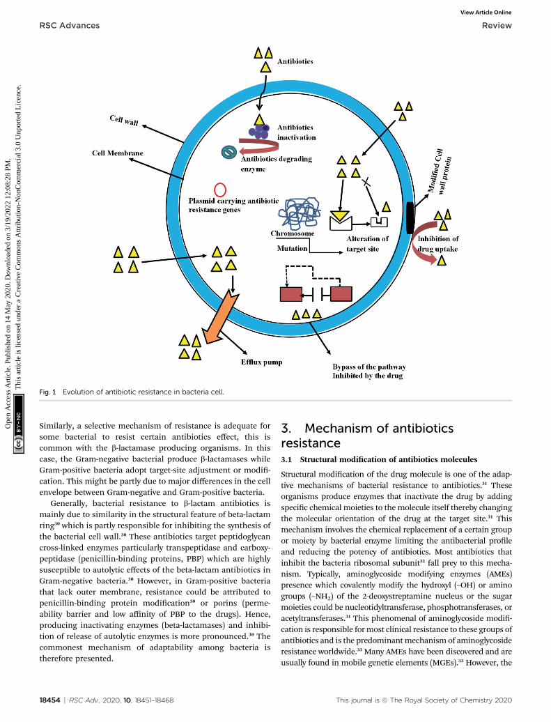
Fig. 1 Evolution of antibiotic resistance in bacteria cell.
RSC Advances Review
Ope
n A
cces
s A
rtic
le. P
ublis
hed
on 1
4 M
ay 2
020.
Dow
nloa
ded
on 3
/19/
2022
12:
08:2
8 PM
. T
his
artic
le is
lice
nsed
und
er a
Cre
ativ
e C
omm
ons
Attr
ibut
ion-
Non
Com
mer
cial
3.0
Unp
orte
d L
icen
ce.
View Article Online
Similarly, a selective mechanism of resistance is adequate forsome bacterial to resist certain antibiotics effect, this iscommon with the b-lactamase producing organisms. In thiscase, the Gram-negative bacterial produce b-lactamases whileGram-positive bacteria adopt target-site adjustment or modi-cation. This might be partly due to major differences in the cellenvelope between Gram-negative and Gram-positive bacteria.
Generally, bacterial resistance to b-lactam antibiotics ismainly due to similarity in the structural feature of beta-lactamring30 which is partly responsible for inhibiting the synthesis ofthe bacterial cell wall.30 These antibiotics target peptidoglycancross-linked enzymes particularly transpeptidase and carboxy-peptidase (penicillin-binding proteins, PBP) which are highlysusceptible to autolytic effects of the beta-lactam antibiotics inGram-negative bacteria.30 However, in Gram-positive bacteriathat lack outer membrane, resistance could be attributed topenicillin-binding protein modication30 or porins (perme-ability barrier and low affinity of PBP to the drugs). Hence,producing inactivating enzymes (beta-lactamases) and inhibi-tion of release of autolytic enzymes is more pronounced.30 Thecommonest mechanism of adaptability among bacteria istherefore presented.
18454 | RSC Adv., 2020, 10, 18451–18468
3. Mechanism of antibioticsresistance3.1 Structural modication of antibiotics molecules
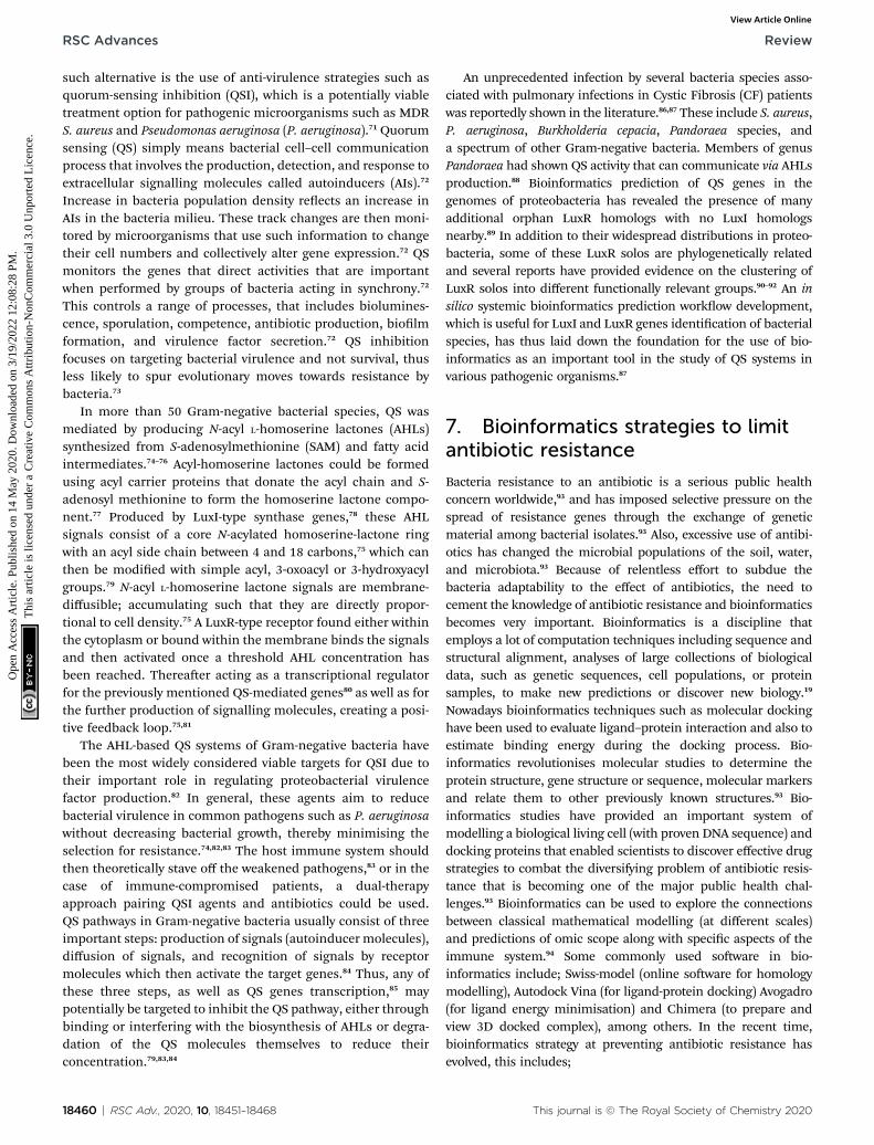
Structural modication of the drug molecule is one of the adap-tive mechanisms of bacterial resistance to antibiotics.31 Theseorganisms produce enzymes that inactivate the drug by addingspecic chemical moieties to the molecule itself thereby changingthe molecular orientation of the drug at the target site.31 Thismechanism involves the chemical replacement of a certain groupor moiety by bacterial enzyme limiting the antibacterial proleand reducing the potency of antibiotics. Most antibiotics thatinhibit the bacteria ribosomal subunit32 fall prey to this mecha-nism. Typically, aminoglycoside modifying enzymes (AMEs)presence which covalently modify the hydroxyl (–OH) or aminogroups (–NH2) of the 2-deoxystreptamine nucleus or the sugarmoieties could be nucleotidyltransferase, phosphotransferases, oracetyltransferases.31 This phenomenal of aminoglycoside modi-cation is responsible formost clinical resistance to these groups ofantibiotics and is the predominantmechanism of aminoglycosideresistance worldwide.33 Many AMEs have been discovered and areusually found in mobile genetic elements (MGEs).33 However, the
This journal is © The Royal Society of Chemistry 2020
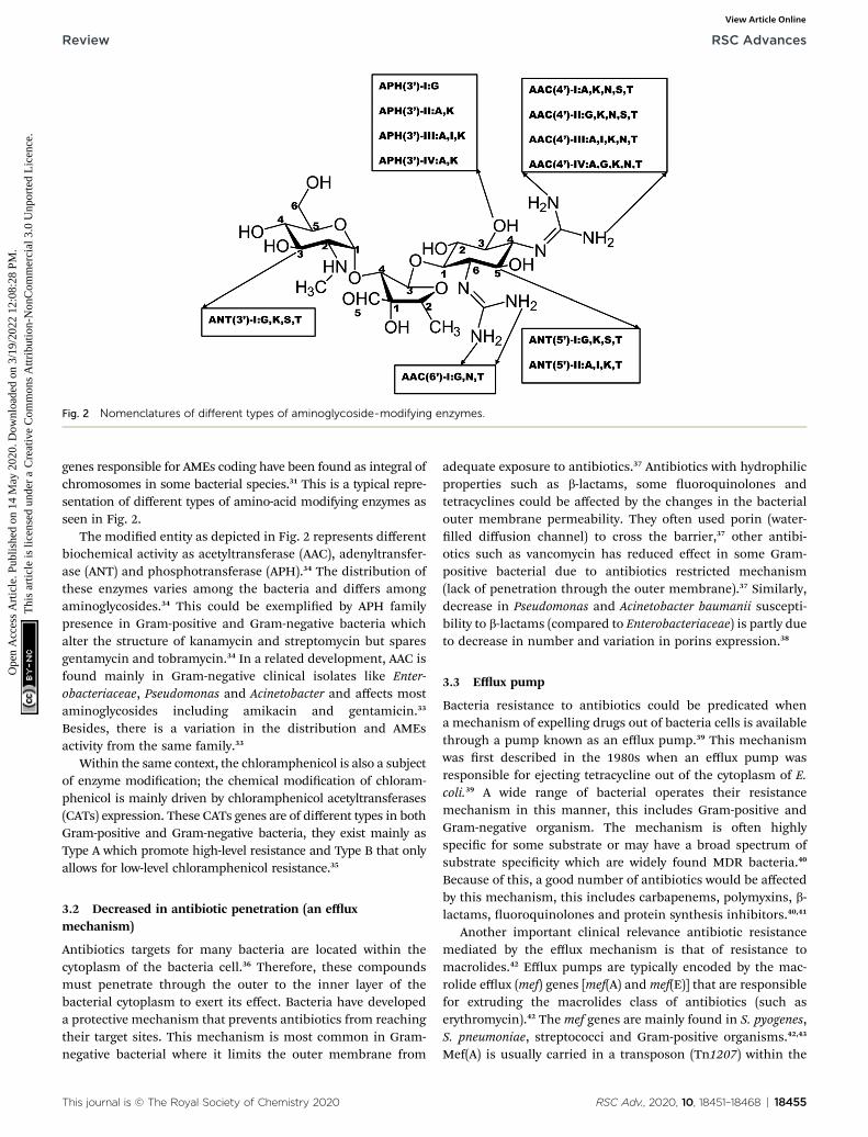
Fig. 2 Nomenclatures of different types of aminoglycoside-modifying enzymes.
Review RSC Advances
Ope
n A
cces
s A
rtic
le. P
ublis
hed
on 1
4 M
ay 2
020.
Dow
nloa
ded
on 3
/19/
2022
12:
08:2
8 PM
. T
his
artic
le is
lice
nsed
und
er a
Cre
ativ
e C
omm
ons
Attr
ibut
ion-
Non
Com
mer
cial
3.0
Unp
orte
d L
icen
ce.
View Article Online
genes responsible for AMEs coding have been found as integral ofchromosomes in some bacterial species.31 This is a typical repre-sentation of different types of amino-acid modifying enzymes asseen in Fig. 2.
The modied entity as depicted in Fig. 2 represents differentbiochemical activity as acetyltransferase (AAC), adenyltransfer-ase (ANT) and phosphotransferase (APH).34 The distribution ofthese enzymes varies among the bacteria and differs amongaminoglycosides.34 This could be exemplied by APH familypresence in Gram-positive and Gram-negative bacteria whichalter the structure of kanamycin and streptomycin but sparesgentamycin and tobramycin.34 In a related development, AAC isfound mainly in Gram-negative clinical isolates like Enter-obacteriaceae, Pseudomonas and Acinetobacter and affects mostaminoglycosides including amikacin and gentamicin.33
Besides, there is a variation in the distribution and AMEsactivity from the same family.33
Within the same context, the chloramphenicol is also a subjectof enzyme modication; the chemical modication of chloram-phenicol is mainly driven by chloramphenicol acetyltransferases(CATs) expression. These CATs genes are of different types in bothGram-positive and Gram-negative bacteria, they exist mainly asType A which promote high-level resistance and Type B that onlyallows for low-level chloramphenicol resistance.35
3.2 Decreased in antibiotic penetration (an effluxmechanism)
Antibiotics targets for many bacteria are located within thecytoplasm of the bacteria cell.36 Therefore, these compoundsmust penetrate through the outer to the inner layer of thebacterial cytoplasm to exert its effect. Bacteria have developeda protective mechanism that prevents antibiotics from reachingtheir target sites. This mechanism is most common in Gram-negative bacterial where it limits the outer membrane from
This journal is © The Royal Society of Chemistry 2020
adequate exposure to antibiotics.37 Antibiotics with hydrophilicproperties such as b-lactams, some uoroquinolones andtetracyclines could be affected by the changes in the bacterialouter membrane permeability. They oen used porin (water-lled diffusion channel) to cross the barrier,37 other antibi-otics such as vancomycin has reduced effect in some Gram-positive bacterial due to antibiotics restricted mechanism(lack of penetration through the outer membrane).37 Similarly,decrease in Pseudomonas and Acinetobacter baumanii suscepti-bility to b-lactams (compared to Enterobacteriaceae) is partly dueto decrease in number and variation in porins expression.38
3.3 Efflux pump
Bacteria resistance to antibiotics could be predicated whena mechanism of expelling drugs out of bacteria cells is availablethrough a pump known as an efflux pump.39 This mechanismwas rst described in the 1980s when an efflux pump wasresponsible for ejecting tetracycline out of the cytoplasm of E.coli.39 A wide range of bacterial operates their resistancemechanism in this manner, this includes Gram-positive andGram-negative organism. The mechanism is oen highlyspecic for some substrate or may have a broad spectrum ofsubstrate specicity which are widely found MDR bacteria.40
Because of this, a good number of antibiotics would be affectedby this mechanism, this includes carbapenems, polymyxins, b-lactams, uoroquinolones and protein synthesis inhibitors.40,41
Another important clinical relevance antibiotic resistancemediated by the efflux mechanism is that of resistance tomacrolides.42 Efflux pumps are typically encoded by the mac-rolide efflux (mef) genes [mef(A) and mef(E)] that are responsiblefor extruding the macrolides class of antibiotics (such aserythromycin).42 The mef genes are mainly found in S. pyogenes,S. pneumoniae, streptococci and Gram-positive organisms.42,43
Mef(A) is usually carried in a transposon (Tn1207) within the
RSC Adv., 2020, 10, 18451–18468 | 18455
RSC Advances Review
Ope
n A
cces
s A
rtic
le. P
ublis
hed
on 1
4 M
ay 2
020.
Dow
nloa
ded
on 3
/19/
2022
12:
08:2
8 PM
. T
his
artic
le is
lice
nsed
und
er a
Cre
ativ
e C
omm
ons
Attr
ibut
ion-
Non
Com
mer
cial
3.0
Unp
orte
d L
icen
ce.
View Article Online
chromosome, Mef(E) is found in the “MEGA-element” (macro-lide efflux genetic assembly), which is a DNA fragment that isnaturally associated with different regions of the bacterialchromosome.42 It is important to note that, macrolide resis-tance caused by this mechanism does not result in cross-resistance to lincosamides and streptogramins (the so-calledMLSB group).42
3.4 Target site interference
Some bacteria can interfere with their target site to negate theeffects of antibiotics. This is usually achieved by preventing theantibiotic from reaching its binding site or modications of thetarget site resulting in a decrease in affinity to the antibioticmolecule.31
3.5 Target site protection
Some of the genetic determinants coding for the proteins thatmediate target protection have been found in the bacterialchromosome, most of which are clinically relevant genes andare involved in target site protection mechanism of resistancewhich is oen carried by MGEs.44 Examples of drugs affected bythis mechanism include tetracycline [Tet(M) and Tet(O)], uo-roquinolones (Qnr) and fusidic acid (FusB and FusC).44
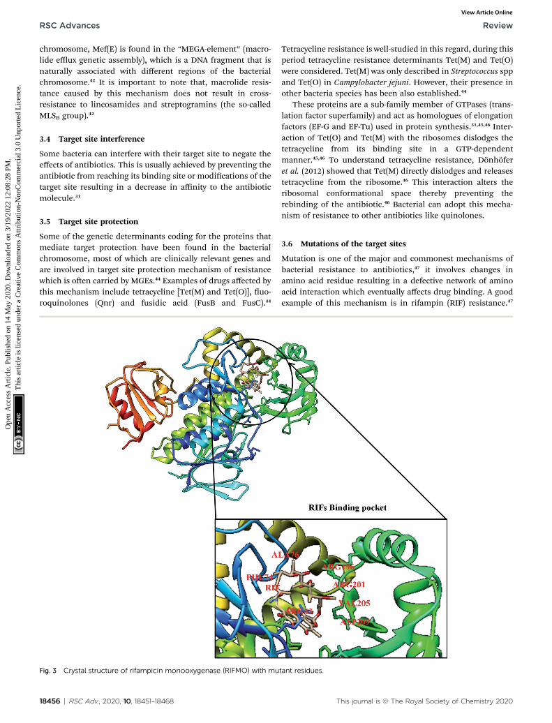
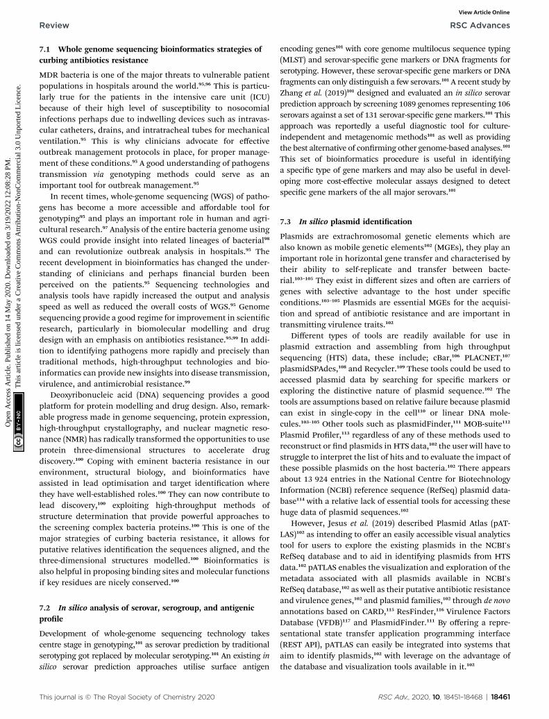
Fig. 3 Crystal structure of rifampicin monooxygenase (RIFMO) with mu
18456 | RSC Adv., 2020, 10, 18451–18468
Tetracycline resistance is well-studied in this regard, during thisperiod tetracycline resistance determinants Tet(M) and Tet(O)were considered. Tet(M) was only described in Streptococcus sppand Tet(O) in Campylobacter jejuni. However, their presence inother bacteria species has been also established.44
These proteins are a sub-family member of GTPases (trans-lation factor superfamily) and act as homologues of elongationfactors (EF-G and EF-Tu) used in protein synthesis.31,45,46 Inter-action of Tet(O) and Tet(M) with the ribosomes dislodges thetetracycline from its binding site in a GTP-dependentmanner.45,46 To understand tetracycline resistance, Donhoferet al. (2012) showed that Tet(M) directly dislodges and releasestetracycline from the ribosome.46 This interaction alters theribosomal conformational space thereby preventing therebinding of the antibiotic.46 Bacterial can adopt this mecha-nism of resistance to other antibiotics like quinolones.
3.6 Mutations of the target sites
Mutation is one of the major and commonest mechanisms ofbacterial resistance to antibiotics,47 it involves changes inamino acid residue resulting in a defective network of aminoacid interaction which eventually affects drug binding. A goodexample of this mechanism is in rifampin (RIF) resistance.47
tant residues.
This journal is © The Royal Society of Chemistry 2020
Review RSC Advances
Ope
n A
cces
s A
rtic
le. P
ublis
hed
on 1
4 M
ay 2
020.
Dow
nloa
ded
on 3
/19/
2022
12:
08:2
8 PM
. T
his
artic
le is
lice
nsed
und
er a
Cre
ativ
e C
omm
ons
Attr
ibut
ion-
Non
Com
mer
cial
3.0
Unp
orte
d L
icen
ce.
View Article Online
Rifampin (a rifamycin) is used as a rst-line antibiotic treat-ment for tuberculosis (TB), and it remains the cornerstone ofcurrent short-term TB treatment.47 Rifamycin-resistant (RIFR)TB,47 results from the RpoB S531L mutation in RNA polymerase(RNAP), this has become a growing problem worldwide.47
Molodtsov et al. (2017) determined the X-ray crystal structuresof the E. coli RNAPs containing the most clinically importantS531L mutation and two other frequently observed RIFR
mutants, RpoB D516V and RpoB H526Y47 have been subjectedto molecular analysis. The structures show that the S531Lmutation impacts subtle if any structural or functional impacton RNAP occurs when RIF is absent,47 this could be illustratedin Fig. 3 with the binding pocket of rifampicin monooxygenase(RIFMO).
However, upon RIF binding, the S531L mutant exhibitsa disordering of the RIFMO binding interface,47 which effec-tively reduces the RIF affinity. In contrast, the H526Y mutationreshapes the RIFMO binding pocket, generating signicantsteric conicts that essentially prevent any RIF binding.47 Whilethe D516V mutant does not exhibit any such gross structuralchanges, certainly the electrostatic surface of the RIFMObinding pocket is dramatically changed, likely resulting in thedecreased affinity for RIFs.47 In a related development, conver-sion of RIF to oxidative products causes a decrease in thepotency of RIFMO.48 Further decomposition of RIF wasobserved in bacterial producing RIFMO and contributes toRIFMO-mediated drug resistance.48
3.7 Enzyme alteration of target sites
Alteration in the target sites of antibiotics is one of the com-monest mechanism of bacterial resistance49 attracting a lot ofsignicance, even the clinical strains showing resistance couldbe found in every class of antibiotic irrespective of the mecha-nism of action.49 Alteration at the target site oen results fromcontinuous mutation of a bacterial gene on the chromosomeand selection when the antibiotics are present.49 For instance,mutations in RNA polymerase and DNA gyrase of susceptiblebacterial result in rifamycins and quinolones resistance,respectively.49 Similarly, acquisition of resistance may involvethe transfer of resistance genes from other organisms throughthe genetic exchange (conjugation, transduction, or trans-formation). Examples of these mechanisms include acquisitionof the mecA genes encoding methicillin resistance in S. aureusand the various van genes in enterococci encoding resistance toglycopeptides.49
In a related development, ribosomal methylation of someantibiotics is catalysed by an enzyme encoded by the erm genes,this includes methylation of erythromycin ribosomes whichresults in macrolide resistance.50 These enzymes are capable ofmono- or demethylation of an adenine residue in positionA2058 of the domain V of the 23rRNA of the 50S ribosomalsubunit.50 Changes resulting from this biochemical process aredirectly responsible for the distortion and re-orientation of thebinding site that affects the drug tness. All the antibiotics withoverlapping binding sites tend to cross-resistance, this is true
This journal is © The Royal Society of Chemistry 2020
with macrolides, lincosamide and streptogramin B with over-lapping binding sites in the 23S rRNA.50
Enzymatic alteration of the target is also involved in Cfr-mediated linezolid resistance.51,52 The plasmid-borne determi-nant gene (cfr gene) was initially described in the year 2000 froma bovine isolate of Staphylococcus sciuri,51 it was rst reported ina human in 2005 in an S. aureus isolated from a patient inColombia.51 Since then, it has been found in several species ofhuman pathogens, including S. aureus, E. faecalis, E. faeciumand some Gram-negative bacteria.51 This gene encodes the Cfrenzyme, which is a member of the S-adenosyl-L-methionine(SAM) methylase family that also confers resistance to pheni-cols, lincosamides, pleuromutilins and streptogramin A.51 Also,cfr has been found to enhance the activities of the variousmobile genetic elements (MGEs) suggesting the relative capacityof transmission of linezolid resistance soon.51
3.8 Target site replacement or circumvention
In applying this strategy, bacterial evolve new targets withsimilar biochemical functions like the original target but arenot susceptible to inhibition of antibiotics.25 This includesmethicillin resistance in S. aureus due to the acquired exoge-nous penicillin-binding protein (PBP) and vancomycin resis-tance in enterococci through modications of thepeptidoglycan structure mediated by the van gene clusters.25
The microorganisms can also avoid antimicrobial action by“circumventing” the susceptible metabolic pathway resulting inan overproduced antibiotic target.25,53 A relevant example of thismechanism includes resistance to trimethoprim–sulfamethox-azole (TMP–SMX) by a “clever” bypass strategy from dihy-dropteroic acid synthase (DHPS) overproduction. DHPS formsdihydrofolate from para-aminobenzoic acid (inhibited by SMX),and dihydrofolate reductase (DHFR) through mutations in thepromoter region of the DNA encoding these enzymes.53,54 Thesemutations result in excessive production of enzymes, thus,“overwhelming” the ability of TMP–SMX to inhibit the folateproduction thereby opening the door for bacterial survival.53,54
4. Pharmacodynamics drugresistance
Pharmacodynamics (PD) of the antibiotic is essentially a matterof the impact of antibiotics on the targeted pathogen.55 It isa complex relationship and very much affiliated with the path-ogen susceptibility to a given antibiotic, it relates the ability ofthe antibiotic to reach the targeted tissue, achieving optimumplasma concentrations at the target site, and patient or hostfactors.55 Patients' underlying comorbidities, immune functionstatus, renal and liver function status, and concomitantadministration of drugs have a major impact on antibiotic PD—in part by affecting pharmacokinetics (PK) and in part byincreasing susceptibility to colonization and bacterial infec-tion,55 decreasing the ability to ght the bacterial infection.55
Toxicity is also an important issue. Therefore, it is importantto balance the need to administer a high enough antibiotic doseto eradicate the pathogen keeping in mind patient safety and
RSC Adv., 2020, 10, 18451–18468 | 18457
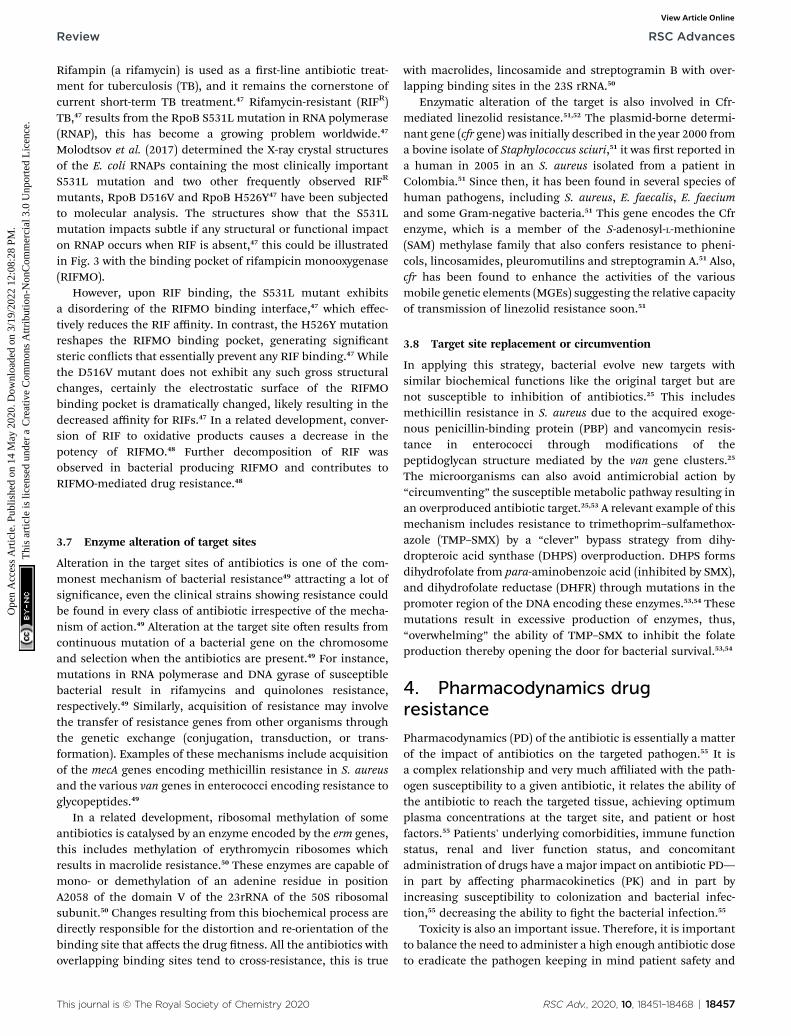
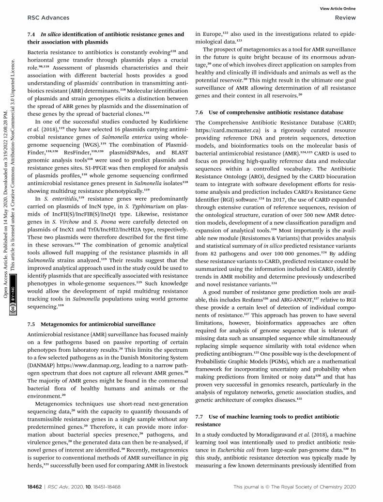
Fig. 4 Important pharmacodynamics parameters describing the effi-cacy of different antibiotics. AUC, area under the concentration–timecurve; Cmax, maximum concentration; Cmin, minimum concentration;MIC, minimum inhibitory concentration; PAE, postantibiotic effect; T,time.
RSC Advances Review
Ope
n A
cces
s A
rtic
le. P
ublis
hed
on 1
4 M
ay 2
020.
Dow
nloa
ded
on 3
/19/
2022
12:
08:2
8 PM
. T
his
artic
le is
lice
nsed
und
er a
Cre
ativ
e C
omm
ons
Attr
ibut
ion-
Non
Com
mer
cial
3.0
Unp
orte
d L
icen
ce.
View Article Online
the ability to tolerate the drug. Because antibiotic PD dependson these various interconnected factors, delivering an effectivedose is more complex than simply choosing a dose that iseffective in well-controlled clinical trials.55 An antibiotic's abilityto resolve an infection depends on a critical drug concentrationbeing reached or exceeded at the infection site and exposure ofthe bacterial to this concentration for a time long enough fororganism eradication to occur from the infection site. There-fore, a decrease or lack of antibiotic minimum inhibitoryconcentration (MIC) required to eradicate bacterial can causebacterial resistance. MIC, dened as the lowest concentration ofan antimicrobial agent that inhibits the growth of the wild typepopulation, assuming nomutations, by 99% (ref. 56 and 57) hasbeen used extensively to classify bacteria as resistant to anantibiotic.57 With an increase in antibiotic concentrations, therst steep decline in colony numbers, representing an approx-imately 1% recovery, corresponds to the MIC.57 Aer exposingcells to antibiotics at MIC levels, there will oen still existresistant mutants population due to spontaneous mutations,usually called single-step resistant mutants.57 As concentrationsincrease beyond the MIC, these single-step mutants will remainuntil a concentration that reduces colony recovery to 0% isachieved.57 Above this concentration, no single-step mutantscan exist. This concentration is the second metric of resistance,the mutant prevention concentration (MPC),57 which could bedened as an interesting tool used in minimising resistanceemergence by modifying antibiotic drug concentrations.55
Based on the experimental result, 3 PD parameters arecommonly used to predict antibiotic efficacy (1) the ratio ofmaximum serum concentration to the minimum inhibitoryconcentration (Cmax/MIC); (2) the ratio of the area under theplasma concentration versus time curve to the minimuminhibitory concentration (AUC/MIC) and (3) the dosing timeinterval that plasma concentrations exceed the MIC (T$MIC).55
The Cmax/MIC ratio showed to predict aminoglycoside efficacy,
18458 | RSC Adv., 2020, 10, 18451–18468
AUC/MIC best describes uoroquinolone, glycopeptide, andketolide efficacy, and T$MIC best describes the efficacy of b-lactams and macrolides as presented in Fig. 4.
Generally, PK and PD (PK/PD) parameters are used to predictantibiotic efficacy with an increasing interest in the use of theseparameters to minimise resistance development.55 Bacterialresistance to uoroquinolone is well described by the ‘‘mutantselection window’’, a developed hypothesis to describe howdrug exposures below themutant prevention concentrationmaycreate conditions for resistant bacterial strains selection.55 TheAUC/MIC ratio has also been used to describe uoroquinolonedrug exposures associated with either increased or decreasedrisk of resistance emergence.
5. Pharmacokinetics drug resistance
The efficiency of antimicrobial treatment could be determinedby both pharmacokinetics and pharmacodynamics.58 Pharma-cokinetics is the study of the bodily absorption, distribution,metabolism and excretion of drugs.59 Antibiotic resistance isrelatively associated with the various mechanisms of drugabsorption, distribution (transport) and elimination (metabo-lism and excretion). Antibiotic PK is oen considered in termsof body's effect on the drug, but physicochemical properties ofthese drugs must also be considered to predict its disposition.60
Most importantly is the relative solubility of the antimicrobial,which can impact signicantly on its volume of distribution,and thus might be vital in selecting agents expected to attainadequate penetration to the site of infection.60 PK parametersthat affect antibiotic efficacy includes;
5.1 Absorption
The ingestion of orally administered antibiotics goes througha normal route like any other oral formulation. However, thiscould change when permeability glycol-protein (p-gp), alsoknown as multidrug-resistant (MDR) protein, found in thegastrointestinal tract (GIT)61 and small intestines (primary sitefor epithelial absorption), is present.61 Co-administration ofantibiotics and anticancer drugs could lead to over-expressionof p-gp thereby decreasing some of these agents' bioavail-ability including azithromycin61 hence, suboptimal plasmaconcentration. Therefore, antibiotics concentration withinbacteria milieu might be quite low, hence encourages bacterialresistance. Genetic polymorphism and change in the bodyphysiological state also play an important role in expressing p-gp in the GIT.62 Thus, causing erratic absorption of some anti-biotics including actinomycin D, erythromycin, gramicidin D,rifampin, salinomycin, sparoxacin and valinomycin.62
Generally, drugs are better absorbed in the small intestine(presence of larger surface area) than in the stomach. Therefore,gastric (stomach) emptying plays a major role in plasma drugconcentrations,63 while eating stimulates gastric acid secretioninto the gastric lumen. Food, especially fatty food, delays gastricemptying, slows and lowers peak plasma concentration, andmay affect the drug's bioavailability and efficacy.63 Eating isa physiological process that stimulates gastric acid secretion
This journal is © The Royal Society of Chemistry 2020
Review RSC Advances
Ope
n A
cces
s A
rtic
le. P
ublis
hed
on 1
4 M
ay 2
020.
Dow
nloa
ded
on 3
/19/
2022
12:
08:2
8 PM
. T
his
artic
le is
lice
nsed
und
er a
Cre
ativ
e C
omm
ons
Attr
ibut
ion-
Non
Com
mer
cial
3.0
Unp
orte
d L
icen
ce.
View Article Online
into the gastric lumen. Antibiotics such as penicillin G, which isunstable in gastric acid, is best administered in the fasting state(i.e., a half-hour before or 2 hours aer a meal). Penicillin V isalso better absorbed in the fasting state.63 Amoxicillin is equallywell absorbed with food or in the fasting state.63 However, whenamoxicillin combines with clavulanate, enhanced absorption ofclavulanate potassium would be observed when it is adminis-tered at the start of a meal. Similarly, macrolides such as azi-thromycin and erythromycin have low bioavailability of about40% that could be further lowered by food availability, thesedrugs are best administered in fasting state for optimumplasma concentration and to avoid drug resistance.63
5.2 Distribution
To optimise dosing regimens of antibiotics, it is imperative tohave a good understanding of pharmacokinetics (PK) andpharmacodynamics (PD). Drug efficacy is usually related tounbound concentrations of drug at the site of action. For anti-biotics, the infection site is typically located outside plasma,and a drug should be transported across the capillarymembranes to reach its target site.64 Disease- and drug-relatedfactors can contribute to differential tissue distribution ofthese drugs, therefore, the impact of protein binding is of primeinterest because the free, unbound drug concentrations at thesite of action/infection are responsible for the drug's effect.64
Drug binding to protein can occur in intra- and extravascularspaces and is an important determinant of a drug's pharma-cokinetics (PK), as it will impact distribution and eliminationprocesses. Tissue binding increases the drug fraction outsideplasma and away from systemic drug elimination organs.64 Theimpact of protein binding on drug efficacy will depend on thebinding limit, PK properties, and intrinsic activity of the drug.65
Despite adequate total plasma levels of highly protein-bounddrugs, free (i.e., active) drug concentration might be less thanthe MIC of the pathogen, which will require the use of higherdoses.66 The clinical signicance of this phenomenon showed invitro failure of cefonicid against S. aureus but was highlyprotein-bound in vivo to cure endocarditis caused by S. aureus.63
When the drug reaches the site of infection, local factorsdetermine its antibacterial activity.67 For example, the amino-glycosides and erythromycin have decreased activity at an acidicpH, such a situation occurs in an abscess.67 The aminoglyco-sides are also less active against facultative organisms in ananaerobic environment because aminoglycosides penetrationinto the bacterial cells depends on an oxygen-dependent reac-tion.67 Other substances such as beta-lactamases and those thatmay cause enzymes deactivation, might be at the site.68
5.3 Elimination
The vital organs responsible for drug elimination in the humansystem include; liver, kidney, bile, lung, and skin. However, theliver and the kidney are responsible for the major eliminationprocess of the drug in the body. Drugs could be eliminated byconverting them to metabolites (mainly in the liver); unchangeddrugs or their metabolites are oen eliminated in faeces orurine by the excretory organs, mainly the kidneys, liver, and
This journal is © The Royal Society of Chemistry 2020
gut.63 Some drugs or their metabolites that are usually excretedin bile would be reabsorbed into the bloodstream and recycledby a process called enterohepatic circulation.63 The amino-glycosides, tetracycline, and vancomycin are primarily excretedby glomerular ltration. More than 80% to 90% of vancomycinis oen recovered unchanged in urine within 24 hours aeradministration of a single dose.69 Only free drugs in the plasmacan pass through the glomerular lter, so drugs such as cef-triaxone that are highly bound to plasma protein stays longer inthe body.69 Most uoroquinolones are primarily eliminated byrenal mechanisms and to a lesser extent, by nonrenal mecha-nisms, such as hepatic metabolism and transepithelial intes-tinal elimination. Fluoroquinolone moxioxacin, however, ismainly eliminated by nonrenal mechanisms such as hepaticmetabolism and transepithelial intestinal elimination.69
Tubular secretion occurs via two active transport mechanisms:one for anions (weak organic acids) and one for cations (weakorganic bases). Competition between drugs for the carriers canoccur within each transport system.69 The organic acid trans-port mechanism contributes to eliminating many beta-lactamantibiotics, uoroquinolones, and some sulfonamides.Competition between probenecid and these beta-lactams, u-oroquinolones, and sulfonamides for the organic acid transportcarriers can prolong the (half-life) duration of these antimi-crobial drugs in the body.69
Polar substances elimination is via the renal system becausethey are not freely diffusible across the tubular membrane andso, remain in the urine despite the concentration gradient thatfavours back-diffusion into the interstitial uid.69 Whilenonpolar drugs are usually eliminated aer the metabolicconversion of the drugs in the liver to more polar metabolites,which cannot diffuse out of the tubular lumen and are thenexcreted in the urine.69 Erythromycin, azithromycin, moxi-oxacin, clindamycin, rifampin, nafcillin, and cefoperazone aremainly excreted by the liver into bile; about 40% dose of cef-triaxone is oen eliminated by the liver bile, but when renalfailure occurs, hepatic excretion increases. Doxycycline is nor-mally eliminated by the gut.69
6. Prevention of resistance
Studies show that drug levels should exceed at least 8 to 10times the MIC to prevent potential antibiotic resistance, thiscould be accomplished by using single daily dosing of an ami-noglycoside, using the most potent uoroquinolone, or usinghigh doses of a beta-lactam.63 In vitro and animal models ofinfection have identied the peak MIC ratios and free-drug 24hour AUC/MIC ratios for uoroquinolones capable to preventemerging resistant subpopulations.63 The minimal preventiondose, which varies among bacterial species, is higher for denserbacterial populations and is oen higher than the ratiosrequired for efficacy.70,70
6.1 Quorum sensing
With bacterial antimicrobial resistance on the rise, the searchfor alternative therapeutic agents remained sustainable. One
RSC Adv., 2020, 10, 18451–18468 | 18459
RSC Advances Review
Ope
n A
cces
s A
rtic
le. P
ublis
hed
on 1
4 M
ay 2
020.
Dow
nloa
ded
on 3
/19/
2022
12:
08:2
8 PM
. T
his
artic
le is
lice
nsed
und
er a
Cre
ativ
e C
omm
ons
Attr
ibut
ion-
Non
Com
mer
cial
3.0
Unp
orte
d L
icen
ce.
View Article Online
such alternative is the use of anti-virulence strategies such asquorum-sensing inhibition (QSI), which is a potentially viabletreatment option for pathogenic microorganisms such as MDRS. aureus and Pseudomonas aeruginosa (P. aeruginosa).71 Quorumsensing (QS) simply means bacterial cell–cell communicationprocess that involves the production, detection, and response toextracellular signalling molecules called autoinducers (AIs).72
Increase in bacteria population density reects an increase inAIs in the bacteria milieu. These track changes are then moni-tored by microorganisms that use such information to changetheir cell numbers and collectively alter gene expression.72 QSmonitors the genes that direct activities that are importantwhen performed by groups of bacteria acting in synchrony.72
This controls a range of processes, that includes biolumines-cence, sporulation, competence, antibiotic production, biolmformation, and virulence factor secretion.72 QS inhibitionfocuses on targeting bacterial virulence and not survival, thusless likely to spur evolutionary moves towards resistance bybacteria.73
In more than 50 Gram-negative bacterial species, QS wasmediated by producing N-acyl L-homoserine lactones (AHLs)synthesized from S-adenosylmethionine (SAM) and fatty acidintermediates.74–76 Acyl-homoserine lactones could be formedusing acyl carrier proteins that donate the acyl chain and S-adenosyl methionine to form the homoserine lactone compo-nent.77 Produced by LuxI-type synthase genes,78 these AHLsignals consist of a core N-acylated homoserine-lactone ringwith an acyl side chain between 4 and 18 carbons,75 which canthen be modied with simple acyl, 3-oxoacyl or 3-hydroxyacylgroups.79 N-acyl L-homoserine lactone signals are membrane-diffusible; accumulating such that they are directly propor-tional to cell density.75 A LuxR-type receptor found either withinthe cytoplasm or bound within the membrane binds the signalsand then activated once a threshold AHL concentration hasbeen reached. Thereaer acting as a transcriptional regulatorfor the previously mentioned QS-mediated genes80 as well as forthe further production of signalling molecules, creating a posi-tive feedback loop.75,81
The AHL-based QS systems of Gram-negative bacteria havebeen the most widely considered viable targets for QSI due totheir important role in regulating proteobacterial virulencefactor production.82 In general, these agents aim to reducebacterial virulence in common pathogens such as P. aeruginosawithout decreasing bacterial growth, thereby minimising theselection for resistance.74,82,83 The host immune system shouldthen theoretically stave off the weakened pathogens,83 or in thecase of immune-compromised patients, a dual-therapyapproach pairing QSI agents and antibiotics could be used.QS pathways in Gram-negative bacteria usually consist of threeimportant steps: production of signals (autoinducer molecules),diffusion of signals, and recognition of signals by receptormolecules which then activate the target genes.84 Thus, any ofthese three steps, as well as QS genes transcription,85 maypotentially be targeted to inhibit the QS pathway, either throughbinding or interfering with the biosynthesis of AHLs or degra-dation of the QS molecules themselves to reduce theirconcentration.79,83,84
18460 | RSC Adv., 2020, 10, 18451–18468
An unprecedented infection by several bacteria species asso-ciated with pulmonary infections in Cystic Fibrosis (CF) patientswas reportedly shown in the literature.86,87 These include S. aureus,P. aeruginosa, Burkholderia cepacia, Pandoraea species, anda spectrum of other Gram-negative bacteria. Members of genusPandoraea had shown QS activity that can communicate via AHLsproduction.88 Bioinformatics prediction of QS genes in thegenomes of proteobacteria has revealed the presence of manyadditional orphan LuxR homologs with no LuxI homologsnearby.89 In addition to their widespread distributions in proteo-bacteria, some of these LuxR solos are phylogenetically relatedand several reports have provided evidence on the clustering ofLuxR solos into different functionally relevant groups.90–92 An insilico systemic bioinformatics prediction workow development,which is useful for LuxI and LuxR genes identication of bacterialspecies, has thus laid down the foundation for the use of bio-informatics as an important tool in the study of QS systems invarious pathogenic organisms.87
7. Bioinformatics strategies to limitantibiotic resistance
Bacteria resistance to an antibiotic is a serious public healthconcern worldwide,93 and has imposed selective pressure on thespread of resistance genes through the exchange of geneticmaterial among bacterial isolates.93 Also, excessive use of antibi-otics has changed the microbial populations of the soil, water,and microbiota.93 Because of relentless effort to subdue thebacteria adaptability to the effect of antibiotics, the need tocement the knowledge of antibiotic resistance and bioinformaticsbecomes very important. Bioinformatics is a discipline thatemploys a lot of computation techniques including sequence andstructural alignment, analyses of large collections of biologicaldata, such as genetic sequences, cell populations, or proteinsamples, to make new predictions or discover new biology.19
Nowadays bioinformatics techniques such as molecular dockinghave been used to evaluate ligand–protein interaction and also toestimate binding energy during the docking process. Bio-informatics revolutionises molecular studies to determine theprotein structure, gene structure or sequence, molecular markersand relate them to other previously known structures.93 Bio-informatics studies have provided an important system ofmodelling a biological living cell (with proven DNA sequence) anddocking proteins that enabled scientists to discover effective drugstrategies to combat the diversifying problem of antibiotic resis-tance that is becoming one of the major public health chal-lenges.93 Bioinformatics can be used to explore the connectionsbetween classical mathematical modelling (at different scales)and predictions of omic scope along with specic aspects of theimmune system.94 Some commonly used soware in bio-informatics include; Swiss-model (online soware for homologymodelling), Autodock Vina (for ligand-protein docking) Avogadro(for ligand energy minimisation) and Chimera (to prepare andview 3D docked complex), among others. In the recent time,bioinformatics strategy at preventing antibiotic resistance hasevolved, this includes;
This journal is © The Royal Society of Chemistry 2020
Review RSC Advances
Ope
n A
cces
s A
rtic
le. P
ublis
hed
on 1
4 M
ay 2
020.
Dow
nloa
ded
on 3
/19/
2022
12:
08:2
8 PM
. T
his
artic
le is
lice
nsed
und
er a
Cre
ativ
e C
omm
ons
Attr
ibut
ion-
Non
Com
mer
cial
3.0
Unp
orte
d L
icen
ce.
View Article Online
7.1 Whole genome sequencing bioinformatics strategies ofcurbing antibiotics resistance
MDR bacteria is one of the major threats to vulnerable patientpopulations in hospitals around the world.95,96 This is particu-larly true for the patients in the intensive care unit (ICU)because of their high level of susceptibility to nosocomialinfections perhaps due to indwelling devices such as intravas-cular catheters, drains, and intratracheal tubes for mechanicalventilation.95 This is why clinicians advocate for effectiveoutbreak management protocols in place, for proper manage-ment of these conditions.95 A good understanding of pathogenstransmission via genotyping methods could serve as animportant tool for outbreak management.95
In recent times, whole-genome sequencing (WGS) of patho-gens has become a more accessible and affordable tool forgenotyping95 and plays an important role in human and agri-cultural research.97 Analysis of the entire bacteria genome usingWGS could provide insight into related lineages of bacterial98
and can revolutionize outbreak analysis in hospitals.95 Therecent development in bioinformatics has changed the under-standing of clinicians and perhaps nancial burden beenperceived on the patients.95 Sequencing technologies andanalysis tools have rapidly increased the output and analysisspeed as well as reduced the overall costs of WGS.95 Genomesequencing provide a good regime for improvement in scienticresearch, particularly in biomolecular modelling and drugdesign with an emphasis on antibiotics resistance.95,99 In addi-tion to identifying pathogens more rapidly and precisely thantraditional methods, high-throughput technologies and bio-informatics can provide new insights into disease transmission,virulence, and antimicrobial resistance.99
Deoxyribonucleic acid (DNA) sequencing provides a goodplatform for protein modelling and drug design. Also, remark-able progress made in genome sequencing, protein expression,high-throughput crystallography, and nuclear magnetic reso-nance (NMR) has radically transformed the opportunities to useprotein three-dimensional structures to accelerate drugdiscovery.100 Coping with eminent bacteria resistance in ourenvironment, structural biology, and bioinformatics haveassisted in lead optimisation and target identication wherethey have well-established roles.100 They can now contribute tolead discovery,100 exploiting high-throughput methods ofstructure determination that provide powerful approaches tothe screening complex bacteria proteins.100 This is one of themajor strategies of curbing bacteria resistance, it allows forputative relatives identication the sequences aligned, and thethree-dimensional structures modelled.100 Bioinformatics isalso helpful in proposing binding sites and molecular functionsif key residues are nicely conserved.100
7.2 In silico analysis of serovar, serogroup, and antigenicprole
Development of whole-genome sequencing technology takescentre stage in genotyping,101 as serovar prediction by traditionalserotyping got replaced by molecular serotyping.101 An existing insilico serovar prediction approaches utilise surface antigen
This journal is © The Royal Society of Chemistry 2020
encoding genes101 with core genome multilocus sequence typing(MLST) and serovar-specic gene markers or DNA fragments forserotyping. However, these serovar-specic gene markers or DNAfragments can only distinguish a few serovars.101 A recent study byZhang et al. (2019)101 designed and evaluated an in silico serovarprediction approach by screening 1089 genomes representing 106serovars against a set of 131 serovar-specic gene markers.101 Thisapproach was reportedly a useful diagnostic tool for culture-independent and metagenomic methods101 as well as providingthe best alternative of conrming other genome-based analyses.101
This set of bioinformatics procedure is useful in identifyinga specic type of gene markers and may also be useful in devel-oping more cost-effective molecular assays designed to detectspecic gene markers of the all major serovars.101
7.3 In silico plasmid identication
Plasmids are extrachromosomal genetic elements which arealso known as mobile genetic elements102 (MGEs), they play animportant role in horizontal gene transfer and characterised bytheir ability to self-replicate and transfer between bacte-rial.103–105 They exist in different sizes and oen are carriers ofgenes with selective advantage to the host under specicconditions.103–105 Plasmids are essential MGEs for the acquisi-tion and spread of antibiotic resistance and are important intransmitting virulence traits.102
Different types of tools are readily available for use inplasmid extraction and assembling from high throughputsequencing (HTS) data, these include; cBar,106 PLACNET,107
plasmidSPAdes,108 and Recycler.109 These tools could be used toaccessed plasmid data by searching for specic markers orexploring the distinctive nature of plasmid sequence.102 Thetools are assumptions based on relative failure because plasmidcan exist in single-copy in the cell110 or linear DNA mole-cules.103–105 Other tools such as plasmidFinder,111 MOB-suite112
Plasmid Proler,113 regardless of any of these methods used toreconstruct or nd plasmids in HTS data,102 the user will have tostruggle to interpret the list of hits and to evaluate the impact ofthese possible plasmids on the host bacteria.102 There appearsabout 13 924 entries in the National Centre for BiotechnologyInformation (NCBI) reference sequence (RefSeq) plasmid data-base114 with a relative lack of essential tools for accessing thesehuge data of plasmid sequences.102
However, Jesus et al. (2019) described Plasmid Atlas (pAT-LAS)102 as intending to offer an easily accessible visual analyticstool for users to explore the existing plasmids in the NCBI'sRefSeq database and to aid in identifying plasmids from HTSdata.102 pATLAS enables the visualization and exploration of themetadata associated with all plasmids available in NCBI'sRefSeq database,102 as well as their putative antibiotic resistanceand virulence genes,102 and plasmid families,102 through de novoannotations based on CARD,115 ResFinder,116 Virulence FactorsDatabase (VFDB)117 and PlasmidFinder.111 By offering a repre-sentational state transfer application programming interface(REST API), pATLAS can easily be integrated into systems thataim to identify plasmids,102 with leverage on the advantage ofthe database and visualization tools available in it.102
RSC Adv., 2020, 10, 18451–18468 | 18461
RSC Advances Review
Ope
n A
cces
s A
rtic
le. P
ublis
hed
on 1
4 M
ay 2
020.
Dow
nloa
ded
on 3
/19/
2022
12:
08:2
8 PM
. T
his
artic
le is
lice
nsed
und
er a
Cre
ativ
e C
omm
ons
Attr
ibut
ion-
Non
Com
mer
cial
3.0
Unp
orte
d L
icen
ce.
View Article Online
7.4 In silico identication of antibiotic resistance genes andtheir association with plasmids
Bacteria resistance to antibiotics is constantly evolving118 andhorizontal gene transfer through plasmids plays a crucialrole.98,118 Assessment of plasmids characteristics and theirassociation with different bacterial hosts provides a goodunderstanding of plasmids' contribution in transmitting anti-biotics resistant (ABR) determinants.118 Molecular identicationof plasmids and strain genotypes elicits a distinction betweenthe spread of ABR genes by plasmids and the dissemination ofthese genes by the spread of bacterial clones.118
In one of the successful studies conducted by Kudirkieneet al. (2018),119 they have selected 16 plasmids carrying antimi-crobial resistance genes of Salmonella enterica using whole-genome sequencing (WGS).119 The combination of Plasmid-Finder,118,120 ResFinder,118,120 plasmidSPAdes, and BLASTgenomic analysis tools118 were used to predict plasmids andresistance genes sites. S1-PFGE was then employed for analysisof plasmids proles,118 whole genome sequencing conrmedantimicrobial resistance genes present in Salmonella isolates118
showing multidrug resistance phenotypically.119
In S. enteritidis,119 resistance genes were predominantlycarried on plasmids of IncN type, in S. Typhimurium on plas-mids of IncFII(S)/IncFIB(S)/IncQ1 type. Likewise, resistancegenes in S. Virchow and S. Poona were carefully detected onplasmids of IncX1 and TrfA/IncHI2/IncHI2A type, respectively.These two plasmids were therefore described for the rst timein these serovars.119 The combination of genomic analyticaltools allowed full mapping of the resistance plasmids in allSalmonella strains analyzed.119 Their results suggest that theimproved analytical approach used in the study could be used toidentify plasmids that are specically associated with resistancephenotypes in whole-genome sequences.119 Such knowledgewould allow the development of rapid multidrug resistancetracking tools in Salmonella populations using world genomesequencing.119
7.5 Metagenomics for antimicrobial surveillance
Antimicrobial resistance (AMR) surveillance has focused mainlyon a few pathogens based on passive reporting of certainphenotypes from laboratory results.20 This limits the spectrumto a few selected pathogens as in the Danish Monitoring System(DANMAP) https://www.danmap.org, leading to a narrow path-ogen spectrum that does not capture all relevant AMR genes.20
The majority of AMR genes might be found in the commensalbacterial ora of healthy humans and animals or theenvironment.20
Metagenomics techniques use short-read next-generationsequencing data,20 with the capacity to quantify thousands oftransmissible resistance genes in a single sample without anypredetermined genes.20 Therefore, it can provide more infor-mation about bacterial species presence,20 pathogens, andvirulence genes,20 the generated data can then be re-analysed, ifnovel genes of interest are identied.20 Recently, metagenomicsis superior to conventional methods of AMR surveillance in pigherds,121 successfully been used for comparing AMR in livestock
18462 | RSC Adv., 2020, 10, 18451–18468
in Europe,122 also used in the investigations related to epide-miological data.123
The prospect of metagenomics as a tool for AMR surveillancein the future is quite bright because of its enormous advan-tage,20 one of which involves direct application on samples fromhealthy and clinically ill individuals and animals as well as thepotential reservoir.20 This might result in the ultimate one goalsurveillance of AMR allowing determination of all resistancegenes and their context in all reservoirs.20
7.6 Use of comprehensive antibiotic resistance database
The Comprehensive Antibiotic Resistance Database (CARD;https://card.mcmaster.ca) is a rigorously curated resourceproviding reference DNA and protein sequences, detectionmodels, and bioinformatics tools on the molecular basis ofbacterial antimicrobial resistance (AMR).124,125 CARD is used tofocus on providing high-quality reference data and molecularsequences within a controlled vocabulary. The AntibioticResistance Ontology (ARO), designed by the CARD biocurationteam to integrate with soware development efforts for resis-tome analysis and prediction includes CARD's Resistance GeneIdentier (RGI) soware.124 In 2017, the use of CARD expandedthrough extensive curation of reference sequences, revision ofthe ontological structure, curation of over 500 new AMR detec-tion models, development of a new classication paradigm andexpansion of analytical tools.124 Most importantly is the avail-able new module (Resistomes & Variants) that provides analysisand statistical summary of in silico predicted resistance variantsfrom 82 pathogens and over 100 000 genomes.124 By addingthese resistance variants to CARD, predicted resistance could besummarized using the information included in CARD, identifytrends in AMR mobility and determine previously undescribedand novel resistance variants.124
A good number of resistance gene prediction tools are avail-able, this includes Resfams126 and ARG-ANNOT,127 relative to RGIthese provide a certain level of detection of individual compo-nents of resistance.127 This approach has proven to have severallimitations, however, bioinformatics approaches are oenrequired for analysis of genome sequence that is tolerant ofmissing data such as unsampled sequence while simultaneouslyreplacing simple sequence similarity with total evidence whenpredicting antibiogram.125One possible way is the development ofProbabilistic Graphic Models (PGMs), which are a mathematicalframework for incorporating uncertainty and probability whenmaking predictions from limited or noisy data128 and that hasproven very successful in genomics research, particularly in theanalysis of regulatory networks, genetic association studies, andgenetic architecture of complex diseases.125
7.7 Use of machine learning tools to predict antibioticresistance
In a study conducted by Moradigaravand et al. (2018), a machinelearning tool was intentionally used to predict antibiotic resis-tance in Escherichia coli from large-scale pan-genome data.120 Inthis study, antibiotic resistance detection was typically made bymeasuring a few known determinants previously identied from
This journal is © The Royal Society of Chemistry 2020
Review RSC Advances
Ope
n A
cces
s A
rtic
le. P
ublis
hed
on 1
4 M
ay 2
020.
Dow
nloa
ded
on 3
/19/
2022
12:
08:2
8 PM
. T
his
artic
le is
lice
nsed
und
er a
Cre
ativ
e C
omm
ons
Attr
ibut
ion-
Non
Com
mer
cial
3.0
Unp
orte
d L
icen
ce.
View Article Online
genome sequencing, this requires the prior knowledge of itsbiological mechanisms.120 To overcome this limitation, machinelearning models were then used to predict resistance to 11compounds across four classes of antibiotics from existing andnovel whole-genome sequences of 1936 E. coli strains.120 Theyconsidered a range of methods and examined population struc-ture, isolation year, gene content, and polymorphism informationas predictors.120 In the end, decision trees with gradient boostedprovides better results compared to alternative models with anaverage accuracy of 0.91 on held-out data (range 0.81–0.97).120
While the bestmodelsmost frequently employed gene content, anaverage accuracy score of 0.79 could be obtained using populationstructure information alone.120 Although, single nucleotide varia-tion data were less useful, and signicantly improved predictiononly for two antibiotics, including ciprooxacin.120 The resultfrom the study demonstrate that whole-genome sequences couldbe used to predict antibiotic resistance in E. coli without a prioriknowledge of the mechanisms and that both genomic andepidemiological data could be informative.120 This paves the wayfor integrating machine learning approaches into diagnostic toolsin the clinic.120
8. Conclusion
Since the discovery of penicillin by Alexander Fleming in 1924,a great deal of research is available from in silico (in vitro) to invivo attempting to improve on the therapeutic outcome andperhaps reduce the incidences of bacterial resistance to anti-biotics. Most bacteria were initially assumed as commensalorganisms and were consider not offending to the humansystem. This is contrary to the current scientic evidence wheredisease-causing bacteria are identied alongside the use ofdrugs in treating disease conditions. Despite the challengesemanating from the clinical use of antibiotics, there isa growing use of these molecules in bacterial infection treat-ment. However, several resistance strains have evolved andthreatened the effectiveness of most antibiotics in the clinicalmanagement of disease conditions.
Molecular understanding of bacterial existence and itsactivities is key in preventing bacterial resistance. In recenttimes, bioinformatics has played a vital role in drug discovery,gene sequencing, gene alignment, and genera proteomicsstudy. Adequate knowledge on the process of bacterial resis-tance can precipitate the bioinformatics approach needed tounravel such resistance cases. Investigation of which is incontinuum to explore different bioinformatics tools leveragingon the available fundamental understanding of the bacterialsystem at the molecular level. Hopefully, this method wouldenable to curb bacteria resistance and potentially lead to animproved therapeutic outcome in treating bacterial infections.
Abbreviations and meanings
AAC
This journal is
Acetyltransferase
ABR Antibiotics resistant AHLs Acyl L-homoserine lactones© The Royal Society of Chemistry 2020
AMEs
Aminoglycoside modifying enzymes ANT Adenyltransferase APH Phosphotransferase ARO Antibiotic resistance ontology AUC Area under curve CARD Comprehensive antibiotic resistance database CATs Chloramphenicol acetyltransferases CF Cystic brosis cfr gene Plasmid-borne determinant gene DANMAP Danish monitoring system DHFR Dihydrofolate reductase DHPS Dihydropteroic acid synthase DNA Deoxyribonucleic acid E. coli Escherichia coli EF-G Elongation factor G EF-Tu Elongation factor Tu FusB Fusidic acid B FusC Fusidic acid C GTPases Guanosine triphosphatase HTS High throughput sequencing ICU Intensive care unit MDR Methicillin-resistant MDR Multidrug-resistant mecA Staphylococcal cassette chromosome mef Macrolide efflux MEGA Macrolide efflux genetic assembly MGEs Mobile genetic elements MIC Minimum inhibitory concentration MIC Minimum inhibitory concentration MLSB Macrolides, lincosamides, streptogramines B-resistance
MLST Multilocus sequence typing MPC Mutant prevention concentration MRSA Methicillin-resistant Staphylococcus aureus NCBI's National centre for biotechnology information's NMR Nuclear magnetic resonance p-gp Permeability glycol-protein PAE Post antibiotic effect pATLAS Plasmid atlas PBP Penicillin-binding proteins PD Pharmacodynamics pH Potentiality of hydrogen ion PK Pharmacokinetics Qnr A plasmid-encoded and chromosomallydetermined protein that protects DNA gyrase andtopoisomerase
QS
Quorum-sensing QSI Quorum-sensing inhibition RefSeq Reference sequence REST API Representational state transfer applicationprogramming interface
RIF Rifampin RIFMO Rifampicin monooxygenase RNAPs Ribonucleic acid proteins RpoB b subunit of bacterial RNA polymerase rRNA Ribosomal ribonucleic acid S. aureus Staphylococcus aureus S. Poona Salmonella Poona S. Virchow Salmonella VirchowRSC Adv., 2020, 10, 18451–18468 | 18463
RSC Advances Review
Ope
n A
cces
s A
rtic
le. P
ublis
hed
on 1
4 M
ay 2
020.
Dow
nloa
ded
on 3
/19/
2022
12:
08:2
8 PM
. T
his
artic
le is
lice
nsed
und
er a
Cre
ativ
e C
omm
ons
Attr
ibut
ion-
Non
Com
mer
cial
3.0
Unp
orte
d L
icen
ce.
View Article Online
S1-PFGE
18464 | RSC
S1-nuclease pulsed-eld gel electrophoresis
SAM S-Adenosyl-L-methionine SAM S-Adenosylmethionine SMX Trimethoprim–sulfamethoxazole TB Tuberculosis Tet Tetracycline Tn1207 Transposon (1207) U.S. United States UN United Nations VFDB Virulence factors database WGS Whole-genome sequencing b-lactamasesBeta-lactamases; enzymes produced by bacteriathat provide multi-resistance to b-lactamantibiotics
Conflict of interest
Authors declare no nancial and intellectual conict ofinterests.
Acknowledgements
The authors acknowledge the School of Health Science,University of KwaZulu-Natal for technical support.
References
1 K. Gould, Antibiotics: from prehistory to the present day, J.Antimicrob. Chemother., 2016, 71, 572–575.
2 M. Wainwright, Moulds in ancient and more recentmedicine, Mycologist, 1989, 3, 21–23.
3 J. Davies and D. Davies, Origins and evolution of antibioticresistance, Microbiol. Mol. Biol. Rev., 2010, 74, 417–433.
4 Biggest Threats and Data|Antibiotic/Antimicrobial Resistance,CDC.
5 S. Sengupta, M. K. Chattopadhyay and H. P. Grossart, Themultifaceted roles of antibiotics and antibiotic resistancein nature, Frontiers in Microbiology, 2013, 4, 47.
6 S. E. Luria and M. Delbruck, Mutations of Bacteria fromVirus Sensitivity to Virus Resistance, Genetics, 1943, 28,491–511.
7 C. L. Ventola, The antibiotic resistance crisis: part 1: causesand threats, P & T, 2015, vol. 40, pp. 277–283.
8 B. Spellberg and D. N. Gilbert, The future of antibiotics andresistance: a tribute to a career of leadership by JohnBartlett, Clin. Infect. Dis., 2014, 59(suppl. 2), S71–S75.
9 M. Baym, T. D. Lieberman, E. D. Kelsic, R. Chait, R. Gross,I. Yelin and R. Kishony, Spatiotemporal microbial evolutionon antibiotic landscapes, Science, 2016, 353, 1147–1151.
10 Scientists Build Giant Petri Dish to Film Bacteria Resistance,American Association for the Advancement of Science.
11 D. Lyddiard, G. L. Jones and B. W. Greatrex, Keeping itsimple: lessons from the golden era of antibioticdiscovery, FEMS Microbiol. Lett., 2016, 363, fnw084.
12 N. Jackson, L. Czaplewski and L. J. V. Piddock, Discoveryand development of new antibacterial drugs: learning
Adv., 2020, 10, 18451–18468
from experience?, J. Antimicrob. Chemother., 2018, 73,1452–1459.
13 The Global Risks Report 2020, World Economic Forum.14 New report calls for urgent action to avert antimicrobial
resistance crisis.15 Biggest Threats and Data|Antibiotic/Antimicrobial Resistance,
CDC.16 J. M. A. Blair, M. A. Webber, A. J. Baylay, D. O. Ogbolu and
L. J. V. Piddock, Molecular mechanisms of antibioticresistance, Nat. Rev. Microbiol., 2015, 13(1), 42–51.
17 C. P. Randall, K. R. Mariner, I. Chopra and A. J. O'Neill, Thetarget of daptomycin is absent from Escherichia coli andother gram-negative pathogens, Antimicrob. AgentsChemother., 2013, 57, 637–639.
18 T. Tsuchidon and M. Takano, Sensitization by heattreatment of Escherichia coli K-12 cells to hydrophobicantibacterial compounds, Antimicrob. Agents Chemother.,1988, 32, 1680–1683.
19 N. M. Luscombe, D. Greenbaum and M. Gerstein, What isbioinformatics? A proposed denition and overview of theeld, Methods Inf. Med., 2001, 40, 346–358.
20 R. S. Hendriksen, V. Bortolaia, H. Tate, G. H. Tyson,F. M. Aarestrup and P. F. McDermott, Using Genomics toTrack Global Antimicrobial Resistance, Frontiers in PublicHealth, 2019, 7, 242.
21 J. A. Carriço, A. J. Sabat, A. W. Friedrich and M. Ramirez,Bioinformatics in bacterial molecular epidemiology andpublic health: databases, tools and the next-generationsequencing revolution, Eurosurveillance, 2013, 14, 4.
22 A. T. M. Saeb, M. Abouelhoda, M. Selvaraju, S. I. Althawadi,M. Mutabagani, M. Adil, A. Al Hokail and H. T. Tayeb, Theuse of next-generation sequencing in the identication ofa fastidious pathogen: a lesson from a clinical setup, Evol.Bioinf. Online, 2017, 13, 1176934316686072.
23 P. M. Hawkey, The origins and molecular basis of antibioticresistance, BMJ, 1998, 317, 657–660.
24 Transposon|genetics|Britannica.com.25 J. M. Munita and C. A. Arias, Mechanisms of Antibiotic
Resistance, Microbiol. Spectrum, 2016, 4, 2.26 E. C. Nannini, K. V. Singh, C. A. Arias and B. E. Murray, In
Vivo Effects of Cefazolin, Daptomycin, and Nafcillin inExperimental Endocarditis with a Methicillin-SusceptibleStaphylococcus aureus Strain Showing an Inoculum Effectagainst Cefazolin, Antimicrob. Agents Chemother., 2013, 57,4276–4281.
27 I. Sultan, S. Rahman, A. T. Jan, M. T. Siddiqui, A. H. Mondaland Q. M. R. Haq, Antibiotics, Resistome and ResistanceMechanisms: A Bacterial Perspective, Frontiers inMicrobiology, 2018, 9, 2066.
28 M. R. Gillings, Integrons: Past, Present, and Future,Microbiol. Mol. Biol. Rev., 2014, 78, 257–277.
29 E. Cambau and L. Gutmann, Mechanisms of Resistance toQuinolones, Drugs, 1993, 45, 15–23.
30 J. Heesemann, Mechanisms of resistance to beta-lactamantibiotics, Infection, 1993, 21(suppl. 1), S4–S9.
This journal is © The Royal Society of Chemistry 2020
Review RSC Advances
Ope
n A
cces
s A
rtic
le. P
ublis
hed
on 1
4 M
ay 2
020.
Dow
nloa
ded
on 3
/19/
2022
12:
08:2
8 PM
. T
his
artic
le is
lice
nsed
und
er a
Cre
ativ
e C
omm
ons
Attr
ibut
ion-
Non
Com
mer
cial
3.0
Unp
orte
d L
icen
ce.
View Article Online
31 J. M. Munita and C. Arias, Mechanisms of AntibioticResistance, Univ. Oklahoma Heal. Sci. Cent., 2016, vol. 23,pp. 1–45.
32 D. N. Wilson, Ribosome-targeting antibiotics andmechanisms of bacterial resistance, Nat. Rev. Microbiol.,2014, 12, 35–48.
33 M. S. Ramirez and M. E. Tolmasky, Aminoglycosidemodifying enzymes, Drug Resist. Updates, 2010, 13, 151–171.
34 C. A. DiazGranados, S. M. Zimmer, K. Mitchel andJ. A. Jernigan, Comparison of Mortality Associated withVancomycin-Resistant and Vancomycin-SusceptibleEnterococcal Bloodstream Infections: A Meta-analysis,Clin. Infect. Dis., 2005, 41, 327–333.
35 S. Schwarz, C. Kehrenberg, B. Doublet and A. Cloeckaert,Molecular basis of bacterial resistance tochloramphenicol and orfenicol, FEMS Microbiol. Rev.,2004, 28, 519–542.
36 B. Samuel, Structure – Medical Microbiology – NCBIBookshelf, 1996.
37 J. M. Pages, C. E. James andM. Winterhalter, The porin andthe permeating antibiotic: a selective diffusion barrier inGram-negative bacteria, Nat. Rev. Microbiol., 2008, 6, 893–903.
38 H. Nikaido, Molecular Basis of Bacterial Outer MembranePermeability Revisited, Microbiol. Mol. Biol. Rev., 2003, 67,593–656.
39 L. McMurry, R. E. Petrucci and S. B. Levy, Active efflux oftetracycline encoded by four genetically differenttetracycline resistance determinants in Escherichia coli,Proc. Natl. Acad. Sci. U. S. A., 1980, 77, 3974–3977.
40 X. Z. Li and H. Nikaido, Efflux-mediated antimicrobialresistance, Drugs, 2004, 64, 159–204.
41 K. V. Singh, G. M. Weinstock and B. E. Murray, AnEnterococcus faecalis ABC homologue (Lsa) is requiredfor the resistance of this species to clindamycin andquinupristin–dalfopristin, Antimicrob. Agents Chemother.,2002, 46, 1845–1850.
42 J. I. Ross, E. A. Eady, J. H. Cove, W. J. Cunliffe, S. Baumbergand J. C. Wootton, Inducible erythromycin resistance instaphlyococci is encoded by a member of the ATP-bindingtransport super-gene family, Mol. Microbiol., 1990, 4,1207–1214.
43 S. Pal, A journey across the sequential development ofmacrolides and ketolides related to erythromycin,Tetrahedron, 2006, 14, 3171–3200.
44 S. R. Connell, D. M. Tracz, K. H. Nierhaus and D. E. Taylor,Ribosomal Protection Proteins and Their Mechanism ofTetracycline Resistance, Antimicrob. Agents Chemother.,2003, 47, 3675–3681.
45 L. K. R. Sharkey and A. J. O'Neill, Antibiotic Resistance ABC-F Proteins: Bringing Target Protection into the Limelight,ACS Infect. Dis., 2018, 4, 239–246.
46 A. Donhofer, S. Franckenberg, S. Wickles,O. Berninghausen, R. Beckmann and D. N. Wilson,Structural basis for TetM-mediated tetracycline resistance,Proc. Natl. Acad. Sci. U. S. A., 2012, 109, 16900–16905.
This journal is © The Royal Society of Chemistry 2020
47 V. Molodtsov, N. T. Scharf, M. A. Stefan, G. A. Garcia andK. S. Murakami, Structural basis for rifamycin resistanceof bacterial RNA polymerase by the three most clinicallyimportant RpoB mutations found in Mycobacteriumtuberculosis, Mol. Microbiol., 2017, 103, 1034–1045.
48 L. K. Liu, Y. Dai, H. Abdelwahab, P. Sobrado andJ. J. Tanner, Structural Evidence for RifampicinMonooxygenase Inactivating Rifampicin by Cleaving ItsAnsa-Bridge, Biochemistry, 2018, 57, 2065–2068.
49 P. A. Lambert, Bacterial resistance to antibiotics: modiedtarget sites, Adv. Drug Delivery Rev., 2005, 57, 1471–1485.
50 R. Leclercq, Mechanisms of Resistance to Macrolides andLincosamides: Nature of the Resistance Elements andTheir Clinical Implications, Clin. Infect. Dis., 2002, 34,482–492.
51 S. M. Toh, L. Xiong, C. A. Arias, M. V. Villegas, K. Lolans,J. Quinn and A. S. Mankin, Acquisition of a naturalresistance gene renders a clinical strain of methicillin-resistant Staphylococcus aureus resistant to the syntheticantibiotic linezolid, Mol. Microbiol., 2007, 64, 1506–1514.
52 Y. Song, Y. Lv, L. Cui, Y. Li, Q. Ke and Y. Zhao, cfr-mediatedlinezolid-resistant clinical isolates of methicillin-resistantcoagulase-negative staphylococci from China, Journal ofGlobal Antimicrobial Resistance, 2017, 8, 1–5.
53 J. Flensburg and O. Skold, Massive overproduction ofdihydrofolate reductase in bacteria as a response to theuse of trimethoprim, Eur. J. Biochem., 1987, 162, 473–476.
54 G. M. Eliopoulos and P. Huovinen, Resistance toTrimethoprim-Sulfamethoxazole, Clin. Infect. Dis., 2001,32, 1608–1614.
55 M. J. Rybak, Pharmacodynamics: relation to antimicrobialresistance, Am. J. Infect. Control, 2006, 34, S38–S45.
56 T. H. Haight and M. Finland, Resistance of Bacteria toErythromycin, Exp. Biol. Med., 1952, 81, 183–188.
57 C. Gianvecchio, N. A. Lozano, C. Henderson, P. Kalhori,A. Bullivant, A. Valencia, L. Su, G. Bello, M. Wong,E. Cook, L. Fuller, J. B. Neal and P. J. Yeh, Variation inMutant Prevention Concentrations, Frontiers inMicrobiology, 2019, 10, 42.
58 Ç. Yılmaz and G. Ozcengiz, Antibiotics: pharmacokinetics,toxicity, resistance and multidrug efflux pumps, Biochem.Pharmacol., 2017, 133, 43–62.
59 M. Boffito, Pharmacokinetic implications of resistance,Antiretrovir. Resist. Clin. Pract, 2006.
60 N. J. Onufrak, A. Forrest and D. Gonzalez, Pharmacokineticand Pharmacodynamic Principles of Anti-infective Dosing,Clin. Ther., 2016, 39, 1930–1947.
61 G. T. Ho, F. M. Moodie and J. Satsangi, Multidrug resistance1 gene (P-glycoprotein 170): an important determinant ingastrointestinal disease?, Gut, 2003, 52, 759–766.
62 M. L. Amin, P-glycoprotein inhibition for optimal drugdelivery, Drug Target Insights, 2013, 7, 27–32.
63 M. E. Levison and J. H. Levison, Pharmacokinetics andPharmacodynamics of Antibacterial Agents, InfectiousDisease Clinics of North America, 2009, 23, 791–815.
64 D. Gonzalez, S. Schmidt and H. Derendorf, Importance ofrelating efficacy measures to unbound drug
RSC Adv., 2020, 10, 18451–18468 | 18465
RSC Advances Review
Ope
n A
cces
s A
rtic
le. P
ublis
hed
on 1
4 M
ay 2
020.
Dow
nloa
ded
on 3
/19/
2022
12:
08:2
8 PM
. T
his
artic
le is
lice
nsed
und
er a
Cre
ativ
e C
omm
ons
Attr
ibut
ion-
Non
Com
mer
cial
3.0
Unp
orte
d L
icen
ce.
View Article Online
concentrations for anti-infective agents, Clin. Microbiol.Rev., 2013, 26, 274–288.
65 L. M. Berezhkovskiy, On the inuence of protein binding onpharmacological activity of drugs, J. Pharm. Sci., 2010, 99,2153–2165.
66 M. W. Garrison, K. Vance-Bryan, T. A. Larson, J. P. Toscanoand J. C. Rotschafer, Assessments of effects of proteinbinding on daptomycin and vancomycin killing ofStaphylococcus aureus by using an in vitropharmacodynamic model, Antimicrob. Agents Chemother.,1990, 34, 1925–1931.
67 L. E. Bryan and H. M. Van Den Elzen, Streptomycinaccumulation in susceptible and resistant strains ofEscherichia coli and Pseudomonas aeruginosa, Antimicrob.Agents Chemother., 1976, 9, 928–938.
68 D. M. Bamberger, B. L. Herndon and P. R. Suvarna, Theeffect of zinc on microbial growth and bacterial killing bycefazolin in a Staphylococcus aureus abscess milieu, J.Infect. Dis., 1993, 168, 893–896.
69 O. Alvarez, J. C. Plaza-Plaza, M. Ramirez, A. Peralta,C. A. Amador and R. Amador, Pharmacokinetic assessmentof vancomycin loading dose in critically ill patients,Antimicrob. Agents Chemother., 2017, 61, e00280-17.
70 P. G. Ambrose, S. M. Bhavnani and R. C. Owens, Clinicalpharmacodynamics of quinolones, Infectious DiseaseClinics of North America, 2003, 17, 529–543.
71 K. Saurav, R. Bar-Shalom, M. Haber, I. Burgsdorf,G. Oliviero, V. Costantino, D. Morgenstern andL. Steindler, In search of alternative antibiotic drugs:Quorum-quenching activity in sponges and their bacterialisolates, Frontiers in Microbiology, 2016, 7, 416.
72 S. T. Rutherford and B. L. Bassler, Bacterial quorumsensing: its role in virulence and possibilities for itscontrol, Cold Spring Harbor Perspect. Med., 2012, 2, 12427.
73 A. R. Padmavathi, D. Bakkiyaraj, N. Thajuddin andS. K. Pandian, Effect of 2,4-di-tert-butylphenol on growthand biolm formation by an opportunistic fungusCandida albicans, Biofouling, 2015, 31, 565–574.
74 R. Hassan, M. I. Shaaban, F. M. Abdel Bar, A. M. El-Mahdyand S. Shokralla, Quorum sensing inhibiting activity ofStreptomyces coelicoavus isolated from soil, Frontiers inMicrobiology, 2016, 7, 659.
75 K. Papenfort and B. L. Bassler, Quorum sensing signal-response systems in Gram-negative bacteria, Nat. Rev.Microbiol., 2016, 14, 576–588.
76 K. Lee and S. S. Yoon, Pseudomonas aeruginosa Biolm,a Programmed Bacterial Life for Fitness, J. Microbiol.Biotechnol., 2017, 27, 1053–1064.
77 D. Safari, N. Kurniati, L. Waslia, M. M. Khoeri, T. Putri,D. Bogaert and K. Trzcinski, Serotype Distribution andAntibiotic Susceptibility of Streptococcus pneumoniaeStrains Carried by Children Infected with HumanImmunodeciency Virus, PLoS One, 2014, 9, e110526.
78 M. Cooley, S. R. Chhabra and P. Williams, N-Acylhomoserine Lactone-Mediated Quorum Sensing: ATwist in the Tail and a Blow for Host Immunity, Chem.Biol., 2008, 15, 1141–1147.
18466 | RSC Adv., 2020, 10, 18451–18468
79 W. Zhang and C. Li, Exploiting quorum sensing interferingstrategies in Gram-negative bacteria for the enhancementof environmental applications, Frontiers in Microbiology,2016, 6, 1535.
80 X. Gao, X. Wang, Q. Mao, R. Xu, X. Zhou, Y. Ma, Q. Liu,Y. Zhang and Q. Wang, VqsA, a novel LysR-typetranscriptional regulator, coordinates quorum sensing(QS) and is controlled by QS to regulate virulence in thepathogen Vibrio alginolyticus, Appl. Environ. Microbiol.,2018, 84, e00444-18.
81 I. Castillo-Juarez, L. E. Lopez-Jacome, G. Soberon-Chavez,M. Tomas, J. Lee, P. Castaneda-Tamez, I. A. Hernandez-Barragan, M. Cruz-Muniz, T. Maeda, T. K. Wood andR. Garcia-Contreras, Exploiting Quorum SensingInhibition for the Control of Pseudomonas aeruginosa andAcinetobacter baumannii Biolms, Curr. Top. Med. Chem.,2017, 17, 1915–1927.
82 P. D. Rekha, H. S. Vasavi, C. Vipin, K. Saptami andA. B. Arun, A medicinal herb Cassia alata attenuatesquorum sensing in Chromobacterium violaceum andPseudomonas aeruginosa, Lett. Appl. Microbiol., 2017, 64,231–238.
83 R. Garcıa-Contreras, Is Quorum Sensing Interferencea Viable Alternative to Treat Pseudomonas aeruginosaInfections?, Frontiers in Microbiology, 2016, 7, 1454.
84 K. Tang and X. H. Zhang, Quorum quenching agents:resources for antivirulence therapy, Mar. Drugs, 2014, 12,3245–3282.
85 B. Subhadra, M. Hwan Oh and C. Hee Choi, Quorumsensing in Acinetobacter: with special emphasis onantibiotic resistance, biolm formation and quorumquenching, AIMS Microbiol., 2016, 2, 27–41.
86 J. C. Davies and B. K. Rubin, Emerging and unusual Gram-negative infections in cystic brosis, Seminars in Respiratoryand Critical Care Medicine, 2007, 28, 312–321.
87 K.-O. Chua, W.-S. See-Too, R. Ee, Y.-L. Lim, W.-F. Yin andK.-G. Chan, In silico Analysis Reveals Distribution ofQuorum Sensing Genes and Consistent Presence of LuxRSolos in the Pandoraea Species, Frontiers in Microbiology,2019, 10, 1758.
88 R. Ee, Y. L. Lim, L. X. Kin, W. F. Yin and K. G. Chan,Quorum sensing activity in Pandoraea pnomenusa RB38,Sensors, 2014, 14, 10177–10186.
89 C. Fuqua, The QscR quorum-sensing regulon ofPseudomonas aeruginosa: an orphan claims its identity, J.Bacteriol., 2006, 9, 3169–3171.
90 S. Brameyer, D. Kresovic, H. B. Bode and R. Heermann,LuxR solos in Photorhabdus species, Front. Cell. Infect.Microbiol., 2014, 4 , 166.
91 H. M. Gan, H. Y. Gan, N. H. Ahmad, N. A. Aziz, A. O. Hudsonand M. A. Savka, Whole genome sequencing and analysisreveal insights into the genetic structure, diversity andevolutionary relatedness of luxI and luxR homologs inbacteria belonging to the Sphingomonadaceae family,Front. Cell. Infect. Microbiol., 2014, 4, 188.
92 S. Subramoni, D. V. F. Salcedo and Z. R. Suarez-Moreno, Abioinformatic survey of distribution, conservation, and
This journal is © The Royal Society of Chemistry 2020
Review RSC Advances
Ope
n A
cces
s A
rtic
le. P
ublis
hed
on 1
4 M
ay 2
020.
Dow
nloa
ded
on 3
/19/
2022
12:
08:2
8 PM
. T
his
artic
le is
lice
nsed
und
er a
Cre
ativ
e C
omm
ons
Attr
ibut
ion-
Non
Com
mer
cial
3.0
Unp
orte
d L
icen
ce.
View Article Online
probable functions of LuxR solo regulators in bacteria,Front. Cell. Infect. Microbiol., 2015, 5, 16.
93 H. Hemlata and A. Tiwari, Applications of bioinformaticstools to combat the antibiotic resistance, in 2015International Conference on So Computing Techniques andImplementations, ICSCTI, 2016, pp. 96–98.
94 N. Rapin, O. Lund, M. Bernaschi and F. Castiglione,Computational immunology meets bioinformatics: theuse of prediction tools for molecular binding in thesimulation of the immune system, PLoS One, 2010, 4, e9862.
95 S. Quainoo, J. P. M. Coolen, S. A. F. T. van Hijum,M. A. Huynen, W. J. G. Melchers, W. van Schaik andH. F. L. Wertheim, Whole-genome sequencing of bacterialpathogens: the future of nosocomial outbreak analysis,Clin. Microbiol. Rev., 2017, 4, 1015–1063.
96 H. Wong, K. Eso, A. Ip, J. Jones, Y. Kwon, S. Powelson, J. deGrood, R. Geransar, M. Santana, A. M. Joffe, G. Taylor,B. Missaghi, C. Pearce, W. A. Ghali and J. Conly, Use ofward closure to control outbreaks among hospitalizedpatients in acute care settings: a systematic review, Syst.Rev., 2015, 4, 152.
97 J. L. Jones, L. Wang, O. Ceric, S. M. Nemser, D. S. Rotstein,D. A. Jurkovic, Y. Rosa, B. Byrum, J. Cui, Y. Zhang,C. A. Brown, A. L. Burnum, S. Sanchez andR. Reimschuessel, Whole genome sequencing conrmssource of pathogens associated with bacterial foodborneillness in pets fed raw pet food, J. Vet. Diagn. Invest., 2019,31, 235–240.
98 S. Biswas, D. Raoult and J. M. Rolain, A bioinformaticapproach to understanding antibiotic resistance inintracellular bacteria through whole genome analysis, Int.J. Antimicrob. Agents, 2008, 32, 207–220.
99 M. Gwinn, D. Maccannell and G. L. Armstrong, Next-Generation Sequencing of Infectious Pathogens, JAMA,2019, 321, 893–894.
100 T. L. Blundell, B. L. Sibanda, R. W. Montalvao,S. Brewerton, V. Chelliah, C. L. Worth, N. J. Harmer,O. Davies and D. Burke, Structural biology andbioinformatics in drug design: opportunities andchallenges for target identication and lead discovery,Philos. Trans. R. Soc., B, 2006, 361, 413–423.
101 X. Zhang, M. Payne and R. Lan, In silico Identication ofSerovar-Specic Genes for Salmonella Serotyping,Frontiers in Microbiology, 2019, 10, 835.
102 T. F. Jesus, B. Ribeiro-Gonçalves, D. N. Silva, V. Bortolaia,M. Ramirez and J. A. Carriço, Plasmid ATLAS: plasmidvisual analytics and identication in high-throughputsequencing data, Nucleic Acids Res., 2019, 47, D188–D194.
103 L. S. Frost, R. Leplae, A. O. Summers and A. Toussaint,Mobile genetic elements: the agents of open sourceevolution, Nat. Rev. Microbiol., 2005, 3, 722–732.
104 M. Shintani, Z. K. Sanchez and K. Kimbara, Genomics ofmicrobial plasmids: classication and identicationbased on replication and transfer systems and hosttaxonomy, Frontiers in Microbiology, 2015, 6, 242.
This journal is © The Royal Society of Chemistry 2020
105 J. P. J. Hall, M. A. Brockhurst and E. Harrison, Sampling themobile gene pool: Innovation via horizontal gene transferin bacteria, Philos. Trans. R. Soc., B, 2017, 372, 20160424.
106 F. Zhou and Y. Xu, cBar: a computer program to distinguishplasmid-derived from chromosome-derived sequencefragments in metagenomics data, Bioinformatics, 2010, 26,2051–2052.
107 V. F. Lanza, M. de Toro, M. P. Garcillan-Barcia, A. Mora,J. Blanco, T. M. Coque and F. de la Cruz, Plasmid Flux inEscherichia coli ST131 Sublineages, Analyzed by PlasmidConstellation Network (PLACNET), a New Method forPlasmid Reconstruction from Whole Genome Sequences,PLoS Genet., 2014, 10, e1004766.
108 D. Antipov, N. Hartwick, M. Shen, M. Raiko, A. Lapidus andP. A. Pevzner, PlasmidSPAdes: assembling plasmids fromwhole genome sequencing data, Bioinformatics, 2016, 32,3380–3387.
109 R. Rozov, A. B. Kav, D. Bogumil, N. Shterzer, E. Halperin,I. Mizrahi and R. Shamir, Recycler: an algorithm fordetecting plasmids from de novo assembly graphs,Bioinformatics, 2017, 33, 475–482.
110 A. Orlek, N. Stoesser, M. F. Anjum, M. Doumith,M. J. Ellington, T. Peto, D. Crook, N. Woodford, A. SarahWalker, H. Phan and A. E. Sheppard, Plasmidclassication in an era of whole-genome sequencing:application in studies of antibiotic resistanceepidemiology, Frontiers in Microbiology, 2017, 8, 182.
111 A. Carattoli, E. Zankari, A. Garcia-Fernandez, M. V. Larsen,O. Lund, L. Villa, F. M. Aarestrup and H. Hasman, In Silicodetection and typing of plasmids using PlasmidFinder andplasmid multilocus sequence typing, Antimicrob. AgentsChemother., 2014, 58, 3895–3903.
112 J. Robertson and J. H. E. Nash, MOB-suite: soware toolsfor clustering, reconstruction and typing of plasmidsfrom dra assemblies, Microb. Genomics, 2018, 4, 1–7.
113 A. Zetner, J. Cabral, L. Mataseje, N. C. Knox, P. Mabon,M. Mulvey and G. Van Domselaar, Plasmid Proler:Comparative Analysis of Plasmid Content in WGS Data,2017, bioRxiv 121350.
114 N. A. O'Leary, M. W. Wright, J. R. Brister, S. Ciufo,D. Haddad, R. McVeigh, B. Rajput, B. Robbertse,B. Smith-White, D. Ako-Adjei, A. Astashyn, A. Badretdin,Y. Bao, O. Blinkova, V. Brover, V. Chetvernin, J. Choi,E. Cox, O. Ermolaeva, C. M. Farrell, T. Goldfarb, T. Gupta,D. Ha, E. Hatcher, W. Hlavina, V. S. Joardar,V. K. Kodali, W. Li, D. Maglott, P. Masterson,K. M. McGarvey, R. Murphy, K. O'Neill, S. Pujar,S. H. Rangwala, D. Rausch, L. D. Riddick, C. Schoch,A. Shkeda, M. S. S. Storz, H. Sun, F. Thibaud-Nissen,I. Tolstoy, R. E. Tully, A. R. Vatsan, C. Wallin, D. Webb,W. Wu, M. J. Landrum, A. Kimchi, T. Tatusova,M. DiCuccio, P. Kitts, T. D. Murphy and K. D. Pruitt,Reference sequence (RefSeq) database at NCBI: currentstatus, taxonomic expansion and functional annotation,Nucleic Acids Res., 2016, 44, D733–D745.
115 D. Wang, J. Zhu, K. Zhou, J. Chen, Z. Yin, J. Feng, L. Ma andD. Zhou, Genetic characterization of novel class 1 Integrons
RSC Adv., 2020, 10, 18451–18468 | 18467
RSC Advances Review
Ope
n A
cces
s A
rtic
le. P
ublis
hed
on 1
4 M
ay 2
020.
Dow
nloa
ded
on 3
/19/
2022
12:
08:2
8 PM
. T
his
artic
le is
lice
nsed
und
er a
Cre
ativ
e C
omm
ons
Attr
ibut
ion-
Non
Com
mer
cial
3.0
Unp
orte
d L
icen
ce.
View Article Online
In0, In1069 and In1287 to In1290, and the inference ofIn1069-associated integron evolution inEnterobacteriaceae, Antimicrobial Resistance & InfectionControl, 2017, 6, 84.
116 E. Zankari, H. Hasman, S. Cosentino, M. Vestergaard,S. Rasmussen, O. Lund, F. M. Aarestrup andM. V. Larsen, Identication of acquired antimicrobialresistance genes, J. Antimicrob. Chemother., 2012, 67,2640–2644.
117 L. Chen, J. Yang, J. Yu, Z. Yao, L. Sun, Y. Shen and Q. Jin,VFDB: a reference database for bacterial virulence factors,Nucleic Acids Res., 2005, 33, 325–328.
118 M. Rozwandowicz, M. S. M. Brouwer, J. Fischer,J. A. Wagenaar, B. Gonzalez-Zorn, B. Guerra, D. J. Meviusand J. Hordijk, Plasmids carrying antimicrobial resistancegenes in Enterobacteriaceae, J. Antimicrob. Chemother.,2018, 73, 1121–1137.
119 E. Kudirkiene, L. A. Andoh, S. Ahmed, A. Herrero-Fresno,A. Dalsgaard, K. Obiri-Danso and J. E. Olsen, The use ofa combined bioinformatics approach to locate antibioticresistance genes on plasmids from whole genomesequences of Salmonella enterica serovars from humans inGhana, Frontiers in Microbiology, 2018, 9, 1010.
120 D. Moradigaravand, M. Palm, A. Farewell, V. Mustonen,J. Warringeri, L. Partsid and A. E. Darling, Prediction ofantibiotic resistance in Escherichia coli from large-scalepan-genome data, PLoS Comput. Biol., 2018, 14, e1006258.
121 P. Munk, V. D. Andersen, L. de Knegt, M. S. Jensen,B. E. Knudsen, O. Lukjancenko, H. Mordhorst, J. Clasen,Y. Agersø, A. Folkesson, S. J. Pamp, H. Vigre andF. M. Aarestrup, A sampling and metagenomicsequencing-based methodology for monitoringantimicrobial resistance in swine herds, J. Antimicrob.Chemother., 2017, 72, 385–392.
122 P. Munk, B. E. Knudsen, O. Lukjacenko, A. S. R. Duarte,L. Van Gompel, R. E. C. Luiken, L. A. M. Smit,H. Schmitt, A. D. Garcia, R. B. Hansen, T. N. Petersen,A. Bossers, E. Ruppe, H. Graveland, A. van Essen,B. Gonzalez-Zorn, G. Moyano, P. Sanders, C. Chauvin,J. David, A. Battisti, A. Caprioli, J. Dewulf, T. Blaha,K. Wadepohl, M. Brandt, D. Wasyl, M. Skarzynska,
18468 | RSC Adv., 2020, 10, 18451–18468
M. Zajac, H. Daskalov, H. W. Saatkamp, K. D. C. Stark,O. Lund, T. Hald, S. J. Pamp, H. Vigre, D. Heederik,J. A. Wagenaar, D. Mevius and F. M. Aarestrup,Abundance and diversity of the faecal resistome inslaughter pigs and broilers in nine European countries,Nat. Microbiol., 2018, 3, 898–908.
123 L. Van Gompel, R. E. C. Luiken, S. Sarrazin, P. Munk,B. E. Knudsen, R. B. Hansen, A. Bossers, F. M. Aarestrup,J. Dewulf, J. A. Wagenaar, D. J. Mevius, H. Schmitt,D. J. J. Heederik, A. Dorado-Garcıa and L. A. M. Smit, Theantimicrobial resistome in relation to antimicrobial useand biosecurity in pig farming, a metagenome-wideassociation study in nine European countries, J.Antimicrob. Chemother., 2019, 74, 865–876.
124 P. A. Brian, R. R. Amogelang, T. Y. L. Tammy, K. T. Kara,B. Megane, E. Arman, H. William, V. N. Anna-Lisa,A. C. Annie, L. Sihan, Y. M. Sally, M. Anatoly, T. Hiu-Ki,E. W. Rak, A. N. Jalees, M. Oloni, D. J. Speicher,A. Florescu, B. Singh, F. Mateusz, H.-K. Anastasia,N. S. Arjun, B. Emily, C. P. Andrew, L. Z. Haley,D. Damion, G. Emma, M. Finlay, W. Geoff,L. G. B. Robert, S. L. B. Fiona, W. L. H. William,V. D. Gary and G. McAndrew, CARD 2020: antibioticresistome surveillance with the comprehensive antibioticresistance database, Nucleic Acids Res., 2019, 48, D1.
125 A. G. McArthur and G. D. Wright, Bioinformatics ofantimicrobial resistance in the age of molecularepidemiology, Curr. Opin. Microbiol., 2015, 27, 45–50.
126 M. K. Gibson, K. J. Forsberg and G. Dantas, Improvedannotation of antibiotic resistance determinants revealsmicrobial resistomes cluster by ecology, ISME J., 2015, 9,207–216.
127 S. K. Gupta, B. R. Padmanabhan, S. M. Diene, R. Lopez-Rojas, M. Kempf, L. Landraud and J. M. Rolain, ARG-annot, a new bioinformatic tool to discover antibioticresistance genes in bacterial genomes, Antimicrob. AgentsChemother., 2014, 58, 212–220.
128 N. A. Sakhanenko and D. J. Galas, Probabilistic logicmethods and some applications to biology and medicine,J. Comput. Biol., 2012, 19, 316–336.
This journal is © The Royal Society of Chemistry 2020