Embed Size (px)
Citation preview
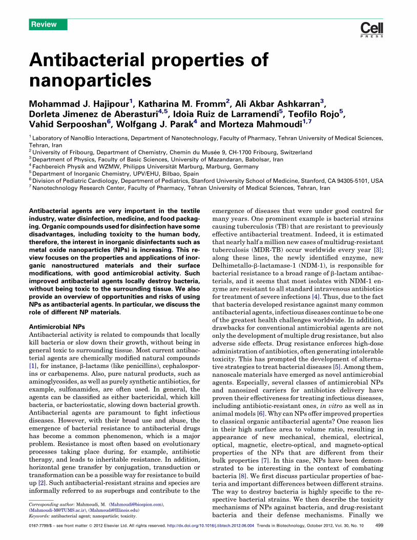
Antibacterial properties ofnanoparticlesMohammad J. Hajipour1, Katharina M. Fromm2, Ali Akbar Ashkarran3,Dorleta Jimenez de Aberasturi4,5, Idoia Ruiz de Larramendi5, Teofilo Rojo5,Vahid Serpooshan6, Wolfgang J. Parak4 and Morteza Mahmoudi1,7
1 Laboratory of NanoBio Interactions, Department of Nanotechnology, Faculty of Pharmacy, Tehran University of Medical Sciences,
Tehran, Iran2 University of Fribourg, Department of Chemistry, Chemin du Muse e 9, CH-1700 Fribourg, Switzerland3 Department of Physics, Faculty of Basic Sciences, University of Mazandaran, Babolsar, Iran4 Fachbereich Physik and WZMW, Philipps Universitat Marburg, Marburg, Germany5 Department of Inorganic Chemistry, UPV/EHU, Bilbao, Spain6 Division of Pediatric Cardiology, Department of Pediatrics, Stanford University School of Medicine, Stanford, CA 94305-5101, USA7 Nanotechnology Research Center, Faculty of Pharmacy, Tehran University of Medical Sciences, Tehran, Iran
Review
Antibacterial agents are very important in the textileindustry, water disinfection, medicine, and food packag-ing. Organic compounds used for disinfection have somedisadvantages, including toxicity to the human body,therefore, the interest in inorganic disinfectants such asmetal oxide nanoparticles (NPs) is increasing. This re-view focuses on the properties and applications of inor-ganic nanostructured materials and their surfacemodifications, with good antimicrobial activity. Suchimproved antibacterial agents locally destroy bacteria,without being toxic to the surrounding tissue. We alsoprovide an overview of opportunities and risks of usingNPs as antibacterial agents. In particular, we discuss therole of different NP materials.
Antimicrobial NPsAntibacterial activity is related to compounds that locallykill bacteria or slow down their growth, without being ingeneral toxic to surrounding tissue. Most current antibac-terial agents are chemically modified natural compounds[1], for instance, b-lactams (like penicillins), cephalospor-ins or carbapenems. Also, pure natural products, such asaminoglycosides, as well as purely synthetic antibiotics, forexample, sulfonamides, are often used. In general, theagents can be classified as either bactericidal, which killbacteria, or bacteriostatic, slowing down bacterial growth.Antibacterial agents are paramount to fight infectiousdiseases. However, with their broad use and abuse, theemergence of bacterial resistance to antibacterial drugshas become a common phenomenon, which is a majorproblem. Resistance is most often based on evolutionaryprocesses taking place during, for example, antibiotictherapy, and leads to inheritable resistance. In addition,horizontal gene transfer by conjugation, transduction ortransformation can be a possible way for resistance to buildup [2]. Such antibacterial-resistant strains and species areinformally referred to as superbugs and contribute to the
Corresponding author: Mahmoudi, M. ([email protected]),([email protected]), ([email protected])Keywords: antibacterial agent; nanoparticle; toxicity.
0167-7799/$ – see front matter � 2012 Elsevier Ltd. All rights reserved. http://dx.doi.org/10.101
emergence of diseases that were under good control formany years. One prominent example is bacterial strainscausing tuberculosis (TB) that are resistant to previouslyeffective antibacterial treatment. Indeed, it is estimatedthat nearly half a million new cases of multidrug-resistanttuberculosis (MDR-TB) occur worldwide every year [3];along these lines, the newly identified enzyme, newDelhimetallo-b-lactamase-1 (NDM-1), is responsible forbacterial resistance to a broad range of b-lactam antibac-terials, and it seems that most isolates with NDM-1 en-zyme are resistant to all standard intravenous antibioticsfor treatment of severe infections [4]. Thus, due to the factthat bacteria developed resistance against many commonantibacterial agents, infectious diseases continue to be oneof the greatest health challenges worldwide. In addition,drawbacks for conventional antimicrobial agents are notonly the development of multiple drug resistance, but alsoadverse side effects. Drug resistance enforces high-doseadministration of antibiotics, often generating intolerabletoxicity. This has prompted the development of alterna-tive strategies to treat bacterial diseases [5]. Among them,nanoscale materials have emerged as novel antimicrobialagents. Especially, several classes of antimicrobial NPsand nanosized carriers for antibiotics delivery haveproven their effectiveness for treating infectious diseases,including antibiotic-resistant ones, in vitro as well as inanimal models [6]. Why can NPs offer improved propertiesto classical organic antibacterial agents? One reason liesin their high surface area to volume ratio, resulting inappearance of new mechanical, chemical, electrical,optical, magnetic, electro-optical, and magneto-opticalproperties of the NPs that are different from theirbulk properties [7]. In this case, NPs have been demon-strated to be interesting in the context of combatingbacteria [8]. We first discuss particular properties of bac-teria and important differences between different strains.The way to destroy bacteria is highly specific to the re-spective bacterial strains. We then describe the toxicitymechanisms of NPs against bacteria, and drug-resistantbacteria and their defense mechanisms. Finally we
6/j.tibtech.2012.06.004 Trends in Biotechnology, October 2012, Vol. 30, No. 10 499
Review Trends in Biotechnology October 2012, Vol. 30, No. 10
provide an outlook on NPs in the environment andecosystems.
Properties of bacteria, and thus the way to destroythem, are highly specific to the respective bacterialstrainsRole of the cell wall
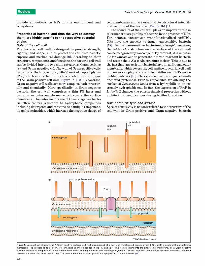
The bacterial cell wall is designed to provide strength,rigidity, and shape, and to protect the cell from osmoticrupture and mechanical damage [9]. According to theirstructure, components, and functions, the bacteria cell wallcan be divided into the two main categories: Gram positive(+) and Gram negative (–). The wall of Gram-positive cellscontains a thick layer (i.e., 20–50 nm) of peptidoglycan(PG), which is attached to teichoic acids that are uniqueto the Gram-positive cell wall (Figure 1a) [10]. By contrast,Gram-negative cell walls are more complex, both structur-ally and chemically. More specifically, in Gram-negativebacteria, the cell wall comprises a thin PG layer andcontains an outer membrane, which covers the surfacemembrane. The outer membrane of Gram-negative bacte-ria often confers resistance to hydrophobic compoundsincluding detergents and contains as a unique component,lipopolysaccharides, which increase the negative charge of
Pep�doglycan
Cytoplasmic membrane
Lipopolysaccharide
Po
Outer membrane
Pep�doglycan
Cytoplasmic membrane
(a)
(b)
Figure 1. Bacterial cell structure. (a) A Gram-positive bacterial cell wall is composed o
membrane. The teichoic acids, as seen, are connected to and embedded in the PG, an
bacterial cell wall is composed of an outer membrane linked by lipoproteins to thin an
between the outer and inner membranes. The outer membrane includes porins and lip
500
cell membranes and are essential for structural integrityand viability of the bacteria (Figure 1b) [11].
The structure of the cell wall plays an important role intolerance or susceptibility of bacteria in the presence of NPs.For instance, vancomycin (van)-functionalized Ag@TiO2
NPs have the capacity to target van-sensitive bacteria[12]. In the van-sensitive bacterium, Desulfotomaculum,the D-Ala-D-Ala structure on the surface of the cell wallcan be recognized by vancomycin. By contrast, it is impossi-ble for vancomycin to penetrate into van-resistant bacteriaand access the D-Ala-D-Ala structure moiety. This is due tothe fact that van-resistant bacteria have an additional outermembrane, which covers the cell surface. Bacterial cell wallproperties can play a crucial role in diffusion of NPs insidebiofilm matrixes [13]. The expression of the major cell-wall-anchored proteinase PrtP is responsible for altering thesurface of Lactococcus lactis from a hydrophilic to an ex-tremely hydrophobic one. In fact, the expression of PrtP inL. lactis 2 changes the physicochemical properties withoutarchitectural modifications during biofilm formation.
Role of the NP type and surface
Species sensitivity is not only related to the structure of thecell wall in Gram-positive and Gram-negative bacteria
rin
Periplasm
Lipoprotein
Teichoicacid
Lipoteichoicacid
TRENDS in Biotechnology
f a thick and multilayered peptidoglycan (PG) sheath outside of the cytoplasmic
d lipoteichoic acids extend into the cytoplasmic membrane. (b) A Gram-negative
d single-layered PG. The PG is placed within the periplasmic space that is formed
opolysaccharide molecules [64].
Review Trends in Biotechnology October 2012, Vol. 30, No. 10
[12]. Several additional factors can influence the suscepti-bility or tolerance of bacteria to NPs. For example, Escher-ichia coli (–) is highly susceptible, whereas Staphylococcusaureus (+) and Bacillus subtilis (+) are less susceptible toCuO NPs [13]. The antibacterial effect of Ag NPs is higherthan Cu NPs against E. coli (–) and S. aureus (+) bacteria[14]. S. aureus (+) and B. subtilis (+) are more susceptiblethan E. coli (–) to NiO and ZnO NPs [13].
Role of growth rate
Another factor that can influence the tolerance of bacteriaagainst NPs is the rate of bacterial growth. Fast-growingbacteria are more susceptible than slow-growing bacteria toantibiotics and NPs [15,16]. It is possible that the toleranceproperty of slow-growing bacteria is related to the expres-sion of stress-response genes [14,17]. Consequently, anti-bacterial effects highly depend on the particular strain.
Role of biofilm formation
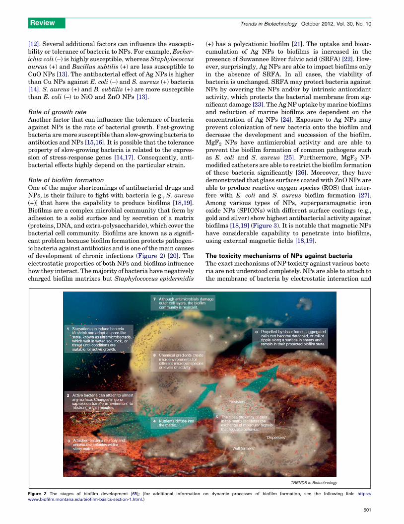
One of the major shortcomings of antibacterial drugs andNPs, is their failure to fight with bacteria [e.g., S. aureus(+)] that have the capability to produce biofilms [18,19].Biofilms are a complex microbial community that form byadhesion to a solid surface and by secretion of a matrix(proteins, DNA, and extra-polysaccharide), which cover thebacterial cell community. Biofilms are known as a signifi-cant problem because biofilm formation protects pathogen-ic bacteria against antibiotics and is one of the main causesof development of chronic infections (Figure 2) [20]. Theelectrostatic properties of both NPs and biofilms influencehow they interact. The majority of bacteria have negativelycharged biofilm matrixes but Staphylococcus epidermidis
Figure 2. The stages of biofilm development [65]; (for additional information o
www.biofilm.montana.edu/biofilm-basics-section-1.html.)
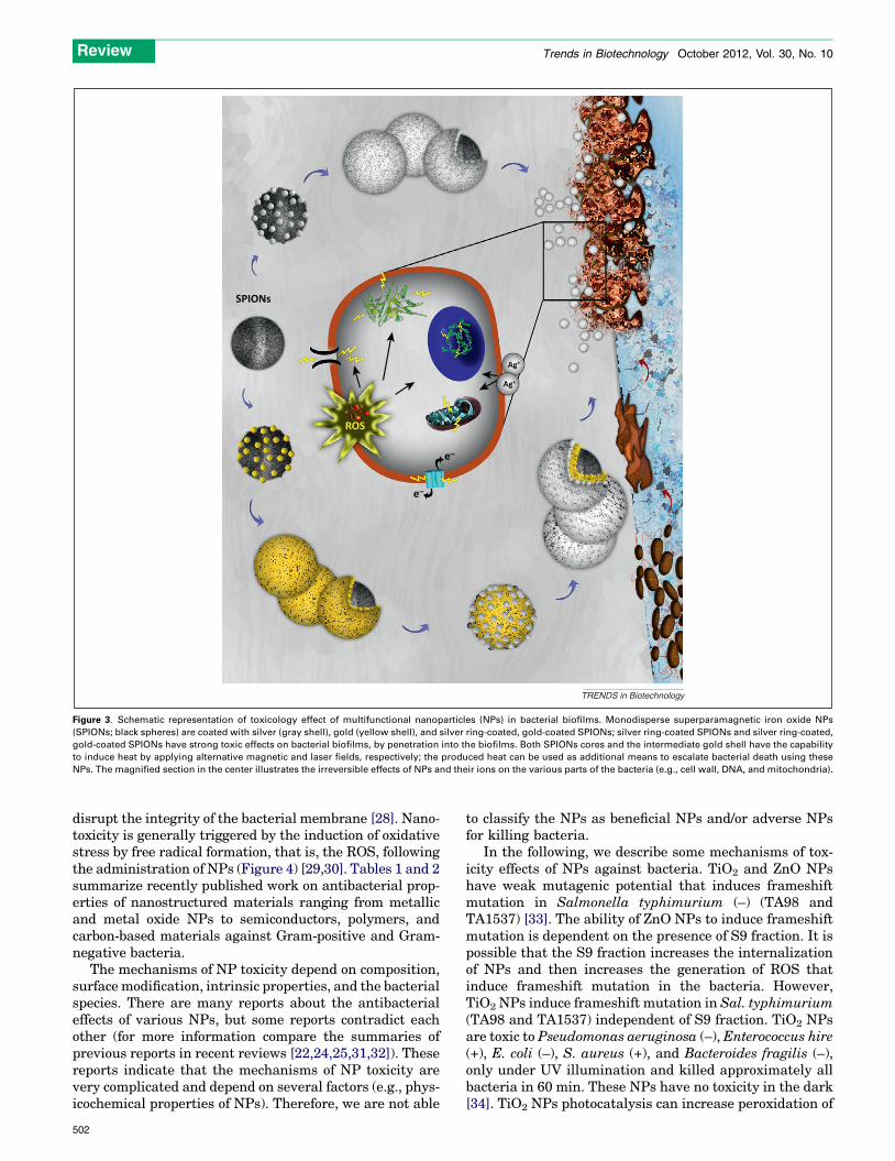
(+) has a polycationic biofilm [21]. The uptake and bioac-cumulation of Ag NPs to biofilms is increased in thepresence of Suwannee River fulvic acid (SRFA) [22]. How-ever, surprisingly, Ag NPs are able to impact biofilms onlyin the absence of SRFA. In all cases, the viability ofbacteria is unchanged. SRFA may protect bacteria againstNPs by covering the NPs and/or by intrinsic antioxidantactivity, which protects the bacterial membrane from sig-nificant damage [23]. The Ag NP uptake by marine biofilmsand reduction of marine biofilms are dependent on theconcentration of Ag NPs [24]. Exposure to Ag NPs mayprevent colonization of new bacteria onto the biofilm anddecrease the development and succession of the biofilm.MgF2 NPs have antimicrobial activity and are able toprevent the biofilm formation of common pathogens suchas E. coli and S. aureus [25]. Furthermore, MgF2 NP-modified catheters are able to restrict the biofilm formationof these bacteria significantly [26]. Moreover, they havedemonstrated that glass surfaces coated with ZnO NPs areable to produce reactive oxygen species (ROS) that inter-fere with E. coli and S. aureus biofilm formation [27].Among various types of NPs, superparamagnetic ironoxide NPs (SPIONs) with different surface coatings (e.g.,gold and silver) show highest antibacterial activity againstbiofilms [18,19] (Figure 3). It is notable that magnetic NPshave considerable capability to penetrate into biofilms,using external magnetic fields [18,19].
The toxicity mechanisms of NPs against bacteriaThe exact mechanisms of NP toxicity against various bacte-ria are not understood completely. NPs are able to attach tothe membrane of bacteria by electrostatic interaction and
TRENDS in Biotechnology
n dynamic processes of biofilm formation, see the following link: https://
501
SPIONs
ROS
Ag+
Ag+
e–
e–
TRENDS in Biotechnology
Figure 3. Schematic representation of toxicology effect of multifunctional nanoparticles (NPs) in bacterial biofilms. Monodisperse superparamagnetic iron oxide NPs
(SPIONs; black spheres) are coated with silver (gray shell), gold (yellow shell), and silver ring-coated, gold-coated SPIONs; silver ring-coated SPIONs and silver ring-coated,
gold-coated SPIONs have strong toxic effects on bacterial biofilms, by penetration into the biofilms. Both SPIONs cores and the intermediate gold shell have the capability
to induce heat by applying alternative magnetic and laser fields, respectively; the produced heat can be used as additional means to escalate bacterial death using these
NPs. The magnified section in the center illustrates the irreversible effects of NPs and their ions on the various parts of the bacteria (e.g., cell wall, DNA, and mitochondria).
Review Trends in Biotechnology October 2012, Vol. 30, No. 10
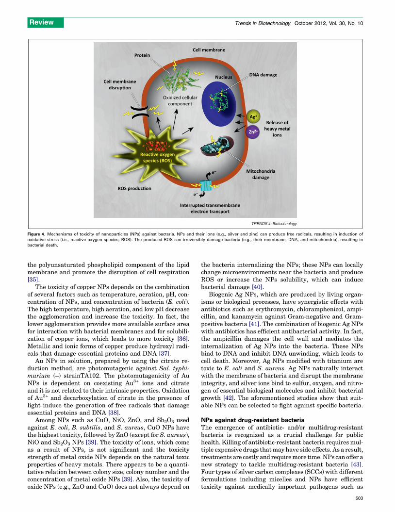
disrupt the integrity of the bacterial membrane [28]. Nano-toxicity is generally triggered by the induction of oxidativestress by free radical formation, that is, the ROS, followingthe administration of NPs (Figure 4) [29,30]. Tables 1 and 2summarize recently published work on antibacterial prop-erties of nanostructured materials ranging from metallicand metal oxide NPs to semiconductors, polymers, andcarbon-based materials against Gram-positive and Gram-negative bacteria.
The mechanisms of NP toxicity depend on composition,surface modification, intrinsic properties, and the bacterialspecies. There are many reports about the antibacterialeffects of various NPs, but some reports contradict eachother (for more information compare the summaries ofprevious reports in recent reviews [22,24,25,31,32]). Thesereports indicate that the mechanisms of NP toxicity arevery complicated and depend on several factors (e.g., phys-icochemical properties of NPs). Therefore, we are not able
502
to classify the NPs as beneficial NPs and/or adverse NPsfor killing bacteria.
In the following, we describe some mechanisms of tox-icity effects of NPs against bacteria. TiO2 and ZnO NPshave weak mutagenic potential that induces frameshiftmutation in Salmonella typhimurium (–) (TA98 andTA1537) [33]. The ability of ZnO NPs to induce frameshiftmutation is dependent on the presence of S9 fraction. It ispossible that the S9 fraction increases the internalizationof NPs and then increases the generation of ROS thatinduce frameshift mutation in the bacteria. However,TiO2 NPs induce frameshift mutation in Sal. typhimurium(TA98 and TA1537) independent of S9 fraction. TiO2 NPsare toxic to Pseudomonas aeruginosa (–), Enterococcus hire(+), E. coli (–), S. aureus (+), and Bacteroides fragilis (–),only under UV illumination and killed approximately allbacteria in 60 min. These NPs have no toxicity in the dark[34]. TiO2 NPs photocatalysis can increase peroxidation of
Cell membrane
DNA damage
Release ofheavy metal
ions
Ag+
e–
e–
Zn2+
Mitochondriadamage
ROS produc�on
Reac�ve oxygenspecies (ROS)
Interrupted transmembraneelectron transport
Nucleus
Oxidized cellularcomponent
Protein
Cell membranedisrup�on
TRENDS in Biotechnology
Figure 4. Mechanisms of toxicity of nanoparticles (NPs) against bacteria. NPs and their ions (e.g., silver and zinc) can produce free radicals, resulting in induction of
oxidative stress (i.e., reactive oxygen species; ROS). The produced ROS can irreversibly damage bacteria (e.g., their membrane, DNA, and mitochondria), resulting in
bacterial death.
Review Trends in Biotechnology October 2012, Vol. 30, No. 10
the polyunsaturated phospholipid component of the lipidmembrane and promote the disruption of cell respiration[35].
The toxicity of copper NPs depends on the combinationof several factors such as temperature, aeration, pH, con-centration of NPs, and concentration of bacteria (E. coli).The high temperature, high aeration, and low pH decreasethe agglomeration and increase the toxicity. In fact, thelower agglomeration provides more available surface areafor interaction with bacterial membranes and for solubili-zation of copper ions, which leads to more toxicity [36].Metallic and ionic forms of copper produce hydroxyl radi-cals that damage essential proteins and DNA [37].
Au NPs in solution, prepared by using the citrate re-duction method, are photomutagenic against Sal. typhi-murium (–) strainTA102. The photomutagenicity of AuNPs is dependent on coexisting Au3+ ions and citrateand it is not related to their intrinsic properties. Oxidationof Au3+ and decarboxylation of citrate in the presence oflight induce the generation of free radicals that damageessential proteins and DNA [38].
Among NPs such as CuO, NiO, ZnO, and Sb2O3 usedagainst E. coli, B. subtilis, and S. aureus, CuO NPs havethe highest toxicity, followed by ZnO (except for S. aureus),NiO and Sb2O3 NPs [39]. The toxicity of ions, which comeas a result of NPs, is not significant and the toxicitystrength of metal oxide NPs depends on the natural toxicproperties of heavy metals. There appears to be a quanti-tative relation between colony size, colony number and theconcentration of metal oxide NPs [39]. Also, the toxicity ofoxide NPs (e.g., ZnO and CuO) does not always depend on
the bacteria internalizing the NPs; these NPs can locallychange microenvironments near the bacteria and produceROS or increase the NPs solubility, which can inducebacterial damage [40].
Biogenic Ag NPs, which are produced by living organ-isms or biological processes, have synergistic effects withantibiotics such as erythromycin, chloramphenicol, ampi-cillin, and kanamycin against Gram-negative and Gram-positive bacteria [41]. The combination of biogenic Ag NPswith antibiotics has efficient antibacterial activity. In fact,the ampicillin damages the cell wall and mediates theinternalization of Ag NPs into the bacteria. These NPsbind to DNA and inhibit DNA unwinding, which leads tocell death. Moreover, Ag NPs modified with titanium aretoxic to E. coli and S. aureus. Ag NPs naturally interactwith the membrane of bacteria and disrupt the membraneintegrity, and silver ions bind to sulfur, oxygen, and nitro-gen of essential biological molecules and inhibit bacterialgrowth [42]. The aforementioned studies show that suit-able NPs can be selected to fight against specific bacteria.
NPs against drug-resistant bacteriaThe emergence of antibiotic- and/or multidrug-resistantbacteria is recognized as a crucial challenge for publichealth. Killing of antibiotic-resistant bacteria requires mul-tiple expensive drugs that may have side effects. As a result,treatments are costly and require more time. NPs can offer anew strategy to tackle multidrug-resistant bacteria [43].Four types of silver carbon complexes (SCCs) with differentformulations including micelles and NPs have efficienttoxicity against medically important pathogens such as
503
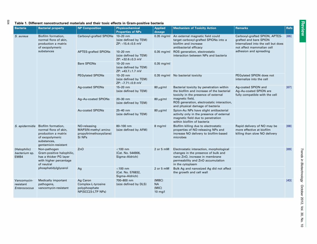
Table 1. Different nanostructured materials and their toxic effects in Gram-positive bacteria
Bacteria Bacterial property NP Composition Physicochemical
Properties of NPs
Applied
dosage
Mechanism of Toxicity Action Remarks Refs
S. aureus Biofilm formation,
normal flora of skin,
production a matrix
of exopolymeric
substances
Carboxyl-grafted SPIONs 10–20 nm
(size defined by TEM)
ZP: –15.4�0.5 mV
0.35 mg/ml An external magnetic field could
target carboxyl-grafted SPIONs into a
biofilm and increase
antibacterial efficacy
Carboxyl-grafted SPION, APTES-
grafted and bare SPION
Internalized into the cell but does
not affect mammalian cell
adhesion and spreading
[66]
APTES-grafted SPIONs 10–20 nm
(size defined by TEM)
ZP: +32.6�0.3 mV
0.35 mg/ml ROS generation, electrostatic
interaction between NPs and bacteria
Bare SPIONs 10–20 nm
(size defined by TEM)
ZP: +43.7�1.7 mV
0.35 mg/ml
PEGylated SPIONs 10–20 nm
(size defined by TEM)
ZP: –7.71�0.9 mV
0.35 mg/ml No bacterial toxicity PEGylated SPION does not
internalize into the cell
Ag-coated SPIONs 15–20 nm
(size defined by TEM)
80 mg/ml Bacterial toxicity by penetration within
the biofilm and increase of the bacterial
toxicity in the presence of external
magnetic field,
ROS generation, electrostatic interaction,
and physical damage of bacteria
Ag-coated SPION and
Ag–Au-coated SPION are
fully compatible with the cell
[67]
Ag–Au-coated SPIONs 20–30 nm
(size defined by TEM)
80 mg/ml
Au-coated SPIONs 25–40 nm
(size defined by TEM)
80 mg/ml Spion-Au NPs have slight antibacterial
activity only in the presence of external
magnetic field due to penetration
within biofilm of bacteria
S. epidermidis Biofilm formation,
normal flora of skin,
production a matrix
of exopolymeric
substances,
gentamicin-resistant
NO-releasing
MAP3(N-methyl amino
propyltrimethoxysilane)
Si NPs
80–100 nm
(size defined by AFM)
8 mg/ml Biofilm killing due to electrostatic
properties of NO-releasing NPs and
increase NO delivery to biofilm-based
microbes
Rapid delivery of NO may be
more effective at biofilm
killing than slow NO delivery
[68]
(Halophilic)
bacterium sp.
EMB4
Non-pathogen
Gram-positive halophilic,
has a thicker PG layer
with higher percentage
of neutral
phosphatidylglycerol
ZnO <100 nm
(Cat. No. 544906,
Sigma–Aldrich)
2 or 5 mM Electrostatic interaction, morphological
changes in the presence of bulk and
nano ZnO, increase in membrane
permeability and ZnO accumulation
in the cytoplasm
[69]
Ag <100 nm
(Cat. No. 576832,
Sigma–Aldrich)
2 or 5 mM Bulk Ag and nanosized Ag did not affect
the growth and cell wall
Vancomycin-
resistant
Enterococcus
Medically important
pathogens,
vancomycin-resistant
Ag Caron
Complex-L-tyrosine
polyphosphate
NP(SCC23-LTP NPs)
700–800 nm
(size defined by DLS)
(MBC)
NA
(MIC)
10 mg/l
[43]
Revie
wT
rends
in B
iote
chnolo
gy
O
cto
ber
2012,
Vol.
30,
No.
10
504
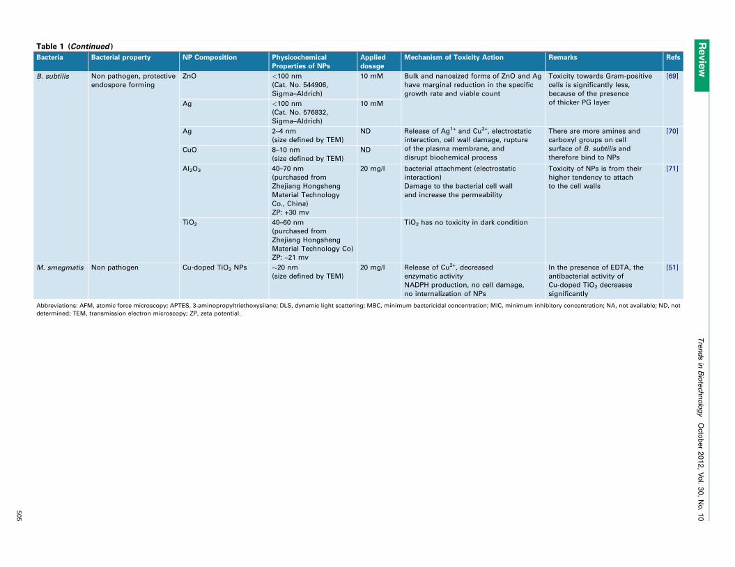
Table 1 (Continued )
Bacteria Bacterial property NP Composition Physicochemical
Properties of NPs
Applied
dosage
Mechanism of Toxicity Action Remarks Refs
B. subtilis Non pathogen, protective
endospore forming
ZnO <100 nm
(Cat. No. 544906,
Sigma–Aldrich)
10 mM Bulk and nanosized forms of ZnO and Ag
have marginal reduction in the specific
growth rate and viable count
Toxicity towards Gram-positive
cells is significantly less,
because of the presence
of thicker PG layer
[69]
Ag <100 nm
(Cat. No. 576832,
Sigma–Aldrich)
10 mM
Ag 2–4 nm
(size defined by TEM)
ND Release of Ag1+ and Cu2+, electrostatic
interaction, cell wall damage, rupture
of the plasma membrane, and
disrupt biochemical process
There are more amines and
carboxyl groups on cell
surface of B. subtilis and
therefore bind to NPs
[70]
CuO 8–10 nm
(size defined by TEM)
ND
Al2O3 40–70 nm
(purchased from
Zhejiang Hongsheng
Material Technology
Co., China)
ZP: +30 mv
20 mg/l bacterial attachment (electrostatic
interaction)
Damage to the bacterial cell wall
and increase the permeability
Toxicity of NPs is from their
higher tendency to attach
to the cell walls
[71]
TiO2 40–60 nm
(purchased from
Zhejiang Hongsheng
Material Technology Co)
ZP: –21 mv
TiO2 has no toxicity in dark condition
M. smegmatis Non pathogen Cu-doped TiO2 NPs �20 nm
(size defined by TEM)
20 mg/l Release of Cu2+, decreased
enzymatic activity
NADPH production, no cell damage,
no internalization of NPs
In the presence of EDTA, the
antibacterial activity of
Cu-doped TiO2 decreases
significantly
[51]
Abbreviations: AFM, atomic force microscopy; APTES, 3-aminopropyltriethoxysilane; DLS, dynamic light scattering; MBC, minimum bactericidal concentration; MIC, minimum inhibitory concentration; NA, not available; ND, not
determined; TEM, transmission electron microscopy; ZP, zeta potential.
Revie
wT
rends
in B
iote
chnolo
gy
O
cto
ber
2012,
Vol.
30,
No.
10
505
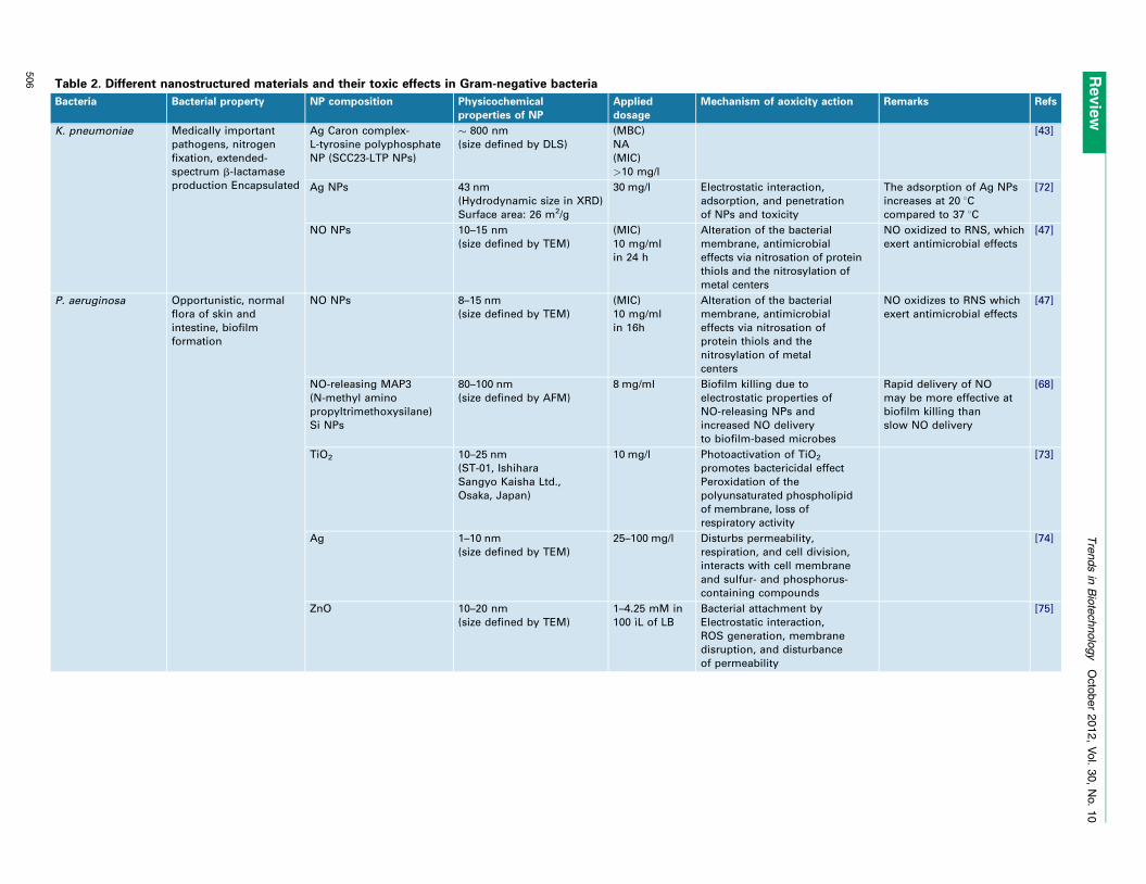
Table 2. Different nanostructured materials and their toxic effects in Gram-negative bacteria
Bacteria Bacterial property NP composition Physicochemical
properties of NP
Applied
dosage
Mechanism of aoxicity action Remarks Refs
K. pneumoniae Medically important
pathogens, nitrogen
fixation, extended-
spectrum b-lactamase
production Encapsulated
Ag Caron complex-
L-tyrosine polyphosphate
NP (SCC23-LTP NPs)
� 800 nm
(size defined by DLS)
(MBC)
NA
(MIC)
>10 mg/l
[43]
Ag NPs 43 nm
(Hydrodynamic size in XRD)
Surface area: 26 m2/g
30 mg/l Electrostatic interaction,
adsorption, and penetration
of NPs and toxicity
The adsorption of Ag NPs
increases at 20 8Ccompared to 37 8C
[72]
NO NPs 10–15 nm
(size defined by TEM)
(MIC)
10 mg/ml
in 24 h
Alteration of the bacterial
membrane, antimicrobial
effects via nitrosation of protein
thiols and the nitrosylation of
metal centers
NO oxidized to RNS, which
exert antimicrobial effects
[47]
P. aeruginosa Opportunistic, normal
flora of skin and
intestine, biofilm
formation
NO NPs 8–15 nm
(size defined by TEM)
(MIC)
10 mg/ml
in 16h
Alteration of the bacterial
membrane, antimicrobial
effects via nitrosation of
protein thiols and the
nitrosylation of metal
centers
NO oxidizes to RNS which
exert antimicrobial effects
[47]
NO-releasing MAP3
(N-methyl amino
propyltrimethoxysilane)
Si NPs
80–100 nm
(size defined by AFM)
8 mg/ml Biofilm killing due to
electrostatic properties of
NO-releasing NPs and
increased NO delivery
to biofilm-based microbes
Rapid delivery of NO
may be more effective at
biofilm killing than
slow NO delivery
[68]
TiO2 10–25 nm
(ST-01, Ishihara
Sangyo Kaisha Ltd.,
Osaka, Japan)
10 mg/l Photoactivation of TiO2
promotes bactericidal effect
Peroxidation of the
polyunsaturated phospholipid
of membrane, loss of
respiratory activity
[73]
Ag 1–10 nm
(size defined by TEM)
25–100 mg/l Disturbs permeability,
respiration, and cell division,
interacts with cell membrane
and sulfur- and phosphorus-
containing compounds
[74]
ZnO 10–20 nm
(size defined by TEM)
1–4.25 mM in
100 ıL of LB
Bacterial attachment by
Electrostatic interaction,
ROS generation, membrane
disruption, and disturbance
of permeability
[75]
Revie
wT
rends
in B
iote
chnolo
gy
O
cto
ber
2012,
Vol.
30,
No.
10
506
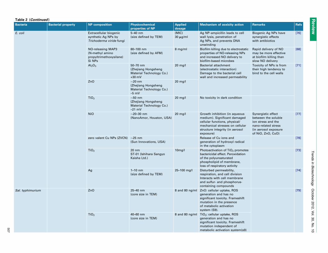
Table 2 (Continued )
Bacteria Bacterial property NP composition Physicochemical
properties of NP
Applied
dosage
Mechanism of aoxicity action Remarks Refs
E. coli Extracellular biogenic
synthetic Ag NPs by
Trichoderma viride fungi
5–40 nm
(size defined by TEM)
(MIC)
30 mg/ml
Ag NP–ampicillin leads to cell
wall lysis, penetration of
Ag NPs, and prevents DNA
unwinding
Biogenic Ag NPs have
synergistic effects
with antibiotics
[76]
NO-releasing MAP3
(N-methyl amino
propyltrimethoxysilane)
Si NPs
80–100 nm
(size defined by AFM)
8 mg/ml Biofilm killing due to electrostatic
properties of NO-releasing NPs
and increased NO delivery to
biofilm-based microbes
Rapid delivery of NO
may be more effective
at biofilm killing than
slow NO delivery
[68]
Al2O3 50–70 nm
(Zhejiang Hongsheng
Material Technology Co.)
+30 mV
20 mg/l Bacterial attachment
(electrostatic interaction)
Damage to the bacterial cell
wall and increased permeability
Toxicity of NPs is from
their high tendency to
bind to the cell walls
[71]
ZnO �20 nm
(Zhejiang Hongsheng
Material Technology Co.)
–5 mV
20 mg/l
TiO2 �50 nm
(Zhejiang Hongsheng
Material Technology Co.)
–21 mV
20 mg/l No toxicity in dark condition
NiO �20–30 nm
(NanoAmor, Houston, USA)
20 mg/l Growth inhibition (in aqueous
medium). Significant damaged
cellular functions, physical/
mechanical stresses on cellular
structure integrity (in aerosol
exposure)
Synergistic effect
between the soluble
ion stress and the
nano-related stress
(in aerosol exposure
of NiO, ZnO, CuO)
[77]
zero valent Cu NPs (ZVCN) �25 nm
(Sun Innovations, USA)
Release of Cu ions and
generation of hydroxyl radical
in the cytoplasm
[78]
TiO2 20 nm
ST-01 (Ishihara Sangyo
Kaisha Ltd.)
10mg/l Photoactivation of TiO2 promotes
bactericidal effect. Peroxidation
of the polyunsaturated
phospholipid of membrane,
loss of respiratory activity
[73]
Ag 1–10 nm
(size defined by TEM)
25–100 mg/l Disturbed permeability,
respiration, and cell division
Interacts with cell membrane
and sulfur- and phosphorus-
containing compounds
[74]
Sal. typhimurium ZnO 25–40 nm
(core size in TEM)
8 and 80 ng/ml ZnO: cellular uptake, ROS
generation and has no
significant toxicity. Frameshift
mutation in the presence
of metabolic activation
system (S9).
[79]
TiO2 40–60 nm
(core size in TEM)
8 and 80 ng/ml TiO2: cellular uptake, ROS
generation and has no
significant toxicity. Frameshift
mutation independent of
metabolic activation system(s9)
Revie
wT
rends
in B
iote
chnolo
gy
O
cto
ber
2012,
Vol.
30,
No.
10
507
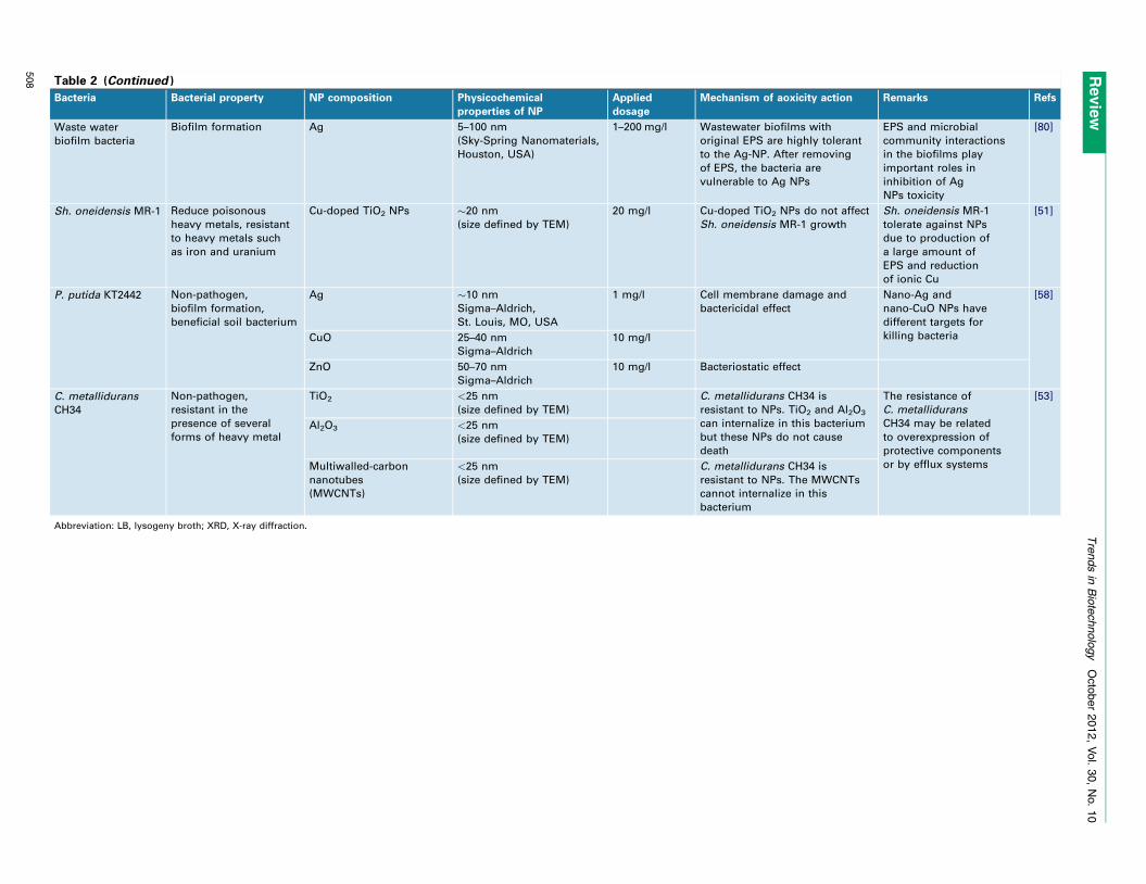
Table 2 (Continued )
Bacteria Bacterial property NP composition Physicochemical
properties of NP
Applied
dosage
Mechanism of aoxicity action Remarks Refs
Waste water
biofilm bacteria
Biofilm formation Ag 5–100 nm
(Sky-Spring Nanomaterials,
Houston, USA)
1–200 mg/l Wastewater biofilms with
original EPS are highly tolerant
to the Ag-NP. After removing
of EPS, the bacteria are
vulnerable to Ag NPs
EPS and microbial
community interactions
in the biofilms play
important roles in
inhibition of Ag
NPs toxicity
[80]
Sh. oneidensis MR-1 Reduce poisonous
heavy metals, resistant
to heavy metals such
as iron and uranium
Cu-doped TiO2 NPs �20 nm
(size defined by TEM)
20 mg/l Cu-doped TiO2 NPs do not affect
Sh. oneidensis MR-1 growth
Sh. oneidensis MR-1
tolerate against NPs
due to production of
a large amount of
EPS and reduction
of ionic Cu
[51]
P. putida KT2442 Non-pathogen,
biofilm formation,
beneficial soil bacterium
Ag �10 nm
Sigma–Aldrich,
St. Louis, MO, USA
1 mg/l Cell membrane damage and
bactericidal effect
Nano-Ag and
nano-CuO NPs have
different targets for
killing bacteria
[58]
CuO 25–40 nm
Sigma–Aldrich
10 mg/l
ZnO 50–70 nm
Sigma–Aldrich
10 mg/l Bacteriostatic effect
C. metallidurans
CH34
Non-pathogen,
resistant in the
presence of several
forms of heavy metal
TiO2 <25 nm
(size defined by TEM)
C. metallidurans CH34 is
resistant to NPs. TiO2 and Al2O3
can internalize in this bacterium
but these NPs do not cause
death
The resistance of
C. metallidurans
CH34 may be related
to overexpression of
protective components
or by efflux systems
[53]
Al2O3 <25 nm
(size defined by TEM)
Multiwalled-carbon
nanotubes
(MWCNTs)
<25 nm
(size defined by TEM)
C. metallidurans CH34 is
resistant to NPs. The MWCNTs
cannot internalize in this
bacterium
Abbreviation: LB, lysogeny broth; XRD, X-ray diffraction.
Revie
wT
rends
in B
iote
chnolo
gy
O
cto
ber
2012,
Vol.
30,
No.
10
508
Review Trends in Biotechnology October 2012, Vol. 30, No. 10
P. aeruginosa (–), Burkholderia cepacia (–), methicillin-re-sistant S. aureus, multidrug-resistant Acinetobacter bau-mannii (–), and Klebsiella pneumoniae (–) in the range of0.5–90 mg/l [43]. The SCCs are able to inhibit the growth ofbio-defense bacteria such B. subtilis and Yersinia pestis (–)[43].
Targeting bactericidal NPs to specific bacteria or specif-ic infected tissue is an efficient prospect in treating infec-tion because this phenomenon minimizes side effects andenhances antibacterial activity [44,45]. In this case, mul-tifunctional NPs can be very useful; for instance, multi-functional IgG–Fe3O4@TiO2 magnetic NPs are able totarget several pathogenic bacteria and have efficient anti-bacterial activity under UV irradiation. The IgG and TiO2
play a critical role in the targeting and killing properties ofthese NPs respectively. These NPs are toxic to Streptococ-cus pyogenes M9022434 and M9141204 [46].
Nitric-oxide-releasing NPs (NO NPs) are broad spec-trum antibacterial agents that are able to inhibit thegrowth of many antibiotic-resistant and sensitive clinicallyisolated bacteria such as K. pneumoniae, Enterococcusfaecalis (+), Str. pyogenes, E. coli, and P. aeruginosa (–).The toxicity of these NPs depends on the delivery of NO tothe target. These NPs are able to change the structure ofthe bacterial membrane and produce reactive nitrogenspecies (RNS), which lead to modification of essentialproteins of bacteria [47]. Beside NO NPs, ZnO NPs aretoxic to antibiotic (methicillin)-resistant bacteria such asStreptococcus agalactiae (+) and S. aureus. These NPs areable to disorganize and damage the cell membrane andincrease the permeability, which leads to cell death. Thepolyvinyl alcohol (PVA)-coated ZnO NPs are able to inter-nalize the bacteria and induce oxidative stress [48]. Thetoxicity of ZnO NPs is concentration-dependent and theseNPs are mildly toxic at low concentration [49].
NPs in water can significantly promote the horizontalconjugative transfer of multidrug-resistance genes medi-ated by the RP4, RK2, and pCF10 plasmids [50]. Here,nanoalumina can promote the conjugative transfer of theRP4 plasmid from E. coli to Salmonella spp. by up to200-fold compared with untreated cells. The nanoaluminais able to induce oxidative stress, damage bacterial cellmembranes, enhance the expression of mating pair forma-tion genes and DNA transfer and replication genes, anddepress the expression of global regulatory genes thatregulate the conjugative transfer of RP4 [50].
Defense mechanisms of tolerant bacteria against NPsSeveral naturally adapted bacteria are tolerant to specifictoxins or NPs that are present in the environment.Cu-doped TiO2 NPs are able to inhibit the growth ofMycobacterium smegmatis (+), but have no effect againstShewanella oneidensis MR-1(–) [51]. These NPs releaseCu2+ ions, which might be the main cause of toxicity,because the antibacterial activity of Cu-doped TiO2 NPswas decreased in the presence of chelating agents such asEDTA. Sh. oneidensis MR-1 has excellent resistant againstseveral concentrations of Cu2+ and Cu-doped TiO2 NPsbecause of the production of extracellular polymeric sub-stances (EPSs) under NP stress. This bacterium is able toabsorb NPs on the cell surface and to decrease the amount
of ionic Cu in the culture medium. Therefore this bacteri-um can be regarded as a promising candidate for cleaningof metal oxide NPs from the environment.
B. subtilis and Pseudomonas putida (–) can physicallyadapt to nC60 [buckminsterfullerene (C60) introduced ascolloidal aggregates in water] [52]. P. putida increasescyclopropan fatty acids and decreases unsaturated fattyacid levels, but B. subtilis increases the transition temper-ature and membrane fluidity in the presence of nC60. Thesephysiological adaptation responses of bacteria help to pro-tect the bacterial membrane against oxidative stress. TiO2
and Al2O3 NPs are able to be internalized by E. coli andCupriavidus metallidurans CH34, but these NPs are toxiconly against E. coli [53]. The resistance mechanism of C.metallidurans CH34 is not yet understood completely. Thetolerance mechanism of this bacterium may be related tophysical properties of their PG layer and/or products ofgenes that are located in the plasmids and are able tostabilize the plasma membrane or efflux of NPs.
Many bacteria are able to tolerate NO NPs using vari-ous mechanisms. For example P. aeruginosa, E. coli, andSal. typhimurium induce the expression of genes that areresponsible for repairing of DNA and altering the metalhomeostasis in the presence of NO NPs [54–56]. In thiscondition, K. pneumoniae produces the enzyme flavohemo-globin, which neutralizes nitrosative stress [57].
NPs against environment and ecosystemsExtensive use of NPs in biological science, medical science,and commercial products leads to leakage and accumula-tion of NPs in the environment (e.g., soil and water).Protection of the environment and beneficial bacteria fromNPs is very important because, for example, the indiscrim-inate use of nanosized Ag materials leads to release of Aginto the environment. The leakage of NPs into the envi-ronment is one of the most serious threats to beneficialmicrobes, microbial communities in ecosystems, and publichealth [58]. Many microbes benefit the environment andthe ecosystem, because they play an important role inbioremediation, element cycling, and nitrogen fixationfor plant growth [59–61]. For instance, in the nitrificationprocess, ammonium nitrogen is converted to nitrite andthen to nitrate by ammonia- and nitrite-oxidizing bacteria,respectively; the nitrifying bacteria are spread in theregions that have a high amount of ammonia; Ag NPs(<5 nm) have toxicity against nitrifying bacteria by inter-action with the bacterial membrane, which contains am-monia-oxidation enzymes and by generation of ROS. Thedeletion of these bacteria from the environment leads todecreased nitrogen removal and interferes with plantgrowth [62]. As another example, the exposure of E. coliand MS2 phages (in a binary system) to Ag NPs and ZnONPs leads to an increase in the transportation of MS2phages into bacteria by 2–6 orders of magnitude. There-fore, Ag NPs and ZnO NPs facilitate the internalization ofMS2 phages into bacteria. This can be a serious problembecause these NPs may mediate the internalization ofphages with drug-resistant genes into the bacteria andthus facilitate multidrug resistance development in thebacteria [63]. Therefore, the scientific community shouldpay attention to the adverse effects of the NPs on the
509
Review Trends in Biotechnology October 2012, Vol. 30, No. 10
environment and human health, in spite of their beneficialcommercial use.
Concluding remarksAntibacterial activities of NPs depend on two main factors:(i) physicochemical properties of NPs and (ii) type of bac-teria. Although there are good trends of correlation in a fewaspects of antibacterial activity of NPs (e.g., for biofilms),individual studies are difficult to generalize. This is mainlydue to the fact that the majority of researchers performexperiments based on available NPs and bacteria, ratherthan targeting specific, desired NPs or bacteria. In partic-ular, often poorly defined and characterized NPs are usedand thus correlation with basic physicochemical propertiesis not possible. Without agreement on standard NPs andbacteria as reference systems, which should be included infuture studies, there is still a long way to go in order tounravel systematically the antibacterial properties of NPs.
AcknowledgmentsThis work was funded in part by BMBF Germany (project UMSICHT).
References1 von Nussbaum, F. et al. (2006) Antibacterial natural products in
medicinal chemistry – exodus or revival? Angew. Chem. Int. Ed. 45,5072–5129
2 Witte, W. (2004) International dissemination of antibiotic resistantstrains of bacterial pathogens. Infect. Genet. Evol. 4, 187–191
3 Oldenburg, A.L. et al. (2004) Magnetic contrast agents for opticalcoherence tomography. Proc. of SPIE 5316, 91–98
4 Rakow, N.A. and Suslick, K.S. (2000) A colorimetric sensor array forodour visualization. Nature 406, 710–713
5 Baker-Austin, C. et al. (2006) Co-selection of antibiotic and metalresistance. Trends Microbiol. 14, 176–182
6 Huh, A.J. and Kwon, Y.J. (2011) ‘‘Nanoantibiotics’’: a new paradigm fortreating infectious diseases using nanomaterials in the antibioticsresistant era. J. Control. Release 156, 128–145
7 Whitesides, G.M. (2005) Nanoscience, nanotechnology, and chemistry.Small 1, 172–179
8 Xia, Y. (2008) Nanomaterials at work in biomedical research. Nat.Mater. 7, 758–760
9 Singleton, P. (2004) Bacteria, In Biology, Biotechnology and Medicine,(6th ed.), John Wiley & Sons Ltd, (West Sussex, England) 570 pp.,ISBN 0-470-09027-8
10 Scott, J.R. and Barnett, T.C. (2006) Surface proteins of gram-positivebacteria and how they get there. Annu. Rev. Microbiol. 60, 397–423
11 Roberts, I.S. (1996) The biochemistry and genetics of capsularpolysaccharide production in bacteria. Annu. Rev. Microbiol. 50,285–315
12 Ashkarran, A.A. et al. (2012) Bacterial effects and protein coronaevaluations: crucial ignored factors for prediction of bio-efficacy ofvarious forms of silver nanoparticles. Chem. Res. Toxicol. 25, 1231–1242
13 Baek, Y.W. and An, Y.J. (2011) Microbial toxicity of metal oxidenanoparticles (CuO, NiO, ZnO, and Sb2O3) to Escherichia coli,Bacillus subtilis, and Streptococcus aureus. Sci. Total Environ. 409,1603–1608
14 Lu, C. et al. (2009) Slow growth induces heat-shock resistance innormal and respiratory-deficient yeast. Mol. Biol. Cell 20, 891–903
15 Brown, M.R. et al. (1988) Resistance of bacterial biofilms to antibiotics:a growth-rate related effect? J. Antimicrob. Chemother. 22, 777–780
16 Mah, T.F. and O’Toole, G.A. (2001) Mechanisms of biofilm resistance toantimicrobial agents. Trends Microbiol. 9, 34–39
17 Stewart, P.S. (2002) Mechanisms of antibiotic resistance in bacterialbiofilms. Int. J. Med. Microbiol. 292, 107–113
18 Mahmoudi, M. and Serpooshan, V. (2012) Silver-coated engineeredmagnetic nanoparticles are promising for the success in the fightagainst antibacterial resistance threat. ACS Nano 6, 2656–2664
510
19 Park, H. et al. (2011) Inactivation of Pseudomonas aeruginosa PA01biofilms by hyperthermia using superparamagnetic nanoparticles.J. Microbiol. Methods 84, 41–45
20 Landini, P. et al. (2010) Molecular mechanisms of compounds affectingbacterial biofilm formation and dispersal. Appl. Microbiol. Biotechnol.86, 813–823
21 Sutherland, I. (2001) Biofilm exopolysaccharides: a strong and stickyframework. Microbiology 147, 3–9
22 Bolla, J.M. et al. (2011) Strategies for bypassing the membrane barrierin multidrug resistant Gram-negative bacteria. FEBS Lett. 585, 1682–1690
23 Fabrega, J. et al. (2009) Silver nanoparticle impact on bacterial growth:effect of pH, concentration, and organic matter. Environ. Sci. Technol.43, 7285–7290
24 Lara, H.H. et al. (2011) Silver nanoparticles are broad-spectrumbactericidal and virucidal compounds. J. Nanobiotechnol. 9, 30
25 Musee, N. et al. (2011) The antibacterial effects of engineerednanomaterials: implications for wastewater treatment plants.J. Environ. Monit. 13, 1164–1183
26 Lellouche, J. et al. (2012) Antibiofilm surface functionalization ofcatheters by magnesium fluoride nanoparticles. Int. J. Nanomed. 7,1175–1188
27 Applerot, G. et al. (2012) ZnO nanoparticle-coated surfaces inhibitbacterial biofilm formation and increase antibiotic susceptibility.RSC Adv. 2, 2314–2321
28 Thill, A. et al. (2006) Cytotoxicity of CeO2 nanoparticles for Escherichiacoli. Physico-chemical insight of the cytotoxicity mechanism. Environ.Sci. Technol. 40, 6151–6156
29 Soenen, S.J. et al. (2011) Cellular toxicity of inorganic nanoparticles:common aspects and guidelines for improved nanotoxicity evaluation.Nano Today 6, 446–465
30 Nel, A.E. et al. (2009) Understanding biophysicochemical interactionsat the nano-bio interface. Nat. Mater. 8, 543–557
31 Allahverdiyev, A.M. et al. (2011) Antimicrobial effects of TiO(2) andAg(2)O nanoparticles against drug-resistant bacteria and leishmaniaparasites. Future microbiol. 6, 933–940
32 Guzman, M. et al. (2012) Synthesis and antibacterial activity of silvernanoparticles against gram-positive and gram-negative bacteria.Nanomedicine 8, 37–45
33 Pan, X. et al. (2010) Mutagenicity evaluation of metal oxidenanoparticles by the bacterial reverse mutation assay. Chemosphere79, 113–116
34 Maness, P-C. et al. (1999) Bactericidal activity of photocatalytic TiO2reaction: toward an understanding of its killing mechanism. Appl.Environ. Microbiol. 65, 4094–4098
35 Wan, Y. et al. (2011) Vancomycin-functionalised Ag@TiO2 phototoxicityfor bacteria. J. Hazard. Mater. 186, 306–312
36 Pramanik, A. et al. (2012) A novel study of antibacterial activity ofcopper iodide nanoparticle mediated by DNA and membrane damage.Colloids Surf. B 96, 50–55
37 Wang, S. et al. (2011) Toxic effects of gold nanoparticles on Salmonellatyphimurium bacteria. Toxicol. Ind. Health 27, 547–554
38 Santo, C.E. et al. (2007) Contribution of copper ion resistance forsurvival of Escherichia coli on metallic copper surfaces. Appl.Environ. Microbiol. 74, 977–986
39 Baek, Y-W. and An, Y-J. (2011) Microbial toxicity of metal oxidenanoparticles (CuO, NiO, ZnO, and Sb2O3) to Escherichia coli,Bacillus subtilis, and Streptococcus aureus. Sci. Total Environ. 409,1603–1608
40 Heinlaan, M. et al. (2008) Toxicity of nanosized and bulk ZnO,CuO and TiO2 to bacteria Vibrio fischeri and crustaceansDaphnia magna and Thamnocephalus platyurus. Chemosphere 71,1308–1316
41 Devi, L.S. and Joshi, S.R. (2012) Antimicrobial and synergistic effectsof silver nanoparticles synthesized using: Soil fungi of high altitudes ofEastern Himalaya. Mycobiology 40, 27–34
42 Juan, L. et al. (2010) Deposition of silver nanoparticles on titaniumsurface for antibacterial effect. Int. J. Nanomed. 5, 261–267
43 Leid, J.G. et al. (2012) In vitro antimicrobial studies of silver carbenecomplexes: activity of free and nanoparticle carbene formulationsagainst clinical isolates of pathogenic bacteria. J. Antimicrob.Chemother. 67, 138–148
Review Trends in Biotechnology October 2012, Vol. 30, No. 10
44 Sondi, I. and Salopek-Sondi, B. (2004) Silver nanoparticles asantimicrobial agent: a case study on E. coli as a model for Gram-negative bacteria. J. Colloid Interface Sci. 275, 177–182
45 Suri, S.S. et al. (2007) Nanotechnology-based drug delivery systems. J.Occup. Med. Toxicol. 2, 16
46 Chen, W.J. et al. (2008) Functional Fe3O4/TiO2 core/shell magneticnanoparticles as photokilling agents for pathogenic bacteria. Small 4,485–491
47 Friedman, A. et al. (2011) Susceptibility of Gram-positive and -negativebacteria to novel nitric oxide-releasing nanoparticle technology.Virulence 2, 217–221
48 Huang, Z. et al. (2008) Toxicological effect of ZnO nanoparticles basedon bacteria. Langmuir 24, 4140–4144
49 Wang, Z.L. and Song, J. (2006) Piezoelectric nanogenerators based onzinc oxide nanowire arrays. Science 312, 242–246
50 Qiu, Z. et al. (2012) Nanoalumina promotes the horizontal transfer ofmultiresistance genes mediated by plasmids across genera. Proc. Natl.Acad. Sci. U.S.A. 109, 4944–4949
51 Wu, B. et al. (2010) Bacterial responses to Cu-doped TiO2nanoparticles. Sci. Total Environ. 408, 1755–1758
52 Fang, J. et al. (2007) Effect of a fullerene water suspension on bacterialphospholipids and membrane phase behavior. Environ. Sci. Technol.41, 2636–2642
53 Simon-Deckers, A. et al. (2009) Size-, composition- and shape-dependenttoxicological impact of metal oxide nanoparticles and carbon nanotubestoward bacteria. Environ. Sci. Technol. 43, 8423–8429
54 Tavares, A.F. et al. (2009) A novel nitroreductase of Staphylococcusaureus with S-nitrosoglutathione reductase activity. J. Bacteriol. 191,3403–3406
55 Jarboe, L.R. et al. (2008) Determination of the Escherichia coli S-nitrosoglutathione response network using integrated biochemicaland systems analysis. J. Biol. Chem. 283, 5148–5157
56 Bang, I.S. et al. (2006) Maintenance of nitric oxide and redoxhomeostasis by the salmonella flavohemoglobin hmp. J. Biol. Chem.281, 28039–28047
57 Frey, A.D. et al. (2002) Bacterial hemoglobins and flavohemoglobins foralleviation of nitrosative stress in Escherichia coli. Appl. Environ.Microbiol. 68, 4835–4840
58 Gajjar, P. et al. (2009) Antimicrobial activities of commercialnanoparticles against an environmental soil microbe, Pseudomonasputida KT2440. J. Biol. Eng. 3, 9
59 Kumar, N. et al. (2011) Perturbation of an arctic soil microbialcommunity by metal nanoparticles. J. Hazard. Mater. 190, 816–822
60 Molina, M.A. et al. (2006) A two-partner secretion system is involved inseed and root colonization and iron uptake by Pseudomonas putidaKT2440. Environ. Microbiol. 8, 639–647
61 Van Wees, S.C. et al. (2008) Plant immune responses triggered bybeneficial microbes. Curr. Opin. Plant Biol. 11, 443–448
62 Choi, O. and Hu, Z. (2008) Size dependent and reactive oxygen speciesrelated nanosilver toxicity to nitrifying bacteria. Environ. Sci. Technol.42, 4583–4588
63 You, J. et al. (2011) Bacteria and bacteriophage inactivation by silverand zinc oxide nanoparticles. Colloids Surf. B: Biointerfaces 85,161–167
64 Cabeen, M.T. and Jacobs-Wagner, C. (2005) Bacterial cell shape. Nat.Rev. Microbiol. 3, 601–610
65 Hall-Stoodley, L. et al. (2004) Bacterial biofilms: from the naturalenvironment to infectious diseases. Nat. Rev. Microbiol. 2, 95–108
66 Subbiahdoss, G. et al. (2012) Magnetic targeting of surface-modifiedsuperparamagnetic iron oxide nanoparticles yields antibacterialefficacy against biofilms of gentamicin-resistant staphylococci. ActaBiomater. 8, 2047–2055
67 Khan, A.U. (2012) Medicine at nanoscale: a new horizon. Int. J.Nanomedicine 7, 2997–2998
68 Hetrick, E.M. et al. (2009) Anti-biofilm efficacy of nitric oxide-releasingsilica nanoparticles. Biomaterials 30, 2782–2789
69 Sinha, R. et al. (2011) Interaction and nanotoxic effect of ZnO and Agnanoparticles on mesophilic and halophilic bacterial cells. Bioresour.Technol. 102, 1516–1520
70 Ruparelia, J.P. et al. (2008) Strain specificity in antimicrobial activityof silver and copper nanoparticles. Acta Biomater. 4, 707–716
71 Jiang, W. et al. (2009) Bacterial toxicity comparison between nano- andmicro-scaled oxide particles. Environ. Pollut. 157, 1619–1625
72 Khan, S.S. et al. (2011) Studies on interaction of colloidal silvernanoparticles (SNPs) with five different bacterial species. ColloidsSurf. B: Biointerfaces 87, 129–138
73 Tsuang, Y.H. et al. (2008) Studies of photokilling of bacteria usingtitanium dioxide nanoparticles. Artif. Organs 32, 167–174
74 Morones, J.R. et al. (2005) The bactericidal effect of silvernanoparticles. Nanotechnology 16, 2346–2353
75 Feris, K. et al. (2010) Electrostatic interactions affect nanoparticle-mediated toxicity to gram-negative bacterium Pseudomonasaeruginosa PAO1. Langmuir 26, 4429–4436
76 Fayaz, A.M. et al. (2010) Biogenic synthesis of silver nanoparticles andtheir synergistic effect with antibiotics: a study against gram-positiveand gram-negative bacteria. Nanomedicine 6, 103–109
77 Wang, Z. et al. (2010) Anti-microbial activities of aerosolized transitionmetal oxide nanoparticles. Chemosphere 80, 525–529
78 Rispoli, F. et al. (2010) Understanding the toxicity of aggregated zerovalent copper nanoparticles against Escherichia coli. J. Hazard. Mater.180, 212–216
79 Kumar, A. et al. (2011) Cellular uptake and mutagenic potentialof metal oxide nanoparticles in bacterial cells. Chemosphere 83,1124–1132
80 Sheng, Z. and Liu, Y. (2011) Effects of silver nanoparticles onwastewater biofilms. Water Res. 45, 6039–6050
511