Embed Size (px)
Citation preview
Therapeutics, Targets, and Chemical Biology
Antiangiogenesis Enhances Intratumoral Drug Retention
Jie Ma, Chong-Sheng Chen, Todd Blute, and David J. Waxman
AbstractThe tumor vasculature delivers nutrients, oxygen, and therapeutic agents to tumor cells. Unfortunately, the
delivery of anticancer drugs through tumor blood vessels is often inefficient and can constitute an importantbarrier for cancer treatment. This barrier can sometimes be circumvented by antiangiogenesis-inducednormalization of tumor vasculature. However, such normalizing effects are transient; moreover, they arenot always achieved, as shown here, when 9L gliosarcoma xenografts were treated over a range of doses with theVEGF receptor-selective tyrosine kinase inhibitors axitinib and AG-028262. The suppression of tumor bloodperfusion by antiangiogenesis agents can be turned to therapeutic advantage, however, through their effects ontumor drug retention. In 9L tumors expressing the cyclophosphamide-activating enzyme P450 2B11, neoadju-vant axitinib treatment combined with intratumoral cyclophosphamide administration significantly increasedtumor retention of cyclophosphamide and its active metabolite, 4-hydroxycyclophosphamide. Similar increaseswere achieved using other angiogenesis inhibitors, indicating that increased drug retention is a general responseto antiangiogenesis. This approach can be extended to include systemic delivery of an anticancer prodrug that isactivated intratumorally, where antiangiogenesis-enhanced retention of the therapeutic metabolite counter-balances the decrease in drug uptake from systemic circulation, as exemplified for cyclophosphamide.Importantly, the increase in intratumoral drug retention induced by neoadjuvant antiangiogenic drug treatmentis shown to increase tumor cell killing and substantially enhance therapeutic activity in vivo. Thus, antiangio-genic agents can be used to increase tumor drug exposure and improve therapeutic activity followingintratumoral drug administration, or following systemic drug administration in the case of a therapeuticagent that is activated intratumorally. Cancer Res; 71(7); 2675–85. �2011 AACR.
Introduction
The tumor vasculature is often characterized by low vascu-larity, poor organization, and abnormal morphology, whichresults in inefficient transport of oxygen and therapeutic agentsinto tumors and constitutes a substantial barrier to effectivecancer treatment (1). Extensive efforts have been made toimprove tumordrug uptake by developing newdelivery vehiclesor increasing tumor vascular patency (2, 3). The growth andexpansion of many tumors is associated with pathologicalangiogenesis stimulated by VEGF, which is a validated ther-apeutic target for cancer treatment. Bevacizumab, a neutraliz-ing antibody for human VEGF, is an antiangiogenic drug thatincreases the survival of patients with metastatic colorectalcancer and non-small-cell lung cancer when given in combina-tion with conventional chemotherapy (4, 5). Small-moleculereceptor tyrosine kinase inhibitors (RTKIs) that inhibit VEGF
receptors (VEGFR) can also be used to induce antiangiogenesis.However, despite efficacy observed in clinical trials with severalsuch RTKIs (6, 7), recent phase III trials showed very limitedbenefits when antiangiogenic RTKIs were combined with con-ventional chemotherapy (8–10). These observations raise thequestion as to how antiangiogenesis treatments affect tumoruptake of other chemotherapeutic agents and the overall anti-tumor activity of combination therapies.
Morphological normalization of surviving tumor blood ves-sels has been observed following treatment with a variety ofantiangiogenic drugs (11). It has been proposed that antiangio-genesis may transiently normalize the tumor vasculature, lead-ing to an increase in tumor drug uptake (12). Like theantiangiogenic antibodies bevacizumab and DC101, the VEGFreceptor-selective RTKI axitinib (13) induces morphologicalnormalization of the tumor vasculature and can increase thetransport efficiency of individual blood vessels that surviveantiangiogenesis treatment (14, 15). However, the total numberof surviving blood vessels decrease and overall tumor bloodperfusion is not improved by axitinib treatment, as indicatedby the increase in tumor hypoxia and the decrease in tumoruptakeof smallmolecules, including 4-OH-CPA, the activemeta-bolite of the anticancer prodrug cyclophosphamide (CPA; refs.14, 16). This lack of improved tumor vascular functionality("normalization") by axitinib and certain other antiangiogenicdrugs (17–20) could be the result of doses that are optimized formaximal angiogenesis inhibition with acceptable host toxicity
Authors' Affiliation: Division of Cell andMolecular Biology, Department ofBiology, Boston University, Boston, Massachusetts
Note: Supplementary data for this article are available at Cancer ResearchOnline (http://cancerres.aacrjournals.org/).
Corresponding Author: David J. Waxman, Department of Biology, Bos-ton University, 5 Cummington Street, Boston, MA 02215. Fax: 617-353-7404. E-mail: [email protected]
doi: 10.1158/0008-5472.CAN-10-3242
�2011 American Association for Cancer Research.
CancerResearch
www.aacrjournals.org 2675
American Association for Cancer Research Copyright © 2011 on April 5, 2011cancerres.aacrjournals.orgDownloaded from
Published OnlineFirst March 31, 2011; DOI:10.1158/0008-5472.CAN-10-3242
but are not optimal for vascular normalization. Supporting thispossibility, increases in chemotherapeutic drug uptake are seenin some tumor models at low but not standard doses of theantiangiogenic agent sunitinib (21, 22), and improved antitumorresponses are reported when bevacizumab is given to patientsat low dose, but not high dose, in combination with conventio-nal chemotherapy (23). An alternative reason why certain VEGFreceptor RTKIs might not induce normalization may due totheir coinhibition of PDGFR-b, which promotes the close asso-ciation between pericytes and endothelial cells that character-izesnormal bloodvessels (24), therebydestabilizing tumorbloodvessels in a way that interferes with tumor vascular normal-ization induced byVEGFdeprivation. These twopossibilities areexamined in the present study, where the impact of antiangio-genesis on tumor drug uptake is investigated over a range ofdoses for both axitinib and AG-028262, an antiangiogenic RTKIwhose specificity for VEGFR-2 as compared with PDGFR-b isapproximately 50-fold greater than that of axitinib (25).
As blood flow to the tumor decreases in response to anti-angiogenesis, blood flow out of the tumor may also decrease.This could potentially increase the retention of therapeuticagents in tumors. Furthermore, for drugs that successfullyextravasate into the tumor extracellular matrix, a decrease intumor interstitial fluid pressure following antiangiogenesistreatment may slow the leakage of drugs from the tumorperiphery to the peritumoral space and could also lead tolonger tumor drug retention. However, the inhibition of VEGFsignaling reduces the permeability of blood vessels and canadversely affect the extravasation of drugs after they are deliv-ered into the tumor through blood circulation (26). Thus, even ifantiangiogenesis increase the retention of therapeutic agents intumor vasculature, it is uncertainwhether this canbe translatedinto increased interstitial drug concentration and higher tumorcell drug exposure. These questions are presently investigatedin a 9L gliosarcoma model that expresses cytochrome P4502B11, which converts the anticancer prodrug CPA to its active,4-hydroxy metabolite (27). Our findings demonstrate that pre-treatment of P450 2B11–expressing tumors with angiogenesisinhibitors significantly increases tumor retention of CPA, aswell as 4-OH-CPA, following intratumoral CPA delivery, leadingto increases in tumor cell apoptosis and antitumor activity.Furthermore, we show that for a prodrug that can be adminis-tered systemically and activated intratumorally, as exemplifiedby CPA, the decrease in tumor drug uptake following angiogen-esis inhibition can be fully reversed by the tumor drug retentioneffect induced by the same antiangiogenesis treatment.
Materials and Methods
Additional details on chemicals and analytical procedures,including drug treatments, 4-OH-CPA and CPA analysis,pharmacokinetic data analysis, endothelial cell chemosensi-tivity to 4-OH-CPA, and analysis of CPA-induced apoptosis areprovided in Supplementary Materials
Tumor cell lines and xenograft models9L rat gliosarcoma cells infected with a retroviral vector
encoding P450 2B11 in combination with P450 reductase (9L/
2B11 cells), and 9L tumor cells infected with the emptyretroviral vector pBabe (9L cells) were described previously(28). Cells were grown in DMEM culture medium containing10% FBS at 37�C in a humidified, 5% CO2 atmosphere.Immune-deficient male Fox Chase ICR scid mice were pur-chased from Taconic, Inc. and housed in the Boston UniversityLaboratory of Animal Care Facility in accordance withapproved protocols and federal guidelines. Autoclaved cagescontaining food and water were changed once a week. Mousebody weight was monitored every 3 to 4 days. On the day oftumor cell inoculation, 9L or 9L/2B11 cells at 70% to 80%confluence were trypsinized and resuspended in FBS-freeDMEM medium. Cells (4 � 106) in a volume of 0.2 mL wereinjected subcutaneously (s.c.) into each flank of a 6-week-oldmale scidmouse. Tumor sizes were measured every 3 to 4 daysusing digital calipers (VWR International) and volumes werecalculated as (3.14/6) � (L � W)3/2.
Tumor growth delay studyMice bearing bilateral 9L/2B11 tumors were randomized
into five groups on the day of initial drug treatment, when theaverage tumor volume reached 800 to 900 mm3 (5–8 mice pergroup), and treated as follows: (i) vehicle injection [5 mL per gbody weight, i.p., once a day (sid) for 4 days], followed by120 mL PBS sid for 2 days, administered at 3 intratumoralinjection sites per tumor at 20 mL per injection and 2 tumors/mouse beginning 24 hours after the last vehicle injection;(ii) axitinib (25mg/kg body weight, i.p., sid for 4 days); (iii) CPA[150 mg CPA/kg body weight in a total vol of 120 mL,administered at 3 intratumoral (i.t.) injection sites per tumorat �20 mL per injection site and 2 tumors/mouse], sid for2 days; (iv) CPA, followed by axitinib given 24 hours after thesecond CPA injection; and (v) axitinib, followed by CPA given24 hour after the fourth axitinib injection. For groups (i) and(ii), the mice were terminated when the tumor volumereached the limit set by institutional guidelines. For groups(iii), (iv), and (v), a second drug treatment cycle was initiatedon day 21, as shown in Figure 6, below, after which the micewere maintained drug-free. Tumor volume and mouse bodyweight were monitored throughout the study.
Statistical analysisResults are expressed as mean � SE based on the indicated
number of tumors or tissue samples per group. Statisticalsignificance of differences was assessed by 2-tailed Student's ttest using GraphPad Prism software, and is indicated by *, P <0.05; **, P < 0.01; and ***, P < 0.001. All drug-treated sampleswere compared with controls, unless indicated otherwise.
Results
VEGFR-selective inhibitors suppress 9L tumor uptakeof CPA and 4-OH-CPA without vascular normalization
The anticancer prodrug CPA is activated by hepatic P450enzymes and then delivered to the tumor via the bloodstream(29). Axitinib treatment for 4 days (25 mg/kg/day, i.p.) sig-nificantly reduced the number of blood vessels in 9L tumorsgrown s.c. in scid mice (Fig. 1A), consistent with our earlier
Ma et al.
Cancer Res; 71(7) April 1, 2011 Cancer Research2676
American Association for Cancer Research Copyright © 2011 on April 5, 2011cancerres.aacrjournals.orgDownloaded from
Published OnlineFirst March 31, 2011; DOI:10.1158/0008-5472.CAN-10-3242
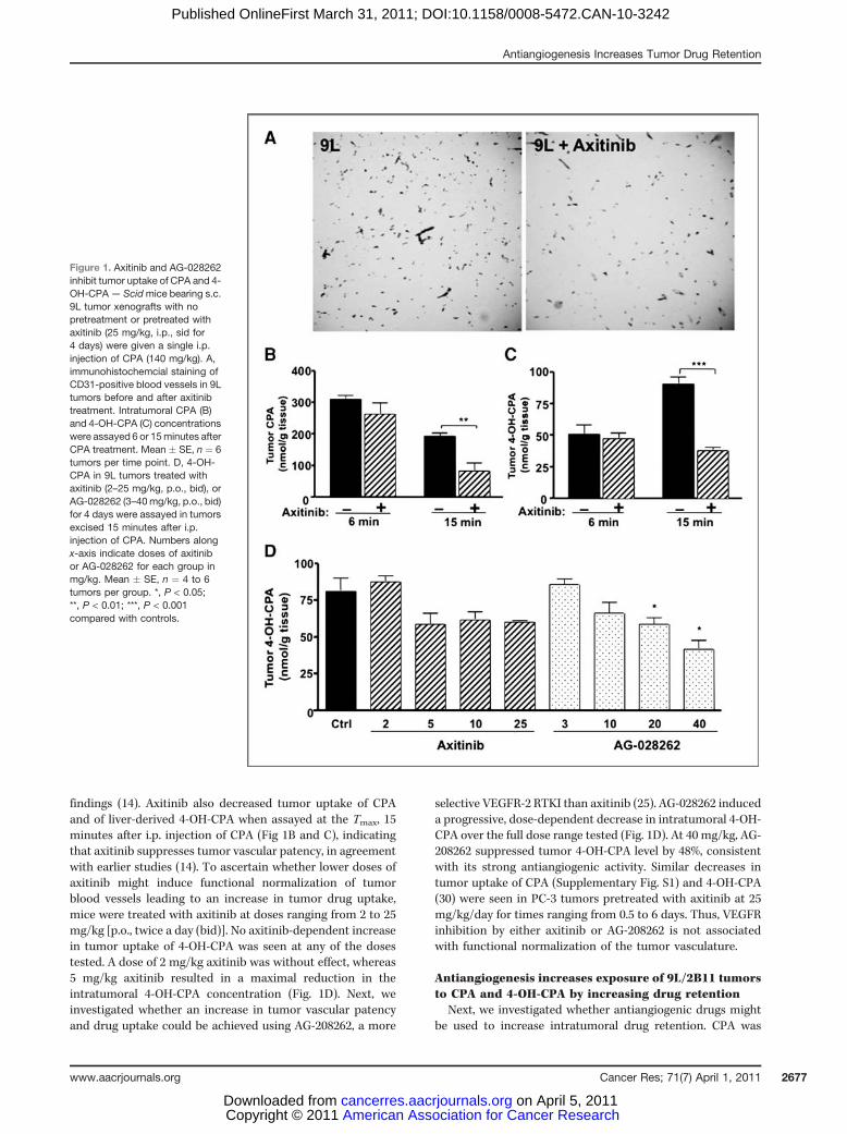
findings (14). Axitinib also decreased tumor uptake of CPAand of liver-derived 4-OH-CPA when assayed at the Tmax, 15minutes after i.p. injection of CPA (Fig 1B and C), indicatingthat axitinib suppresses tumor vascular patency, in agreementwith earlier studies (14). To ascertain whether lower doses ofaxitinib might induce functional normalization of tumorblood vessels leading to an increase in tumor drug uptake,mice were treated with axitinib at doses ranging from 2 to 25mg/kg [p.o., twice a day (bid)]. No axitinib-dependent increasein tumor uptake of 4-OH-CPA was seen at any of the dosestested. A dose of 2 mg/kg axitinib was without effect, whereas5 mg/kg axitinib resulted in a maximal reduction in theintratumoral 4-OH-CPA concentration (Fig. 1D). Next, weinvestigated whether an increase in tumor vascular patencyand drug uptake could be achieved using AG-208262, a more
selective VEGFR-2 RTKI than axitinib (25). AG-028262 induceda progressive, dose-dependent decrease in intratumoral 4-OH-CPA over the full dose range tested (Fig. 1D). At 40 mg/kg, AG-208262 suppressed tumor 4-OH-CPA level by 48%, consistentwith its strong antiangiogenic activity. Similar decreases intumor uptake of CPA (Supplementary Fig. S1) and 4-OH-CPA(30) were seen in PC-3 tumors pretreated with axitinib at 25mg/kg/day for times ranging from 0.5 to 6 days. Thus, VEGFRinhibition by either axitinib or AG-208262 is not associatedwith functional normalization of the tumor vasculature.
Antiangiogenesis increases exposure of 9L/2B11 tumorsto CPA and 4-OH-CPA by increasing drug retention
Next, we investigated whether antiangiogenic drugs mightbe used to increase intratumoral drug retention. CPA was
Figure 1. Axitinib and AG-028262inhibit tumor uptake of CPA and 4-OH-CPA— Scidmice bearing s.c.9L tumor xenografts with nopretreatment or pretreated withaxitinib (25 mg/kg, i.p., sid for4 days) were given a single i.p.injection of CPA (140 mg/kg). A,immunohistochemcial staining ofCD31-positive blood vessels in 9Ltumors before and after axitinibtreatment. Intratumoral CPA (B)and 4-OH-CPA (C) concentrationswere assayed 6 or 15minutes afterCPA treatment. Mean � SE, n ¼ 6tumors per time point. D, 4-OH-CPA in 9L tumors treated withaxitinib (2–25 mg/kg, p.o., bid), orAG-028262 (3–40mg/kg, p.o., bid)for 4 days were assayed in tumorsexcised 15 minutes after i.p.injection of CPA. Numbers alongx-axis indicate doses of axitinibor AG-028262 for each group inmg/kg. Mean � SE, n ¼ 4 to 6tumors per group. *, P < 0.05;**, P < 0.01; ***, P < 0.001compared with controls.
Antiangiogenesis Increases Tumor Drug Retention
www.aacrjournals.org Cancer Res; 71(7) April 1, 2011 2677
American Association for Cancer Research Copyright © 2011 on April 5, 2011cancerres.aacrjournals.orgDownloaded from
Published OnlineFirst March 31, 2011; DOI:10.1158/0008-5472.CAN-10-3242
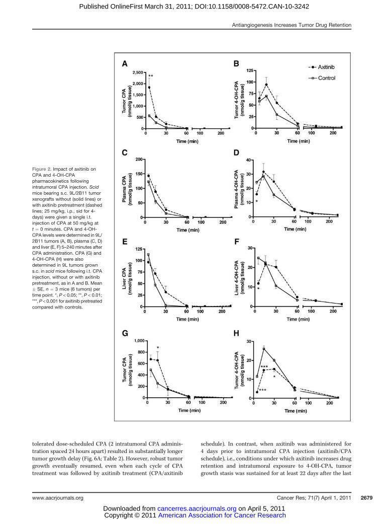
injected intratumorally into 9L/2B11 tumors, where CYP2B11,a cytochrome P450 enzyme, catalyzes intratumoral metabo-lism of CPA to its active metabolite, 4-OH-CPA. CPA that exitsthe tumor intact can be converted by liver P450 enzymes to 4-OH-CPA, a portion of which may then reenter the tumor (27).Five minutes after CPA injection, intratumoral CPA levelswere 580 mmol/L in untreated tumors as compared with 1,840mmol/L in axitinib-pretreated tumors (Fig. 2A). However, theinitial tumor level of 4-OH-CPA was not significantly higher inthe axitinib-pretreated tumors (Fig. 2B), indicating that axi-tinib does not increase the intrinsic rate of intratumoral CPA4-hydroxylation, despite the 3.2-fold increase in initial intra-tumoral CPA concentration. This finding can be explained byCYP2B11 being already saturated at the concentration of 580mmol/L CPA that is reached when CPA is injected i.t. withoutprior axitinib treatment [c.f., Km (CPA) ¼ 70 mmol/L in 9L/2B11 cells; ref 28]. As the intratumoral CPA levels declinedwith time due to a combination of drug efflux and intratu-moral conversion to 4-OH-CPA, we observed an increase inoverall intratumoral 4-OH-CPA exposure with axitinib treat-ment (Fig. 2B). Total intratumoral exposure to CPA, and 4-OH-CPA was 76% to 77% higher in axitinib-pretreated mice, asindicated by area under the curve (AUC; Table 1). This findingis consistent with axitinib slowing drug efflux from thetumors. After 30 minutes, intratumoral 4-OH-CPA levelsdeclined both with and without axitinib-pretreatment, reflect-ing depletion of the intratumoral pool of CPA available formetabolism to 4-OH-CPA. The Cmax of tumor exposure to 4-OH-CPA was also increased with axitinib pretreatment, by42% (Table 1B). CPA and 4-OH-CPA levels were substantiallylower in plasma and liver than in the tumor (Fig. 2C–F), as isexpected for an intratumoral route of drug delivery. Moreover,at the 5-minute time point, plasma and liver 4-OH-CPA levelswere significantly lower in axitinib-pretreated mice than incontrols (P < 0.05), indicating that the greater retention of CPAand 4-OH-CPA by the axitinib-pretreated tumor renders CPAless available for hepatic metabolism and also reduces theefflux of tumor-derived 4-OH-CPA during this initial timeperiod. By 30 minutes, however, plasma and liver levels ofCPA and 4-OH-CPA were higher in the axitinib-pretreatedmice, reflecting the delay in drug release from the tumors.
To confirm the role of intratumoral P450 metabolism in theobserved retention of 4-OH-CPA, we used mice bearing wild-type 9L tumors to investigate the impact of axitinib pretreat-ment on the intratumoral pharmacokinetics of CPA and 4-OH-following i.t. CPA injection. Wild-type 9L tumors do notexpress significant levels of P450 enzymes and cannot meta-bolize CPA to 4-OH-CPA. As anticipated, we observed higherlevels of intratumoral CPA in the axitinib-pretreated tumors atboth 6 and 15 minutes, reflecting increased tumor drugretention (Fig. 2G), however, intratumoral 4-OH-CPA levelswere lower through 30 minutes, reflecting the inhibition oftumor uptake of 4-OH-CPA formed in the liver (Fig. 2H).
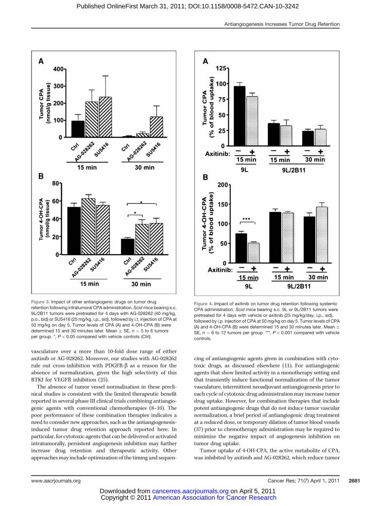
Two other antiangiogenic drugs, AG-028262 and SU5416,were investigated for their effects on intratumoral drug reten-tion in mice bearing 9L/2B11 tumors (Fig. 3). Four days ofpretreatment with AG-028262 or SU5416 had no significanteffect on intratumoral CPA or 4-OH-CPA levels 15 minutes
after intratumoral CPA injection, although a trend of increas-ing drug retention was apparent for CPA (Fig. 3A). However, 30minutes after CPA injection, when intratumoral CPA concen-trations decreased well below the Km for metabolism byCYP2B11 (c.f.; Fig. 2A), both antiangiogenic drugs significantlyincreased intratumoral 4-OH-CPA compared with controls (P< 0.05; Fig. 3B). Thus, prolonged tumor drug retention mayrepresent a common response to antiangiogenesis treatmentsinvolving these RTKIs.
Although axitinib decreased tumor levels of both CPA and4-OH-CPA in mice bearing wild-type 9L tumors following i.p.CPA administration (Fig. 4A and B, left set of bars; also seeFig. 1B and C; refs. 11, 14), axitinib effected no such decrease intumor 4-OH-CPA levels inmice bearing 9L/2B11 tumors either15 or 30 minutes after i.p. injection of CPA (Fig. 4B). This canbe explained by the increased retention of CPA that enters theaxitinib-pretreated tumors, which makes CPA more availablefor metabolism by the tumor cell-expressed P450 2B11enzyme, and by the increased retention of tumor cell-derived4-OH-CPA. This increased retention of CPA and 4-OH-CPA inpart compensates for the decreased tumor uptake of 4-OH-CPA that is formed in the liver. Thus, antiangiogenesis notonly increases the exposure of tumor cells to drugs deliveredintratumorally, but can also increase intratumoral metabo-lism, and exposure, to the activated form of a prodrug that isadministered systemically.
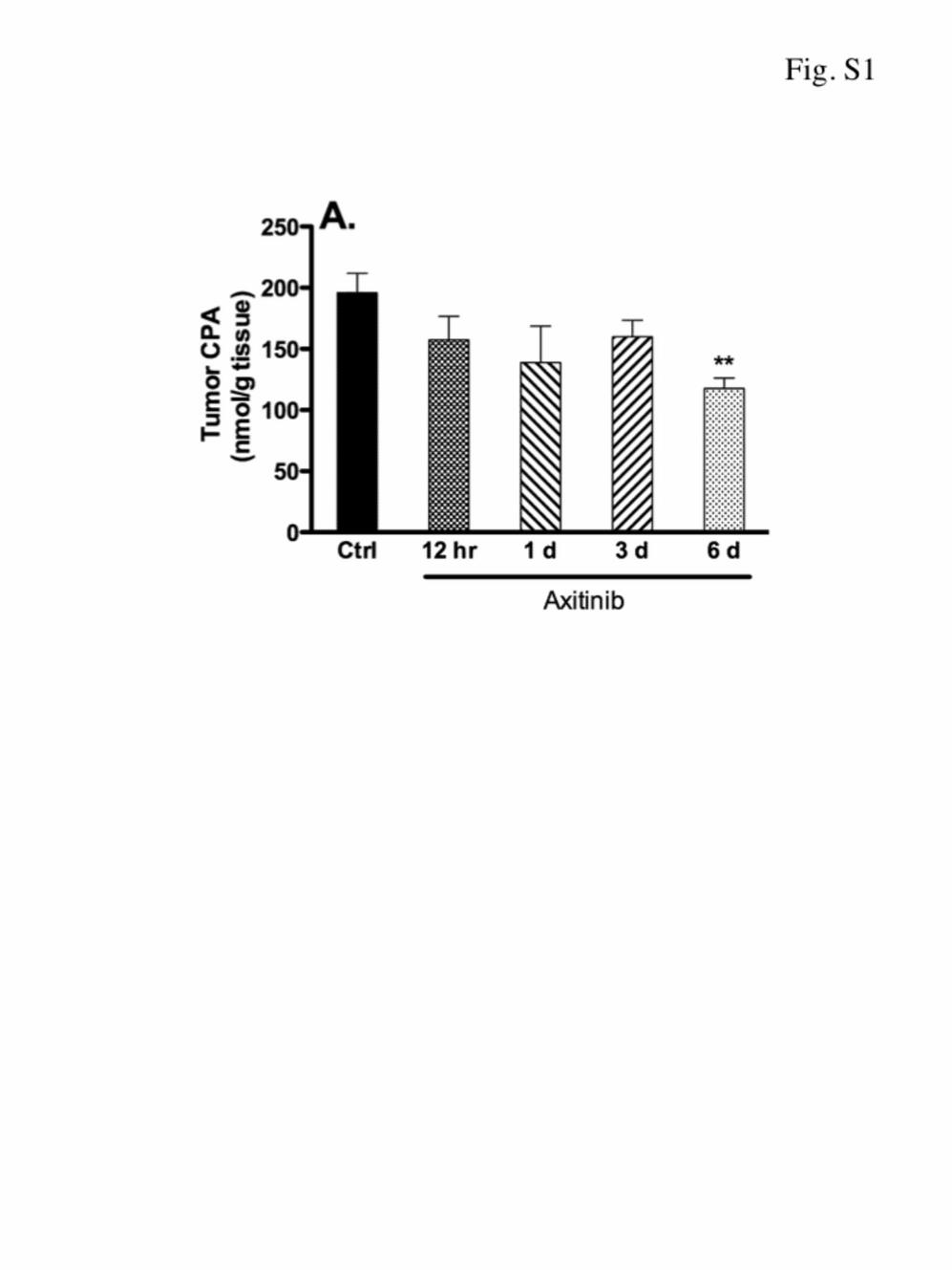
In control experiments, we observed that the CPA 4-hydro-xylase activity of 9L/2B11 tumor-derived microsomes was notaltered by axitinib pretreatment (Supplementary Fig. S2).Hepatic cytochrome P450-catalyzed CPA 4-hydroxylation isalso unaffected by axitinib treatment (14). Thus, the increasein intratumoral concentration of 4-OH-CPA seen followingaxitinib treatment cannot be explained by an increase in theintrinsic CPA 4-hydroxylase activity of either the liver ortumor.
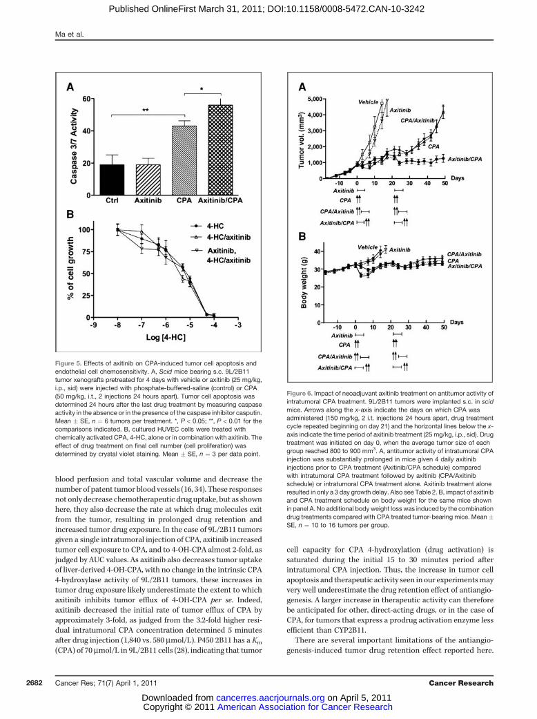
Axitinib enhances CPA-induced tumor cell apoptosisThe impact of the enhanced drug retention following
axitinib treatment on tumor cell apoptosis was investigatedby measuring caspase activity in tumor samples excised 24hours after the last drug treatment. Basal 9L/2B11 tumorcaspase activity was unaffected by axitinib treatment (Fig. 5A).Intratumoral administration of CPA (50 mg/kg, 2 injections 24hours apart) significantly increased caspase activity. However,the highest caspase activity was observed when mice bearingthe intratumoral CPA-treated 9L/2B11 tumors were pre-treated with axitinib (Fig. 5A). Thus, the prolonged exposureof tumor cells to 4-OH-CPA translates into a significantincrease in tumor cell apoptosis. Axitinib had no effect onthe intrinsic sensitivity of endothelial cells to activated CPA, asdetermined using cultured HUVEC cells (Fig. 5B).
Axitinib enhances the antitumor activity ofintratumoral CPA injection
The therapeutic impact of axitinib-enhanced tumor drugretention was investigated in a tumor growth delay study.Four days of axitinib treatment resulted in a transient (�3day) delay in 9L/2B11 tumor growth, whereas maximum
Ma et al.
Cancer Res; 71(7) April 1, 2011 Cancer Research2678
American Association for Cancer Research Copyright © 2011 on April 5, 2011cancerres.aacrjournals.orgDownloaded from
Published OnlineFirst March 31, 2011; DOI:10.1158/0008-5472.CAN-10-3242
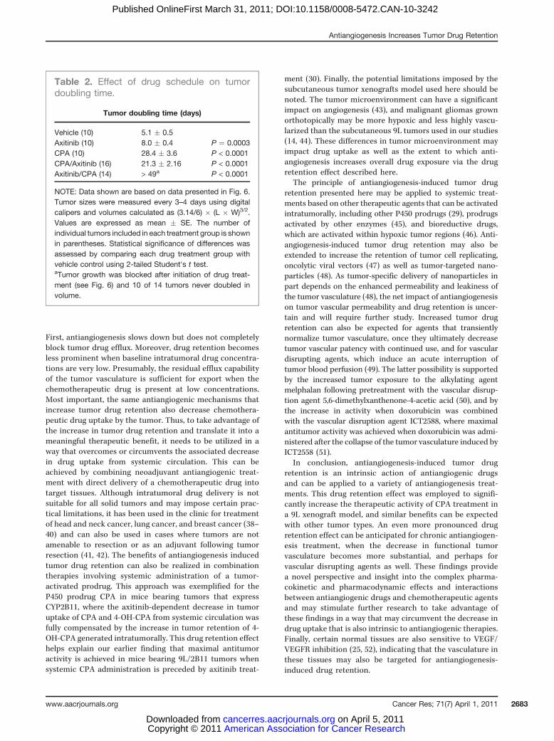
tolerated dose-scheduled CPA (2 intratumoral CPA adminis-tration spaced 24 hours apart) resulted in substantially longertumor growth delay (Fig. 6A; Table 2). However, robust tumorgrowth eventually resumed, even when each cycle of CPAtreatment was followed by axitinib treatment (CPA/axitinib
schedule). In contrast, when axitinib was administered for4 days prior to intratumoral CPA injection (axitinib/CPAschedule), i.e., conditions under which axitinib increases drugretention and intratumoral exposure to 4-OH-CPA, tumorgrowth stasis was sustained for at least 22 days after the last
Figure 2. Impact of axitinib onCPA and 4-OH-CPApharmacokinetics followingintratumoral CPA injection. Scidmice bearing s.c. 9L/2B11 tumorxenografts without (solid lines) orwith axitinib pretreatment (dashedlines; 25 mg/kg, i.p., sid for 4-days) were given a single i.t.injection of CPA at 50 mg/kg att ¼ 0 minutes. CPA and 4-OH-CPA levels were determined in 9L/2B11 tumors (A, B), plasma (C, D)and liver (E, F) 5–240 minutes afterCPA administration. CPA (G) and4-OH-CPA (H) were alsodetermined in 9L tumors growns.c. in scid mice following i.t. CPAinjection, without or with axitinibpretreatment, as in A and B. Mean� SE, n ¼ 3 mice (6 tumors) pertime point. *, P < 0.05; **, P < 0.01;***,P < 0.001 for axitinib pretreatedcompared with controls.
Antiangiogenesis Increases Tumor Drug Retention
www.aacrjournals.org Cancer Res; 71(7) April 1, 2011 2679
American Association for Cancer Research Copyright © 2011 on April 5, 2011cancerres.aacrjournals.orgDownloaded from
Published OnlineFirst March 31, 2011; DOI:10.1158/0008-5472.CAN-10-3242
cycle of drug treatment (Fig. 6A; P < 0.05, 1-way ANOVA, day 0to day 49 for axitinib/CPA schedule vs. CPA/axitinib schedule).Similar body weight profiles were observed for the CPA mono-therapy and for both axitinib-CPA schedules (Fig. 6B), indicat-ing that no additional toxicities were associated with theaxitinib/CPA schedule. A small body weight loss occurred aftereach CPA treatment, as is typical for this cytotoxic drug. Thus,the extended drug retention associated with neoadjuvant axi-tinib treatment significantly enhances CPA antitumor activity.
Discussion
In the present study, we investigated how VEGF receptor-targeted antiangiogenic agents modulate chemotherapeuticdrug delivery and drug retention by the tumor. Antiangiogen-esis was found to decrease tumor uptake of the anticancerprodrug CPA and its active metabolite, 4-OH-CPA, consistentwith the established requirement for a functional tumorvasculature and blood flow for effective drug delivery. How-ever, in mice bearing tumors that express the CPA-activatingcytochrome P450 enzyme CYP2B11, neoadjuvant axitininbtreatment significantly increased the AUC of intratumoral4-OH-CPA exposure following intratumoral CPA administra-tion, leading to an increase in tumor cell apoptosis and amajor increase in antitumor activity, as seen in tumor growthdelay studies. Increased tumor drug retention was also seenwith two other antiangiogenic agents, indicating that thiseffect is a general response to tumor antiangiogenesis. Impor-tantly, the increases in therapeutic activity were achieveddespite the transient nature of the tumor drug retention effectof neoadjuvant axitinib treatment, and they cannot beexplained by an increase in tumor cell or endothelial cellchemosensitivity following axitinib treatment (14). Further-
more, no increase in antitumor activity was seen when CPAtreatment preceded axitinib administration, consistent withthe proposed mechanism for the improved therapeuticresponse, namely, antiangiogenesis-dependent drug retentionleading to increased tumor cell exposure to 4-OH-CPA. Finally,when CPA was administered systemically and activated intra-tumorally, the decreases in tumor uptake of both CPA and 4-OH-CPA due to antiangiogenesis were counter-balanced, andfully compensated for, by antiangiogenesis-induced drugretention. Thus, when a tumor-activated prodrug is adminis-tered systemically, antiangiogenesis-induced drug retentioncan counteract the decrease in drug uptake while at the sametime retaining the therapeutic benefits of antiangiogenesis-induced tumor cell starvation.
Antiangiogenesis induces morphological normalization ofthe tumor vasculature, which involves pruning of immatureblood vessels, a decrease in blood vessel tortuosity and dila-tion, and a closer association between pericytes and tumorendothelial cells (12). This can lead to functional improve-ments, as shown by the increases in tumor vascular patencyand drug uptake and decreases in tumor hypoxia reported inpreclinical studies with several antiangiogenic agents (31).However, these effects are short lived and they disappear withcontinued antiangiogenic drug treatment (32, 33). Moreover,for axitinib (15, 34) and certain other antiangiogenic drugs (17,35, 36), although morphological normalization of the tumorvasculature and improved functionality of individual bloodvessels may occur, overall tumor vascular patency and capa-city for drug uptake actually decrease. Presently, we investi-gated the hypothesis that tumor blood vessel normalizationleading to increased drug uptake can be achieved by reducingthe dose of the antiangiogenic RTKI; however, we obtainedno evidence for such functional normalization of tumor
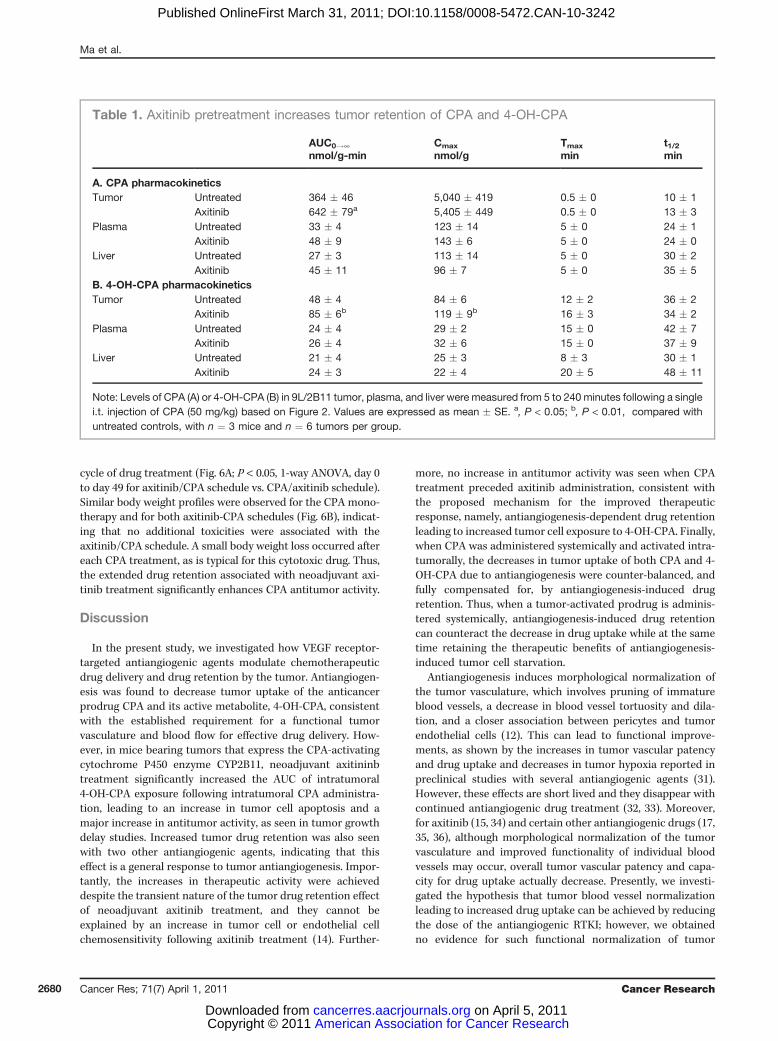
Table 1. Axitinib pretreatment increases tumor retention of CPA and 4-OH-CPA
AUC0!¥ Cmax Tmax t1/2nmol/g-min nmol/g min min
A. CPA pharmacokineticsTumor Untreated 364 � 46 5,040 � 419 0.5 � 0 10 � 1
Axitinib 642 � 79a 5,405 � 449 0.5 � 0 13 � 3Plasma Untreated 33 � 4 123 � 14 5 � 0 24 � 1
Axitinib 48 � 9 143 � 6 5 � 0 24 � 0Liver Untreated 27 � 3 113 � 14 5 � 0 30 � 2
Axitinib 45 � 11 96 � 7 5 � 0 35 � 5B. 4-OH-CPA pharmacokineticsTumor Untreated 48 � 4 84 � 6 12 � 2 36 � 2
Axitinib 85 � 6b 119 � 9b 16 � 3 34 � 2Plasma Untreated 24 � 4 29 � 2 15 � 0 42 � 7
Axitinib 26 � 4 32 � 6 15 � 0 37 � 9Liver Untreated 21 � 4 25 � 3 8 � 3 30 � 1
Axitinib 24 � 3 22 � 4 20 � 5 48 � 11
Note: Levels of CPA (A) or 4-OH-CPA (B) in 9L/2B11 tumor, plasma, and liver were measured from 5 to 240 minutes following a singlei.t. injection of CPA (50 mg/kg) based on Figure 2. Values are expressed as mean � SE. a, P < 0.05; b, P < 0.01, compared withuntreated controls, with n ¼ 3 mice and n ¼ 6 tumors per group.
Ma et al.
Cancer Res; 71(7) April 1, 2011 Cancer Research2680
American Association for Cancer Research Copyright © 2011 on April 5, 2011cancerres.aacrjournals.orgDownloaded from
Published OnlineFirst March 31, 2011; DOI:10.1158/0008-5472.CAN-10-3242
vasculature over a more than 10-fold dose range of eitheraxitinib or AG-028262. Moreover, our studies with AG-028262rule out cross-inhibition with PDGFR-b as a reason for theabsence of normalization, given the high selectivity of thisRTKI for VEGFR inhibition (25).The absence of tumor vessel normalization in these precli-
nical studies is consistent with the limited therapeutic benefitreported in several phase III clinical trials combining antiangio-genic agents with conventional chemotherapies (8–10). Thepoor performance of these combination therapies indicates aneed to consider new approaches, such as the antiangiogenesis-induced tumor drug retention approach reported here. Inparticular, for cytotoxic agents that can bedelivered or activatedintratumorally, persistent angiogenesis inhibition may furtherincrease drug retention and therapeutic activity. Otherapproachesmay include optimization of the timing and sequen-
cing of antiangiogenic agents given in combination with cyto-toxic drugs, as discussed elsewhere (11). For antiangiogenicagents that show limited activity in a monotherapy setting andthat transiently induce functional normalization of the tumorvasculature, intermittent neoadjuvant antiangiogenesis prior toeach cycle of cytotoxic drug administrationmay increase tumordrug uptake. However, for combination therapies that includepotent antiangiogenic drugs that do not induce tumor vascularnormalization, a brief period of antiangiogenic drug treatmentat a reduced dose, or temporary dilation of tumor blood vessels(37) prior to chemotherapy administration may be required tominimize the negative impact of angiogenesis inhibition ontumor drug uptake.
Tumor uptake of 4-OH-CPA, the active metabolite of CPA,was inhibited by axitinib and AG-028262, which reduce tumor
Figure 4. Impact of axitinib on tumor drug retention following systemicCPA administration. Scid mice bearing s.c. 9L or 9L/2B11 tumors werepretreated for 4 days with vehicle or axitinib (25 mg/kg/day, i.p., sid),followed by i.p. injection of CPA at 50mg/kg on day 5. Tumor levels of CPA(A) and 4-OH-CPA (B) were determined 15 and 30 minutes later. Mean �SE, n ¼ 6 to 12 tumors per group. ***, P < 0.001 compared with vehiclecontrols.
Figure 3. Impact of other antiangiogenic drugs on tumor drugretention following intratumoral CPA administration. Scidmice bearing s.c.9L/2B11 tumors were pretreated for 4 days with AG-028262 (40 mg/kg,p.o., bid) or SU5416 (25 mg/kg, i.p., sid), followed by i.t. injection of CPA at50 mg/kg on day 5. Tumor levels of CPA (A) and 4-OH-CPA (B) weredetermined 15 and 30 minutes later. Mean � SE, n ¼ 5 to 6 tumorsper group. *, P < 0.05 compared with vehicle controls (Ctrl).
Antiangiogenesis Increases Tumor Drug Retention
www.aacrjournals.org Cancer Res; 71(7) April 1, 2011 2681
American Association for Cancer Research Copyright © 2011 on April 5, 2011cancerres.aacrjournals.orgDownloaded from
Published OnlineFirst March 31, 2011; DOI:10.1158/0008-5472.CAN-10-3242
blood perfusion and total vascular volume and decrease thenumber of patent tumor blood vessels (16, 34). These responsesnot only decrease chemotherapeutic druguptake, but as shownhere, they also decrease the rate at which drug molecules exitfrom the tumor, resulting in prolonged drug retention andincreased tumor drug exposure. In the case of 9L/2B11 tumorsgiven a single intratumoral injection of CPA, axitinib increasedtumor cell exposure to CPA, and to 4-OH-CPA almost 2-fold, asjudged by AUC values. As axitinib also decreases tumor uptakeof liver-derived 4-OH-CPA, with no change in the intrinsic CPA4-hydroxylase activity of 9L/2B11 tumors, these increases intumor drug exposure likely underestimate the extent to whichaxitinib inhibits tumor efflux of 4-OH-CPA per se. Indeed,axitinib decreased the initial rate of tumor efflux of CPA byapproximately 3-fold, as judged from the 3.2-fold higher resi-dual intratumoral CPA concentration determined 5 minutesafter drug injection (1,840 vs. 580 mmol/L). P450 2B11 has a Km
(CPA) of 70 mmol/L in 9L/2B11 cells (28), indicating that tumor
cell capacity for CPA 4-hydroxylation (drug activation) issaturated during the initial 15 to 30 minutes period afterintratumoral CPA injection. Thus, the increase in tumor cellapoptosis and therapeutic activity seen in our experimentsmayvery well underestimate the drug retention effect of antiangio-genesis. A larger increase in therapeutic activity can thereforebe anticipated for other, direct-acting drugs, or in the case ofCPA, for tumors that express a prodrug activation enzyme lessefficient than CYP2B11.
There are several important limitations of the antiangio-genesis-induced tumor drug retention effect reported here.
Figure 6. Impact of neoadjuvant axitinib treatment on antitumor activity ofintratumoral CPA treatment. 9L/2B11 tumors were implanted s.c. in scidmice. Arrows along the x-axis indicate the days on which CPA wasadministered (150 mg/kg, 2 i.t. injections 24 hours apart, drug treatmentcycle repeated beginning on day 21) and the horizontal lines below the x-axis indicate the time period of axitinib treatment (25 mg/kg, i.p., sid). Drugtreatment was initiated on day 0, when the average tumor size of eachgroup reached 800 to 900 mm3. A, antitumor activity of intratumoral CPAinjection was substantially prolonged in mice given 4 daily axitinibinjections prior to CPA treatment (Axitinib/CPA schedule) comparedwith intratumoral CPA treatment followed by axitinib (CPA/Axitinibschedule) or intratumoral CPA treatment alone. Axitinib treatment aloneresulted in only a 3 day growth delay. Also see Table 2. B, impact of axitiniband CPA treatment schedule on body weight for the same mice shownin panel A. No additional bodyweight loss was induced by the combinationdrug treatments compared with CPA treated tumor-bearing mice. Mean �SE, n ¼ 10 to 16 tumors per group.
Figure 5. Effects of axitinib on CPA-induced tumor cell apoptosis andendothelial cell chemosensitivity. A, Scid mice bearing s.c. 9L/2B11tumor xenografts pretreated for 4 days with vehicle or axitinib (25 mg/kg,i.p., sid) were injected with phosphate-buffered-saline (control) or CPA(50 mg/kg, i.t., 2 injections 24 hours apart). Tumor cell apoptosis wasdetermined 24 hours after the last drug treatment by measuring caspaseactivity in the absence or in the presence of the caspase inhibitor casputin.Mean � SE, n ¼ 6 tumors per treatment. *, P < 0.05; **, P < 0.01 for thecomparisons indicated. B, cultured HUVEC cells were treated withchemically activated CPA, 4-HC, alone or in combination with axitinib. Theeffect of drug treatment on final cell number (cell proliferation) wasdetermined by crystal violet staining. Mean � SE, n ¼ 3 per data point.
Ma et al.
Cancer Res; 71(7) April 1, 2011 Cancer Research2682
American Association for Cancer Research Copyright © 2011 on April 5, 2011cancerres.aacrjournals.orgDownloaded from
Published OnlineFirst March 31, 2011; DOI:10.1158/0008-5472.CAN-10-3242
First, antiangiogenesis slows down but does not completelyblock tumor drug efflux. Moreover, drug retention becomesless prominent when baseline intratumoral drug concentra-tions are very low. Presumably, the residual efflux capabilityof the tumor vasculature is sufficient for export when thechemotherapeutic drug is present at low concentrations.Most important, the same antiangiogenic mechanisms thatincrease tumor drug retention also decrease chemothera-peutic drug uptake by the tumor. Thus, to take advantage ofthe increase in tumor drug retention and translate it into ameaningful therapeutic benefit, it needs to be utilized in away that overcomes or circumvents the associated decreasein drug uptake from systemic circulation. This can beachieved by combining neoadjuvant antiangiogenic treat-ment with direct delivery of a chemotherapeutic drug intotarget tissues. Although intratumoral drug delivery is notsuitable for all solid tumors and may impose certain prac-tical limitations, it has been used in the clinic for treatmentof head and neck cancer, lung cancer, and breast cancer (38–40) and can also be used in cases where tumors are notamenable to resection or as an adjuvant following tumorresection (41, 42). The benefits of antiangiogenesis inducedtumor drug retention can also be realized in combinationtherapies involving systemic administration of a tumor-activated prodrug. This approach was exemplified for theP450 prodrug CPA in mice bearing tumors that expressCYP2B11, where the axitinib-dependent decrease in tumoruptake of CPA and 4-OH-CPA from systemic circulation wasfully compensated by the increase in tumor retention of 4-OH-CPA generated intratumorally. This drug retention effecthelps explain our earlier finding that maximal antitumoractivity is achieved in mice bearing 9L/2B11 tumors whensystemic CPA administration is preceded by axitinib treat-
ment (30). Finally, the potential limitations imposed by thesubcutaneous tumor xenografts model used here should benoted. The tumor microenvironment can have a significantimpact on angiogenesis (43), and malignant gliomas grownorthotopically may be more hypoxic and less highly vascu-larized than the subcutaneous 9L tumors used in our studies(14, 44). These differences in tumor microenvironment mayimpact drug uptake as well as the extent to which anti-angiogenesis increases overall drug exposure via the drugretention effect described here.
The principle of antiangiogenesis-induced tumor drugretention presented here may be applied to systemic treat-ments based on other therapeutic agents that can be activatedintratumorally, including other P450 prodrugs (29), prodrugsactivated by other enzymes (45), and bioreductive drugs,which are activated within hypoxic tumor regions (46). Anti-angiogenesis-induced tumor drug retention may also beextended to increase the retention of tumor cell replicating,oncolytic viral vectors (47) as well as tumor-targeted nano-particles (48). As tumor-specific delivery of nanoparticles inpart depends on the enhanced permeability and leakiness ofthe tumor vasculature (48), the net impact of antiangiogenesison tumor vascular permeability and drug retention is uncer-tain and will require further study. Increased tumor drugretention can also be expected for agents that transientlynormalize tumor vasculature, once they ultimately decreasetumor vascular patency with continued use, and for vasculardisrupting agents, which induce an acute interruption oftumor blood perfusion (49). The latter possibility is supportedby the increased tumor exposure to the alkylating agentmelphalan following pretreatment with the vascular disrup-tion agent 5,6-dimethylxanthenone-4-acetic acid (50), and bythe increase in activity when doxorubicin was combinedwith the vascular disruption agent ICT2588, where maximalantitumor activity was achieved when doxorubicin was admi-nistered after the collapse of the tumor vasculature induced byICT2558 (51).
In conclusion, antiangiogenesis-induced tumor drugretention is an intrinsic action of antiangiogenic drugsand can be applied to a variety of antiangiogenesis treat-ments. This drug retention effect was employed to signifi-cantly increase the therapeutic activity of CPA treatment ina 9L xenograft model, and similar benefits can be expectedwith other tumor types. An even more pronounced drugretention effect can be anticipated for chronic antiangiogen-esis treatment, when the decrease in functional tumorvasculature becomes more substantial, and perhaps forvascular disrupting agents as well. These findings providea novel perspective and insight into the complex pharma-cokinetic and pharmacodynamic effects and interactionsbetween antiangiogenic drugs and chemotherapeutic agentsand may stimulate further research to take advantage ofthese findings in a way that may circumvent the decrease indrug uptake that is also intrinsic to antiangiogenic therapies.Finally, certain normal tissues are also sensitive to VEGF/VEGFR inhibition (25, 52), indicating that the vasculature inthese tissues may also be targeted for antiangiogenesis-induced drug retention.
Table 2. Effect of drug schedule on tumordoubling time.
Tumor doubling time (days)
Vehicle (10) 5.1 � 0.5Axitinib (10) 8.0 � 0.4 P ¼ 0.0003CPA (10) 28.4 � 3.6 P < 0.0001CPA/Axitinib (16) 21.3 � 2.16 P < 0.0001Axitinib/CPA (14) > 49a P < 0.0001
NOTE: Data shown are based on data presented in Fig. 6.Tumor sizes were measured every 3–4 days using digitalcalipers and volumes calculated as (3.14/6) � (L � W)3/2.Values are expressed as mean � SE. The number ofindividual tumors included in each treatment group is shownin parentheses. Statistical significance of differences wasassessed by comparing each drug treatment group withvehicle control using 2-tailed Student's t test.aTumor growth was blocked after initiation of drug treat-ment (see Fig. 6) and 10 of 14 tumors never doubled involume.
Antiangiogenesis Increases Tumor Drug Retention
www.aacrjournals.org Cancer Res; 71(7) April 1, 2011 2683
American Association for Cancer Research Copyright © 2011 on April 5, 2011cancerres.aacrjournals.orgDownloaded from
Published OnlineFirst March 31, 2011; DOI:10.1158/0008-5472.CAN-10-3242
Disclosure of Potential Conflicts of Interests
No potential conflicts of interests were disclosed.
Acknowledgemnts
We thank Pfizer Global Research and Development for providing axitinib andAG-028262, and Dr. Dana Hu-Lowe for useful discussions.
Grant Support
Supported in part by NIH grant CA49248 (to D.J. Waxman).The costs of publication of this article were defrayed in part by the payment
of page charges. This article must therefore be hereby marked advertisement inaccordance with 18 U.S.C. Section 1734 solely to indicate this fact.
Received September 3, 2010; revised December 9, 2010; accepted January 3,2011; published OnlineFirst March 29, 2011.
References1. Minchinton AI, Tannock IF. Drug penetration in solid tumours. Nat Rev
Cancer 2006;6:583–92.2. Moses MA, Brem H, Langer R. Advancing the field of drug delivery:
taking aim at cancer. Cancer Cell 2003;4:337–41.3. Sonveaux P. Provascular strategy: Targeting functional adaptations of
mature blood vessels in tumors to selectively influence the tumorvascular reactivity and improve cancer treatment. Radiother Oncol2008;86:300–13.
4. Hurwitz H, Fehrenbacher L, Novotny W, Cartwright T, Hainsworth J,Heim W, et al. Bevacizumab plus irinotecan, fluorouracil, and leucov-orin for metastatic colorectal cancer. N Engl J Med 2004;350:2335–42.
5. Sandler A, Gray R, Perry MC, Brahmer J, Schiller JH, Dowlati A, et al.Paclitaxel-carboplatin alone or with bevacizumab for non-small-celllung cancer. N Engl J Med 2006;355:2542–50.
6. Escudier B, Eisen T, Stadler WM, Szczylik C, Oudard S, Siebels M,et al. Sorafenib in advanced clear-cell renal-cell carcinoma. N Engl JMed 2007;356:125–34.
7. Motzer RJ, Hutson TE, Tomczak P, Michaelson MD, Bukowski RM,Rixe O, et al. Sunitinib versus interferon alfa in metastatic renal-cellcarcinoma. N Engl J Med 2007;356:115–24.
8. Hauschild A, Agarwala SS, Trefzer U, Hogg D, Robert C, Hersey P,et al. Results of a phase III, randomized, placebo-controlled study ofsorafenib in combination with carboplatin and paclitaxel as second-line treatment in patients with unresectable stage III or stage IVmelanoma. J Clin Oncol 2009;27:2823–30.
9. Scagliotti G, Novello S, von Pawel J, Reck M, Pereira JR, Thomas M,et al. Phase III study of carboplatin and paclitaxel alone or withsorafenib in advanced non-small-cell lung cancer. J Clin Oncol2010;28:1835–42.
10. Herbst RS, Sun Y, Eberhardt WE, Germonpr�e P, Saijo N, Zhou C, et al.Vandetanib plus docetaxel versus docetaxel as second-line treatmentfor patients with advanced non-small-cell lung cancer (ZODIAC):adouble-blind, randomised, phase 3 trial. Lancet Oncol 2010;11:619–26.
11. Ma J, Waxman DJ. Combination of antiangiogenesis with chemother-apy for more effective cancer treatment. Mol Cancer Ther2008;7:3670–84.
12. Jain RK. Normalization of tumor vasculature: an emerging concept inantiangiogenic therapy. Science 2005;307:58–62.
13. Hu-Lowe DD, Zou HY, Grazzini ML, Hallin ME, Wickman GR, Amund-son K, et al. Nonclinical antiangiogenesis and antitumor activities ofaxitinib (AG-013736), an oral, potent, and selective inhibitor of vas-cular endothelial growth factor receptor tyrosine kinases 1, 2, 3. ClinCancer Res 2008;14:7272–83.
14. Ma J, Waxman DJ. Modulation of the antitumor activity of metronomiccyclophosphamide by the angiogenesis inhibitor axitinib. Mol CancerTher 2008;7:79–89.
15. Nakahara T, Norberg SM, Shalinsky DR, Hu-Lowe DD, McDonald DM.Effect of inhibition of vascular endothelial growth factor signaling ondistribution of extravasated antibodies in tumors. Cancer Res2006;66:1434–45.
16. Fenton BM, Paoni SF. The addition of AG-013736 to fractionatedradiation improves tumor response without functionally normalizingthe tumor vasculature. Cancer Res 2007;67:9921–8.
17. Franco M, Man S, Chen L, Emmenegger U, Shaked Y, Cheung AM,et al. Targeted anti-vascular endothelial growth factor receptor-2therapy leads to short-term and long-term impairment of vascular
function and increase in tumor hypoxia. Cancer Res 2006;66:3639–48.
18. Ma J, Pulfer S, Li S, Chu J, Reed K, Gallo JM. Pharmacodynamic-mediated reduction of temozolomide tumor concentrations by theangiogenesis inhibitor TNP-470. Cancer Res 2001;61:5491–8.
19. Williams KJ, Telfer BA, Brave S, Kendrew J, Whittaker L, Stratford IJ,et al. ZD6474, a potent inhibitor of vascular endothelial growth factorsignaling, combined with radiotherapy: schedule-dependentenhancement of antitumor activity. Clin Cancer Res 2004;10:8587–93.
20. Tailor TD, Hanna G, Yarmolenko PS, Dreher MR, Betof AS, Nixon AB,et al. Effect of pazopanib on tumor microenvironment and liposomedelivery. Mol Cancer Ther 2010;9:1798–808.
21. Mendel DB, Laird AD, Xin X, Louie SG, Christensen JG, Li G, et al. Invivo antitumor activity of SU11248, a novel tyrosine kinase inhibitortargeting vascular endothelial growth factor and platelet-derivedgrowth factor receptors: determination of a pharmacokinetic/pharma-codynamic relationship. Clin Cancer Res 2003;9:327–37.
22. Zhou Q, Guo P, Gallo JM. Impact of angiogenesis inhibition bysunitinib on tumor distribution of temozolomide. Clin Cancer Res2008;14:1540–9.
23. Kabbinavar F, Hurwitz HI, Fehrenbacher L, Meropol NJ, Novotny WF,Lieberman G, et al. Phase II, randomized trial comparing bevacizumabplus fluorouracil (FU)/leucovorin (LV) with FU/LV alone in patients withmetastatic colorectal cancer. J Clin Oncol 2003;21:60–5.
24. Abramsson A, Lindblom P, Betsholtz C. Endothelial and nonendothe-lial sources of PDGF-B regulate pericyte recruitment and influencevascular pattern formation in tumors. J Clin Invest 2003;112:1142–51.
25. Mancuso MR, Davis R, Norberg SM, O'Brien S, Sennino B, NakaharaT, et al. Rapid vascular regrowth in tumors after reversal of VEGFinhibition. J Clin Invest 2006;116:2610–21.
26. Dvorak HF. Vascular permeability factor/vascular endothelial growthfactor: a critical cytokine in tumor angiogenesis and a potential targetfor diagnosis and therapy. J Clin Oncol 2002;20:4368–80.
27. Chen CS, Jounaidi Y, Su T, Waxman DJ. Enhancement of intratumoralcyclophosphamide pharmacokinetics and antitumor activity in a P4502B11-based cancer gene therapy model. Cancer Gene Ther 2007;14:935–44.
28. Jounaidi Y, Chen C-S, Veal GJ, Waxman DJ. Enhanced antitumoractivity of P450 prodrug-based gene therapy using the low Kmcyclophosphamide 4-hydroxylase P450 2B11. Mol Cancer Ther2006;5:541–55.
29. Roy P, Waxman DJ. Activation of oxazaphosphorines by cytochromeP450: application to gene-directed enzyme prodrug therapy for can-cer. Toxicol In Vitro 2006;20:176–86.
30. Ma J, Waxman DJ. Dominant effect of antiangiogenesis in combina-tion therapy involving cyclophosphamide and axitinib. Clin CancerRes 2009;15:578–88.
31. Fukumura D, Duda DG, Munn LL, Jain RK. Tumor microvasculatureand microenvironment: novel insights through intravital imaging inpre-clinical models. Microcirculation 2010;17:206–25.
32. Winkler F, Kozin SV, Tong RT, Chae SS, Booth MF, Garkavtsev I, et al.Kinetics of vascular normalization by VEGFR2 blockade governs braintumor response to radiation: role of oxygenation, angiopoietin-1, andmatrix metalloproteinases. Cancer Cell 2004;6:553–63.
33. Ansiaux R, Baudelet C, Jordan BF, Crokart N, Martinive P, DeWever J,et al. Mechanism of reoxygenation after antiangiogenic therapy using
Ma et al.
Cancer Res; 71(7) April 1, 2011 Cancer Research2684
American Association for Cancer Research Copyright © 2011 on April 5, 2011cancerres.aacrjournals.orgDownloaded from
Published OnlineFirst March 31, 2011; DOI:10.1158/0008-5472.CAN-10-3242
SU5416 and its importance for guiding combined antitumor therapy.Cancer Res 2006;66:9698–704.
34. Inai T, Mancuso M, Hashizume H, Baffert F, Haskell A, Baluk P, et al.Inhibition of vascular endothelial growth factor (VEGF) signaling incancer causes loss of endothelial fenestrations, regression of tumorvessels, and appearance of basement membrane ghosts. Am J Pathol2004;165:35–52.
35. Riesterer O, Honer M, JochumW, Oehler C, Ametamey S, Pruschy M.Ionizing radiation antagonizes tumor hypoxia induced by antiangio-genic treatment. Clin Cancer Res 2006;12:3518–24.
36. Claes A, Wesseling P, Jeuken J, Maass C, Heerschap A, LeendersWP. Antiangiogenic compounds interfere with chemotherapy of braintumors due to vessel normalization. Mol Cancer Ther 2008;7:71–8.
37. Martinive P, De Wever J, Bouzin C, Baudelet C, Sonveaux P, Gr�egoireV, et al. Reversal of temporal and spatial heterogeneities in tumorperfusion identifies the tumor vascular tone as a tunable variable toimprove drug delivery. Mol Cancer Ther 2006;5:1620–7.
38. Almond BA, Hadba AR, Freeman ST, Cuevas BJ, York AM, DetrisacCJ, et al. Efficacy of mitoxantrone-loaded albumin microspheres forintratumoral chemotherapy of breast cancer. J Control Release2003;91:147–55.
39. Celikoglu F, Celikoglu SI, Goldberg EP. Bronchoscopic intratumoralchemotherapy of lung cancer. Lung Cancer 2008;61:1–12.
40. Duvillard C, Polycarpe E, Romanet P, Chauffert B. [Intratumoralchemotherapy:experimental data and applications to head and necktumors]. Ann Otolaryngol Chir Cervicofac 2007;124:53–60.
41. Menei P, Jadaud E, Faisant N, Boisdron-Celle M, Michalak S, FournierD, et al. Stereotaxic implantation of 5-fluorouracil-releasing micro-spheres in malignant glioma. Cancer 2004;100:405–10.
42. Vogl TJ, Muller PK, Mack MG, Straub R, Engelmann K, Neuhaus P.[Therapeutic options in non-resectable liver metastases. Percuta-neous radiological interventions]. Chirurg 1999;70:133–40.
43. Fidler IJ. Angiogenic heterogeneity: regulation of neoplastic angio-genesis by the organ microenvironment. J Natl Cancer Inst2001;93:1040–1.
44. Amberger-Murphy V. Hypoxia helps glioma to fight therapy. CurrCancer Drug Targets 2009;9:381–90.
45. Portsmouth D, Hlavaty J, Renner M. Suicide genes for cancer therapy.Mol Aspects Med 2007;28:4–41.
46. Tredan O, Garbens AB, Lalani AS, Tannock IF. The hypoxia-activatedProDrug AQ4N penetrates deeply in tumor tissues and complementsthe limited distribution of mitoxantrone. Cancer Res 2009;69:940–7.
47. Libertini S, Iacuzzo I, Perruolo G, Scala S, Ieranò C, Franco R, et al.Bevacizumab increases viral distribution in human anaplasticthyroid carcinoma xenografts and enhances the effects of E1A-defective adenovirus dl922–947. Clin Cancer Res 2008;14:6505–14.
48. Byrne JD, Betancourt T, Brannon-Peppas L. Active targeting schemesfor nanoparticle systems in cancer therapeutics. Adv Drug Deliv Rev2008;60:1615–26.
49. Tozer GM, Kanthou C, Lewis G, Prise VE, Vojnovic B, Hill SA. Tumourvascular disrupting agents: combating treatment resistance. Br JRadiol 2008;81Spec No 1:S12–20.
50. Pruijn FB, van Daalen M, Holford NH, Wilson WR. Mechanisms ofenhancement of the antitumour activity of melphalan by the tumour-blood-flow inhibitor 5,6-dimethylxanthenone-4-acetic acid. CancerChemother Pharmacol 1997;39:541–6.
51. Atkinson JM, Falconer RA, Edwards DR, Pennington CJ, Siller CS,Shnyder SD, et al. Development of a novel tumor-targeted vasculardisrupting agent activated by membrane-type matrix metalloprotei-nases. Cancer Res 2010;70:6902–12.
52. Kamba T, Tam BY, Hashizume H, Haskell A, Sennino B, MancusoMR,et al. VEGF-dependent plasticity of fenestrated capillaries in thenormal adult microvasculature. Am J Physiol Heart Circ Physiol2006;290:H560–76.
Antiangiogenesis Increases Tumor Drug Retention
www.aacrjournals.org Cancer Res; 71(7) April 1, 2011 2685
American Association for Cancer Research Copyright © 2011 on April 5, 2011cancerres.aacrjournals.orgDownloaded from
Published OnlineFirst March 31, 2011; DOI:10.1158/0008-5472.CAN-10-3242
2011;71:2675-2685. Published OnlineFirst March 31, 2011.Cancer Res Jie Ma, Chong-Sheng Chen, Todd Blute, et al. Antiangiogenesis Enhances Intratumoral Drug Retention
Updated Version 10.1158/0008-5472.CAN-10-3242doi:
Access the most recent version of this article at:
MaterialSupplementary
htmlhttp://cancerres.aacrjournals.org/content/suppl/2011/03/25/0008-5472.CAN-10-3242.DC1.Access the most recent supplemental material at:
Cited Articles http://cancerres.aacrjournals.org/content/71/7/2675.full.html#ref-list-1
This article cites 52 articles, 29 of which you can access for free at:
E-mail alerts related to this article or journal.Sign up to receive free email-alerts
SubscriptionsReprints and
[email protected] Department atTo order reprints of this article or to subscribe to the journal, contact the AACR
To request permission to re-use all or part of this article, contact the AACR Publications
American Association for Cancer Research Copyright © 2011 on April 5, 2011cancerres.aacrjournals.orgDownloaded from
Published OnlineFirst March 31, 2011; DOI:10.1158/0008-5472.CAN-10-3242
Ma et al, Supplementary Material
Supplementary Materials and Methods Chemicals - Axitinib and AG-028262 were obtained from Pfizer Global Research and Development (San Diego, CA). SU5416, CPA, NADPH and semicarbazide hydrochloride were purchased from Sigma-Aldrich Co. (St. Louis, MO). 4-hydroperoxycyclophosphamide (4-HC) was obtained from Dr. Ulf Niemeyer (Baxter Oncology GmbH, Frankfurt, Germany). Carboxymethyl cellulose (low viscosity) was purchased from MP Biomedicals (Solon, OH). Polyethylene glycol 400 (PEG-400) was purchased from Fisher Scientific (Hampton, NH). Caspase-GloTM 3/7 assay kit was purchasd from Promega (Madison, WI). Casputin, a selective inhibitor of caspase 3 and caspase 7, was purchased from BIOMOL International (Plymouth Meeting, PA). Protease inhibitor cocktail tablets were purchased from Roche Diagnostics (Mannheim, Germany). DMEM culture medium and fetal bovine serum (FBS) were purchased from Invitrogen (Carlsbad, CA). Human umbilical vein endothelial cells (HUVEC) and EGM-2 BulletKit culture medium were purchased from Cambrex Bio Science (East Rutherford, NJ). Drug treatments and tissue 4-OH-CPA analysis – In one treatment schedule, mice bearing 9L tumors were randomized to different groups on the day of initial drug treatment when the average tumor volume reached ~500 mm3. Axitinib (0.4 mg/ml to 5 mg/ml) and AG-028262 (0.2 mg/ml to 8 mg/ml) were suspended in 0.5% carboxymethyl cellulose and administered orally using a 20-gauge gavage feeding needle in a volume of 5 !l/g body weight every 12 hr for 4 days. Axitinib was administered at 2, 5, 10, or 25 mg/kg body weight per dose, and AG-028262 at 3, 10, 20, or 40 mg/kg body weight per dose. This treatment schedule and route was selected to mimic clinical studies, where axitinib is administered orally twice daily (1), and based on the finding that anti-angiogenesis-induced tumor vascular normalization is typically observed during a 2-7 day period of time following the initial drug treatment (2-4). The impact of these anti-angiogenic agents on the uptake of 4-OH-CPA by 9L tumors was determined as follows. Freshly prepared CPA dissolved in PBS (140 mM NaCl, 10 mM Na2HPO4, 2.7 mM KCl, 1.8 mM KH2PO4) was filtered through a 0.2 µm acrodisc syringe filter (Pall Corp., Ann Arbor, MI) and a single test dose of CPA (either 50 or 140 mg/kg body weight, as indicated) was administered by i.p. injection to tumor-bearing mice 24 hr after the last axitinib or AG-028262 treatment. Mice were killed 6 or 15 min after CPA injection, the latter time corresponding to the Tmax of plasma and liver 4-OH-CPA (5,6). Blood, liver and tumor samples were collected and assayed for 4-OH-CPA and/or CPA, as described below.
In a second treatment schedule, male scid mice implanted s.c. with 9L/2B11 tumor cells were randomized to different groups on the day of initial drug treatment, when the average tumor volume reached ~ 800 mm3. This tumor size was chosen to facilitate intratumoral drug delivery. Axitinib was suspended at 5 mg/ml of polyethylene glycol 400 and sonicated at room temperature for 10-20 min to obtain a fine suspension. The suspension was adjusted to pH 2-3 using 0.1 N HCl, sonicated for an additional 20 min, then stored at 4°C in the dark up to 4-5 days. On the day of dosing, a final 3:7 (v/v) ratio of polyethylene glycol 400: H2O was obtained by adding acidified water (pH 2-3) with brief vortexing. Axitinib was administered to the tumor-bearing mice by daily i.p. injection at 25 mg/kg body weight and in a volume of 5 µl per g body weight. AG-028262 was prepared as described above and administered at 40 mg/kg body weight per injection (p.o., bid). SU5416 was dissolved in DMSO and injected at 25 mg/kg body weight (i.p., sid). CPA was administered 24 hr after the last anti-angiogenesis treatment, or to untreated
Ma et al, Supplementary Material
controls. Intratumoral delivery of CPA was achieved using a syringe pump (cat. # 70-2212, Harvard Apparatus, Holliston, MA) set to deliver 1 µl per sec. The total CPA dose given to each mouse (50 mg/kg body weight) was dissolved in a volume of ~120 µl PBS and administered intratumorally by injection at three sites per tumor over a period of ~1 min in each of 2 tumors/mouse. Blood, tumor, and liver tissues were collected at times ranging from 5 to 240 min after the last CPA injection, typically for n = 3 mice and n = 6 tumors per time point. HPLC and MS analysis of CPA and 4-OH-CPA – Tissue samples (tumor, plasma, liver) were processed and analyzed for 4-OH-CPA by HPLC after derivatization (7,8). Tissue recovery of 4-OH-CPA was 60 + 3% with a sensitivity of 1 µM under these conditions (8). CPA levels in tumor plasma and liver were determined using liquid chromatography-tandem mass spectrometry, essentially as described (9). Briefly, 100 µl of tumor or liver homogenate, or 25 µl of plasma, was mixed with 100 µl of 10 ng/µl or 1 ng/µl ifosfamide solution (internal standard). The sample was adjusted to a total volume of 200 µl, and 800 µl of acetonitrile was added to precipitate the protein. Samples were vortexed and centrifuged and the supernatant was removed and evaporated under a stream of N2 at 40°C. The pellet was then reconstituted in 100 µl of HPLC mobile phase and stored at –20°C until analysis. CPA was analyzed using a Waters 600 controller coupled to an API 2000 tandem mass spectrometer (Applied Biosystems, Foster City, CA). Chromatography was carried out using a Luna C18(2) column (5 µm, 150 x 3.0 mm) (Phenomenex, Torrance, CA) with an isocratic mobile phase consisting of 35% acetonitrile in 20 mM ammonium acetate buffer (pH 5.0) and a flow rate of 0.4 ml/min. CPA was determined in the positive electrospray ionization mode at 350°C capillary temperature, 5.0 kv ionization voltage, and 20v collision energy. Nitrogen was used as the collision gas at a setting of 12 (arbitrary units). Multiple reaction monitoring data were acquired with the following variables: the CPA transition was m/z 260.9 > 140.1 and ifosfamide was m/z 260.9 > 154.1, with a dwell time of 600 milliseconds for both. Selected ion monitoring was used for CPA (m/z 140.1) and ifosfamide (m/z 154.1); retention times were 4.2 min for CPA and 4.0 min for ifosfamide. CPA was quantified using Analyst software (Applied Biosystems, Foster City, CA). Data are expressed as nmol CPA or 4-OH-CPA per g of tissue (i.e., µM), mean ± SE based on n = 3 individual mice or n = 6 individual tumors per time point. Pharmacokinetic data analysis - Pharmacokinetics data were analyzed as described (5). Using 6 pharmacokinetic time course data points collected for each of 3 individual mice (3 blood, 3 liver and 6 tumor samples at each time point). Data were randomly assigned into three separate time course data sets for plasma and liver, and six separate time course data sets for the tumors. Each data set was used to calculate AUC, t1/2, Cmax and Tmax values using WinNonlin software version 1.5 (Scientific Consulting Inc, Apex, NC) with a simple noncompartment model. The initial intratumoral concentration of CPA (at t = 0.5 min) was set to the mean CPA concentration within each of the six tumor samples and was calculated based on the quantity of CPA injected divided by the tumor’s weight. Pharmacokinetic parameters were calculated based on with the descriptive statistics module of WinNonlin software. Statistical comparisons using a nonparametric t-test were performed using GraphPad Prism software version 4 (GraphPad, Inc., San Diego, CA). Data are expressed as nmol CPA or 4-OH-CPA per g of tissue (i.e., µM), mean ± SE based on n = 3 individual mice or n = 6 individual tumors per time point. CPA 4-hydroxylase activity was assayed in tissues obtained from mice bearing 9L/2B11 tumors treated with vehicle (5 µl per g body weight, i.p. sid) or axitinib (25 mg/kg body weight, i.p. sid) for 4
Ma et al, Supplementary Material
days. Twenty four hr after the last drug treatment, microsomes were prepared from fresh tumor tissue and CPA 4-hydroxylase activity was assayed by HPLC following incubation in vitro with 0.2 or 2 mM of CPA (6). Endothelial cell chemosensitivity to 4-OH-CPA - HUVEC cells were grown in EGM-2 culture medium containing 2% FBS at 37oC in a humidified, 5% CO2 atmosphere. 4-HC, a chemically activated derivative of CPA that spontaneously decomposes to 4-OH-CPA in aqueous solution, was used to assay the chemosensitivity of cultured HUVEC cells to 4-OH-CPA. Cells were treated with one of the following schedules: 1) 4-HC alone at 0 to 100 µM for 4 days; 2) concurrent 4-HC and axitinib (2 µM) for 4 days; 3) two days of axitinib pretreatment followed by 4 days of co-treatment with 4-HC and axitinib. Relative cell number was determined after 4 days by crystal violet staining. Data are expressed as mean ± SE based on n = 3 individual wells per point. Apoptosis in CPA-induced tumors - Mice bearing 9L/2B11 tumors were treated with one of the following schedules: 1) untreated controls; 2) axitinib (25 mg/kg body weight, i.p., sid, for 4 days) followed by PBS (120 µl/mouse, administered on day 5 at 20 µl per injection at each of 3 intratumoral sites per tumor and 2 tumors/mouse, and repeated on day 6); 3) vehicle (5 µl per g body weight, i.p., sid, for 4 days) followed by CPA (a total of 50 mg CPA/kg body weight, administered on day 5 in a total vol of 120 µl/mouse at 20 µl per injection at each of 3 intratumoral sites per tumor and 2 tumors/mouse, and repeated on day 6); 4) axitinib daily for 4 days followed by CPA on days 5 and 6. Tumors were collected 24 hr after the last intratumoral injection and homogenized in ice-cold KPi buffer (100 mM potassium phosphate, 1 mM EDTA, 1 mM dithiothreitol, 1 mM Na3VO4, and 1 tablet of protease inhibitor cocktail/50 ml). Following an initial centrifugation (12,000 rpm in a Beckman Coulter microfuge) for 20 min at 4oC, the supernatant was centrifuged at 35,000 rpm in a Sorval T-1270 rotor for 1 hr at 4oC. Supernatant protein concentrations were determined by Bradford assay. Caspase activity was determined using the Caspase-Glo 3/7 assay kit and the manufacturer’s protocol, with the following modifications. Protein samples in a 96-well plate (50 µg in 50 µl) were incubated with 10 µl of potassium phosphate buffer or casputin stock solution (2 mg/ml) at room temparature for 15 min. 60 µl of reconstituted caspase 3/7 substrate was then added to each well. After a brief shaking, the plate was incubated at room temparature for 30 min and luciferase activity was determined using a Victor-3 Multilabel Counter (Perkin Elmer). Caspase 3/7 activity was calculated as the difference of luciferase activity in the presence vs. absence of casputin. Data are expressed as mean ± SE values based on n = 6 individual tumors per treatment group. References for Supplementary Materials and Methods 1. Rixe O, Bukowski RM, Michaelson MD, et al. Axitinib treatment in patients with cytokine-refractory metastatic renal-cell cancer: a phase II study. Lancet Oncol 2007; 8: 975-84. 2. Dickson PV, Hamner JB, Sims TL, et al. Bevacizumab-induced transient remodeling of the vasculature in neuroblastoma xenografts results in improved delivery
Ma et al, Supplementary Material
and efficacy of systemically administered chemotherapy. Clin Cancer Res 2007; 13: 3942-50. 3. Wildiers H, Guetens G, De Boeck G, et al. Effect of antivascular endothelial growth factor treatment on the intratumoral uptake of CPT-11. Br J Cancer 2003; 88: 1979-86. 4. Winkler F, Kozin SV, Tong RT, et al. Kinetics of vascular normalization by VEGFR2 blockade governs brain tumor response to radiation: role of oxygenation, angiopoietin-1, and matrix metalloproteinases. Cancer Cell 2004; 6: 553-63. 5. Chen CS, Jounaidi Y, Su T, Waxman DJ. Enhancement of intratumoral cyclophosphamide pharmacokinetics and antitumor activity in a P450 2B11-based cancer gene therapy model. Cancer Gene Ther 2007; 14: 935-44. 6. Ma J, Waxman DJ. Modulation of the antitumor activity of metronomic cyclophosphamide by the angiogenesis inhibitor axitinib. Mol Cancer Ther 2008; 7: 79-89. 7. Chen CS, Lin JT, Goss KA, He YA, Halpert JR, Waxman DJ. Activation of the anticancer prodrugs cyclophosphamide and ifosfamide: identification of cytochrome P450 2B enzymes and site-specific mutants with improved enzyme kinetics. Mol Pharmacol 2004; 65: 1278-85. 8. Yu LJ, Drewes P, Gustafsson K, Brain EGC, Hecht JED, Waxman DJ. In vivo modulation of alternative pathways of P450-catalyzed cyclophosphamide metabolism: impact on pharmacokinetics and antitumor activity. J Pharmacol Exp Ther 1999; 288: 928-37. 9. Pass GJ, Carrie D, Boylan M, et al. Role of hepatic cytochrome p450s in the pharmacokinetics and toxicity of cyclophosphamide: studies with the hepatic cytochrome p450 reductase null mouse. Cancer Res 2005; 65: 4211-7. 10. Ma J, Waxman DJ. Dominant effect of antiangiogenesis in combination therapy involving cyclophosphamide and axitinib. Clin Cancer Res 2009; 15: 578-88.
Ma et al, Supplementary Material
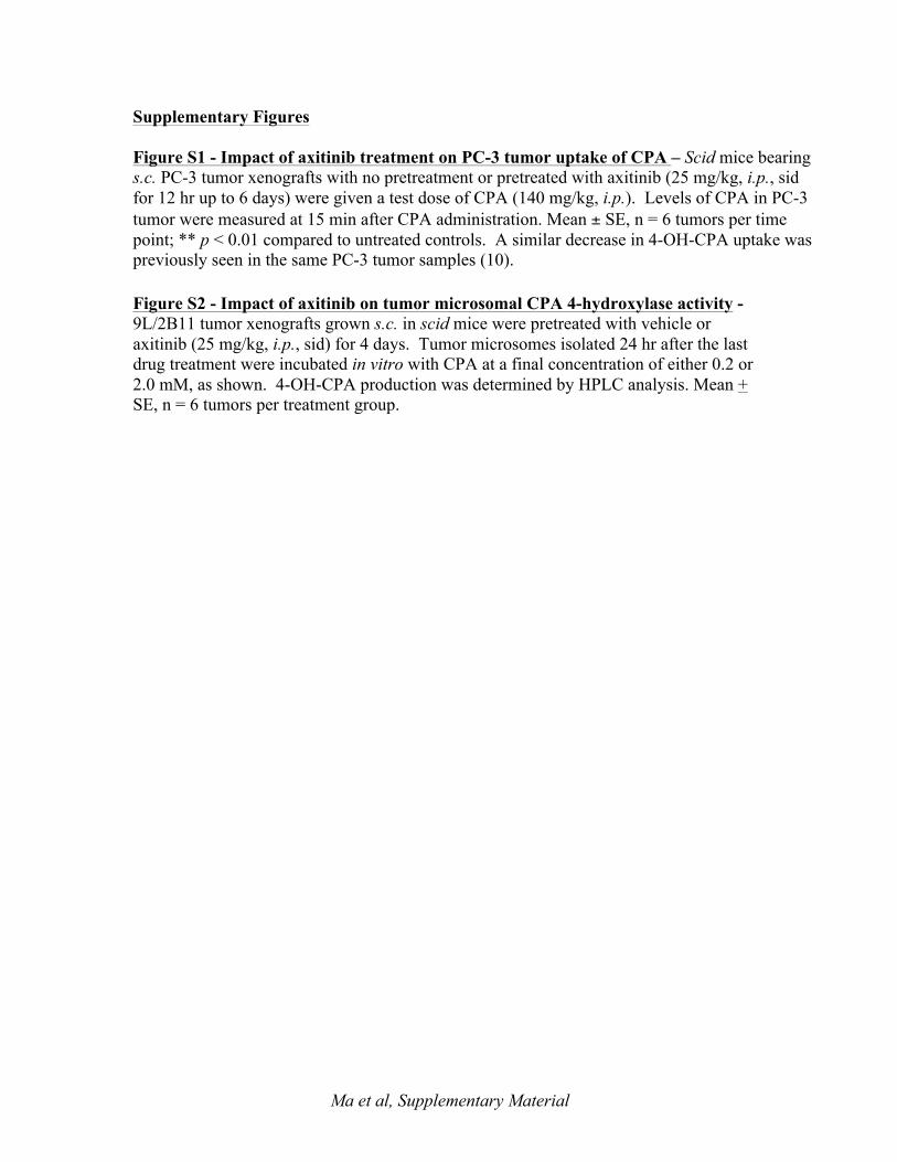
Supplementary Figures Figure S1 - Impact of axitinib treatment on PC-3 tumor uptake of CPA – Scid mice bearing s.c. PC-3 tumor xenografts with no pretreatment or pretreated with axitinib (25 mg/kg, i.p., sid for 12 hr up to 6 days) were given a test dose of CPA (140 mg/kg, i.p.). Levels of CPA in PC-3 tumor were measured at 15 min after CPA administration. Mean ± SE, n = 6 tumors per time point; ** p < 0.01 compared to untreated controls. A similar decrease in 4-OH-CPA uptake was previously seen in the same PC-3 tumor samples (10). Figure S2 - Impact of axitinib on tumor microsomal CPA 4-hydroxylase activity - 9L/2B11 tumor xenografts grown s.c. in scid mice were pretreated with vehicle or axitinib (25 mg/kg, i.p., sid) for 4 days. Tumor microsomes isolated 24 hr after the last drug treatment were incubated in vitro with CPA at a final concentration of either 0.2 or 2.0 mM, as shown. 4-OH-CPA production was determined by HPLC analysis. Mean + SE, n = 6 tumors per treatment group.


![Review Article - Hindawi Publishing Corporationdownloads.hindawi.com/journals/jo/2012/193436.pdftherapy [22]. Furthermore, antiangiogenesis can normalize tumor vasculature and decrease](https://img.pdfslide.us/doc/110x75/5f4d5f35bd4e976d402d7ac5/review-article-hindawi-publishing-therapy-22-furthermore-antiangiogenesis.jpg)