Embed Size (px)
Citation preview
Human Journals
Research Article
February 2018 Vol.:11, Issue:3
© All rights are reserved by MADHURI. Y et al.
Anti-Ulcer Activity of Hibiscus sabdariffa on Albino Rats
www.ijppr.humanjournals.com
Keywords: Hibiscus sabdariffa, antiulcer activity, pylorus
ligation method, ethanol-induced method, ranitidine, ulcer
index
ABSTRACT
To study the antiulcer activity of Hibiscus sabdariffa stems
using different models of gastric ulceration in rats. Antiulcer
activity of Hibiscus sabdariffa stem extract was studied in rats
by administration of ethanol-induced method and by pyloric
ligation method. Stem extract was administered in the dose of
250 mg/kg and 500 mg/kg orally 30 min prior to the ulcer
induction. The antiulcer activity was assessed by determining
and comparing the ulcer index in the test drug group with that
of the ulcerated control group. Ranitidine was used as a
reference drug. The ulcer index in the stem extract treated
animals was found to be significantly less in all the models
compared to ulcerated control animals. The antiulcer property
was more prominent in animals in whom ulcers were induced
by ethanol-induced and pyloric ligation. Ranitidine 50 mg/kg
produced a significant gastric ulcer protection when compared
with the control group. The antiulcer activity of Hibiscus
sabdariffa was, however, less than that of ranitidine. Our results
suggest that Hibiscus sabdariffa stem extract possess significant
antiulcer property which could be either due to the
cytoprotective action of the drug or by strengthing of gastric
mucosa and thus enhancing mucosal defense.
MADHURI. Y*, NARENDRA BABU. A
1, NANDA
KUMAR. E2, YANADAIAH. P
2
1. Chalapathi Institute of Pharmaceutical Sciences,
Chalapathi nagar, Lam, Guntur-522034.
2. Narayana Pharmacy College, Chinthareddypalem,
Nellore-524002.
Submission: 24 January 2018
Accepted: 29 January 2018
Published: 28 February 2018
www.ijppr.humanjournals.com
Citation: MADHURI. Y et al. Ijppr.Human, 2018; Vol. 11 (3): 13-26. 14
INTRODUCTION:
Ulcers are deep lesions penetration through the entire thickness of the gastrointestinal tract
(g.i.t) mucosa and muscularis mucosa. Peptic ulcer has unquestionably been a disease of the
twentieth century. Epidemiological data for this disease and its complications have shown
striking geographical variations in incidence and prevalence. There are different types of
ulcers; most common are peptic ulcer, gastric ulcer which appeared to be due to damage to
the lining of the stomach and duodenal ulcer, which was associated with excessive acid
secretion by the stomach. The etiology of peptic ulcer was fiercely debated. It is believed that
peptic ulcers developed due to an imbalance between aggressive factors (mucin, bicarbonate,
prostaglandins) leading to an interruption in the mucosal integrity1. Various factors are
implicated that play a pivotal role in the pathogenesis of ulceration like sedentary lifestyle,
alcohol intake, spicy food, drugs and various bacterial infections. Moreover, several
endogenous substances have been identified and are reported to be involved in the production
of gastrointestinal lesions in animals. The more important ones include some of the bacterial
infection, various drugs and chemicals, gastric secretion, lipid metabolites, neuropeptides,
inflammatory mediators and reactive free radicals. Oxidative stress has emerged as one of the
major pathogenic factors in the progression of ulcer that directly impaired the cellular
functions and promotes cellular organelles damage in the cell, including mitochondria,
lysosomes, and nucleus. Also, NO is accepted as the vital mediator of GIT mucosal defenses
as decreased NO generation or synthesis contribute to the pathogenesis of ulceration. The
present study summarises the ulcerogenic mechanism of these substances and the enable us to
understand the better etiology of peptic ulcer2.
MATERIALS AND METHODS:
The study was conducted on Wistar albino rats of weight 200±30 gm and maintained under
standard conditions. The major chemicals used are ranitidine, anesthetic ether, and
chloroform.
About 100 gm of powdered Hibiscus sabdariffa stems were packed in a thick paper and it
was subject to extraction by using soxhlet extraction method for 72 hrs until the marc become
colorless. Then the extract was concentrated under reduced pressure and dried in the vacuum
condition to get a semi-solid mass3.
www.ijppr.humanjournals.com
Citation: MADHURI. Y et al. Ijppr.Human, 2018; Vol. 11 (3): 13-26. 15
A. PRELIMINARY PHYTOCHEMICAL SCREENING:
The preliminary phytochemical investigations were carried out for the qualitative detection of
phytoconstituents. Qualitative tests were conducted for all the extracts to identify the various
phytoconstituents. The various tests and reagents used are given below and the observations
are recorded in table4.
Alkaloids:
(a) Dragendorff’s test: 1 ml of extract, add 1 ml of Dragendorff’s reagent (potassium
bismuth iodide solution). An orange-red precipitate indicates the presence of alkaloids.
(b) Mayer’s test: 1 ml of extract, add 1 ml of Mayer’s reagent (potassium mercuric iodide
solution). Whitish or cream colored precipitate indicates the presence of alkaloids.
(c) Hager’s test: 1 ml of extract, add 3 ml of Hager’s reagent (saturated aqueous solution of
picric acid). Yellow colored precipitate indicates the presence of alkaloids
(d) Wagner’s test: 1 ml of extract, add 2 ml of Wagner's reagent (iodine in potassium
iodide). Reddish brown colored precipitate indicates the presence of alkaloids
Carbohydrates and Glycosides:
(a) Molisch's test: Two ml of the prepared filtrate were mixed with 0.2 ml of an alcoholic
solution of α-naphthol 10% in addition to 2 ml of sulphuric acid, a bluish violet zone is
formed this indicates the presence of carbohydrates and /or glycosides.
(b) Fehling's test: In a test tube 5 ml of the filtrate were treated with 5 ml Fehling's solutions
(A & B) and heated; the appearance of a red precipitate indicates the presence.
(c) Benedict's test: To 1 ml of the filtrate, 5 ml of Benedict's reagent were added. The
mixture was heated; the appearance of red precipitate indicated the presence5.
Flavones and flavonoids:
One ml of 10% ethanolic extract of the studied plant was mixed with 0.5 ml of hydrochloric
acid (10%) and magnesium metal. A developed reddish color indicates the presence of
flavonoids. Five ml of 1% hydrochloric acid extract were shaken with sodium hydroxide; a
yellow color appeared indicating the presence of compound flavonoids.
www.ijppr.humanjournals.com
Citation: MADHURI. Y et al. Ijppr.Human, 2018; Vol. 11 (3): 13-26. 16
Proteins and amino acids:
Five ml of extract, 2 drops of freshly prepared 0.2 percent ninhydrin reagent was added and
heated. The appearance of the blue color indicates the presence of proteins, peptides or amino
acids.
Saponins:
One g of the plant under investigation was boiled with 10 ml water for few minutes and
filtrated. The filtrate was vigorously shaken. The persistent froth (1 cm height)was observed
for 1 hr indicates the presence of saponins.
Steroids:
For testing the presence of unsaturated sterols and triterpenes, 1g of the air-dried powder of
the studied plant was extracted with few ml of ethanol then filtrated and the filtrate was
evaporated to dryness. The residue was dissolved in 10 ml chloroform, filtered and the filtrate
was divided into two equal portions for preceding the following tests4,
(a) Libermann-Burchard test:
To the first portion of chloroform filtrate 1 ml of acetic acid anhydride was added, followed
by 2 ml of sulphuric acid down the wall of the test tube. The appearance of reddish-violet
color at the junction of two layers and a bluish-green color in acetic acid layer indicates the
presence.
To the second portion of chloroform filtrate, an equal volume of sulphuric acid was added.
The appearance of a red color indicated the presence.
Tannins:
About 2 g of the air-dried powder of the plant was extracted with ethanol (50 %) and tested
for the presence of tannins using the following tests.
One drop of ferric chloride was added to 2 ml of the extract, the appearance of bluish or
greenish black coloration indicates the presence of pyrogallol or catechol tannins,
respectively.
www.ijppr.humanjournals.com
Citation: MADHURI. Y et al. Ijppr.Human, 2018; Vol. 11 (3): 13-26. 17
Five ml of the alcoholic extract of the studied plant were mixed with 2 ml vanillin
Hydrochloric acid solution if a precipitate was formed. This indicates the presence of gallic
acid6.
B. METHODOLOGY:
Animal models used in the screening of antiulcer activity:
Various screening models are used for the screening of the antiulcer activity. It helps to
understand the etiology of the ulcer and screening of antiulcer agents.
Aspirin-induced ulcers
Ethanol-induced ulcers
Pylorus ligation induced ulcers
Water immersion stress-induced ulcers
Indomethacin-induced ulcers
Histamine-induced ulcers
Reserpine-induced ulcers
Serotonin-induced ulcers7
From this, majorly used models are explained below
1. Ethanol-induced ulcers method7:
Albino rats of either sex weighing between (150-200 gm) are divided into the group. The
animals are fasted for 24 hours with free access water. Animals are given test drugs or
standard drug. 1 hour later 1ml/200gm of 99.80% alcohol is administered orally to each
animal. The animals were anesthetized 1 h later with ether and stomach was incised along the
greater curvature and ulceration was scored. The number of ulcers and the length of each
ulcer were measured. Ulcer index was calculated using severity scores and the average
number of ulcers per animal. Severity scores as below.
www.ijppr.humanjournals.com
Citation: MADHURI. Y et al. Ijppr.Human, 2018; Vol. 11 (3): 13-26. 18
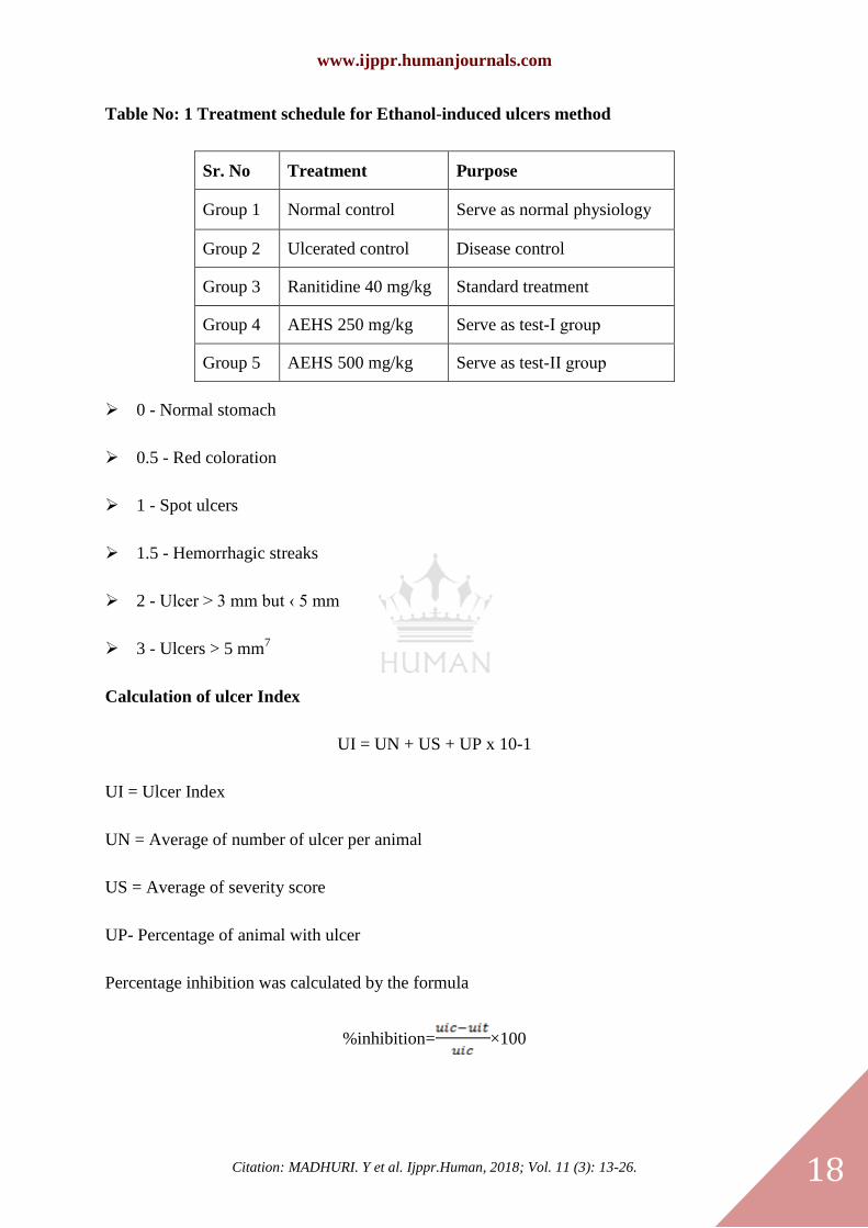
Table No: 1 Treatment schedule for Ethanol-induced ulcers method
Sr. No Treatment Purpose
Group 1 Normal control Serve as normal physiology
Group 2 Ulcerated control Disease control
Group 3 Ranitidine 40 mg/kg Standard treatment
Group 4 AEHS 250 mg/kg Serve as test-Ӏ group
Group 5 AEHS 500 mg/kg Serve as test-ӀӀ group
0 - Normal stomach
0.5 - Red coloration
1 - Spot ulcers
1.5 - Hemorrhagic streaks
2 - Ulcer > 3 mm but ‹ 5 mm
3 - Ulcers > 5 mm7
Calculation of ulcer Index
UI = UN + US + UP x 10-1
UI = Ulcer Index
UN = Average of number of ulcer per animal
US = Average of severity score
UP- Percentage of animal with ulcer
Percentage inhibition was calculated by the formula
%inhibition= ×100
www.ijppr.humanjournals.com
Citation: MADHURI. Y et al. Ijppr.Human, 2018; Vol. 11 (3): 13-26. 19
Histopathological studies were conducted by fixing stomach tissues in 10% formalin for 24 h.
The formalin-fixed specimens are embedded in paraffin and section (3-5μm) and stained with
hematoxylin and eosin dye. The histochemical sections are evaluated by light microscopy8.
2. Pylorus ligation induced ulcers7:
Albino Wister rats of either sex weighing between (150-200 gm) are divided into groups of
animals. In this method, albino rats are fasted in individual cages for 24 hours. Test drug or
standard drug or control vehicle is administered 30 minutes prior to pyloric ligation. Under
light ether anesthesia, the abdomen is opened and the pylorus was ligated. The abdomen is
then sutured. At the end of 4 hours after ligation, the animals are sacrificed with the excess of
anesthetic ether, and the stomach is dissected out gastric juice is collected were drained into
tubes and were centrifuged at 1000 rpm for 10 minutes and the volume is noted. The pH of
gastric juice is recorded by pH meter. Then the contents are subjected to analysis for free and
total acidity. The stomachs are then washed with running water to see for ulcers in the
glandular portion of the stomach. The numbers of ulcers per stomach are noted and severity
of the ulcers scored microscopically with the help of 10x lens.
Histopathological studies were conducted by fixing stomach tissues in 10% formalin for 24 h.
The formalin-fixed specimens are embedded in paraffin and section (3-5μm) and stained with
hematoxylin and eosin dye. The histochemical sections are evaluated by light microscopy.
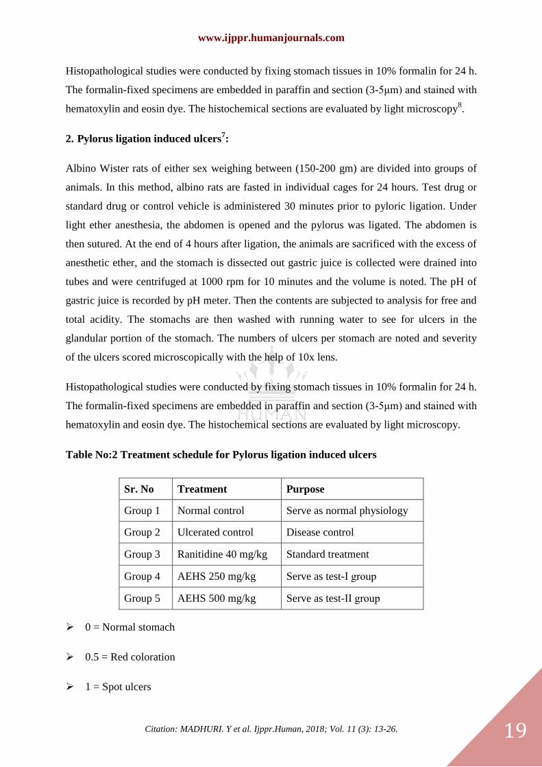
Table No:2 Treatment schedule for Pylorus ligation induced ulcers
Sr. No Treatment Purpose
Group 1 Normal control Serve as normal physiology
Group 2 Ulcerated control Disease control
Group 3 Ranitidine 40 mg/kg Standard treatment
Group 4 AEHS 250 mg/kg Serve as test-Ӏ group
Group 5 AEHS 500 mg/kg Serve as test-ӀӀ group
0 = Normal stomach
0.5 = Red coloration
1 = Spot ulcers
www.ijppr.humanjournals.com
Citation: MADHURI. Y et al. Ijppr.Human, 2018; Vol. 11 (3): 13-26. 20
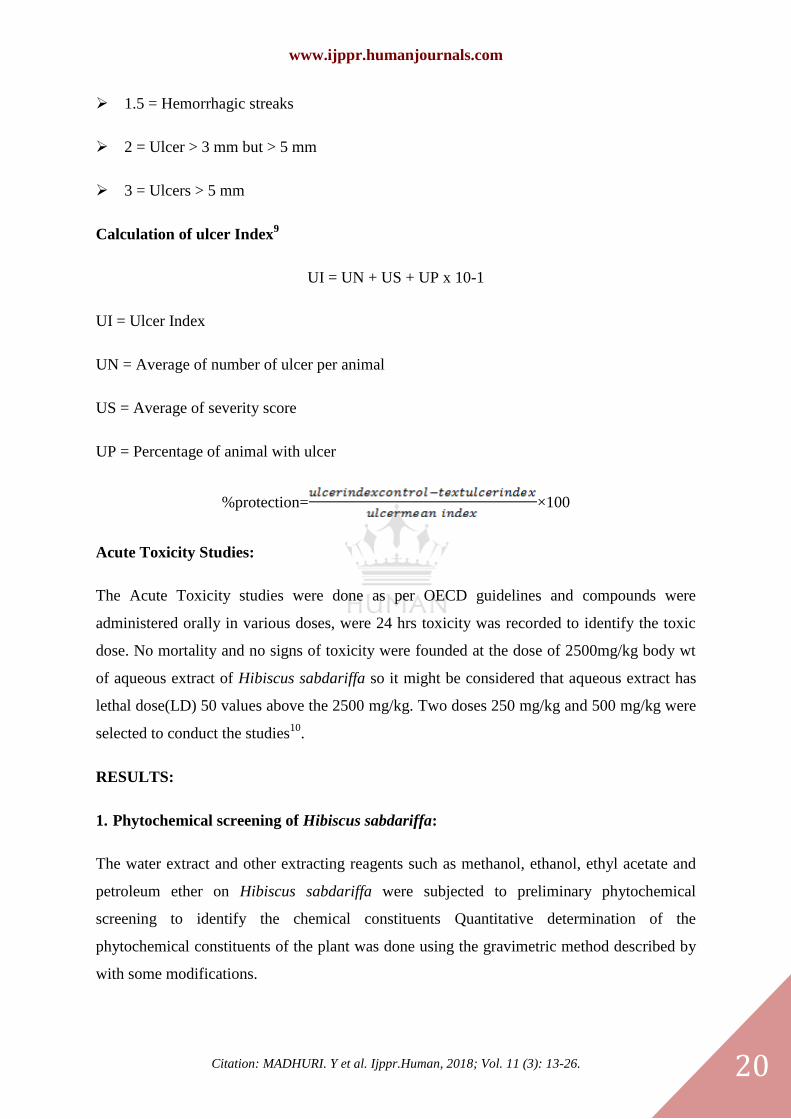
1.5 = Hemorrhagic streaks
2 = Ulcer > 3 mm but > 5 mm
3 = Ulcers > 5 mm
Calculation of ulcer Index9
UI = UN + US + UP x 10-1
UI = Ulcer Index
UN = Average of number of ulcer per animal
US = Average of severity score
UP = Percentage of animal with ulcer
%protection= ×100
Acute Toxicity Studies:
The Acute Toxicity studies were done as per OECD guidelines and compounds were
administered orally in various doses, were 24 hrs toxicity was recorded to identify the toxic
dose. No mortality and no signs of toxicity were founded at the dose of 2500mg/kg body wt
of aqueous extract of Hibiscus sabdariffa so it might be considered that aqueous extract has
lethal dose(LD) 50 values above the 2500 mg/kg. Two doses 250 mg/kg and 500 mg/kg were
selected to conduct the studies10
.
RESULTS:
1. Phytochemical screening of Hibiscus sabdariffa:
The water extract and other extracting reagents such as methanol, ethanol, ethyl acetate and
petroleum ether on Hibiscus sabdariffa were subjected to preliminary phytochemical
screening to identify the chemical constituents Quantitative determination of the
phytochemical constituents of the plant was done using the gravimetric method described by
with some modifications.
www.ijppr.humanjournals.com
Citation: MADHURI. Y et al. Ijppr.Human, 2018; Vol. 11 (3): 13-26. 21
Table No: 3 Phytochemical screening of the stem extracts of Hibiscus sabdariffa 11
Sr. No. Test for Result
1. Carbohydrates +
2. Proteins -
3. Alkaloids +
4. Phenols +
5. Tannins +
6. Saponins _
7. Glycosides +
8. Gum +
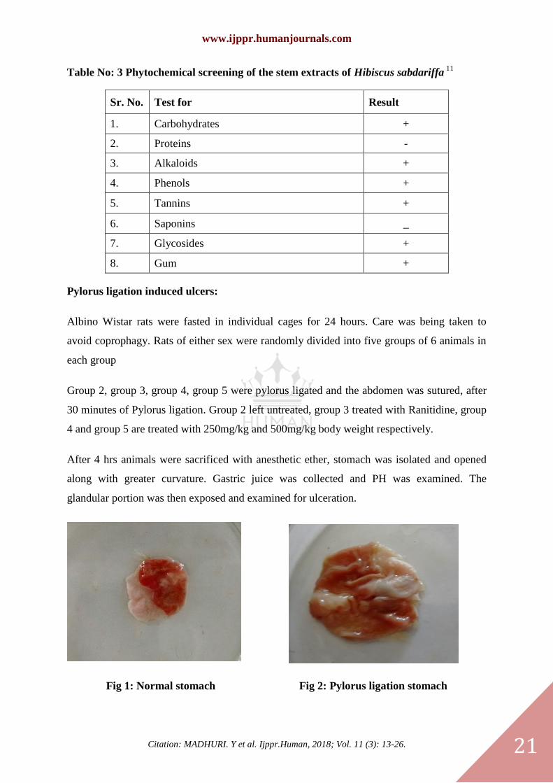
Pylorus ligation induced ulcers:
Albino Wistar rats were fasted in individual cages for 24 hours. Care was being taken to
avoid coprophagy. Rats of either sex were randomly divided into five groups of 6 animals in
each group
Group 2, group 3, group 4, group 5 were pylorus ligated and the abdomen was sutured, after
30 minutes of Pylorus ligation. Group 2 left untreated, group 3 treated with Ranitidine, group
4 and group 5 are treated with 250mg/kg and 500mg/kg body weight respectively.
After 4 hrs animals were sacrificed with anesthetic ether, stomach was isolated and opened
along with greater curvature. Gastric juice was collected and PH was examined. The
glandular portion was then exposed and examined for ulceration.
Fig 1: Normal stomach Fig 2: Pylorus ligation stomach
www.ijppr.humanjournals.com
Citation: MADHURI. Y et al. Ijppr.Human, 2018; Vol. 11 (3): 13-26. 22
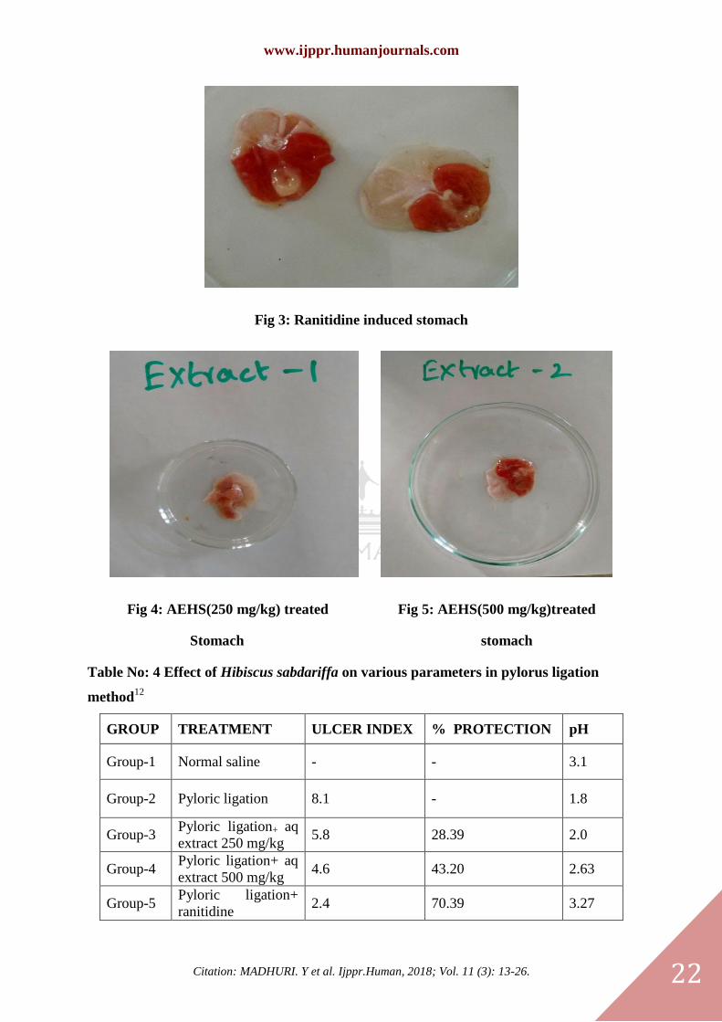
Fig 3: Ranitidine induced stomach
Fig 4: AEHS(250 mg/kg) treated Fig 5: AEHS(500 mg/kg)treated
Stomach stomach
Table No: 4 Effect of Hibiscus sabdariffa on various parameters in pylorus ligation
method12
GROUP TREATMENT ULCER INDEX % PROTECTION pH
Group-1 Normal saline - - 3.1
Group-2 Pyloric ligation 8.1 - 1.8
Group-3 Pyloric ligation+ aq
extract 250 mg/kg 5.8 28.39 2.0
Group-4 Pyloric ligation+ aq
extract 500 mg/kg 4.6 43.20 2.63
Group-5 Pyloric ligation+
ranitidine 2.4 70.39 3.27
www.ijppr.humanjournals.com
Citation: MADHURI. Y et al. Ijppr.Human, 2018; Vol. 11 (3): 13-26. 23
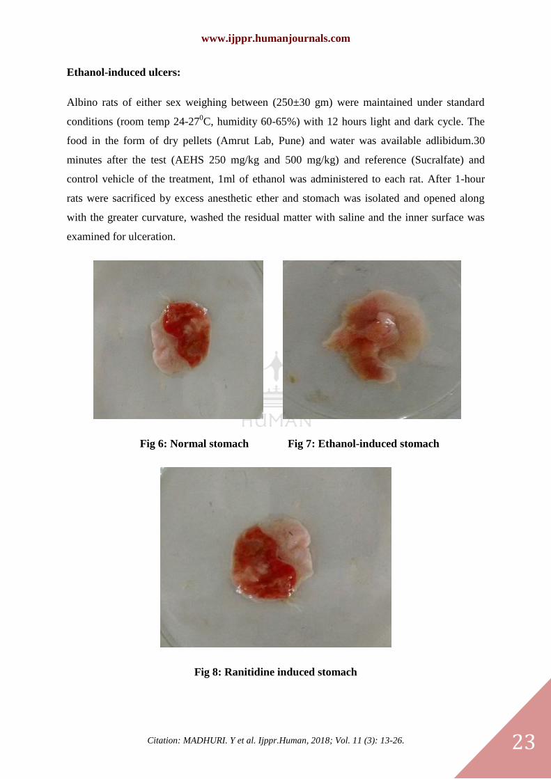
Ethanol-induced ulcers:
Albino rats of either sex weighing between (250±30 gm) were maintained under standard
conditions (room temp 24-270C, humidity 60-65%) with 12 hours light and dark cycle. The
food in the form of dry pellets (Amrut Lab, Pune) and water was available adlibidum.30
minutes after the test (AEHS 250 mg/kg and 500 mg/kg) and reference (Sucralfate) and
control vehicle of the treatment, 1ml of ethanol was administered to each rat. After 1-hour
rats were sacrificed by excess anesthetic ether and stomach was isolated and opened along
with the greater curvature, washed the residual matter with saline and the inner surface was
examined for ulceration.
Fig 6: Normal stomach Fig 7: Ethanol-induced stomach
Fig 8: Ranitidine induced stomach
www.ijppr.humanjournals.com
Citation: MADHURI. Y et al. Ijppr.Human, 2018; Vol. 11 (3): 13-26. 24
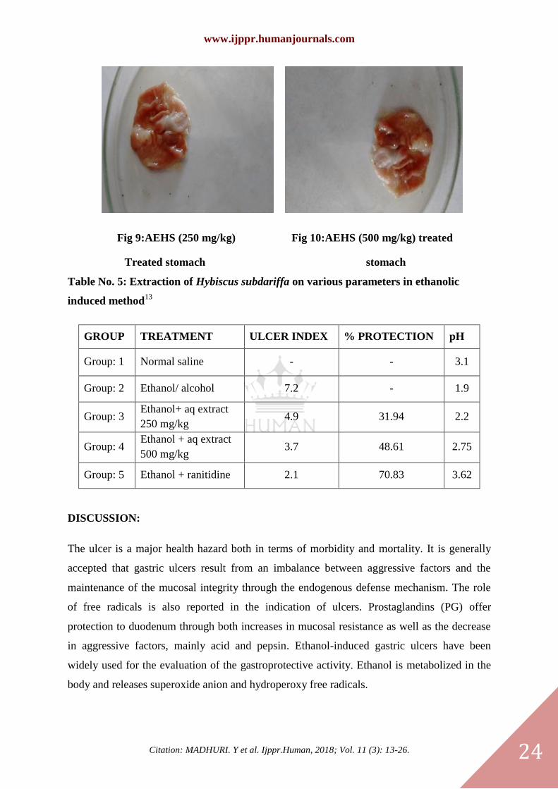
Fig 9:AEHS (250 mg/kg) Fig 10:AEHS (500 mg/kg) treated
Treated stomach stomach
Table No. 5: Extraction of Hybiscus subdariffa on various parameters in ethanolic
induced method13
GROUP TREATMENT ULCER INDEX % PROTECTION pH
Group: 1 Normal saline - - 3.1
Group: 2 Ethanol/ alcohol 7.2 - 1.9
Group: 3 Ethanol+ aq extract
250 mg/kg 4.9 31.94 2.2
Group: 4 Ethanol + aq extract
500 mg/kg 3.7 48.61 2.75
Group: 5 Ethanol + ranitidine 2.1 70.83 3.62
DISCUSSION:
The ulcer is a major health hazard both in terms of morbidity and mortality. It is generally
accepted that gastric ulcers result from an imbalance between aggressive factors and the
maintenance of the mucosal integrity through the endogenous defense mechanism. The role
of free radicals is also reported in the indication of ulcers. Prostaglandins (PG) offer
protection to duodenum through both increases in mucosal resistance as well as the decrease
in aggressive factors, mainly acid and pepsin. Ethanol-induced gastric ulcers have been
widely used for the evaluation of the gastroprotective activity. Ethanol is metabolized in the
body and releases superoxide anion and hydroperoxy free radicals.
www.ijppr.humanjournals.com
Citation: MADHURI. Y et al. Ijppr.Human, 2018; Vol. 11 (3): 13-26. 25
The incidence of ethanol-induced ulcers is predominant in the glandular part of the stomach.
It was reported to stimulate the formation of leukotriene C4 (LTC4), mast cell secretory
products and reactive oxygen species resulting in the damage of rat gastric mucosa. It has
been found that oxygen-derived free radicals are implicated in the mechanism of acute and
chronic ulceration in the gastric mucosa and scavenging these free radicals can play an
appreciable role in healing these ulcers. When aspirin is in the lipid-soluble undissociated
form it can damage the gastric mucosa. Aspirin causes a dose-dependent reduction in
mucosal prostaglandins – PGE2 and PGI2 bio-synthesis accompanied by an increase in the
mean area of gastric ulcerations.
Investigation of aqueous extract of H. sabdariffa (AEHS) in the present study provides
sample indications of its strong gastric anti-ulcerogenic property. The observation in the
present study, a significant decrease in the ulcer index in this model suggests the ability of
Roselle extract is involved in decreasing the gastric acid secretion. Furthermore, AEHS also
showed a significant effectiveness by inhibiting basal gastric acid secretion and ulcer
formation in the pylorus-ligated rat model. The ulcer index in pylorus ligation method was
found to be 8.1 at PH 1.8, Our extract at concentration 250mg/kg, the ulcer index is 5.8 and
percentage protection were found to be 28.39% at PH 2.0. At 500mg/kg, the ulcer index is 4.6
&%protection is 43.20% at PH 2.63. These results show less antiulcer activity compared with
Salah Alqasoumi et. al. The present study compared with Ranitidine (Std, the UI 2.4 & %
protection is 70.39%), our extract shows less activity.
AEHS significantly prevented gastric lesions induced by ethanol, the most commonly
employed tests in the evaluation of anti-ulcer/cytoprotective activity. The ulcer index in
ethanol-induced method was found to be 7.2. Our extract at concentration 250 mg/kg, the
ulcer index is 4.9 and percentage protection was found to be 31.94%. At 500mg/kg the ulcer
index is 3.7 &% protection is 48.61%. The present study in accordance with Rachhadiya
Rakesh et.al, (ulcer index is 5.1&2.0 at 250,500mg/kg respectively). The present study
compared with Ranitidine (Std, the UI 2.4 & %protection is 70.39%), our extract shows less
activity.
www.ijppr.humanjournals.com
Citation: MADHURI. Y et al. Ijppr.Human, 2018; Vol. 11 (3): 13-26. 26
CONCLUSION:
From the study, we concluded that aqueous extract of Hibiscus sabdariffa has potent antiulcer
activity. From the result, it was proved that AEHS in the dose of 250 mg/kg shows 28.39%
protection from ulcers, whereas 500 mg/kg shows almost double protection.
REFERENCES:
1. Kaur Amandeep, A review on etiology and pathogenesis.IRJP; 3(6); 2012.
2. K. PURO, Medicinal Uses of Roselle Plant (Hibiscus sabdariffa L.): A Mini Review. Indian Journal of Hill
Farming 27(1): June 2014; 27 (1); 81-90.
3. Dinesh Kumar Patidar, anti-ulcer activity of aqueous extract of Murraya koenigii in albino rats, International
Journal of Pharma and Bio Sciences; 2 (1); Jan-mar 2011.
4. Ine Da-Costa-Rocha, Hibiscus sabdariffa L. –A phytochemical and pharmacological review, Food
Chemistry; Volume 165; 15 December 2014; 424–443.
5. Abba Pacome Obouayeba, Phytochemical Analysis, Purification and Identification of Hibiscus
Anthocyanins, RJPBCS; 3(2); June-August 2015; 156-168.
6. Ali Abdella Eltayeib, Phytochemical and Chemical Composition of Water Extract of Hibiscus Sabdariffa
(Red Karkade Calyces) in North Kordofan State-Sudan, International Journal of Advanced Research in
Chemical Science (IJARCS); 1 (6); August 2014; 10-13.
7. Shirisha Bongu, Animal Models In Experimental Gastric Ulcer Screening-A Review;2; (2);2012; 82-87.
8. Ghangale G. R, Evaluation of Antiulcer Activity of Ocimum sanctum in Rats. Veterinary World, 2(12); 465-
466.
9. N. L. Dashputre, Evaluation of Anti-Ulcer Activity of Methanolic Extract of Abutilon indicum Linn Leaves
in Experimental Rats. International Journal of Pharmaceutical Sciences and Drug Research 2011; 3(2); 97-100.
10. Rachhadiya Rakesh M, Evaluation of Antiulcer activity of castor oil rats IJRAP 2011; 2(4); 1349-1353.
11. Abba Pacome Obouayeba, Phytochemical and Antioxidant Activity of Roselle(Hibiscus Sabdariffa L.) Petal
Extracts, Research Journal of Pharmaceutical, Biological and Chemical Sciences; 5(2); June-august 2015; 1453-
1465
12. Baral Sanja Raj, Evaluation of Antiulcer activity of Ethanolic extract of Dalbergia sissoo leaves in
Experimental animals; IRJP; 4(12); 2013.
13. Raju. D, Evaluation of Anti-ulcer activity of methanolic extract of Terminalia chebula fruits in experimental
rats J. Pharm. Sci. & Res; 1(3); 2009; 101-107.