Embed Size (px)
Citation preview

Anti-Transforming Growth Factor (TGF)-,B Antibodies Inhibit Breast Cancer CellTumorigenicity and Increase Mouse Spleen Natural Killer Cell ActivityImplications for a Possible Role of Tumor Cell/Host TGF-,B Interactionsin Human Breast Cancer Progression
Carlos L. Arteaga,** Stephen D. Hurd,* Angela R. Winnier,* Mahlon D. Johnson,$ Brian M. Fendly,11 and James T. Forbes*'Departments of *Medicine, *Cell Biology, and §Pathology, Vanderbilt University School of Medicine,Nashville, Tennessee 37232; and IlGenentech Inc., South San Francisco, California 94080
Abstract
TGF-0 effects on angiogenesis, stroma formation, and immunefunction suggest its possible involvement in tumor progression.This hypothesis was tested using the 2G7 IgG2b, whichneutralizes TGF-fil, -,82, and -,83, and the MDA-231 humanbreast cancer cell line. Inoculation of these cells in athymicmice decreases mouse spleen natural killer (NK) cell activity.Intraperitoneal injections of 2G7 starting 1 d after intraperito-neal inoculation of tumor cells suppressed intraabdominal tu-mor and lung metastases, whereas the nonneutralizing anti-TGF-,8 12H5 IgG2a had no effect. 2G7 transiently inhibitedgrowth of established MDA-231 subcutaneous tumors. Histo-logically, both 2G7-treated and control tumors were identical.Intraperitoneal administration of 2G7 resulted in a marked in-crease in mouse spleen NK cell activity. 2G7 did not inhibitMDA-231 primary tumor or metastases formation, nor did itstimulate NKcell-mediated cytotoxicity in beige NK-deficientnude mice. Finally, serum-free conditioned medium fromMDA-231 cells inhibited the NKcell activity of human bloodlymphocytes. This inhibition was blocked by the neutralizinganti-TGF-ft 2G7 antibody but not by a nonspecific IgG2. Thesedata support a possible role for tumor cell TGF-ft in the progres-sion of mammarycarcinomas by suppressing host immune sur-veillance. (J. Clin. Invest. 1993. 92:2569-2576.) Key words:breast neoplasms * transforming growth factor-# * nude micedimmunologic surveillance * natural killer activity
Introduction
The TGF-#s comprise a family of several multifunctional struc-turally related polypeptides first identified for their ability toinduce anchorage-independent growth of nontransformed fi-broblasts (1, 2). The multiplicity of TGF-# actions in nearly allcell types (3) suggests that this molecule has a pivotal role inseveral physiological and pathological processes. The escape of
This work was presented in part at the 83rd Annual Meeting of theAmerican Association for Cancer Research, San Diego, CA, 1992, andpublished in abstract form (Proc. Am. Assoc. CancerRes. 1992. 33:80).
Address correspondence to Carlos L. Arteaga, M.D., Division ofMedical Oncology, Vanderbilt University, 22nd Avenue South, 1956TVC, Nashville, TN 37232-5536.
Received for publication 6 May 2993 and in revised form 29 June1993.
many tumor cells from TGF-,B-mediated growth inhibition aswell as the ability of this polypeptide to suppress the immunesystem, to induce angiogenesis, and to promote the formationof stroma have suggested a possible role of TGF-# on the main-tenance and progression of transformed cells in an intacthost (4).
Expression of TGF-fll, -#2, and -#3 mRNAshas been de-tected in human breast cancer cells in culture (5, 6). ExogenousTGF-#lI and -#2 inhibit the growth of most breast cancer celllines in vitro (7-9). Furthermore, anti-TGF-# l and -#2 neutral-izing antisera stimulated the proliferation ofhormone-indepen-dent human breast cancer cell lines and downregulated basalTGF-,lB mRNAlevels (10), supporting a functional TGF-#-mediated negative autocrine pathway in breast cancer cell cul-ture systems. However, the biological and clinical implicationsof these in vitro data are unclear. For example, exogenousTGF-#l fails to inhibit the growth of (in vitro sensitive) humanbreast cancer cells in athymic mice (1 1). Moreover, higher lev-els of TGF-#l expression have been associated with states ofenhanced breast cancer cell tumorigenesis or with a more ag-gressive breast cancer phenotype ( 1 2-19).
A causal association between higher levels of TGF-# 1 ex-pression and enhanced tumorigenicity of human breast cancercells is supported by a recent report in which MCF-7 cells trans-fected with a TGF-# 1 expression vector escaped from hormonedependence when inoculated in castrated athymic mice (20).Tumor formation by the MCF-7/TGF-#I transfectants wasblocked by the anti-TGF-# 2G7 monoclonal antibody that neu-tralizes TGF-#,l, -#2, and -#3 (21). Moreover, administrationof human recombinant TGF-#l transiently supported estro-gen-independent growth of parental MCF-7 cells in castratednude mice (20). To follow this recent observation, we nowreport a series of experiments using the naturally transformedMDA-23 1 human breast cancer cell line and anti-TGF-# neu-tralizing antibodies in athymic mice. Our results show a cleardissociation between the in vitro and the animal model databut continue to support a causal association between TGF-#and the progression of human breast carcinoma cells in vivo.
Methods
Cell line and antibodies. The MDA-23 1 human breast cancer cell linewas purchased from the American Type Culture Collection (Rockville,MD) and maintained in improved minimum essential medium(GIBCO BRL, Gaithersburg, MD) supplemented with 10%FCS(Haz-leton, Lenexa, KS). These cells express high levels of TGF-#lI and -,32(5, 6), and are inhibited by picomolar concentrations of TGF-l and-f#2 in vitro (7-9). The 2G7 IgG2b and the 12H5 IgG2a were raisedagainst human recombinant TGF-,B1 and have been characterized pre-viously (21). Both immunoprecipitated '25I-TGF-#l1 but only 2G7 didso with labeled TGF-fl2 (21). 2G7 neutralized the growth inhibitory
Transforming Growth Factor-# and HumanBreast Cancer Progression 2569
J. Clin. Invest.© The American Society for Clinical Investigation, Inc.0021-9738/93/12/1569/08 $2.00Volume 92, December 1993, 2569-2576

activity of TGF-01, -#2, and -#3 on mink lung MvI Lu epithelial cellsbut 12H5 was devoid of neutralizing activity (21).
DNAsynthesis. 4 X 10⁴ MDA-23 1 cells/well were plated in 24-wellplates and treated the following day with human recombinant TGF-#lIfor 18 h. In some cases TGF-#lI was preincubated overnight at 4VCwithdifferent concentrations of 2G7 or a nonspecific IgG2 (Sigma ChemicalCo., St. Louis, MO)before addition to the cells. Monolayers were thenlabeled with 0.5 1tCi/ml [3H]thymidine (82.2 Ci/mmol; NENProducts,Boston, MA) for 2 h, and the rate of DNAsynthesis was estimated bymeasuring acid-precipitable radioactivity as described previously (8).
Anchorage-independent growth assay. A 1 -ml top layer containing asingle-cell suspension of 104 cells, 0.8 agarose (Sea-Plaque; FMCCorp.BioProducts, Rockland, ME), IMEM, 10% calf serum (CS; Hazleton),and 10 mMHepes, with or without different concentrations of 2G7,was added to a 1-ml bottom layer of 0.8% agarose/10% CS in 35-mmdishes. Dishes were incubated in a humidified 5% CO2 incubator at370C, and colonies measuring > 50 ,m were counted after 10-14 dusing an Omnicon feature analysis stem model II image analyzer(Bausch & Lomb, Rochester, NY).
Collection of conditioned medium (CM).' MDA-231 cells wereplated in 100-mm tissue culture dishes in growth medium and, when50-75% confluent, washed twice with PBSand then changed to 7 ml ofserum-free IMEM. After 24 h, the CMwas collected, supplementedwith pepstatin, aprotinin, and leupeptin (1 ,ug/ml each), centrifuged at3,000 rpm to remove cellular debris, and tested the same day on ahuman lymphocyte NKcytotoxicity assay (below).
Experiments in athymic mice. 3-4-wk-old female athymic mice(Harlan Sprague Dawley, Madison, WI) or 5-9-wk-old NIH-3 nudebeige NK-deficient female mice (Taconic, Germantown, NY) were in-oculated intraperitoneally or subcutaneously in the flank just caudal tothe forelimb with 5 X 1o6 exponentially growing MDA-23 1 cells. Micewere then injected serially intraperitoneally at the indicated intervalswith the monoclonal antibodies 12H5 or 2G7 or PBS in a 0.5-ml vol-ume using a 26-gauge needle. At the completion of the experiment,mice were killed by cervical dislocation. In those mice inoculated withtumor cells intraperitoneally, multiple specimens from different sites inthe abdominal cavity and the lungs were collected, fixed in 10%forma-lin, paraffin embedded, stained with hematoxylin and eosin, and evalu-ated by light microscopy (M. D. Johnson and C. L. Arteaga) At leastthree histological sections were prepared from each lung, two fromopposite sides and one from the middle of each paraffin block. Subcuta-neous tumors were removed and processed similarly to the intraperito-neal tumors. Tumor diameters were measured with calipers and tumorvolume calculated by the formula:
volume = width2 X length/2.
Factor VIII and collagen immunohistochemistry. For in situ detec-tion of factor VIII, 5-jim-thick sections from formalin-fixed, paraffin-embedded MDA-23 1 subcutaneous xenografts were predigested with0.1% trypsin/0. 1%CaC12. This was followed by a peroxidase-antiperox-idase (PAP) histochemical procedure using a 1:200 dilution of the anti-human von Willebrand factor purified Ig fraction of rabbit antiserum(Dakopatts, Glostrup, Denmark) as previously described (22). Nonim-mune rabbit Ig fraction was used as control. For detection of collagenin the MDA-231 xenografts, similar sections were analyzed using aMasson's Trichrome histochemical stain procedure (23).
Mouse spleen NKcell cytotoxicity assay. Whole mouse spleens wereaseptically removed and forced through a sterile stainless mesh to ob-tain a single-cell suspension. The Yac- 1 mouse lymphoma cell line wasused as target cells in the cytotoxicity assay. 107 Yac- 1 cells in suspen-sion culture were labeled with 200 ,uCi/ml of Na25'CrO4 (AmershamCorp., Arlington Heights, IL) for 60 min at 37°C as described (24).Unbound radioactivity was removed after two washes. Live effectorspleen cells and target cells (in ratios of 10:1, 50:1, and 100:1) wereadded to microtiter wells in a final volume of 0.2 ml/well in quadrupli-
1. Abbreviation used in this paper: CM, conditioned medium.
cate. Wells containing target cells alone served as controls for spontane-ous 5Cr release (SR), and 0.1 ml of labeled target cells was counted by yscintillography to determine total potential release (TR) of radioactiv-ity. The plates were incubated in a humidified 5%C02 incubator for 4 hat 370C. After centrifugation at 750 g for 10 min at room temperature,0. 1-ml aliquots of the supernatants (SN) were removed for determina-tion of radioactivity. Percent cytotoxicity was calculated from theformula:
100 x[(cpm in SN X 2) - (SR x 2)]lOX[TR -(SR X2)]
Humanperipheral blood lymphocyte NKcytotoxicity assay. Singledonor peripheral blood lymphocytes were separated by Ficoll-Hypa-que discontinuous gradient centrifugation, washed twice, and treatedlike mouse splenocytes to obtain a single-cell suspension. Cell suspen-sions were then treated in a 5%C02 incubator for 18 h at 370C withdifferent volumes of serum-free medium conditioned by MDA-231cells. HumanK-562 erythroleukemia cells were labeled with 5"Cr simi-lar to the Yac- I mouse lymphoma cells (above) and used as target in thecytotoxicity assay. After two washes, adjusted equal concentrations oflive effector cells (1ym phocytes) were added to 5Cr-labeled K-562 cellsfor 4 h at 370C. Lymphocyte-mediated NKactivity was estimated asradioisotope release from target cells as described above.
Results
Neutralizing anti-TGF-f3 antibodies stimulate growth of MDA-231 breast cancer cells in vitro. Initially, we tested whether the2G7 IgG2b blocked the inhibitory effect of exogenous TGF-# 1on sensitive MDA-23 1 cells. Picomolar concentrations of hu-man recombinant TGF-,3l inhibited DNAsynthesis in MDA-231 cells. Preincubation with 2G7 but not with a nonspecificIgG2 blocked this inhibition with total abrogation of the TGF-,3l inhibitory effect at 100 gg/ml (Fig. 1). 2G7 by itself did notchange the basal rate of [3Hjthymidine incorporation in .thesecells. Before the experiments in nude mice we examined theability of anti-TGF-# antibodies on basal growth of MDA-23 1cells. Similar to previously reported anti-TGF-p polyclonal an-tibodies (10), the 2G7 monoclonal induced a marked increase
0 0
4~~~~~~~0, 80C.)X ° 60 )\ g/mlL~0
z o 0
X: 20_No2G7
320 _1 10 0.025 0.25 2.
TGF,81 (ng/ml)
Figure 1. Neutralization of TGF-#,l-induced inhibition of DNAsyn-thesis in MDA-23 1 cells with anti-TGF-# antibodies. [3H]Thymidineincorporation in MDA-23 1 cells was measured as described in Meth-ods 18 h after the addition of human recombinant TGF-#1. TGF-,BIwas preincubated overnight with different concentrations of 2G7 or10 tg/ml of a nonspecific IgG2 (No 2G7) before addition to the cellmonolayers. Each data point represents the mean of triplicate wells.All SE were < 10%.
2570 C. L. Arteaga, S. D. Hurd, A. R. Winnier, M. D. Johnson, B. M. Fendly, and J. T Forbes

in MDA-231 colony formation in a dose-dependent fashion(Fig. 2). Similar results were obtained in a monolayer growthassay in serum-free medium (not shown).
Antibody-mediated TGF-f3 blockade inhibits growth of hu-manbreast cancer cells in athymic mice. For these experimentswe inoculated the highly tumorigenic MDA-23 1 cells intraperi-toneally. When injected intraperitoneally, this cell line rapidlyforms extensive intraabdominal tumors and readily metasta-sizes to the lungs (C. L. Arteaga and C. K. Osborne, unpub-lished data). This site of injection was chosen initially for tworeasons: first, to asure a high local concentration of anti-TGF-3antibodies (also injected intraperitoneally) in the tumor cellenvironment; and second, to anticipate a possible differentialeffect of the antibodies on tumors at the injection site vs. meta-static tumors. Every-other-day intraperitoneal injections of a200-/ig dose of 2G7 started 1 d after intraperitoneal inoculationof tumor cells suppressed the development of MDA-231 in-traabdominal tumor and detectable lung metastases. Wearbi-trarily chose this initial dose of antibody because in prelimi-nary experiments plasma from animals treated with 200 /ig2G7 i.p. 48 h before blood collection inhibited binding of 1251ITGF-p2 to AKR-2B mouse fibroblasts (not shown). All ani-mals treated with the nonneutralizing anti-TGF-f 1 2H5 IgG2aor PBSexhibited extensive omental seeding by tumor cells andmetastases in both lungs at 3 wk (Figs. 3 and 4).
To confirm this result we performed a second experimentwith different doses of 2G7 given every 3 d. As shown in Table Iand Fig. 4, all mice treated with PBSor the lowest dose of 2G7developed intraabdominal tumor and all but one exhibitedlarge hematogenous lung metastases. Intravascular foci of tu-mor cells were identified in the lungs of one of four micetreated with 20 jig of 2G7 every 3 d (Fig. 4, third panel). At thehighest dose of antibody, only one of four mice developed asmall focus of tumor cells in the abdominal wall and noneexhibited histologically identifiable lung metastases at 3 wk.
Wenext studied the effect of TGF-# blockade with systemicadministration of 2G7 on established subcutaneous MDA-23 1tumors. Treatment was started when subcutaneous tumors hadreached a volume of - 100 mm3(Fig. 5, day 8). Serial injec-
700
600
500-0toA 400Lu 300-z
02000
100
0 0.1 10 1002G7 (pg/ml)
Figure 2. Effect of 2G7 on MDA-23 1 colony formation. A single-cellsuspension of 104 MDA-231 cells was plated in 0.8% agarose/10%calf serum/ 10 mMHepes in the absence or presence of different con-centrations of the 2G7 IgG2 as described in Methods. Colonies mea-
suring 2 50 gm were counted 10 d later with an automated imageanalyzer. Each bar represents the mean±SE of three dishes.
TREATMENT
IP TUMORS
LUNG METASTASES
MDA-231 cells (5 x 10⁶)P
PBS 12H5 2G7(200yg ipq2d)
6/6 6/6 o/6
6/6 6/6 0/6
Figure 3. Neutralizing anti-TGF-# antibodies suppress MDA-23 1 in-traabdominal tumor and lung metastases. MDA-23 1 cells were inoc-ulated intraperitoneally in female nude mice. The following day,every-other-day intraperitoneal injections of PBSor 200 ,ug of the 2G7or 1 2H5 monoclonal antibodies were started. Six mice were in eachtreatment group. After 10 doses (3 wk) mice were killed. The contentsof the abdominal cavity and both lungs were examined macroscopi-cally and microscopically for tumor formation as described inMethods.
tions of 2G7 at a distant site (intraperitoneally) every 3 d fromdays 8 to 20 transiently inhibited MDA-231 tumor growthcompared with the nonneutralizing TGF-p monoclonal 12H5and PBS (Fig. 5, *P < 0.01 on days 14 and 17, Student's t test).Two tumors from each of the three treatment groups were har-vested on day 20 and processed for light microscopy as well asfactor VIII and Masson's Trichrome stains. All tumors weremoderately or poorly differentiated adenocarcinomas. Endo-thelial cells (identified by factor VIII immunostaining), pres-ence of extracellular matrix and collagen (using Masson'sTrichrome stain), and infiltration with mouse lymphocytes orother stromal cells appeared scarce in all tumors (data notshown). 2G7-treated tumors were histologically and immuno-histochemically indistinguishable from 1 2H5- and PBS-treatedtumors.
2G7 upregulates mouse spleen NK activity and lacks anantitumor effect in NK-deficient nude mice. Once we failed torecognize any differences in the degree of tumor angiogenicactivity or tumor stroma induced by TGF-,B blockade, weexam-ined the effect of 2G7 on mouse immune function, which, inturn, may explain the potent antitumor effect. Since T cellfunction is deficient while NKcell function is normal in athy-mic mice (25) and because of the reported effects of TGF-,B onNK cell activity (26), we focused on the latter. Three dailyinjections of 200 ,ug of 2G7 induced a marked increase inmouse spleen NKactivity compared with PBSor to a nonspe-cific control IgG2 in tumor-free athymic mice (Fig. 6). Further-more, the sole presence of subcutaneous MDA-23 1 tumors in-hibited basal mouse spleen NKactivity 10 d after tumor cellinoculation (Fig. 6). Daily intraperitoneal injections of 2G7 onmice bearing subcutaneous MDA-23 1 tumors on days 7, 8, and9 after tumor cell inoculation did not change the tumor-in-duced inhibition of NKactivity (not shown).
We next tested whether coadministration of exogenousTGF-,B1 could reverse the 2G7-induced stimulation of NKac-tivity. In this second experiment, spleen NK activity wasgreater than sixfold higher in mice treated with 2G7 than in
Transforming Growth Factor-/3 and HumanBreast Cancer Progression 2571

0 2 20 200
Figure 4. Dose-dependent inhibition of MDA-23 1 lung metastases by anti-TGF-f antibodies. 1 d after intraperitoneal inoculation of MDA-23 1cells into nude mice, every-3-d doses of 2G7 (2, 20, and 200 ,g) or PBSwere started and continued for 3 wk as described in Methods and TableI. After death of the animals, three histological sections were prepared from each lung and examined for the presence of tumor cells. Mice treatedwith PBS and the low dose of antibody exhibited dense infiltration of their lung parenchyma by large MDA-23 1 metastases. Small metastaseswere detected in one of four of the mice treated with 20 ,ug of 2G7 every 3 d (X 125).
those treated with the nonneutralizing 12H5 control IgG2a(Fig. 7). Simultaneous administration of human recombinantTGF-f31 partially blocked 2G7-induced upregulation of NKcell activity while it did not affect those levels measured in12H5-treated mice (Fig. 7).
Finally, we proposed that if the stimulation of NKactivitywere critical for the antitumor effect mediated by the neutraliz-ing anti-TGF-fl monoclonal, the latter would not exert an anti-tumor effect in an NK-deficient host. A similar experiment tothe one shown in Fig. 3 was performed in beige NKcell-defi-cient athymic mice. In a preliminary experiment, all six NK-animals rapidly developed intraabdominal tumors and lungmetastases after an intraperitoneal inoculum of 5 X 10⁶ MDA-231 cells. In a second experiment 12 NK- mice were treatedwith either 2G7 or 1 2H5 (200 ,g i.p. every 2 d for eight doses; n= 6 per group) starting 1 d after tumor cell inoculation. All 12mice treated either with 2G7 or 1 2H5 developed extensive in-traperitoneal tumors and large metastatic foci in the lungs. Incontrast to the results with NK+ athymic mice (Figs. 6 and 7),spleen NK cell activity against 5'Cr-labeled Yac- 1 lymphoma
cells from both tumor-free and tumor-bearing beige NK- micetreated with either 2G7 or 1 2H5 was undetectable.
MDA-231 cell-CM contains TGF-3 activity that inhibitshuman lymphocyte-mediated NK cell activity. To follow theobserved inhibition of mouse spleen NKcell activity induced
soo0
66
g
400'
300-
200'
100'
Table I. Dose-dependent Inhibition of MDA-231 IntraabdominalTumor and Metastases Formation by Anti-TGF-f3 Antibodies
Treatment Primary tumor Lung metastases
PBS 4/4 4/42 ,gg 2G7 4/4 3/420 1Ag 2G7 2/4 1/4200M2G7 1/4 0/4
MDA-23 1 cells were inoculated intraperitoneally into female athymicmice. The following day serial intraperitoneal injections of PBS or2G7 were initiated and continued every 3 d for a total of 3 wk (n = 4per group). At 3 wk, mice were killed and their abdominal contentsand both lungs examined macro and microscopically for the presenceof gross and/or microscopic tumor as described in Methods.
-0-- <- PBS
el 12H50 2G7
4 8 1 2 1 6 20 24 28
DAYSPOSTINOCULATION
|- 2G7---Figure 5. Neutralizing anti-TGF-# antibodies transiently inhibitgrowth of established MDA-231 subcutaneous tumors. Nude micewere inoculated subcutaneously with MDA-231 cells as described inMethods. After 8 d all mice had tumors with an approximate volumeof 100 mm3. Intraperitoneal administration of 200 ,g of 2G7 every 3d from days 8 to 20 transiently inhibited MDA-23 1 tumor growthcompared with the nonneutralizing TGF-,B antibody 12H5 and PBS(*P < 0.01 on days 14 and 17, Student's t test). Each data point rep-resents the mean±SE of six mice.
2572 C. L. Arteaga, S. D. Hurd, A. R. Winnier, M. D. Johnson, B. M. Fendly, and J. T. Forbes

35
30
E-
0E-0
E-E-
z
w.
25
20
15
10
5
00 25 50 75 100
EFFECTOR: TARGET
Figure 6. Effect of 2G7 and of MDA-23 1 tumors on mouse spleenNK cell activity. 5-wk-old female nude mice were inoculated subcu-taneously with 5 x 10⁶ MDA-23 1 cells and tumors allowed to formfor 10 d. In other cases, tumor-free mice were treated intraperitoneallythree times daily with 200 tig of 2G7 or a nonspecific IgG2 (SigmaChemical Co.) or PBS. 1 d after the last injection or 10 d after subcu-taneous tumor cell inoculation, mouse spleens (n = 3 per group) were
removed and tested in an NKcell cytotoxicity assay against syngeneicYac- 1 lymphoma cells as described in Methods. Mice injected with2G7, normal IgG2, or PBS were tumor free.
from MDA-23 1 cells and added it to human peripheral bloodlymphocytes from a healthy donor. CMbut not control me-dium induced a dose-dependent decrease in lymphocyte-me-diated NK activity against syngeneic K-562 erythroleukemiacells (Fig. 8). Transient acidification followed by neutralizationof the CMbefore addition to the effector lymphocytes en-hanced the inhibitory effect on NK activity > 20-fold (notshown). This change induced by acidification was consistentwith the possibility that the NK inhibitory activity present inthe CMwas mediated by TGF-f.
To confirm this latter possibility, we incubated the CMwith the neutralizing anti-TGF-3 2G7 antibody before addi-tion to the human lymphocytes. 2G7 but not a nonspecificIgG2 partially blocked the CM-induced inhibition of lympho-cyte-mediated NKactivity (Fig. 9). This result was consistentin both concentrations of CMtested (25 and 50%) and in allthree effector/target cell ratios examined (Fig. 9).
Discussion
We have presented a series of experiments that address thebiological role of TGF-fl in the tumorigenicity of human breastcancer cells. Antibody-induced blockade of all three mamma-lian TGF-f isoforms with the monoclonal antibody 2G7 inhib-ited MDA-23 1 breast cancer cells in athymic mice. Similar to aprevious report (24), the establishment of tumors in the ani-mals downregulated mouse spleen NKcell activity. However,administration of the neutralizing TGF-# antibody enhancedthis activity in tumor-free animals. 2G7-mediated tumor cellinhibition was not seen in NK-deficient nude mice. Taken to-
70by MDA-23 1 tumors (Fig. 6), we tested the effect of secretedfactors by these cells on the NKactivity mediated by humaneffector cells. For this purpose we collected serum-free CM
30
xX0
O 20F-
.,I
60
E-_-4C)
0
0L)
so
50
40
30
20
12H5 TGF-1 2G7 TGF-.8I+ +
12H5 2G7
Figure 7. Exogenous TGF-f31 abrogates 2G7-induced upregulation ofmouse spleen NK cell activity. 5-wk-old female nude mice were
treated intraperitoneally three times daily with 200 pg of 2G7 or
12H5. In some cases, the antibodies were administered simulta-neously with 1 pig of human recombinant TGF-f31. Two mice were
in each treatment group. On day 4, spleens were removed and sple-nocyte NKcell activity was measured as described in Methods usingsyngeneic 5"Cr-labeled Yac- I lymphoma cells as target.
10-10 0 1 0 20 30 40 50 60
MEDIUMCONCENTRATION(%)
Figure 8. MDA-23 1 cell serum-free CMinhibits human lymphocyte-mediated NK cell activity. Human peripheral blood lymphocyteswere incubated for 18 h at 370C in a 5%C02 incubator with differentconcentrations of unconditioned (control) or conditioned mediumfrom MDA-23 I cells. Lymphocytes were then washed and tested inan NKcell cytotoxicity assay using syngeneic 'Cr-labeled K-562 eryth-roleukemia cells as target as described in Methods. Results using a
50:1 effector/target cell ratio are shown.
Transforming Growth Factor-#3 and HumanBreast Cancer Progression 2573
I_
* PBSv 2G7V nl IgGo Tumor
w
0 25 50 75 100
EFFECTOR TARG
0 25 50 75 100
* unconditioned medium
hETV MDA-231 CM+ 207
V MDA-231Ca+ normIgG
TUMORCELL t Angiogenesist Connective
tissue andfibroblasts
Disregulation ofProteolysis
, Immunesurveillance
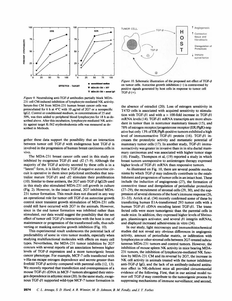
Figure 10. Schematic illustration of the proposed net effect of TGF-flon tumor cells. Autocrine growth inhibition (-) is contravened bypositive signals generated by host cells in response to tumor cellTGF-# (+).
Figure 9. Neutralizing anti-TGF-# antibodies partially block MDA-231 cell CM-induced inhibition of lymphocyte-mediated NKactivity.Serum-free CMfrom MDA-23 1 human breast cancer cells waspreincubated for 6 h at 4VC with 10 ,g/ml of 2G7 or a nonspecificIgG2. Control or conditioned medium, in concentrations of 25 and50%, was then added to peripheral blood lymphocytes for 18 h as de-scribed above. After this incubation, lymphocyte-mediated NKactiv-ity against target K-562 erythroleukemia cells was measured as de-scribed in Methods.
gether these data support the possibility that an interactionbetween tumor cell TGF-f with endogenous host TGF-j3 isinvolved in the progression of human breast carcinoma cells invivo.
The MDA-231 breast cancer cells used in this study are
inhibited by exogenous TGF-f31 and -p32 (7-9). Although themajority of the TGF-fl activity secreted by these cells is in a
"latent" form, it is likely that a TGF-fl-negative autocrine cir-cuit is operative in them since polyclonal antibodies that neu-tralize mature TGF-f31 and -32 stimulate their proliferation(10). Similar to these antisera, the 2G7 anti-TGF-,3 IgG2b usedin this study also stimulated MDA-23 1 cell growth in culture(Fig. 2). However, in the intact animal, 2G7 inhibited MDA-231 tumor formation. This result does not discard in any wayan operational role for tumor cell TGF-i3 on autocrine growthcontrol since transient growth stimulation of MDA-23 1 cellscould still have occurred with 2G7 in the animals. However,since in the end tumor formation was inhibited rather thanstimulated, our data would suggest the possibility that the neteffect of tumor cell TGF-i's interaction with the host is one ofmaintenance or progression of the transformed cells, thus sub-verting or masking autocrine growth inhibition (Fig. 10).
This experimental result underscores the potential lack ofpredictability of some in vitro tumor models, especially whenstudying multifunctional molecules with effects on many celltypes. Nevertheless, the MDA-231 tumor inhibition by 2G7concurs with several reports of an association between higherlevels of TGF-,3 expression and a more tumorigenic breastcancer phenotype. For example, MCF-7 cells transfected withv-Ha-ras escape estrogen dependence and secrete greater thanfivefold TGF-13 activity compared with parental cells (12, 13).Werecently reported that transfection and overexpression of a
mouse TGF-#3I cDNAin MCF-7 tumors abrogated their estro-gen dependence in athymic mice (20). In the same study, exoge-nous TGF-1 I supported wild-type MCF-7 tumor formation in
the absence of estradiol (20). Loss of estrogen sensitivity inT47D cells is associated with acquired sensitivity to stimula-tion with TGF-3l1 and with a > 100-fold increase in TGF-,3lmRNAlevels (14). TGF-#3I mRNAtranscripts are more abun-dant in tumor than in nontumor mammary tissues (15), and78%of estrogen receptor/progesterone receptor (ER/PgR)-neg-ative but only 13%of ER/PgR-positive tumors exhibited a highlevel of immunoreactive TGF-f31 protein (16). TGF-l I in-creases the proteolytic activity and metastatic potential ofmammarytumor cells (17). In another study, TGF-flI immu-noreactivity was greater in invasive than in in situ ductal mam-mary carcinomas and was associated with higher tumor stage(18). Finally, Thompson et al, (19) reported a study in whichbreast tumors unresponsive to antiestrogen therapy expressedhigher levels of TGF-,B1 mRNAthan responsive tumors.
As illustrated on Fig. 10, there are several possible mecha-nisms by which TGF-f3 may indirectly contribute to the estab-lishment and progression of tumor cells in an intact host. These
include the induction of angiogenesis (27), the formation ofconnective tissue and deregulation of pericellular proteolysis(27-29), the recruitment of stromal cells (29, 30), and the sup-
pression of several elements of the host's immune response (26,31-33). Arrick et al. (34) recently confirmed some of these bytransfecting human ElA-transformed 293 tumor cells with ahuman TGF-fll cDNA encoding latent TGF-fl1. The trans-fected cells were more tumorigenic than the parental cells innude mice. In addition, they expressed higher levels of fibrino-gen, plasminogen activator, and several 131 integrin mRNAs,and displayed increased adhesiveness in vitro (34).
In our study, light microscopy and immunohistochemicalstudies did not reveal any obvious differences in angiogenicactivity, amount of pericellular matrix, or infiltration withlymphocytes or other stromal cells between 2G7-treated subcu-taneous MDA-23 1 tumors and control tumors. However, theinhibition of mouse spleen NKactivity in mice bearing MDA-231 tumors, the inhibition of lymphocyte-mediated NKfunc-tion by MDA-23 1 CMand its reversal by 2G7, the increase inNKcell activity in animals treated with the tumor inhibitoryanti-TGF-# IgG, and the lack of an antibody-induced antitu-mor effect in NK-deficient mice all provided circumstantialevidence of the following. First, that in our animal model tu-mor cell TGF-# may contribute to the tumorigenic process bysuppressing mechanisms of immune surveillance; and second,
2574 C. L. Arteaga, S. D. Hurd, A. R. Winnier, M. D. Johnson, B. M. Fendly, and J. T Forbes
t:0
0
E-
a.
0
1 UL4
50%
75
V
50-
25-
0
that in an intact animal, the endogenously produced TGF-fregulates immune function in an autocrine fashion. Since 2G7was probably blocking the autocrine/paracrine effects of TGF-# on both tumor and host cells, it is difficult to dissect thecontribution of each (or all) of these blocks to the net effect oftumor inhibition. Whether upregulation of NK function byblocking endogenous TGF-f (with antibodies or other inhibi-tors) would be a practical approach to prevent or inhibit tumorprogression, and whether this would only be effective againsttumors expressing high levels of immunosuppressive cyto-kines, are speculations that require further testing.
Other genetically manipulated cell systems also support anassociation between tumor cell TGF-f3 and immune suppres-sion. UV-induced fibrosarcoma cells transfected with a mouseTGF-#3I cDNA exhibited enhanced tumorigenicity in nudemice and, different from the parental cells, were unable to in-duce cytotoxic mouse T lymphocyte responses (35). CHOcellstransfected with a TGF-3li expression vector encoding for la-tent TGF-fll decreased NK cell activity and rapidly formedtumors in nude mice (36). Furthermore, MATLyLu rat pros-tate cancer cells (37) and MCF-7 human breast carcinoma cells(20) became more tumorigenic or metastatic after transfectionwith a mouse TGF-f 1 expression vector. Of note, some of thesecells were sensitive to TGF-3 inhibition in vitro (37) but stillbehaved more tumorigenic in the animal after transfection.Wuet al. (38) reported the opposite result in that FET humancolon cancer cells transfected with an antisense expression vec-tor for TGF-,Bl escaped TGF-f3-mediated autocrine growthcontrol and uniformly formed tumors in nude mice. In the lastthree studies, however, immunological functions on animalsbearing TGF-3 1 -overexpressing or TGF-p3I -antisensed tumorswere not reported.
A more clinico-biological precedent to our findings is theidentification of TGF-fl2 as the glioblastoma-derived T cellsuppresor factor, which explains the cellular immunodefi-ciency state exhibited by some patients with this commontypeof brain tumor (39-41). Obviously, the possible identity of theTGF-# isoform(s) involved in MDA-231 tumor formation isbeyond the scope of this report. Experiments with TGF-pl -spe-cific neutralizing antibodies are currently in progress to addressthat question.
Because of our inability to assess the relative contributionof 2G7-induced effects on tumor cells vs. host cells to the netoutcome of MDA-23 1 tumor growth inhibition, we examinedthe effect of MDA-23 1 cells on NKfunction in vitro. Concor-dant with the animal data, medium conditioned by the MDA-231 cells inhibited human lymphocyte-mediated NKactivity,and this inhibition was reversed by the 2G7 IgG2b but not by anonspecific IgG2 (Figs. 8 and 9). The biological implications ofthis observation in the context of breast carcinomas are unclearbut suggest that TGF-,3 secreted by tumor cells mayhave immu-nosuppressive effects in diseases other than glioblastoma. Fu-ture studies of TGF-3 isoform and receptor localization in tu-mor and especially in host cells along different stages of theprogressive transformation of human mammary epithelialcells will shed light on some of these questions.
AcknowledgmentsThis work was supported by a Department of Veteran Affairs MeritReview grant (C. L. Arteaga) and a University Research Council grant(C. L. Arteaga). C. L. Arteaga is recipient of a Research Associate Ca-reer Development Award from the Department of Veteran Affairs.
References
1. Moses, H. L., E. B. Branum, J. A. Proper, and R. A. Robinson. 1981.Transforming growth factor production by chemically transformed cells. CancerRes. 41:2842-2848.
2. Roberts, A. B., M. A. Anzano, L. C. Lamb, J. M. Smith, and M. B. Sporn.1981. Newclass of transforming growth factors potentiated by epidermal growthfactor: isolation from non-neoplastic tissues. Proc. Nadl. Acad. Sci. USA.78:5339-5343.
3. Roberts, A. B., and M. B. Sporn. 1990. The transforming growth factor-fls.In Handbook of Experimental Pharmacology, vol. 95. M. B. Sporn and A. B.Roberts, editors. Springer-Verlag, Heidelberg. 419-472.
4. Roberts, A. B., N. L. Thompson, U. Heine, C. Flanders, and M. B. Sporn.1988. Transforming growth factor-e: possible roles in carcinogenesis. Br. J.Cancer. 57:594-600.
5. Zajchowski, D., V. Band, N. Pauzie, A. Tager, M. Stampfer, and R. Sager.1988. Expression of growth factors and oncogenes in normal and tumor-derivedhuman mammaryepithelial cells. Cancer Res. 48:7041-7047.
6. Arrick, B. A., M. Korc, and R. Derynck. 1990. Differential regulation ofexpression of three transforming growth factor /3 species in human breast cancercell lines by estradiol. Cancer Res. 50:299-303.
7. Knabbe, C., M. E. Lippman, L. M. Wakefield, K. C. Flanders, A. Kasid, R.Derynck, and R. B. Dickson. 1987. Evidence that transforming growth factor-e isa hormonally regulated negative growth factor in human breast cancer cells. Cell.48:417-428.
8. Arteaga, C. L., A. K. Tandon, D. D. Von Hoff, and C. K. Osborne. 1988.Transforming growth factor fl: potential autocrine growth inhibitor of estrogenreceptor-negative human breast cancer cells. Cancer Res. 48:3898-3904.
9. Zugmaier, G., B. W. Ennis, B. Deschauer, D. Katz, C. Knabbe, G. Wilding,P. Daly, M. E. Lippman, and R. B. Dickson. 1989. Transforming growth factorstype (l 1 and fl2 are equipotent growth inhibitors of human breast cancer cell lines.J. Cell. Physiol. 141:353-361.
10. Arteaga, C. L., R. J. Coffey, T. C. Dugger, C. M. McCutchen, H. L. Moses,and R. M. Lyons. 1990. Growth stimulation of human breast cancer cells withanti-transforming growth factor f3 antibodies: evidence for negative autocrineregulation by transforming growth factor fl. Cell Growth & Differ. 1:367-374.
11. Zugmaier, G., S. Paik, G. Wilding, C. Knabbe, M. Bano, R. Lupu, B.Deschauer, S. Simpson, R. B. Dickson, and M. Lippman. 1991. Transforminggrowth factor /B1 induces cachexia and systemic fibrosis without an antitumoreffect in nude mice. Cancer Res. 51:3590-3594.
12. Dickson, R. B., A. Kasid, K. K. Huff, S. E. Bates, C. Knabbe, D. Bronzert,E. P. Gelman, and M. E. Lippman. 1987. Activation of growth factor secretion intumorigenic states of breast cancer induced by 1 7fl-estradiol or v-Ha-ras onco-gene. Proc. Nail. Acad. Sci. USA. 84:837-841.
13. Kasid, A., C. Knabbe, and M. E. Lippman. 1987. Effect of v-rasH onco-gene transfection on estrogen-independent tumorigenicity of estrogen-dependentbuman breast cancer cells. Cancer Res. 47:5733-5738.
14. Daly, R. J., R. J. B. King, and P. D. Darbre. 1990. Interaction of growthfactors during progression towards steroid independence in T-47-D human breastcancer cells. J. Cell. Biochem. 43:199-21 1.
15. Barrett-Lee, P., M. Travers, Y. Luqmani, and R. C. Coombes. 1990.Transcripts for transforming growth factors in human breast cancer: clinical corre-lates. Br. J. Cancer. 61:612-617.
16. King, R. J. B., D. Y. Wang, R. J. Daly, and P. D. Darbre. 1989. Ap-proaches to studying the role of growth factors in the progression of breast tumorsfrom the steroid sensitive to insensitive state. J. Steroid Biochem. 34:133-138.
17. Welch, D. R., A. Fabra, and M. Nakajima. 1990. Transforming growthfactor fl stimulates mammaryadenocarcinoma cell invasion and metastatic po-tential. Proc. Nail. Acad. Sci. USA. 87:7678-7682.
18. Walker, R. A., and S. J. Dearing. 1992. Transforming growth factor 1 inductal carcinoma in situ and invasive carcinomas of the breast. Eur. J. Cancer.28:641-644.
19. Thompson, A. M., D. J. Kerr, and C. M. Steel. Transforming growthfactor ft1 is implicated in the failure oftamoxifen therapy in human breast cancer.Br. J. Cancer. 63:609-614.
20. Arteaga, C. L., T. Carty-Dugger, H. L. Moses, S. Hurd, and J. Pietenpol.1993. Transforming growth factor d1 can induce estrogen-independent tumorige-nicity of human breast cancer cells in athymic mice. Cell Growth & Differ. 4:193-201.
21. Lucas, C., L. N. Bald, B. M. Fendly, M. Mora-Worms, I. S. Figari, E. J.Patzer, and M. A. Palladino. 1990. The autocrine production of transforminggrowth factor-# l during lymphocyte activation: a study with a monoclonal anti-body-based ELISA. J. Immunol. 145:1415-1422.
22. Sehested, M., and K. Hou-Jensen. 1981. Factor VIII-related antigen as an
endothelial cell marker in benign and malignant diseases. Pathol. Anat. 391:217-225.
23. Masson, P. 1929. Trichrome stainings and their preliminary technique. J.Tech. Methods. 12:75-90.
24. Fraker, L. D., S. A. Halter, and J. T. Forbes. 1986. Effects of orally admin-
Transforming Growth Factor-,8 and HumanBreast Cancer Progression 2575
istered retinol on natural killer cell activity in wild type BALB/c and congenitallyathymic BALB/c mice. Cancer Immunol. Immunother. 21:114-118.
25. Fogh, J., and B. C. Giovanella. 1978. The Nude Mouse in Experimentaland Clinical Research. Academic Press, NewYork. 502-521.
26. Rook, A. H., J. H. Kehrl, L. M. Wakefield, A. B. Roberts, M. B. Sporn,D. B. Burlington, H. C. Lane, and A. S. Fauci. 1986. Effects of transforminggrowth factor fi on the functions of natural killer cells: depressed cytolytic activityand blunting of interferon responsiveness. J. Immunol. 136:3916-3920.
27. Roberts, A. B., M. B. Sporn, R. K. Assoian, J. M. Smith, N. S. Roche,L. M. Wakefield, U. I. Heine, L. A. Liotta, V. Falanga, J. H. Kehrl, and A. S.Fauci. 1986. Transforming growth factor type-fl: rapid induction of fibrosis andangiogenesis in vivo and stimulation of collagen formation in vitro. Proc. Nat!.Acad. Sci. USA. 83:4167-4171.
28. Keski-Oja, J., F. Blasi, E. B. Leof, and H. L. Moses. 1988. Regulation ofthe synthesis and activity of urokinase plasminogen activator in A549 humanlung carcinoma cells by transforming growth factor-a. J. Cell Biol. 106:451-459.
29. Laiho, M., and J. Keski-Oja. 1989. Growth factors in the regulation ofpericellular proteolysis: a review. Cancer Res. 49:2533-2553.
30. Postlethwaite, A. E., J. Keski-Oja, H. L. Moses, and A. H. Kang. 1985.Stimulation of the chemotactic migration of human fibroblasts by transforminggrowth factor fl. J. Exp. Med. 165:251-256.
31. Kehrl, J. H., L. M. Wakefield, A. B. Roberts, S. Jakolew, M. Alvarez-Mon,R. Derynck, M. B. Sporn, and A. S. Fauci. 1986. Production of transforminggrowth factor fl by T lymphocytes and its potential in the regulation of T cellgrowth. J Exp. Med. 163:1037-1050.
32. Kehrl, J. H., A. B. Roberts, L. M. Wakefield, S. Jakolew, M. B. Sporn, andA. S. Fauci. 1986. Transforming growth factor fl is an important immunomodu-latory protein for human B lymphocytes. J. Immunol. 137:3855-3860.
33. Tsunawaki, S., M. Sporn, A. Ding, and C. Nathan. 1988. Deactivation ofmacrophages by transforming growth factor-fl. Nature (Lond.). 334:260-262.
34. Arrick, B. A., A. R. Lopez, F. Elfman, R. Ebner, C. H. Damsky, and R.
Derynck. 1992. Altered metabolic and adhesive properties and increased tumori-genesis associated with increased expression of transforming growth factor fl 1. J.Cell Biol. 1 8:715-726.
35. Torre-Amione, G., R. D. Beauchamp, H. Koeppen, B. H. Park, H.Schreiber, H. L. Moses, and D. A. Rowley. 1990. A highly immunogenic tumortransfected with a murine transforming growth factor type 3ll cDNA escapesimmune surveillance. Proc. Natl. Acad. Sci. USA. 87:1486-1490.
36. Wallick, S. C., I. S. Figari, R. E. Morris, A. D. Levinson, and M. A.Palladino. 1990. Immunoregulatory role of transforming growth factor ,B (TGF-f3) in development of killer cells: comparison of active and latent TGF-fl 1. J. Exp.Med. 172:1777-1784.
37. Steiner, M. S., and E. R. Barrack. 1992. Transforming growth factor-f,overproduction in prostate cancer: effects on growth in vivo and in vitro. Mol.Endocrinol. 6:15-25.
38. Wu, S., D. Theodorescu, R. S. Kerbel, J. K. V. Willson, K. M. Mulder,L. E. Humphrey, and M. G. Brattain. 1992. TGF-fl, is an autocrine-negativegrowth regulator of human colon carcinoma FET cells in vivo as revealed bytransfection of an antisense expression vector. J. Cell Biol. 1 16:187-196.
39. Wrann, M., S. Bodmer, R. de Martin, S. Siepl, R. Hofer-Warbinek, K.Frei, E. Hofer, and A. Fontana. 1987. T cell suppressor factor from human glio-blastoma cells is a 1 2.5-kd protein closely related to transforming growth factor-,B.EMBO(Eur. Mol. Biol. Organ.) J. 6:1633-1636.
40. de Martin, R., B. Haendler, R. Hofer-Warbinek, H. Gaugitsch, M. Wrann,H. Schlusener, J. M. Seifert, S. Bodmer, A. Fontana, and E. Hofer. 1987. Comple-mentary DNAfor human glioblastoma-derived T cell suppressor factor, a novelmember of the transforming growth factor-f gene family. EMBO(Eur. Mol. Biol.Organ.) J. 6:3673-3677.
41. Kuppner, M. C., M.-F. Hamou, S. Bodmer, A. Fontana, and N. De Tribo-let. 1988. The glioblastoma-derived T-cell suppressor factor/transforming growthfactor beta2 inhibits the generation of lymphokine-activated killer (LAK) cells.Int. J. Cancer. 45:562-567.
2576 C. L. Arteaga, S. D. Hurd, A. R. Winnier, M. D. Johnson, B. M. Fendly, and J. T. Forbes