Embed Size (px)
Citation preview
Anti-resorptive agent-related osteonecrosis of the jaw: Position Paper 2017 of the
Japanese Allied Committee on Osteonecrosis of the Jaw
Japanese Allied Committee on Osteonecrosis of the Jaw: Toshiyuki Yoneda1,a, Hiroshi
Hagino1,b, Toshitsugu Sugimoto1,c, Hiroaki Ohta2,d, Shunji Takahashi1,e, Satoshi Soen2,f, Akira
Taguchi3,g, Toshihiko Nagata4,i, Masahiro Urade5,j, Takahiko Shibahara5,k, Satoru Toyosawa6,h
1The Japanese Society for Bone and Mineral Research, 2The Japan Osteoporosis Society,
3The Japanese Society of Oral and Maxillofacial Radiology, 4The Japanese Society of
Periodontology, 5The Japanese Society of Oral and Maxillofacial Surgeons, 6The Japanese
Society of Oral Pathology,
aDivision of Hematology and Oncology, Indiana University School of Medicine, bSchool of
Health Science, Faculty of Medicine, Tottori University, cInternal Medicine 1, Shimane
University Faculty of Medicine, dClinical Research Centers for Medicine, International
University of Health and Welfare, eDepartment of Medical Oncology, The Cancer Institute
Hospital Of JFCR, fDepartment of Orthopedic Surgery and Rheumatology, Kindai University
Nara Hospital, gDepartment of Hard Tissue Research, Graduate School of Oral Medicine,
Matsumoto Dental University, hDepartment of Oral Pathology, Osaka University Graduate
School of Dentistry, iDepartment of Periodontology and Endodontology, School of Dentistry,
Tokushima University, jDepartment of Oral and Maxillofacial Surgery, Hyogo College of
Medicine, kDepartment of Oral & Maxillo-Facial Surgery, Tokyo Dental College.
___________________________________________________________________
This is the author's manuscript of the article published in final edited form as:
Yoneda, T., Hagino, H., Sugimoto, T., Ohta, H., Takahashi, S., Soen, S., ... & Toyosawa, S. (2017). Antiresorptive agent-related osteonecrosis of the jaw: Position Paper 2017 of the Japanese Allied Committee on Osteonecrosis of the Jaw. Journal of bone and mineral metabolism, 35(1), 6-19. https://doi.org/10.1007/s00774-016-0810-7
─ 2 ─
Disclosure of Conflicts of Interest (COI) by Authors
Toshiyuki Yoneda: Consultant fee (Daiichi-Sankyo); Hiroaki Ohta: Lecture fee (Pfizer),
Manuscript fee (Medical Review Co., Ltd.); Toshitsugu Sugimoto: Lecture fee, Consultant fee
(Asahi Kasei Pharma, Pfizer), Research grant (Astellas Pharma, Eisai, Ono Pharmaceutical,
Daiichi-sankyo, Chugai Pharmaceutical, Eli Lilly Japan); Satoshi Soen: Lecture fee (Asahi
Kasei Pharma, Astellas Pharma, Eisai, MSD, Ono Pharmaceutical, Daiichi-Sankyo, Takeda
Pharmaceutical, Chugai Pharmaceutical, Teijin Pharma), Research Grant (Eisai, Daiichi-
Sankyo, Takeda Pharmaceutical); Shunji Takahashi: Lecture fee (Eisai, Daiichi-Sankyo),
Research grant (AstraZeneca, Daiichi-Sankyo, Chugai Pharmaceutical, Novartis Pharma,
Bayer, Parexel International); Akira Taguchi: Lecture fee (Asahi Kasei Pharma, MSD, Ono
Pharmaceutical, Daiichi-Sankyo, Takeda Pharmaceutical, Chugai Pharmaceutical, Teijin
Pharma), Consultant fee (Asahi Kasei Pharma); Hiroshi Hagino: Lecture fee (Asahi Kasei
Pharma, EA Pharma, MSD, Daiichi-Sankyo, Taisho Toyama Pharmaceutical, Takeda
Pharmaceutical, Chugai Pharmaceutical, Pfizer), Research Grant (Chugai Pharmaceutical);
Masahiro Urade: None; Takahiko Shibahara: None; Satoru Toyosawa: None; Toshihiko
Nagata: None
─ 3 ─
Abstract
Anti-resorptive agent-related osteonecrosis of the jaw (ARONJ) is an intractable, although
rarely occurs, complication in cancer patients with bone metastases and patients with
osteoporosis who are treated with anti-resorptives including bisphosphonates and denosumab.
Despite that more than 10 years have passed since the first cases of BRONJ was reported,
our understanding of the epidemiology and pathophysiology of ARONJ still remains limited
and data of ARONJ supported by evidence-based medicine are still poorly accumulated.
However, diagnosis and staging of ARONJ, identification of risk factors, and development of
preventive and therapeutic approaches have significantly advanced over the last a decade.
The Position Paper 2017 is an updated version of the Position Paper 2010 of the Japanese
Allied Committee on Osteonecrosis of the Jaw”, which is now consisted of six Japanese
academic societies. The Position Paper 2017 describes new diagnostic definition for ARONJ
according to AAOMS proposal, summarizes our current understandings of the
pathophysiology of ARONJ based on literature search and suggests how physicians and
dentists/oral surgeons should manage ARONJ, Further, the appropriateness of
discontinuation of anti-resorptives (drug holiday) before, during and after invasive dental
treatments is extensively discussed. More importantly, the manuscript also proposes for the
first time the importance of interactive communication and co-operation between physicians
and dentists/oral surgeons for successful treatment of ARONJ patients. It is expected that the
Position Paper 2017 will be a guide to improve the management of ARONJ patients in Japan.
─ 4 ─
I. Background
Bisphosphonates (BPs), which possess high chemical affinity to bone and specifically inhibit
osteoclastic bone resorption, have been widely and safely used for the treatment of bone
metastases and osteoporosis in which osteoclastic bone resorption is excessively increased.
In 2003, Marx first reported many cases of BP-related osteonecrosis of the jaw (BRONJ) in
cancer patients with bone metastasis and patients with osteoporosis who were treated with
BPs [1]. BRONJ is a rare but an intractable disease. Since its pathophysiology remains
unclear, physicians, dentists and oral surgeons have had difficulties in the management of
BRONJ patients from early days until recently. However, our understanding of BRONJ is
gradually and consistently advancing by analytical reviews of accumulating clinical and
preclinical data on BRONJ over the last several years. In this context, it is particularly notable
that recent clinical studies have showed that the occurrence of BRONJ is significantly
decreased by blocking oral infection via extensive oral health control [2-4], suggesting that
infection is a key step of the development of BRONJ.
Denosumab, a human IgG2 monoclonal antibody against receptor activator of nuclear factor-
kappa B ligand (RANKL) [5], is a new therapeutic agent for osteoporosis and bone metastases
with the half-life of approximately one month. Different from BPs that promote apoptosis in
osteoclasts, denosumab inhibits osteoclastic bone resorption without causing apoptosis in
osteoclasts. Further, denosumab does not deposit and persist in bones for a long period of
time as do BPs, and thus the effects of denosumab are reversible. These pharmacological
properties of denosumab initially led us to assume that ONJ unlikely occurs by treatment with
denosumab. To our surprise, however, patients treated with denosumab also developed ONJ
(DRONJ) clinically indistinguishable from BRONJ at almost the same incidence as BRONJ [6].
Since both BP and denosumab, which show anti-bone resorption effects via different
molecular mechanism of action, are associated with ONJ, anti-resorptive agent-related ONJ
(ARONJ) [7] has been suggested as a comprehensive term representing both BRONJ and
─ 5 ─
DRONJ. Meanwhile, the American Association of Oral and Maxillofacial Surgeons (AAOMS)
proposes the term, medication-related ONJ (MRONJ), based on the observations that anti-
angiogenic inhibitors and molecularly-targeted drugs such as tyrosine kinase inhibitors also
are infrequently associated with ONJ or increase the incidence of BRONJ/DRONJ in cancer
patients receiving BPs or denosumab, although global consensus has not been established
yet [8]. In this position paper, the term ARONJ will be used according to the proposal of the
International Task Force on Osteonecrosis of the Jaw [2] of which the Japanese Society for
Bone and Mineral Research is a member.
Since the first position paper on BRONJ was published by the Japanese Allied Committee on
Osteonecrosis of the Jaw in 2010 [9], it has been passed 6 years, during which DRONJ has
emerged, numerous and diverse ARONJ cases have been reported, and clinical and
preclinical studies on ONJ have been accumulating, thereby increasing our understanding of
ONJ and improving management of ONJ. The Position Paper 2017 is an updated and revised
version of the Position Paper 2010 attempting to provide the latest clinical and basic
information of ARONJ and propose a consensus for management of ARONJ in Japan.
The paper is compiled by the Japanese Allied Committee on Osteonecrosis of the Jaw, which
consists of diverse members of bone specialists, including physicians, orthopedic surgeons,
rheumatologists, obstetricians, medical oncologists, oral surgeons, periodontologists, dental
radiologists, oral pathologists, and cancer biologists. The Japanese Allied Committee on
Osteonecrosis of the Jaw was organized through a collaboration of six academic societies:
the Japanese Society for Bone and Mineral Research, Japan Osteoporosis Society, Japanese
Society of Periodontology, Japanese Society of Oral and Maxillofacial Radiology, Japanese
Society of Oral and Maxillofacial Surgeons, and the Japanese Society of Clinical Oral
Pathology.
II. Anti-resorptive agent-related osteonecrosis of the jaw (ARONJ)
─ 6 ─
1. Uniqueness of the jaw bone
There are several unique anatomical and microbiological characteristics in the jaw bone that
could be responsible for the specific occurrence of ARONJ in jaw bones. These characteristics
are not found in bones in other parts of the body.
1) The teeth erupt on the jaw bone breaking through the oral epithelium, allowing infectious
factors, agents and microbes in the oral cavity directly invade into the jaw bone via the
gap between epithelium and teeth or via root canal.
2) The oral mucosa covering the jaw bone is thin and infection caused by mucosal injury
spreads to the jaw bone beneath the mucosa.
3) More than 800 types of resident bacteria (1011 to 1012/cm3) inhabit in dental plaques as
sources of infection in the oral cavity.
4) Inflammations due to tooth decay, pulpitis, periapical lesions, and periodontal diseases
extend to the jaw bone.
5) The jaw bone exposes to the oral cavity following invasive dental treatments including
tooth extraction, leading to infection.
Thus, the environments around jaw bone have a predisposition to readily get bacterial infection
[10], which may be the reason why ARONJ occurs specifically in the jaw bone.
2. Diagnosis of ARONJ
AAOMS proposed additional diagnostic criteria for ONJ in 2014 and the Allied Committee
agreed to adopt these new diagnostic criteria. Accordingly, ARONJ is definitely diagnosed
when the following three conditions are met.
1) Patients have history of treatment with BP or denosumab.
─ 7 ─
2) Patients have no history of radiation therapy to the jaw bone. Bone lesions of ARONJ
must be differentiated from cancer metastases to the jaw bone by histological
examinations.
3) Exposure of alveolar bone in the oral cavity, jaw and/or face is continuously observed for
longer than 8 weeks after first detection by medical or dental experts. Or the bone is
palpable in the intra- or extra-oral fistula for longer than 8 weeks [8]. These criteria do not
apply to a patient in Stage 0.
3. Incidence of ARONJ
The incidence of ARONJ varies depending on studies. There are no reliable epidemiologic
data that are derived from evidence-based medicine. This position paper follows the data cited
by the International Task Force on ONJ [2].
1) Patients with osteoporosis
① BRONJ
The incidence is 1.04 to 69 per 100,000 patients treated with oral administration per year,
and 0 to 90 per 100,000 patients treated with intravenous administration per year. The
incidence of ONJ in osteoporotic patients treated with oral/intravenous nitrogen-
containing BPs ranges from 0.001% to 0.01%, which is estimated to be almost the same
or slightly higher than the incidence (0.001%) of ONJ in the general population.
② DRONJ
The incidence is 0 to 30.2 per 100,000 patients per year.
2) Cancer patients
─ 8 ─
The incidence of ONJ in cancer patients is higher than that in patients with osteoporosis.
Prospective studies of the incidence of ONJ have been conducted in cancer patients treated
with zoledronic acid or denosumab. Of 5,723 patients with breast, prostate and other solid
cancers and multiple myeloma, 52 patients (1.8%) treated with denosumab and 37 patients
(1.3%) treated with zoledronic acid (i.e., 89 cancer patients in total) developed ONJ in 3 year
follow-up [6, 11].
3) Incidence of ARONJ in Japan
① BRONJ
In nationwide surveys, The Japanese Society of Oral and Maxillofacial Surgeons found
263 patients with BRONJ from the year of 2006 to 2008 [12] and 4,797 from the year of
2011 to 2013 [13]. Approximately 40% of patients with BRONJ from 2006 to 2008 and
half of those from 2011 to 2013 developed BRONJ following oral BP administration [12,
13]. These results in Japan differ from the results obtained in other countries showing
higher incidence of BRONJ in patients treated with intravenous BPs than oral BPs. The
incidence of BRONJ in Japan is unknown, since data of the total number of patients
treated with BPs are unavailable at the present time.
② DRONJ
A study conducted by a pharmaceutical company (Daiichi Sankyo) after denosumab was
launched in the market reported that 120 cancer patients treated with denosumab from
April 17, 2012 to July 31, 2015 developed DRONJ, and 58 of these patients had been
treated with BPs before denosumab.
Twenty patients with osteoporosis treated with denosumab from June 11, 2013 to
December 31, 2015 developed DRONJ, and 15 of them had received BPs before
denosumab.
─ 9 ─
4. Clinical manifestations and staging of ARONJ
Clinical manifestations and staging of ARONJ are summarized in Table 1. Paresthesia in the
chin, including the lower lip (Vincent’s symptom) of patients treated with BP is an early sign of
ARONJ before alveolar bone exposure is detected.
1) Stage 0
The 2012 Position Paper (in Japanese) proposed that cases with ONJ-like clinical
manifestations but no alveolar bone exposure can be diagnosed as Stage 0 ONJ. It is reported
that Stage 0 ONJ accounts for 25-30% of ONJ, however, that half of Stage 0 cases heal
without progression to Stage 1 [14]. Accordingly, the International Task Force on ONJ does
not include Stage 0 in ONJ, concerning it may cause over-diagnosis [2, 15]. On the other hand,
the AAOMS proposes that Stage 0 should be diagnosed and treated as a pre-ONJ [8]. This
Position Paper includes Stage 0 in ONJ in agreement with the proposal of AAOMS from
therapeutic points of view. However, it is strongly recommended that the diagnosis of Stage 0
should be cautiously made to avoid over-diagnosis.
2) Differences in clinical characteristics between BRONJ and DRONJ
Clinical manifestations are indistinguishable between BRONJ and DRONJ at the present time.
There is not sufficient information accumulated yet on imaging and histopathological
characteristics of DRONJ that allows us to compare with those of BRONJ [16
Incidence of DRONJ in cancer patients is reported to be less than 2% and equivalent to that
of BRONJ [6, 11]. In contrast, incidence of DRONJ in osteoporosis patients is unknown.
3) Serum biochemical markers for bone turnover and ARONJ
Value of serum biochemical markers of bone turnover is decreased by the treatment with BP
and denosumab. Thus, it is expected that these markers are potentially useful for diagnosis,
follow-up and assessment of therapeutic effects in patients with ARONJ [17]. However, most
─ 10 ─
clinical studies found no significant correlation between changes in these serum bone turnover
markers and the occurrence and progression or healing of ARONJ [2]. Therefore, currently-
used bone turnover markers unlikely have diagnostic value for ARONJ.
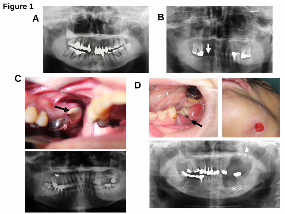
4) Imaging
For patients under the treatment with low-dose anti-resorptives and with no evident clinical
manifestations of ARONJ, intraoral and panoramic radiographs in conjunction with clinical
manifestations are sufficient for diagnosis (Figure 1). Intraoral radiographs, which have high
resolution, can reveal the site of infection in detail.
For cancer patients being treated with high-dose anti-resorptives, it is recommended that
intraoral radiographs of all existing teeth and panoramic radiographs should be undergone to
identify potential sites of infection even if they have no signs of ONJ, since they potentially
have increased risk for developing ONJ.
For patients who are clinically suspected to have developed ONJ, computed tomography (CT)
and dental cone-beam CT are helpful to detect early changes in trabecular and cortical bones
of the jaws and assess the sequestra, fistula formation, periosteal responses and involved
teeth. However, use of dental cone-beam CT is limited to localized lesions and supplemental
to CT. CT must be combined with intraoral and panoramic radiographs. For cases in which
differential diagnosis between ONJ and malignant tumors are required, use of CT and MRI,
rather than dental cone-beam CT, is recommended.
MRI, which allows assessment of changes in the bone marrow, may be useful for diagnostic
evaluation of ONJ. For patients with ONJ who are under conservative and/or surgical
treatments, the characteristics and extents of bone changes surrounding exposed bone can
be assessed by CT and dental CT. MRI is useful for assessment of surrounding soft tissues,
in addition to bones. Existing teeth that may be causes of infection can be detected by intraoral
radiographs [18].
─ 11 ─
Recent studies have proposed that hybrid SPECT/CT may be useful for distinguishing ONJ
lesions from unaffected healthy bone at the time of surgical interventions of ONJ [19, 20].
Simple PET using 18F-FDG and PET/CT may be also useful for assessment of ONJ lesions.
At present, no specific differences in images between BRONJ and DRONJ have been
described.
5) Histopathological findings in ARONJ
Although histopathological definition for diagnosis of ARONJ is yet to be established, several
features of ARONJ are noted. The major histopathological finding of BRONJ is characterized
by chronic osteomyelitis accompanied with osteonecrosis. In BRONJ lesions, there are
relatively large osteoclasts detaching from the bone surfaces, which is similarly seen in bones
treated with BPs [21] (Figure 2A). Trabecular bone exhibits mosaic-pattern lines of bone
remodeling (Figure 2B), with increased thickness and decreased osteon density (osteon
number/bone area). These histological views resemble those of bones in which osteoclastic
bone resorption is inhibited by BP [22]. On the other hand, trabeculae bones with active
inflammation demonstrate extensive osteoclastic bone resorption with resorption lacunae. Of
note, Actinomyces colonies (resident bacteria in the oral cavity) are frequently present in
contact with necrotic bones in ONJ lesions (Figure 2B, insert), raising the possibility that
Actinomyces play a role in the pathogenesis of BRONJ [23].
A histopathological study on DRONJ published by a Japanese group showed that numbers of
osteoclasts are decreased and immature osteoclasts with few nuclei are increased in DRONJ
lesions presumably due to the action of denosumab [24]. However, whether these
histopathological features are unique to DRONJ and distinct from those of BRONJ are
currently unknown.
5. Risk factors for ARONJ
─ 12 ─
Proposed risk factors for ARONJ are listed in Table 2. Of these risk factors, invasive dental
treatments such as tooth extraction, dental implant and apical/periodontal surgery are
definitely local risk factors for ARONJ. It should, however, be noted that the list is not made
based on robust medical evidence but the summary of published reports the Allied Committee
investigated. Here, risk factors that are newly listed are discussed.
1) Dental implants and ARONJ
Recent reports suggest that implants inserted before patients with cancer or osteoporosis are
treated with BPs are unlikely associated with subsequent occurrence of BRONJ if oral health
is appropriately managed [25, 26]. However, dental implantation performed during or after BP
treatment is a potential risk factor for BRONJ.
It is unknown whether dental implants are risk factors in patients receiving denosumab. Dental
implant for cancer patients who are on the treatment with anti-resorptives is not recommended
and alternative dental measures are recommended. On the other hand, dental implant in
patients with osteoporosis may be performed in case physicians and dentists agree that dental
implants are essential to improve systemic and oral health of patients.
2) New therapeutic agents and ARONJ
In Table 2, denosumab, ibandronate and anti-angiogenic agents and tyrosine kinase inhibitors
are newly listed. Since the Position Paper 2010 was published, denosumab was launched in
2012 in Japan and has been widely used in the treatment of bone metastases and
osteoporosis with infrequent occurrence of DRONJ.
Ibandronate was also launched as a new BP for the treatment of osteoporosis in Japan. A
prospective study performed in Japan showed that there is no significant difference in the
incidence of ONJ between osteoporotic patients treated with intravenous and oral ibandronate
[27], suggesting that the route of administration does not influence the occurrence of BRONJ
associated with ibandronate.
─ 13 ─
Anti-angiogenic agents and tyrosine kinase inhibitors, which are essentially administered as
adjuvants in the treatment of cancer patients, have been shown to cause, although very rarely,
by themselves or increase the incidence of ARONJ due to BP or denosumab [28].
6. Mechanism of ARONJ
The mechanism responsible for ARONJ is not fully understood. In particular, it is a major
question why the incidence of ARONJ is so low in osteoporotic patients, despite that millions
of patients receive BP or denosumab. It is also unknown why ARONJ spontaneously develops
in patients who are treated with anti-resorptives, but receive no invasive dental treatments.
These issues can’t be explained only by the mechanisms currently proposed. Animal models
in which ONJ is induced by administration of BP or RANKL inhibitors, followed by tooth
extraction, have recently been developed in mice, rats, dogs and pigs [29-31]. It is expected
that these animal models allow us to determine the pathophysiology and mechanism of
ARONJ and develop new therapeutic interventions for ARONJ. Here, the mechanisms already
described in the literature are summarized. Complex interactions of these mechanisms
together with additional yet-unknown mechanisms are assumed to lead to the development
and progression of ARONJ [29].
1) Inhibition of bone remodeling and excessive inhibition of osteoclast activity by anti-
resorptives
2) Increased predisposition to oral bacterial infection due to BP administration
3) BP suppression of remodeling and migration of oral epithelial cells
4) Changes in immune surveillance by anti-resorptives
5) Anti-angiogenic effects of BP
6) Others
─ 14 ─
III. Dental treatments and discontinuation of anti-resorptives
1. Dental treatments of patients who are to receive anti-resorptives
Before starting administration of anti-resorptives, physicians need to explain to patients not
only the benefits of anti-resorptives for bone metastases and osteoporosis but also risks of
anti-resorptives for ONJ. It is wise to request patients to visit a dentist to control oral health to
prevent the occurrence of ONJ. During dental treatments of these patients, interactive
communication and close cooperation between physicians and dentists are essential. It is
most appropriate that physicians inform dentists of the current status, clinical courses,
therapeutic history and prognosis of the primary disease. Ideally, all dental treatments are
expected to be completed 2 weeks before starting anti-resorptive treatment. However, in case
anti-resorptive treatment can’t be delayed because of progression of bone metastases or high
risk for fracture, administration of anti-resorptives in parallel with dental treatments may be
acceptable. During treatment with anti-resorptives, physicians instruct patients to routinely visit
a dentist to have oral examination. Dentists should inform physicians of the results of oral
examinations and dental treatments as soon as they are done, so that there is no delay in anti-
resorptive administration by physicians. It is also helpful if physicians inquire patients the
status of their oral cavity and teeth at their visit.
2. Dental treatments of patients receiving anti-resorptives
1) Discontinuation of BPs before starting dental treatments
It is controversial whether discontinuation (drug holiday) of BPs for a certain period of time
before starting invasive dental treatments is effective at preventing or decreasing the
occurrence of BRONJ. The arguments are as follows:
① There is little clinical evidence that short-term discontinuation of BPs helps prevent
the occurrence of BRONJ resulting from invasive dental treatments.
─ 15 ─
② Based on the physiochemical properties of BPs that deposit and persist in the bone
for a long period of time [5], it appears unlikely that short-term drug holiday of BPs
prevents BRONJ.
③ Survey conducted by the Japan Osteoporosis Society showed no changes in
incidence of BRONJ in osteoporotic patients even if BPs or denosumab are
discontinued before dental treatments [32, 33].
④ In osteoporotic patients who had BP drug holiday, there are exacerbations of
osteoporosis including decreased bone mineral density and increased incidence of
fractures [32-34].
⑤ Given extremely low incidence of BRONJ in osteoporosis, the benefits of BP for
fracture prevention outweigh the risks for BRONJ [35].
⑥ Several recent studies reported that infection is a key event for BRONJ and that
extensive infection control before invasive dental treatments decreases BRONJ [3].
Of particular note, this study also shows that BRONJ did not occur even in cancer
patients who previously had BRONJ at other sites in the oral cavity if infection is
properly controlled. These results suggest that infection control is most important
for prevention of BRONJ.
⑦ The American Dental Association estimated an incidence of ARONJ in patients
with osteoporosis is, at the highest, up to 0.1% and suggests that the benefits of
anti-resorptives for fracture prevention outweigh the risks for ARONJ.
Discontinuation of anti-resorptives is unlikely to decrease the risk for ARONJ, but
rather increase negative effects such as increased fractures [7].
These pieces of background collectively suggest that discontinuation (drug holiday) of BP
before starting invasive dental treatments is not logically supported.
In contrast, however, the advisory board of the Food and Drug Administration (FDA) (http://
─ 16 ─
www.fda.gov/downloads/AdvisoryCommittees/CommitteesMeetingMaterials/Drugs/DrugSafe
tyandRiskManagementAdvisoryCommittee/UCM270958.pdf), AAOMS [8, 17] and other
groups [36, 37] described that the incidence of BRONJ increased in patients with osteoporosis
who are treated with BPs for longer than 4 years in retrospective studies with small number of
cases. From these results, AAOMS recommended that discontinuation of anti-resorptives for
approximately 2 months before invasive dental treatments needs to be considered in case
patients receive anti-resorptives for longer than 4 years and have low risk for fractures but
potential high risk for BRONJ with a consultation with physicians [8]. The Japanese Society of
Oral and Maxillofacial Surgeons, and other academic societies including the Korean Society
for Bone and Mineral Research, the Korean Association of Oral and Maxillofacial Surgeons
[38], and the International Association of Oral and Maxillofacial Surgeons (IAOMS) support
the AAOMS proposal. Thus, no consensus has been reached yet regarding whether drug
holiday of BP before invasive dental treatments is adequate and necessary for prevention of
BRONJ. Prospective clinical studies should be performed under the cooperation of allied
teams of physicians, dentists and oral surgeons, hopefully at international levels to include as
many as BRONJ cases, to address this important issue. Whatever the results of the studies,
however, it is most important that invasive dental treatments for patients who are receiving
anti-resorptives are conducted with careful and meticulous surgical techniques under
elaborative planning in conjunction with extensive control of oral infection.
2) Suggested dental treatments of patients with cancer and osteoporosis who are receiving
BPs
There are many review articles that propose the practical approaches to prevent the
occurrence of BRONJ in patients who are receiving BPs during dental treatments [2-4, 7, 8].
The Position Paper 2017 proposes the followings as an example. Dental experts will need to
educate patients on the importance of daily oral sanitation including how to clean the oral
cavity after each meal and rinse their mouth with antibacterial mouthwash. In parallel, dentists
make efforts to eliminate causes of infection such as dental plaque, calculus, tooth decays,
─ 17 ─
remaining roots, periodontitis, apical lesions, ill-fitting dentures, crowns and inlays as much as
possible. Subsequently, dentists can begin conservative dental treatments without
discontinuation of BPs. In case, however, invasive dental treatments such as removal of teeth
responsible for BRONJ are inevitable, antibacterial agents are administered to patients in
advance and invasive dental treatments should be restricted to as minimum extent and area
as possible without discontinuation of BPs. At the end of the invasive treatments, remaining
sharp edges of alveolar bones should be smoothened, and surgical wounds are to be closed
primarily with mucoperiosteal flap lined by the periosteum.
3) Suggested dental treatment of patients with cancer and osteoporosis who are receiving
denosumab
Denosumab has significantly superior benefits for cancer patients with bone metastases than
does zoledronic acid [11]. The incidence of DRONJ was found to be equivalent to that of
BRONJ in cancer patients [6]. Occurrence of DRONJ in patients with osteoporosis under
treatment with denosumab is also reported in Japan, although the incidence is extremely low
[39]. Similar to patients treated with BPs, dentists conduct conservative dental treatments
without drug holiday. Invasive dental treatments, if inevitable, can be conducted without drug
holiday following appropriate infection control. A recent case report showed uneventful healing
of tooth extraction sockets by closing the sockets with oral mucosa to prevent secondary
infection in patients under denosumab [40]. Interestingly, another case report from Japan
described that DRONJ in colon cancer patients with bone metastases healed after
discontinuation of denosumab [41], suggesting that the actions of denosumab are reversible.
Given that denosumab is administered to osteoporotic patients once every 6 months and the
half-life of denosumab is approximately one month, there is room to consider the timing and
plan of dental treatments between the 6 month intervals.
Of note, intriguing experimental results in a mouse model in which administration of
osteoprotegerin (OPG)-Fc or zoledronic acid caused ONJ were recently reported [31]. In this
─ 18 ─
model, ONJ was spontaneously healed by discontinuation of OPG-Fc, while ONJ was not
healed by discontinuation of zoledronic acid. Since OPG-Fc has RANKL inhibitory action
similar to denosumab, these findings are consistent with the notion that the effects of
denosumab are reversible.
4) Discontinuation of anti-resorptives after invasive dental treatments
Anti-resorptives may interfere with the healing of surgical wounds, especially epithelialization
of wounds [29]. In this case, it may be required to temporarily discontinue the administration
of anti-resorptives or change to alternative therapeutic drugs unassociated with ONJ until
surgical wounds completely heal. Continuation or discontinuation of anti-resorptives needs to
be decided depends on fracture risk evaluated by the “Guidelines on the prevention and
treatment of osteoporosis 2015” [42] and the status of the healing of surgical wounds in the
oral cavity under agreement between physicians and dentists.
5) Timing of resuming the administration of anti-resorptives
Timing of re-starting administration of anti-resorptives after drug holiday is dependent on the
balance between healing of surgical wounds and control of the primary disease. If fracture risk
or bone metastasis is well-controlled, it is recommended that the treatment with anti-
resorptives is resumed approximately two months after invasive dental treatments, when the
alveolar bones damaged are expected to heal. However, if fracture risk is high or bone
metastasis progresses during drug holiday and re-administration of anti-resorptives is urgent,
and if there are no signs of infection around surgical wounds, two weeks after the invasive
dental treatments, when epithelialization of the surgical site is almost complete, may be the
earliest timing. Dentists are expected to immediately inform physicians of healing of surgical
wounds, so that administration of anti-resorptives is resumed without delay.
3. Dental treatment of pediatric patients treated with BPs for osteogenesis imperfecta (OI)
Intravenous injection of pamidronate is most commonly used for treatment of pediatric patients
─ 19 ─
with OI. There are no reports of BRONJ in these patients following dental treatments including
tooth extraction [43, 44]. Denosumab has also beneficial effects on OI [45]. It is unknown
whether these pediatric OI patients developed DRONJ following invasive dental treatments.
IV. Management of ARONJ
Management of BRONJ by dentists and oral surgeons has markedly improved. In contrast,
there is still much less information on the management of DRONJ. Since inhibitory effects of
denosumab are transient and reversible, it seems that prognosis of DRONJ is less serious
than BRONJ. However, since the differences in the pathophysiological characteristics
between DRONJ and BRONJ are currently unclear, it is recommended that BRONJ and
DRONJ are essentially treated in similar manners.
The therapeutic recommendations described below are a summary of case studies and
opinions of experts previously published and are not validated by evidence-based medicine.
1. Goal of treatment of ARONJ
Treatment of ARONJ should be performed along with the following three principles:
1) Blockade of ONJ extension
2) Maintenance of QOL of patients by relieving symptoms including pain, pus discharge and
paresthesia and by control of infection
3) Education and routine follow-up for oral health care in patients by dental experts.
2. Treatment of ARONJ
1) Choice of conservative or surgical treatments
Treatment of ARONJ varies with the stage of the disease. However, regardless of stages, it is
required to treat dental and periodontal diseases, maintain and improve oral health with
─ 20 ─
antibacterial mouthwash, and systemically administer antibacterial agents. Importantly,
isolated sequestra must be eliminated to promote healing of soft tissues and prevent further
extension of ONJ.
Therapeutic strategies according to ARONJ stage are summarized in Table 3. Until several
years ago the first-line treatment for ARONJ was conservative approaches and surgical
therapies were performed only when ONJ advancement and prevailing of infection could not
be prevented by conservative ways. However, many recent clinical case studies showed that
Stage 2 and Stage 3 ARONJ are cured better by surgical than conservative therapies [46],
leading to a trend to prefer surgical approaches to conservative ones [3, 4, 8, 13]. To make
surgical approaches successful, complete elimination of ARONJ lesions and closure of
surgical wounds with systemic administration of antibacterial agents are critical. If patients
have a history of malignant tumor, all necrotic bones removed will need to be examined by
histopathology to exclude that excised ARONJ lesions are tumor metastases to the jaws.
Isolated sequestra should be completely removed without exposing healthy bones
surrounding ARONJ lesions, regardless of disease stage. Further, since tooth extraction per
se is unlikely to exacerbate existing ARONJ lesions, removal of affected teeth in exposed
necrotic bones is recommended. For Stage 1 ARONJ, however, conservative approaches are
still recommended [47]. Notably, however, the same authors recommended surgical therapies
if ARONJ advances to Stage 2 or 3 [48]. Thus, improvement of surgical techniques and control
of infection are requisites for better and successful management of ARONJ.
2) Administration of antibacterial agents
There are no evidence-based recommendations for what kinds of antibacterial agents are
chosen and how long these agents are administered for ARONJ treatment. The survey
conducted by the Japanese Society of Oral and Maxillofacial Surgeons showed that various
classes of antibacterial agents were given intravenously, orally or mixed for a varying period
of time. Of interest, a study from a Japanese group reported that 2- to 10-week administration
─ 21 ─
of sitafloxacin resolved or cured some cases of Stage 2 and Stage 3 BRONJ [49]. However,
long-term administration of antibacterial agents may not be recommended considering
acquisition of drug-resistance in the future use of antibacterial agents.
3) Treatment with parathyroid hormone (Teriparatide)
Systemic administration of low-doses of recombinant parathyroid hormone (teriparatide) is
shown to resolve ONJ symptoms and promote cure [50]. Studies in Japan have also shown
that bone regeneration and healing in ONJ lesions are improved by teriparatide [51, 52].
However, these results are all derived from case reports but not prospective randomized
controlled studies and thus do not provide yet robust medical evidence to endorse the use of
teriparatide for ARONJ treatment. It should also be noted that administration of teriparatide is
a contra-indication for patients with metastatic bone tumor and there is also strict restriction
for its total dose and period of administration. Thus, the advantages and benefits of teriparatide
administration in the treatment of ARONJ still need to be validated.
4) Other treatments
Low-intensityl laser therapy, hyperbaric oxygen therapy and local administration of platelet-
derived growth factor (PDGF) have also been attempted, but their efficacies are uncertain at
present [2, 4].
3. Administration of anti-resorptives to patients under ARONJ treatments
It is unknown whether discontinuation or continuation of anti-resorptives is adequate in
patients who suffer from ARONJ. Discontinuation of anti-resorptives in cancer patients with
progressive bone metastases seems inadequate. On the other hand, for patients with
osteoporosis, excluding those with high fracture risk, discontinuation of anti-resorptives may
be recommended until the treatments of ARONJ are completed. In support of validity of drug
holiday during ARONJ treatments, a study reported that resolution of ARONJ is faster by six
months in patients who had drug holiday than those who continued to receive anti-resorptives
─ 22 ─
after surgical treatments for ARONJ [53].
4. Importance of the cooperation between physicians and dentists in ARONJ treatment
Survey results of the Japan Osteoporosis Society indicated that dentists frequently requested
discontinuation of anti-osteoporotic agents unrelated with ARONJ and nearly 30% of these
agents were drugs other than BPs or denosumab [32, 33]. Therefore, dentists should re-
recognize that not all anti-osteoporotic agents are associated with ARONJ. On the other hand,
the same survey showed that 62% of physicians have not requested oral health care to
dentists and 72% have not consulted with dentists before starting administration of anti-
resorptives [33]. These results suggest a lack of communications and interactions between
physicians and dentists in the treatments of patients with ARONJ, creating the circumstances
in which incidence of ARONJ is still increasing in Japan. ARONJ is a disease that possesses
both medical and dental aspects that require harmonious and systematic managements by
both physicians and dentists. The Allied Committee strongly recommends that the team
consisting of medical and dental experts is organized for establishment of preventive and
therapeutic approaches for ARONJ.
V. Future perspectives
Despite that more than 10 years have passed since the first report of BRONJ was published,
our understanding of the epidemiology and pathophysiology of ARONJ still remains limited. It
is almost certain that new anti-resorptive agents with distinct molecular mechanisms of actions
and pharmacokinetics from those of currently-available anti-resorptives will emerge and be
used for treatment of osteoporosis and bone metastases in the near future, inducing the
occurrence of ONJ of different clinical characteristics from BRONJ and DRONJ. To properly
control these future situations, identification of risk factors and understanding of
pathophysiological mechanisms of ARONJ are mandatory. The most enigmatic issue at
present is why the incidence of ARONJ in patients with osteoporosis is only one per 10,000 to
─ 23 ─
100,000 people per year. If the risk factors listed in Table 2 are associated with the occurrence
of ARONJ, the incidence of ARONJ are most likely much higher, suggesting that yet-unknown
mechanisms or risk factors contribute to ARONJ. Further, the mechanism of ARONJ that
spontaneously occurs in patients receiving anti-resorptives without invasive dental treatments
also needs to be uncovered.
Pre-clinical animal models are an essential tool to determine the mechanism of diseases to
enable to design mechanism-based therapeutic interventions. Several animal models of
ARONJ have been developed over the last several years and have significantly advanced our
understanding of the pathophysiology of ARONJ [29-31]. However, these animal models only
partially represent the pathologic conditions of human ARONJ and clinical relevance of these
animal models is yet far satisfactory. Thus, animal models of ARONJ that more closely
resemble human ARONJ need to be established to further advance ARONJ research and
treatments. It is also noted that there are many hurdles to overcome to extrapolate the results
obtained in animal models to patients.
There are also many challenging issues to be addressed. At cellular levels, our understandings
of the effects of anti-resorptives on the differentiation, proliferation and motility of oral epithelial
cells that play a critical role in closure of tooth extraction sockets to protect alveolar bone from
exposure to oral cavity are poor. Further, responses of immune cells and hematopoietic stem
cells in bone marrow that directly or indirectly contribute to bone remodeling to anti-resorptives
are also still unclear.
Clinical issues to be addressed include, 1) significance and effects of drug holiday of anti-
resorptives with respect to ARONJ prevention, 2) choice and regimen of antibacterial agents
for ARONJ prevention, 3) drug resistance induced as a consequence of long-term use of
antibacterial agents, 4) development of imaging techniques for better detection of margins of
ARONJ lesions to facilitate surgical treatments, 5) definitive criteria for making decisions for
conservative, surgical or combined approaches for ARONJ treatment according to the stage
─ 24 ─
of ARONJ, 6) evidence-based effectiveness of medicinal therapies for ARONJ including
teriparatide and other bone-modifying agents, and 7) validation of therapeutic value of
hyperbaric oxygen and low-intensity laser.
Recently, necrosis of the external auditory canal associated with the treatment with BPs have
been reported, although cases are extremely few [54], The Pharmaceuticals and Medical
Devices Agency (PMDA) of Japan lists it as a serious adverse effect and alerts users to those
adverse effects.
Patients should not receive any disadvantages during the treatments of osteoporosis, bone
metastases and ARONJ due to a lack of communications between physicians and dentists
[55]. One reason for increasing occurrence of ARONJ in Japan could be attributable to poor
interest and understanding of dental treatments by physicians. In the meantime, dentists must
recognize that the incidence for ARONJ is extremely low and anti-resorptives are safe and
beneficial drugs for fracture prevention and suppression of bone metastases. Dentists are
expected to correctly and accurately know the mechanism of action and indication of anti-
resorptives and should not turn down dental treatments of ARONJ patients by unnecessarily
and non-scientifically concerning ARONJ occurrence. It is inappropriate to request physicians
for modification of therapeutic strategies of osteoporosis during dental treatments. The Allied
Committee proposes to establish intimate cooperative environments that allow physicians and
dentists to share the epidemiologic, pathophysiologic, diagnostic and therapeutic information
of patients to provide the best treatments for patients with ARONJ.
VI. Conclusion
ARONJ, although rarely occurs, is an intractable complication in cancer patients with bone
metastases and patients with osteoporosis who are treated with anti-resorptives. Data of
ARONJ supported by evidence-based medicine are still poorly accumulated. However,
diagnosis and staging of ARONJ, identification of risk factors, and development of preventive
─ 25 ─
and therapeutic approaches have significantly advanced over the last a decade. In particular,
the reports showing that extensive infection control in the oral cavity before invasive dental
treatments decreases or prevents occurrence of ARONJ are encouraging for physicians who
prescribe anti-resorptives and dentists/oral surgeons who treat ARONJ. For successful
treatment of each individual case of ARONJ, the best therapeutic options should be chosen
with informed consent under the agreement of a collaborative team of physicians, dentists,
oral surgeons and co-medical and -dental staffs who share consolidated information on the
patient.
Finally, it should be noted that this Position Paper 2017 describes summaries of bodies of
current information on ARONJ available in literature but does not provide proposals supported
by evidence-based medicine.
─ 26 ─
References
1. Marx RE (2003) Pamidronate (Aredia) and zoledronate (Zometa) induced avascular
necrosis of the jaws: a growing epidemic. J Oral Maxillofac Surg 61:1115-1117
2. Khan AA, Morrison A, Hanley DA, Felsenberg D, McCauley LK, O'Ryan F, Reid IR,
Ruggiero SL, Taguchi A, Tetradis S, Watts NB, Brandi ML, Peters E, Guise T, Eastell R,
Cheung AM, Morin SN, Masri B, Cooper C, Morgan SL, Obermayer-Pietsch B, Langdahl
BL, Al Dabagh R, Davison KS, Kendler DL, Sándor GK, Josse RG, Bhandari M, El
Rabbany M, Pierroz DD, Sulimani R, Saunders DP, Brown JP, Compston J; International
Task Force on Osteonecrosis of the Jaw (2015) Diagnosis and management of
osteonecrosis of the jaw: a systematic review and international consensus. J Bone Miner
Res 30:3-23
3. Otto S, Tröltzsch M, Jambrovic V, Panya S, Probst F, Ristow O, Ehrenfeld M, Pautke C
(2015) Tooth extraction in patients receiving oral or intravenous bisphosphonate
administration: A trigger for BRONJ development? J Craniomaxillofac Surg 43:847-854
4. Campisi G, Fedele S, Fusco V, Pizzo G, Di Fede O, Bedogni A (2014) Epidemiology,
clinical manifestations, risk reduction and treatment strategies of jaw osteonecrosis in
cancer patients exposed to antiresorptive agents. Future Oncol 10:257-275
5. Baron R, Ferrari S, Russell RG (2011) Denosumab and bisphosphonates: different
mechanisms of action and effects. Bone 48:677-692
6. Saad F, Brown JE, Van Poznak C, Ibrahim T, Stemmer SM, Stopeck AT, Diel IJ, Takahashi
S, Shore N, Henry DH, Barrios CH, Facon T, Senecal F, Fizazi K, Zhou L, Daniels A,
Carrière P, Dansey R (2012) Incidence, risk factors, and outcomes of osteonecrosis of the
jaw: integrated analysis from three blinded active-controlled phase III trials in cancer
patients with bone metastases. Ann Oncol 23:1341-1347
7. Hellstein JW, Adler RA, Edwards B, Jacobsen PL, Kalmar JR, Koka S, Migliorati CA, Ristic
H; American Dental Association Council on Scientific Affairs Expert Panel on
Antiresorptive Agents (2011) Managing the care of patients receiving antiresorptive
─ 27 ─
therapy for prevention and treatment of osteoporosis: executive summary of
recommendations from the American Dental Association Council on Scientific Affairs. J
Am Dent Assoc 142:1243-1251
8. Ruggiero SL, Dodson TB, Fantasia J, Goodday R, Aghaloo T, Mehrotra B, O'Ryan F
(2014) American Association of Oral and Maxillofacial Surgeons Position Paper on
Medication-Related Osteonecrosis of the Jaw-2014 Update. J Oral Maxillofac Surg
72:1938-1956
9. Yoneda T, Hagino H, Sugimoto T, Ota H, Takahashi S, Soen S, Taguchi A, Toyosawa S,
Nagata T, Urade M (2010) Bisphosphonate-related osteonecrosis of the jaw: Position
paper from the allied task force committee of Japanese Society for Bone and Mineral
Research, Osteoporosis Society Japan, Japanese Society of Periodontology, Japanese
Society for Oral and Maxillofacial Radiology and Japanese Society of Oral and
Maxillofacial Surgeons. J Bone Miner Metab 28: 365-383
10. Hinson AM, Smith CW, Siegel ER, Stack BC Jr (2014) Is bisphosphonate-related
osteonecrosis of the jaw an infection? A histological and microbiological ten-year summary.
Int J Dent 2014:452737
11. Lipton A, Fizazi K, Stopeck AT, Henry DH, Brown JE, Yardley DA, Richardson GE, Siena
S, Maroto P, Clemens M, Bilynskyy B, Charu V, Beuzeboc P, Rader M, Viniegra M, Saad
F, Ke C, Braun A, Jun S (2012) Superiority of denosumab to zoledronic acid for prevention
of skeletal-related events: a combined analysis of 3 pivotal, randomised, phase 3 trials.
Eur J Cancer 48:3082-3092
12. Urade M, Tanaka N, Furusawa K, Shimada J, Shibata T, Kirita T, Yamamoto T, Ikebe T,
Kitagawa Y, Fukuta J (2011) Nationwide survey for bisphosphonate-related osteonecrosis
of the jaws in Japan. J Oral Maxillofac Surg 69:e364-e371
13. Japanese Society of Oral and Maxillofacial Surgeons (2015) Nationwide survey for
BRONJ management. (In Japanese)
14. Fedele S, Porter SR, D'Aiuto F, Aljohani S, Vescovi P, Manfredi M, Arduino PG, Broccoletti
R, Musciotto A, Di Fede O, Lazarovici TS, Campisi G, Yarom N (2010) Nonexposed variant
─ 28 ─
of bisphosphonate-associated osteonecrosis of the jaw: a case series. Am J Med
123:1060-1064
15. Khan A, Morrison A, Cheung A, Hashem W, Compston J (2016) Osteonecrosis of the Jaw
(ONJ) diagnosis and management in 2015. Osteoporos Int 27:853-859,
16. Boquete-Castro A, Gómez-Moreno G, Calvo-Guirado JL, Aguilar-Salvatierra A, Delgado-
Ruiz RA (2016) Denosumab and osteonecrosis of the jaw. A systematic analysis of events
reported in clinical trials. Clin Oral Implants Res 27:367-375
17. Marx RE, Cillo JE Jr, Ulloa JJ (2007) Oral bisphosphonate-induced osteonecrosis: risk
factors, prediction of risk using serum CTX testing, prevention, and treatment. J Oral
Maxillofac Surg 65:2397-2410
18. Taguchi A, Akiyama H, Koseki T, Shimizutani K (2013) Recognition of bisphosphonate-
related osteonecrosis of the jaw among oral and maxillofacial radiologists: results from a
questionnaire-based survey in Japan. Oral Radiol 29: 98-104
19. Miyashita H, Shiba H, Kawana H, Nakahara T (2015) Clinical utility of three-dimensional
SPECT/CT imaging as a guide for the resection of medication-related osteonecrosis of
the jaw. Int J Oral Maxillofac Surg 44:1106-1109
20. Assaf AT, Zrnc TA, Remus CC, Adam G, Zustin J, Heiland M, Friedrich RE, Derlin T (2015)
Intraindividual comparison of preoperative (99m)Tc-MDP SPECT/CT and intraoperative
and histopathological findings in patients with bisphosphonate- or denosumab-related
osteonecrosis of the jaw. J Craniomaxillofac Surg 43:1461-1469
21. Weinstein RS, Roberson PK, Manolagas SC (2009) Giant osteoclast formation and long-
term oral bisphosphonate therapy. N Engl J Med 360:53-62
22. Favia G, Pilolli GP, Maiorano E (2009) Histologic and histomorphometric features of
bisphosphonate-related osteonecrosis of the jaws: an analysis of 31 cases with confocal
laser scanning microscopy. Bone 45:406-413
23. De Ceulaer J, Tacconelli E, Vandecasteele SJ (2014) Actinomyces osteomyelitis in
bisphosphonate-related osteonecrosis of the jaw (BRONJ): the missing link? Eur J Clin
Microbiol Infect Dis 33:1873-1880
─ 29 ─
24. Matsushita Y, Hayashida S, Morishita K, Sakamoto H, Naruse T, Sakamoto Y, Yamada SI,
Yanamoto S, Fujita S, Ikeda T, Umeda M (2016) Denosumab-associated osteonecrosis of
the jaw affects osteoclast formation and differentiation: Pathological features of two cases.
Mol Clin Oncol 4:191-194
25. Holzinger D, Seemann R, Matoni N, Ewers R, Millesi W, Wutzl A (2014) Effect of dental
implants on bisphosphonate-related osteonecrosis of the jaws. J Oral Maxillofac Surg
72:1937, e1-e8
26. Matsuo A, Hamada H, Takahashi H, Okamoto A, Kaise H, Chikazu D (2016) Evaluation of
dental implants as a risk factor for the development of bisphosphonate-related
osteonecrosis of the jaw in breast cancer patients. Odontology 104:363-371
27. Nakamura T, Ito M, Hashimoto J, Shinomiya K, Asao Y, Katsumata K, Hagino H, Inoue T,
Nakano T, Mizunuma H (2015) MOVEST Study Group. Clinical efficacy and safety of
monthly oral ibandronate 100 mg versus monthly intravenous ibandronate 1 mg in
Japanese patients with primary osteoporosis. Osteoporos Int 26:2685-2693
28. Ramírez L, López-Pintor RM, Casañas E, Arriba L, Hernández G (2015) New non-
bisphosphonate drugs that produce osteonecrosis of the jaws. Oral Health Prev Dent
13:385-393
29. Allen MR (2015) Medication-Related Osteonecrosis of the Jaw: Basic and Translational
Science Updates. Oral Maxillofac Surg Clin North Am 27:497-508
30. Aghaloo TL, Cheong S, Bezouglaia O, Kostenuik P, Atti E, Dry SM, Pirih FQ, Tetradis S
(2014) RANKL inhibitors induce osteonecrosis of the jaw in mice with periapical disease.
J Bone Miner Res 29:843-854
31. de Molon RS, Shimamoto H, Bezouglaia O, Pirih FQ, Dry SM, Kostenuik P, Boyce RW,
Dwyer D, Aghaloo TL, Tetradis S (2015) OPG-Fc but not zoledronic acid discontinuation
reverses osteonecrosis of the jaws (ONJ) in mice. J Bone Miner Res 30:1627-1640
32. Taguchi A, Shiraki M, Tsukiyama M, Miyazaki T, Soen S, Ohta H, Nakamura T, Orimo H
(2015) Impact of osteonecrosis of the jaw on osteoporosis treatment in Japan: Results of
a questionnaire-based survey by the adequate treatment of osteoporosis (A-TOP)
─ 30 ─
research group. Calcif Tissue Int 97:542-550
33. Taguchi A, Shiraki M, Sugimoto M, Ohta H, Soen S (2016) Lack of cooperation between
physicians and dentists during osteoporosis treatment may increase fractures and
osteonecrosis of the jaw. Curr Med Res Opin 32:1261-1268
34. Curtis JR, Westfall AO, Cheng H, Deizell E, Saag KG (2008) Risk of hip fracture after
bisphosphonate discontinuation: implications for a drug holiday. Osteoporos Int 19:1613-
1620
35. Black DM, Rosen CJ (2016) Postmenopausal osteoporosis. N Engl J Med 374:254-262
36. Chiu WY, Chien JY, Yang WS, Juang JM, Lee JJ, Tsai KS (2014) The risk of osteonecrosis
of the jaws in Taiwanese osteoporotic patients treated with oral alendronate or raloxifene.
J Clin Endocrinol Metab 99:2729-2735
37. Barasch A, Cunha-Cruz J, Curro FA, Hujoel P, Sung AH, Vena D, Voinea-Griffin AE;
CONDOR Collaborative Group, Beadnell S, Craig RG, DeRouen T, Desaranayake A,
Gilbert A, Gilbert GH, Goldberg K, Hauley R, Hashimoto M, Holmes J, Latzke B, Leroux
B, Lindblad A, Richman J, Safford M, Ship J, Thompson VP, Williams OD, Yin W (2011)
Risk factors for osteonecrosis of the jaws: a case-control study from the CONDOR dental
PBRN. J Dent Res 90:439-444
38. Kim KM, Rhee Y, Kwon YD, Kwon TG, Lee JK, Kim DY (2015) Medication related
osteonecrosis of the jaw: 2015 Position statement of the Korean Society for Bone and
Mineral Research and the Korean Association of Oral and Maxillofacial Surgeons. J Bone
Metab 22:151-165
39. Sugimoto T, Matsumoto T, Hosoi T, Miki T, Gorai I, Yoshikawa H, Tanaka Y, Tanaka S,
Fukunaga M, Sone T, Nakano T, Ito M, Matsui S, Yoneda T, Takami H, Watanabe K,
Osakabe T, Shiraki M, Nakamura T (2015) Three years of denosumab treatment in
Japanese postmenopausal women and men with osteoporosis: Results from a 1-year
open-label extension of the denosumab fracture intervention randomized placebo
controlled trial (DIRECT). Osteoporosis Int 26:765-774
40. Matsumoto A, Sasaki M, Schmelzeisen R, Oyama Y, Mori Y, Voss PJ. Primary wound
─ 31 ─
closure after tooth extraction for prevention of medication-related osteonecrosis of the jaw
in patients under denosumab. Clin Oral Invest PMID: 26924135. DOI: 10.1007/s00784-
016-1762-y
41. Ohga N, Yamazaki Y, Tsuboi K, Kitagawa Y (2015) Healing of osteonecrosis of the jaw
(ONJ) after discontinuation of denosumab in a patient with bone metastases of colorectal
cancer: a case report and hypothesis. Quintessence Int 46:621-626
42. Guidelines on the prevention and treatment of osteoporosis 2015. The Committee for
Development of the Guidelines on the Prevention and Treatment of Osteoporosis. Japan
Osteoporosis Society. (In Japanese)
43. Chahine C, Cheung MS, Head TW, Schwartz S, Glorieux FH, Rauch F (2008) Tooth
extraction socket healing in pediatric patients treated with intravenous pamidronate. J
Pediatr 153:719-720
44. Maines E, Monti E, Doro F, Morandi G, Cavarzere P, Antoniazzi F (2012) Children and
adolescents treated with neridronate for osteogenesis imperfecta show no evidence of
any osteonecrosis of the jaw. J Bone Miner Metab 30:434-438
45. Hoyer-Kuhn H, Semler O, Schoenau E (2014) Effect of denosumab on the growing
skeleton in osteogenesis imperfecta. J Clin Endocrinol Metab 99:3954-3955
46. Carlson ER (2014) Management of antiresorptive osteonecrosis of the jaws with primary
surgical resection. J Oral Maxillofac Surg 72:655-657
47. Bodem JP, Kargus S, Engel M, Hoffmann J, Freudlsperger C (2015) Value of nonsurgical
therapeutic management of stage I bisphosphonate-related osteonecrosis of the jaw. J
Craniomaxillofac Surg 43:1139-1143
48. Bodem JP, Schaal C, Kargus S, Saure D, Mertens C, Engel M, Hoffmann J, Freudlsperger
C (2016) Surgical management of bisphosphonate-related osteonecrosis of the jaw
stages II and III. Oral Surg Oral Med Oral Pathol Oral Radiol 121:367-372
49. Ikeda T, Kuraguchi J, Kogashiwa Y, Yokoi H, Satomi T, Kohno N (2015) Successful
treatment of bisphosphonate-related osteonecrosis of the jaw (BRONJ) patients with
sitafloxacin: new strategies for the treatment of BRONJ. Bone 73:217-222
─ 32 ─
50. Chan HL, McCauley LK (2013) Parathyroid hormone applications in the craniofacial
skeleton. J Dent Res 92:18-25
51. Ohbayashi Y, Miyake M, Sawai F, Minami Y, Iwasaki A, Matsui Y (2013) Adjunct
teriparatide therapy with monitoring of bone turnover markers and bone scintigraphy for
bisphosphonate-related osteonecrosis of the jaw. Oral Surg Oral Med Oral Pathol Oral
Radiol 115:e31-e37
52. Kakehashi H, Ando T, Minamizato T, Nakatani Y, Kawasaki T, Ikeda H, Kuroshima S,
Kawakami A, Asahina I (2015) Administration of teriparatide improves the symptoms of
advanced bisphosphonate-related osteonecrosis of the jaw: preliminary findings. Int J Oral
Maxillofac Surg 44:1558-1564
53. Hinson AM, Siegel ER, Stack BC Jr (2015) Temporal correlation between bisphosphonate
termination and symptom resolution in osteonecrosis of the jaw: a pooled case report
analysis. J Oral Maxillofac Surg 73:53-62
54. Thorsteinsson AL, Vestergaard P, Eiken P (2014) External auditory canal and middle ear
cholesteatoma and osteonecrosis in bisphosphonate-treated osteoporosis patients: a
Danish national register-based cohort study and literature review. Osteoporos Int 25:1937-
1944
55. Akintoye SO, Hersh EV (2016) Impact of communication between physicians and dentists
on the incidence of jaw osteonecrosis caused by bone anti-resorptives. Curr Med Res
Opin 18:1-2
─ 33 ─
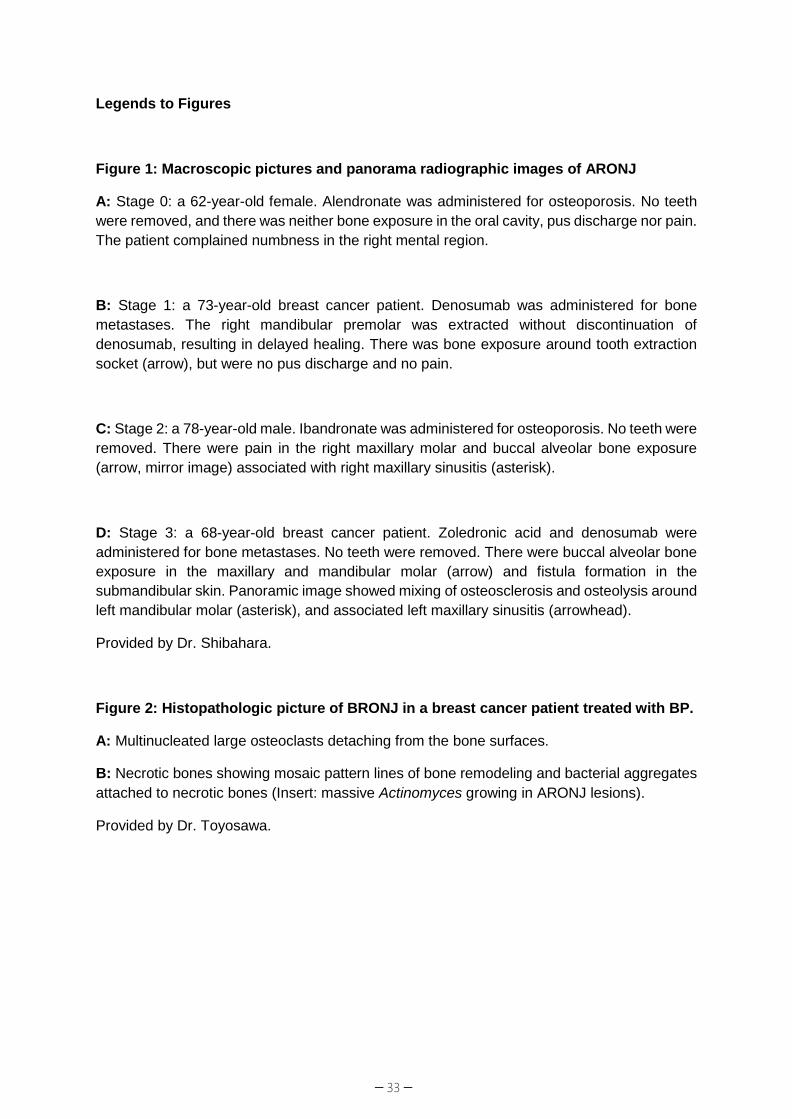
Legends to Figures
Figure 1: Macroscopic pictures and panorama radiographic images of ARONJ
A: Stage 0: a 62-year-old female. Alendronate was administered for osteoporosis. No teeth were removed, and there was neither bone exposure in the oral cavity, pus discharge nor pain. The patient complained numbness in the right mental region.
B: Stage 1: a 73-year-old breast cancer patient. Denosumab was administered for bone metastases. The right mandibular premolar was extracted without discontinuation of denosumab, resulting in delayed healing. There was bone exposure around tooth extraction socket (arrow), but were no pus discharge and no pain.
C: Stage 2: a 78-year-old male. Ibandronate was administered for osteoporosis. No teeth were removed. There were pain in the right maxillary molar and buccal alveolar bone exposure (arrow, mirror image) associated with right maxillary sinusitis (asterisk).
D: Stage 3: a 68-year-old breast cancer patient. Zoledronic acid and denosumab were administered for bone metastases. No teeth were removed. There were buccal alveolar bone exposure in the maxillary and mandibular molar (arrow) and fistula formation in the submandibular skin. Panoramic image showed mixing of osteosclerosis and osteolysis around left mandibular molar (asterisk), and associated left maxillary sinusitis (arrowhead).
Provided by Dr. Shibahara.
Figure 2: Histopathologic picture of BRONJ in a breast cancer patient treated with BP.
A: Multinucleated large osteoclasts detaching from the bone surfaces.
B: Necrotic bones showing mosaic pattern lines of bone remodeling and bacterial aggregates attached to necrotic bones (Insert: massive Actinomyces growing in ARONJ lesions).
Provided by Dr. Toyosawa.

─ 34 ─
[Contact information: Office of the Japanese Society for Bone and Mineral Research. E-mail
*
Figure 1
*
A B
C D
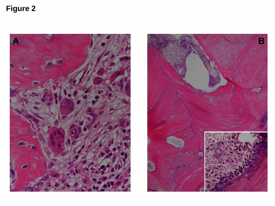
Figure 2
A B
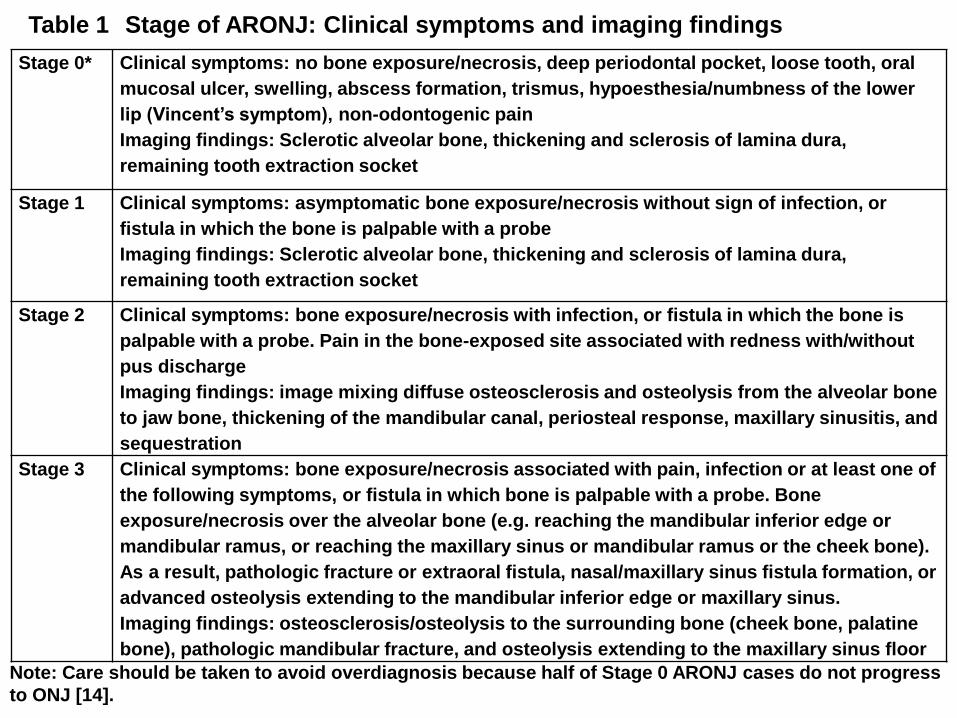
Stage 0* Clinical symptoms: no bone exposure/necrosis, deep periodontal pocket, loose tooth, oral mucosal ulcer, swelling, abscess formation, trismus, hypoesthesia/numbness of the lower lip (Vincent’s symptom), non-odontogenic painImaging findings: Sclerotic alveolar bone, thickening and sclerosis of lamina dura, remaining tooth extraction socket
Stage 1 Clinical symptoms: asymptomatic bone exposure/necrosis without sign of infection, or fistula in which the bone is palpable with a probeImaging findings: Sclerotic alveolar bone, thickening and sclerosis of lamina dura, remaining tooth extraction socket
Stage 2 Clinical symptoms: bone exposure/necrosis with infection, or fistula in which the bone is palpable with a probe. Pain in the bone-exposed site associated with redness with/without pus dischargeImaging findings: image mixing diffuse osteosclerosis and osteolysis from the alveolar bone to jaw bone, thickening of the mandibular canal, periosteal response, maxillary sinusitis, and sequestration
Stage 3 Clinical symptoms: bone exposure/necrosis associated with pain, infection or at least one of the following symptoms, or fistula in which bone is palpable with a probe. Bone exposure/necrosis over the alveolar bone (e.g. reaching the mandibular inferior edge or mandibular ramus, or reaching the maxillary sinus or mandibular ramus or the cheek bone). As a result, pathologic fracture or extraoral fistula, nasal/maxillary sinus fistula formation, or advanced osteolysis extending to the mandibular inferior edge or maxillary sinus.Imaging findings: osteosclerosis/osteolysis to the surrounding bone (cheek bone, palatine bone), pathologic mandibular fracture, and osteolysis extending to the maxillary sinus floor
Table 1 Stage of ARONJ: Clinical symptoms and imaging findings
Note: Care should be taken to avoid overdiagnosis because half of Stage 0 ARONJ cases do not progress to ONJ [14].
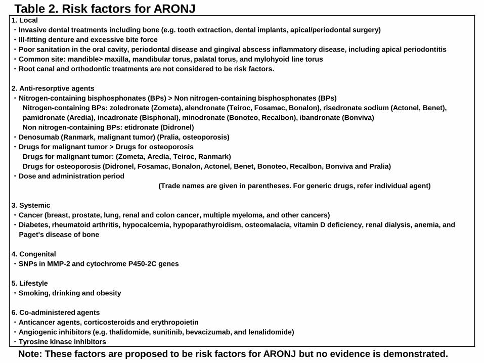
1. Local・Invasive dental treatments including bone (e.g. tooth extraction, dental implants, apical/periodontal surgery)・Ill-fitting denture and excessive bite force・Poor sanitation in the oral cavity, periodontal disease and gingival abscess inflammatory disease, including apical periodontitis・Common site: mandible> maxilla, mandibular torus, palatal torus, and mylohyoid line torus・Root canal and orthodontic treatments are not considered to be risk factors.
2. Anti-resorptive agents・Nitrogen-containing bisphosphonates (BPs) > Non nitrogen-containing bisphosphonates (BPs)
Nitrogen-containing BPs: zoledronate (Zometa), alendronate (Teiroc, Fosamac, Bonalon), risedronate sodium (Actonel, Benet), pamidronate (Aredia), incadronate (Bisphonal), minodronate (Bonoteo, Recalbon), ibandronate (Bonviva)Non nitrogen-containing BPs: etidronate (Didronel)
・Denosumab (Ranmark, malignant tumor) (Pralia, osteoporosis)・Drugs for malignant tumor > Drugs for osteoporosis
Drugs for malignant tumor: (Zometa, Aredia, Teiroc, Ranmark)Drugs for osteoporosis (Didronel, Fosamac, Bonalon, Actonel, Benet, Bonoteo, Recalbon, Bonviva and Pralia)
・Dose and administration period(Trade names are given in parentheses. For generic drugs, refer individual agent)
3. Systemic・Cancer (breast, prostate, lung, renal and colon cancer, multiple myeloma, and other cancers)・Diabetes, rheumatoid arthritis, hypocalcemia, hypoparathyroidism, osteomalacia, vitamin D deficiency, renal dialysis, anemia, and
Paget's disease of bone
4. Congenital・SNPs in MMP-2 and cytochrome P450-2C genes
5. Lifestyle・Smoking, drinking and obesity
6. Co-administered agents・Anticancer agents, corticosteroids and erythropoietin・Angiogenic inhibitors (e.g. thalidomide, sunitinib, bevacizumab, and lenalidomide)・Tyrosine kinase inhibitors
Table 2. Risk factors for ARONJ
Note: These factors are proposed to be risk factors for ARONJ but no evidence is demonstrated.
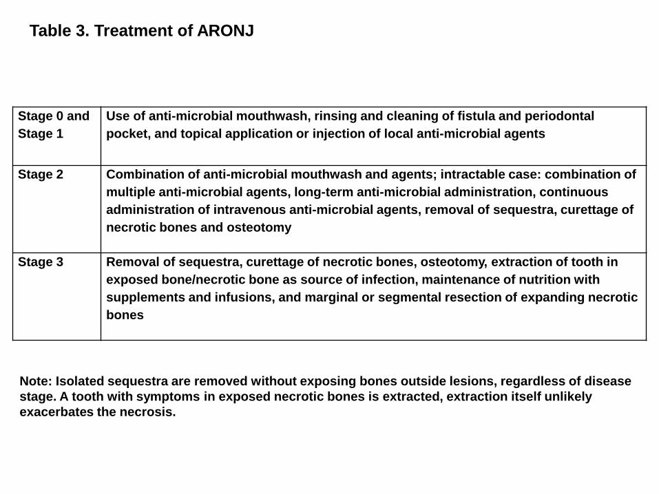
Stage 0 and Stage 1
Use of anti-microbial mouthwash, rinsing and cleaning of fistula and periodontal pocket, and topical application or injection of local anti-microbial agents
Stage 2 Combination of anti-microbial mouthwash and agents; intractable case: combination of multiple anti-microbial agents, long-term anti-microbial administration, continuous administration of intravenous anti-microbial agents, removal of sequestra, curettage of necrotic bones and osteotomy
Stage 3 Removal of sequestra, curettage of necrotic bones, osteotomy, extraction of tooth in exposed bone/necrotic bone as source of infection, maintenance of nutrition with supplements and infusions, and marginal or segmental resection of expanding necrotic bones
Table 3. Treatment of ARONJ
Note: Isolated sequestra are removed without exposing bones outside lesions, regardless of disease stage. A tooth with symptoms in exposed necrotic bones is extracted, extraction itself unlikely exacerbates the necrosis.