Embed Size (px)
Citation preview
Anti-CENP-B (IgG) ELISA
For the quantitative and qualitative detection of IgG antibodies against centromere
protein B (Cenp-B) in human serum
Catalog Number: 35-CENHU-E01 Size: 96 wells Version: 002: 2007-08-28 – ALPCO 8/24/2010
For Research Use Only. Not For Use In Diagnostic Procedures.
1/8
1. Intended Use The Anti-CENP-B (IgG) ELISA is a solid phase enzyme linked immunosorbent assay employing purified recombinant human 80 kDa centromere protein B (Cenp-B) for the quantitative and qualitative detection of IgG antibodies against Cenp-B in human serum. The assay ensures the highest sensitivity for the detection of antibodies against Cenp-B, which serves as an aid in the study of systemic sclerosis and CREST syndrome. This kit is for research use only. It is not for use in diagnostic procedures.
2. Application and Principle of the Assay The centromere is the primary constriction site of eukaryo tic chromosomes where sister chromatids appear most tightly paired. It is responsible for the coordinated segregation of chromosomes in mitosis and meiosis. The centromere is a complex area composed of particular nucleotidsequences, and various proteins. The three main centromere proteins are Cenp-A (17 kDa), Cenp-B (80 kDa), and Cenp-C (140 kDa). Systemic sclerosis is a multisystem autoimmune disease with system fibrosis of the connective tissue and typical occurrence of autoantibodies against centromere proteins as well as DNA topoisomerase I (Scl 70). Antibodies against Cenp-B are characteristic for CREST syndrome, a slower progressing variant of systemic sclerosis. The CREST syndrome exhibits symptoms of Calcinosis cutis, Raynaud’s syndrome, Esophageal motility disturbances, Sclerodactyly, and Talangiectasis. Up to 70-80% of CREST syndrome patients and 25% of patients with Raynaud syndrome exhibit centromere antibodies.
Principle of the test
Serum samples diluted 1:101 are incubated in the microplate coated with the specific antigen. Antibodies, if present in the sample, bind to the antigen. The unbound fraction is washed off in the following step. Afterwards, anti-human immunoglobulins conjugated to horseradish peroxidase (conjugate) are incubated and react with the antigen-antibody complex of the samples in the microplate. Unbound conjugate is washed off in the following step. Addition of TMB subs trate generates an enzymatic colorimetric (blue) reaction, which is stopped by diluted acid (color changes to yellow). The rate of color formation from the chromogen is a function of the amount of conjugate bound to the antigen-antibody complex and this is proportional to the initial concentration of the respective antibodies in the sample.
3. Kit Contents To be diluted: 5X Sample Buffer 1 vial, 20 ml – 5X concentrated (capped white: yellow solution) Containing: Tris, NaCl, BSA, sodium azide < 0.1% (preservative)
50X Wash Buffer 1 vial, 20 ml – 50X concentrated (capped white: green solution) Containing: Tris, NaCl, Tween 20, sodium azide < 0.1% (preservative)
2/8
Ready to use: Negative Control 1 vial, 1.5 ml (capped green: colorless solution)
Containing: Human serum (diluted), sodium azide < 0.1% (preservative)
Positive Control 1 vial, 1.5 ml (capped red: yellow solution) Containing: Human serum (diluted), sodium azide < 0.1% (preservative)
Cut-off Calibrator 1 vial, 1.5 ml (capped blue: yellow solution) Containing: Human serum (diluted), sodium azide < 0.1% (preservative)
Calibrators 6 vials, 1.5 ml each 0, 3, 10, 30, 100, 300 U/ml (color increasing with concentration: yellow solutions)
Containing: Human serum (diluted), sodium azide < 0.1% (preservative)
Conjugate 1 vial, 15 ml IgG (capped blue: blue solution)
Containing: Anti-human immunoglobulins conjugated to horseradish peroxidase
TMB Substrate 1 vial, 15 ml (capped black) Containing: Stabilized TMB/H2O2
Stop Solution 1 vial, 15 ml (capped white: colorless solution) Containing: 1 M Hydrochloric acid
Microtiter plate 12 x 8 well strips with breakaway microwells
Coating: see paragraph 1
Materials required but not provided: Microtiter plate reader 450 nm reading filter and optional 620 nm reference filter (600-690 nm). Glassware (100-1,000 ml), test tubes for dilutions. Vortex mixer, precision pipettes (10; 100; 200; 500; 1,000 µl) or adjustable multipipette (100-1,000 ml). Microplate washing device (300 µl repeating or multichannel pipette or automated system), absorbent paper.The assay is designed to be used with purified water according to the definition of the United States Pharmacopeia (USP 26 - NF 21) and the European Pharmacopeia (Eur.Ph. 4th ed.).
4. Storage and Shelf Life Store all reagents and the microplate at 2-8°C/35-46°F, in the original containers. Once prepared, diluted solutions are stable for at least 1 month at 4°C/39°F. Reagents and the microplate should only be used within the expiry date indicated on each component. Avoid exposing TMB solution to intense light. Store the microplate in the designated bag, including the desiccant, and seal tightly.
5. Precautions of Use 5.1 Health hazard data This product is for RESEARCH USE ONLY. Only staff trained and specially advised in the appropriate methods may perform the kit. Although this product is not considered particularly toxic or dangerous under conditions of normal use, refer to the following for maximum safety:
3/8
Recommendations and precautions This kit contains potentially hazardous components. Although the reagents are not classified as being irritants to eyes and skin, it is recommended to avoid contact with eyes and skin and wear disposable gloves. WARNING: Calibrators, Controls, and Buffers contain sodium azide (NaN3) as a preservative. NaN3 may be toxic if ingested or absorbed by skin or eyes. NaN3 may react with lead or copper plumbing to form highly explosive metal azides. On disposal, flush with a large volume of water to prevent azide build-up. Please refer to decontamination procedures as outlined by CDC or other local/national guidelines. Do not smoke, eat, or drink when manipulating the kit. Do not pipette by mouth.
All human source material used in kit breagents (e.g., controls, standards) has been tested by approved methods and found negative for HBsAg, Hepatitis C, and HIV1. No test can guarantee the absence of viral agents in such material completely; thus, handle controls, standards, and samples as if capable of transmitting infectious disease.
5.2 General Directions for Use Do not mix or substitute reagents or microplates from different lot numbers. This may lead to variation in the results. Allow all c omponents to reach room temperature (20-32°C/68- 89.6°F) before use, mix well, and follow the recommended incubation scheme for an optimum performance of the test. Incubation: It is recommended to test performance at 30°C/86°F for automated systems. Never expose components to temperatures higher than 37°C/98.6° F. Always pipette the substrate solution with only clean tips. Protect this reagent from light. Never pipette the conjugate with tips used previously with other reagents.
6. Sample Collection, Handling, and Storage It is recommended to use newl y collected serum samples. Bl ood withdrawal must follow national requirements. Do not use icteric, lipemic, hemolyzed, or bacterially contaminated samples. Sera wit h particles should be cleared by low speed centrifugation (<1,000 x g). Blood samples should be collected in clean, dry, and empty tubes. After separation, the serum samples should either be used immediately, stored at 2-8°C/35-46°F for up to three days, or frozen at -20°C/-4°F for longer periods.
7. Assay Procedure 7.1 Preparations prior to pipetting Dilute concentrated reagents: Dilute the concentrated sample buffer 1:5 with distilled water (e.g., 20 ml + 80 ml). Dilute the concentrated wash buffer 1:50 with distilled water (e.g., 20 ml + 980 ml).
4/8
Samples Dilute serum samples 1:101 with sample buffer (1X) [e.g., 10 µl serum + 1,000 µl sample buffer (1X).] Mix well. Washing Prepare 20 ml of diluted wash buffer (1X) per 8 wells or 200 ml for 96 wells (e.g., 4 ml concentrate + 196 ml distilled water). Automated washing Consider excess volumes required for setting up the instrument and dead volume of robot pipette. Manual washing Discard liquid from wells by inverting the plate. Knock the microwell frame vigorously on clean absorbent paper with wells facing down. Pipette 300 µl of diluted wash buffer into each well, wait for 20 seconds. Repeat the whole procedure twice again. Microplate Calculate the number of wells required for the test. Remove unused wells from the frame, replace and store in the provided plastic bag, and seal tightly together with the desiccant (2-8°C/35-46°F).
7.2 Work flow - For pipetting scheme see Annex A. It is recommended to pipette samples and calibrators in duplicate. The cut-off calibrator should be used for qualitative testing only.
Pipette 100 µl of each diluted serum sample into the designated microwells.
Pipette 100 µl of each calibrator OR cut-off calibrator and negative and positive controls into the designated wells.
Incubate for 30 minutes at room temperature (20-32°C/68-89.6°F).
Wash 3X with 300 µl washing buffer (diluted 1:50).
Pipette 100 µl of conjugate into each well.
Incubate for 30 minutes at room temperature (20-32°C/68-89.6°F).
Wash 3X with 300 µl washing buffer (diluted 1:50).
Pipette 100 µl of TMB substrate into each well.
Incubate for 30 minutes at room temperature (20-32°C/68-89.6°F), protected from intense light.
Pipette 100 µl of stop solution into each well in the same order as when the TMB substrate was pipetted.
Incubate for a minimum of 5 minutes.
Agitate plate carefully for 5 seconds.
Read absorbance at 450 nm (optionally 450/620 nm) within 30 minutes.
5/8
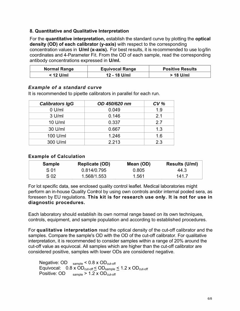
8. Quantitative and Qualitative Interpretation For the quantitative interpretation, establish the standard curve by plotting the optical density (OD) of each calibrator (y-axis) with respect to the corresponding concentration values in U/ml (x-axis). For best results, it is recommended to use log/lin coordinates and 4-Parameter Fit. From the OD of each sample, read the corresponding antibody concentrations expressed in U/ml.
Normal Range Equivocal Range Positive Results < 12 U/ml 12 - 18 U/ml > 18 U/ml
Example of a standard curve It is recommended to pipette calibrators in parallel for each run.
Calibrators IgG OD 450/620 nm CV % 0 U/ml 0.049 1.9 3 U/ml 0.146 2.1 10 U/ml 0.337 2.7 30 U/ml 0.667 1.3
100 U/ml 1.246 1.6 300 U/ml 2.213 2.3
Example of Calculation
Sample Replicate (OD) Mean (OD) Results (U/ml) S 01 0.814/0.795 0.805 44.3 S 02 1.568/1.553 1.561 141.7
For lot specific data, see enclosed quality control leaflet. Medical laboratories might perform an in-house Quality Control by using own controls and/or internal pooled sera, as foreseen by EU regulations. This kit is for research use only. It is not for use in diagnostic procedures.
Each laboratory should establish its own normal range based on its own techniques, controls, equipment, and sample population and according to established procedures.
For qualitative interpretation read the optical density of the cut-off calibrator and the samples. Compare the sample's OD with the OD of the cut-off calibrator. For qualitative interpretation, it is recommended to consider samples within a range of 20% around the cut-off value as equivocal. All samples which are higher than the cut-off calibrator are considered positive, samples with lower ODs are considered negative.
Negative: OD sample < 0.8 x ODcut-off Equivocal: 0.8 x ODcut-off < ODsample < 1.2 x ODcut-off Positive: OD sample > 1.2 x ODcut-off
6/8
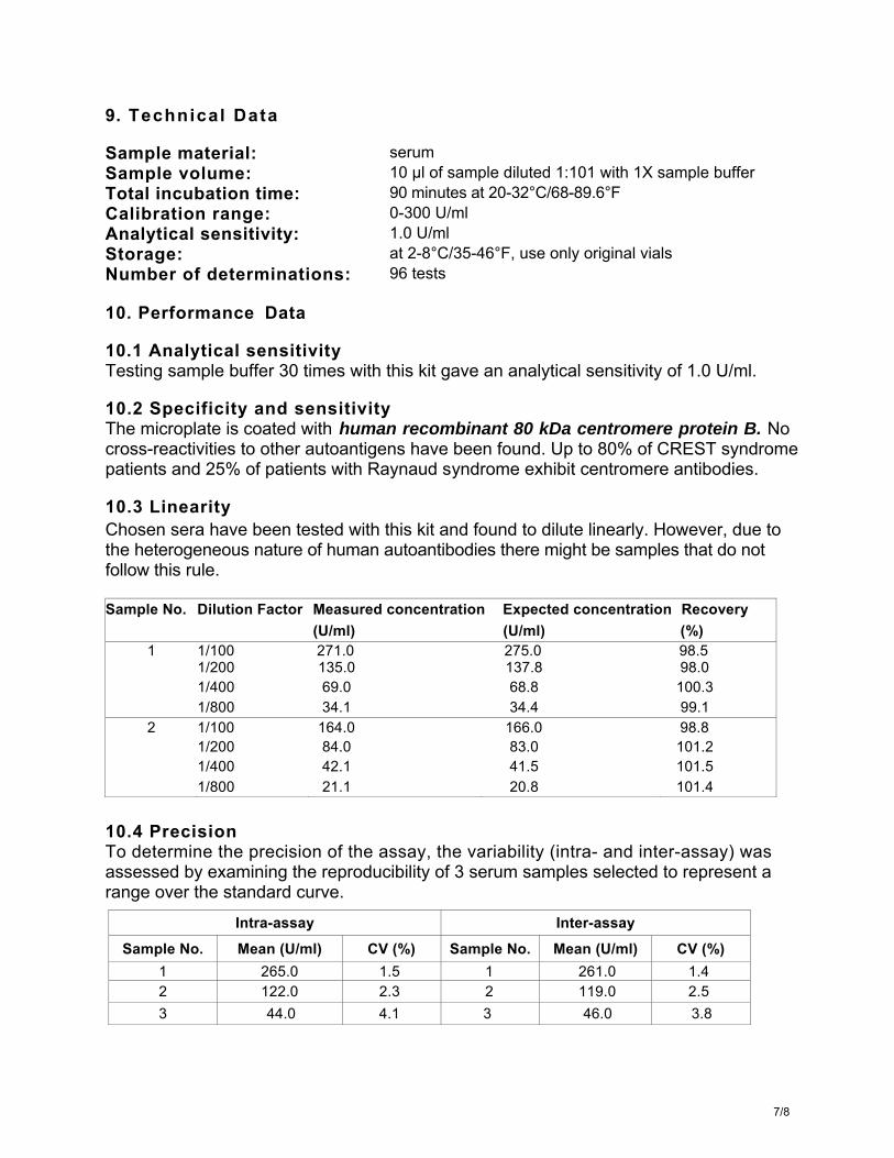
9. Technical Data
Sample material: serum Sample volume: 10 µl of sample diluted 1:101 with 1X sample buffer Total incubation time: 90 minutes at 20-32°C/68-89.6°F Calibration range: 0-300 U/ml Analytical sensitivity: 1.0 U/ml Storage: at 2-8°C/35-46°F, use only original vials Number of determinations: 96 tests
10. Performance Data
10.1 Analytical sensitivity Testing sample buffer 30 times with this kit gave an analytical sensitivity of 1.0 U/ml.
10.2 Specificity and sensitivity The microplate is coated with human recombinant 80 kDa centromere protein B. No cross-reactivities to other autoantigens have been found. Up to 80% of CREST syndrome patients and 25% of patients with Raynaud syndrome exhibit centromere antibodies.
10.3 Linearity Chosen sera have been tested with this kit and found to dilute linearly. However, due to the heterogeneous nature of human autoantibodies there might be samples that do not follow this rule.
Sample No. Dilution Factor Measured concentration Expected concentration Recovery (U/ml) (U/ml) (%)
1 1/100 271.0 275.0 98.5 1/200 135.0 137.8 98.0 1/400 69.0 68.8 100.3 1/800 34.1 34.4 99.1
2 1/100 164.0 166.0 98.8 1/200 84.0 83.0 101.2 1/400 42.1 41.5 101.5 1/800 21.1 20.8 101.4
10.4 Precision To determine the precision of the assay, the variability (intra- and inter-assay) was assessed by examining the reproducibility of 3 serum samples selected to represent a range over the standard curve.
Intra-assay Inter-assay Sample No. Mean (U/ml) CV (%) Sample No. Mean (U/ml) CV (%)
1 265.0 1.5 1 261.0 1.4 2 122.0 2.3 2 119.0 2.5 3 44.0 4.1 3 46.0 3.8
7/8

10.5 Calibration This assay is calibrated against reference sera from the CDC Atlanta (Centers for Disease Control and Prevention). The results are expressed in U/ml.
11. Literature 1. Earnshaw, WC, Bordwell, B, Marino, C, Rothfield, N. (1986):
The three human chromosomal autoantigens are recognized by sera from patients with anticentromere antibodies. J Clin Invest 77: 426-430.
2. von Mühlen, CA, Tan, EM (1995): Autoantibodies in the diagnosis of system rheumatic diseases. Semin Arthritis Rheum 24: 323-358.
3. Tan, EM (1989): Antinuclear antibodies: diagnostic markers for autoimmune diseases and probes for cell biology. Adv Immunol 44: 93-151.
4. Fritzler, MJ (1993): Autoantibodies in scleroderma. J Dermatol 24: 323-358.
Annex A: Pipetting scheme It is suggested to pipette the calibrators, controls, and samples as follows: For quantitative interpretation use the calibrators to establish a standard curve, for qualitative interpretation use the cut-off calibrator.
For quantitative interpretation use calibrators to establish a standard curve
For qualitative interpretation use cut-off calibrator
1 2 3 4 5 6 7 8 9 10 11 12 A CalA CalE S1 NC S2
B CalA CalE S1 NC S2
C CalB CalF S2 CC S3
D CalB CalF S2 CC S3
E CalC PC S3 PC
F CalC PC S3 PC
G CalD NC S1
H CalD NC S1 CalA: calibrator A, CalB: calibrator B, CalC: calibrator C, CalD: calibrator D, CalE: calibrator E, CalF: calibrator F PC: positive control NC: negative control CC: cut-off calibrator S1: sample 1 S2: sample 2 S3: sample 3
8/8