Embed Size (px)
Citation preview

Contents lists available at ScienceDirect
Materials Science & Engineering C
journal homepage: www.elsevier.com/locate/msec
Anti-biofouling microfiltration membranes based on 1-vinyl-3-butylimidazolium chloride grafted PVDF with improved bactericidalproperties and vitro biocompatibilityXiaowei Zhang, Yuanyuan Liang⁎, Chunjun Ni, Yongjin Li⁎
College of Material, Chemistry and Chemical Engineering, Hangzhou Normal University, Hangzhou 311121, People's Republic of China
A R T I C L E I N F O
Keywords:Polyvinylidene fluoride1-vinyl-3-butylimidazolium chlorideAnti-biofoulingBactericidal propertiesBiocompatibility
A B S T R A C T
Polyvinylidene fluoride (PVDF) porous membranes have been widely used as the filtration and separation in-dustry. Herein, novel microfiltration membranes based on 1-vinyl-3-butylimidazolium chloride ([VBIm][Cl])grafted PVDF (PVDF-g-[VBIm][Cl]) were prepared via the non-solvent induced phase separation method. Thechemical composition and microstructure of PVDF-g-[VBIm][Cl] membranes were characterized by Fouriertransform infrared spectroscopy, X-ray photoelectron spectroscopy, Scanning electron microscopy and Watercontact angle measurements. The results showed that an increasing in [VBIm][Cl] grafting content leads to theincreasing hydrophilicity and wetting capacity of the PVDF-g-[VBIm][Cl] porous membranes. The anti-bio-fouling properties of membranes were evaluated by measuring the water flux before and after Bovine serumalbumin solution treatment. It was found that the modified membranes presented a good anti-biofoulingproperty. The degree of irreversible flux loss caused by protein adsorption dramatically reduced from 42.1% to2.9% compared with the pristine hydrophobic PVDF membranes. Meanwhile, these PVDF-g-[VBIm][Cl] mem-branes also exhibited excellent bactericidal properties against both gram-positive bacteria Staphylococcus saureusand gram-negative bacteria Escherichia coli, while PVDF membranes did not show any antibacterial activity. Thevitro biocompatibility of the modified membranes was studied by hemolysis analysis, the platelet adhesionobservation, thromboelastography assay and cytotoxicity assay. It was found that the incorporation of [VBIm][Cl] into PVDF membranes has less effect on the hemolysis and cytotoxicity of PVDF membranes. Furthermore,both hydrophilicity and charges of the membrane surface played important role in the adhesion and activation ofplatelet cells, which consequently affected the clotting process of whole blood. The membrane with appropriate[VBIm][Cl] grafting ratio (2.94 wt.%) exhibited good hemocompatibility with less blood coagulation effect. Asan ultrafiltration membrane, PVDF-g-[VBIm][Cl] membranes have potential applications in the biomedical fielddue to the improved antibacterial property and biocompatibility.
1. Introduction
Poly(vinylidene fluoride) (PVDF) is widely used in the fabrication ofmembranes for water or blood purification due to its excellent me-chanical properties, chemical resistance, thermal stability and proces-sability [1–4]. However, PVDF membranes show a strong nonspecificadsorption of microorganisms, cells and proteins and suffer from socalled “biofouling” when encountering living system mainly due to itshydrophobicity, which limits its application [5–8]. Specifically, theattachment of microbial cells coming from bacteria or fungi usuallycauses membrane blocking and decreasing flux. What's more, theseattached microorganisms also convert soluble salts and nutrients toinsoluble ones, which intensifying the fouling of PVDF membranes.
These biofilms resulting from microorganisms or cells also have beenfound in the application of polymer hemodialysis catheter, contributingto bloodstream infections [9–12]. Therefore, significant attention hasbeen paid to developing anti-biofouling membranes by introducingnon-fouling [8,13–15] or charged molecules [16,17] to minimize thebacterial adhesion or attachment to membranes. It has also been ob-served that biofouling of membranes can be effectively prevented byincorporating biocidal agents such as copper or silver [18,19] intomembranes. These biocidal agents have the function of killing thebacteria attached to the membranes surface and inhibiting the growthof bacteria. Although the membrane biofouling is still a complicatedphenomenon and many factors are involved, developing membraneswith both anti-adhesion and anti-bacteria would be an effective way to
https://doi.org/10.1016/j.msec.2020.111411Received 26 March 2020; Received in revised form 6 August 2020; Accepted 19 August 2020
⁎ Corresponding authors.E-mail addresses: [email protected] (Y. Liang), [email protected] (Y. Li).
Materials Science & Engineering C 118 (2021) 111411
Available online 22 August 20200928-4931/ © 2020 Elsevier B.V. All rights reserved.
T

control membrane biofouling.Ionic liquids (ILs), a class of organic salts with melting points at or
below 100 °C, have a variety of applications including alternative sol-vents [20], catalysts [21], gas separation [22], fuel cells [23] andpharmaceuticals [24] because of their unique structures and properties.In particular, ILs with cations of ammonium [16], imidazolium [25,26],pyridinium [27], quinolinium [28], and phosphonium [29] exhibitexcellent antimicrobial activities against a broad range of pathogenicmicrobes including gram-positive bacteria, gram-negative bacteria andfungus. However, one of main challenges for these cationic ILs is theirhigh toxicity to mammalian cells, which greatly restrains their appli-cations [30]. Recently, cationic type poly(ionic liquid)s (PILs) haveshown attractive antimicrobial activities. Yan's group has system-atically studied the structure-antibacterial activity relationship on PILsmembranes obtained from quaternary ammonium [31], imidazolium-type [32], and pyrrolidinium-type [33] ILs monomers. They synthe-sized a series of mono- and bis-imidazolium or pyrrolidinium ILmonomers and their analogous PILs via reversible addition fragmen-tation chain transfer polymerization. Remarkably, pyrrolidinium basedPILs membranes [33] were prepared via in situ photo-cross-linkingpolymerization followed by anion-exchange with L-proline and L-tryptophan. The resultant membranes showed high antimicrobial ac-tivities and low cytotoxicity. However, the polymerization or copoly-merization of ionic ILs monomers is relatively complicated and timeconsuming. Moreover, the resulting casted polymer membranes areusually weak and brittle, extra steps such as chemical crosslinking aredemanded to satisfy the real-world applications. Nevertheless, theseresults indicated that introducing the cation containing ILs into mem-branes to develop anti-biofouling membranes with improved bio-compatibility is still a very attractive and promising prospect that willboost the membrane applications.
Therefore, in this work, a facile and scalable approach to fabricateanti-biofouling membranes with bactericidal properties and bio-compatibility by introducing ILs to PVDF was explored. Firstly, weimmobilized 1-vinyl-3-butylimidazolium chloride ([VBIm][Cl]) ontoPVDF by irradiation-induced graft polymerization according to ourprevious work [26,34]. The prepared [VBIm][Cl] grafted PVDF (PVDF-g-[VBIm][Cl]) was then used to fabricate microfiltration membranesvia non-solvent induced phase separation method, which is the mostwidely used technique for casting polymeric membranes for commer-cial use. Finally, the interplay between the chemical structures andantibacterial versus hemolytic properties of membranes based on theamphiphilic [VBIm][Cl] grafted PVDF were investigated. It is shownthat the prepared PVDF-g-[VBIm][Cl] membranes have potential to beapplied in medical material due to the improved antibacterial propertyand biocompatibility.
2. Experimental section
2.1. Materials
The poly(vinylidene fluoride) (PVDF) (Mw ~ 209,000, Mw/Mn = 2.0) was obtained from Kureha Chemicals (Japan). The ionic li-quid, 1-vinyl-3-butylimidazolium chloride was provided by LanzhouGreenchem. ILs. LICP. CAS.(China). Poly(vinyl pyrrolidone) (PVP) K30(Mw = 50,000), bovine serum albumin (BSA, 98%, Mn = 66,430 Da)was purchased from J&K Scientific (China). Mouse fibroblast NIH 3T3cell line was purchased from the Cell bank of the Chinese Academy ofScience (Shanghai, China). N, N-dimethyl formamide (DMF), methanol(CH3OH) were obtained from sinopharm chemical reagent Co., Ltd.(China). All the solutions were prepared with deionized water.
2.2. Synthesis of PVDF-g-[VBIm][Cl]
The material PVDF-g-[VBIm][Cl] with different grafting ratios waspre-prepared by irradiation-induced graft polymerization, according to
our previous work [26,34]. Briefly, the PVDF/[VBIm][Cl] blends wereobtained by melt-compounding at 180 °C using a Haake Polylab QCmixer and prepared into films through hot-press. Then the PVDF/[VBIm][Cl] blend films were exposed to the electron beam at 45 kGydosage at room temperature. Under the action of an electron beam, freeradicals were produced on the PVDF molecular chain, initiated additionto double bonds of ILs. The irradiated blends had been extracted withCH3OH for 72 h, in order to remove the residual ILs or IL homo-polymers. Here, the grafting efficiency (GE) and final grafting ratio(GR) could be calculated according to following equations:
=×
×GE m mm i
1 100%1 2
1 (1)
= ×GR GE w (2)
where GE was grafting efficiency of ionic liquid, m1 and m2 were themass of PVDF-g-[VBIm][Cl]film before and after extraction, the re-duced mass was regarded as the part of the ionic liquid that didn't graft,i was the mass fraction of ionic liquid in PVDF/[VBIm][Cl] blend, GRwas the final grafting ratio of ionic liquid, w was the feed ratio of ionicliquid to PVDF in melt mixing.
2.3. Preparation of PVDF-g-[VBIm][Cl] membranes
The conventional non-solvent induced phase separation method wasemployed to fabricate PVDF-g-[VBIm][Cl] membranes. Briefly, pristineor grafted PVDF and PVP were dissolved in DMF at 80 °C with a weightratio of 0.95: 0.05: 5. After eliminating bubbles by ultrasonication for30 min, the solution was carefully poured and cast onto a glass plate.Subsequently, the liquid membrane with glass plate immersed into thedeionized water bath set at 25 °C. The membrane was taken out when itwas completely solidified, and washed with running deionized water tothoroughly remove the residual solvent. The membranes were kept infresh deionized water for further characterization.
2.4. Characterizations of membranes
Fourier transform infrared spectroscopy (FTIR) of PVDF-g-[VBIm][Cl] composites was performed by a Bruker VERTEX 70v spectrometer.Scanning electron microscopy (SEM, S-4800, Hitachi, Japan) was usedto observe membrane morphological structure. The accelerating vol-tage is 3 kV and working distance is 8–10 cm. To observe the cross-section, membranes were fractured in liquid nitrogen. We used animage analysis software (Image J, NIH) to count the area fraction ofpores on the membrane surface. Briefly, the SEM images of surface wereimported into the software, then the pores were marked and the areafractions of pores were calculated. And the porosity of membrane wascalculated by the following equations, measuring though the dry-wetmethod:
=×
×porosity m mS d
( )/ 100%3 4(3)
where m3 was the mass of the membrane saturated completely withdeionized water, m4 was the mass of the membrane after completelydried, ρ was the density of water, S was the area of the membrane, dwas the average thickness of the membrane.
The chemical composition of membrane surface was analyzed by X-ray photoelectron spectroscopy (XPS, ESCALAB 250Xi, with an Al Kalpha X-ray source (1486.8 eV), Thermo Scientific, USA). The contactangle instrument (DSA 100, Data-Physical, Germany) was used toevaluate membranes surface hydrophilicity with purified water at am-bient temperature. The contact angle value was recorded every 30 s andat least 11 readings collected for each membrane type. The membranesurface water infiltration was evaluated through the change of contactangle value with time. The zeta potential on membrane surface, basedon streaming potential and streaming current measurements, was
X. Zhang, et al. Materials Science & Engineering C 118 (2021) 111411
2
measured with a solid surface zeta potential analyzer (Surpass, AntonPaar, Austria). The test condition was 1 mM KCl aqueous solution at25 °C and pH of 7.4.
2.5. Filtration measurement and antifouling properties evaluation
The flux of membrane was measured by a dead-end filtrationequipment which was home-made (as shown in Fig. S1). The samplemembrane was mounted on a sand core with an active transport area of1.767 cm2 and pre-pressured at 0.1 MPa for 30 min, using the deionizedwater, to approach a stable flux at room temperature. Subsequently, thetime required to permeate through 10 mL deionized water was re-corded, the flux for deionized water (J, Lm−2 h−1) was calculated bythe following formula, and five readings collected to acquire theaverage value:
=×
J VA t (5)
Where V was the filtrated volume of the deionized water or BSAsolution (L), A was the effective permeable area (m2) of the membrane,t was the time (h) that took to flow through a certain volume of thefluid.
Subsequently, the deionized water was replaced by the BSA solu-tion, the flux for BSA solution (JB, Lm−2 h−1) was measured in thesame way as above. Then the contaminated membrane was taken outand dipped into deionized water and cleaned with ultrasonic washer for5 min, and then rinsed under flowing deionized water for 5 min.Finally, the water flux of the washed membrane was measured again.
In order to quantitatively analyze antifouling properties of mem-branes, two parameters were calculated through following expressions:
= ×FRR JJ
100%w
w
2
1 (6)
= ×R J JJ
100%irw w
w
1 2
1 (7)
where FRR was the flux recovery ratio; Rir was the degree of irreversibleflux loss. Jw1 and Jw2 was the flux of deionized water before and afterthe membranes were treated with BSA solution, respectively.
2.6. Antibacterial test
The antibacterial activities of PVDF or PVDF-g-[VBIm][Cl] mem-branes against Staphylococcus aureus (ATCC 6538p) and Escherichia coli(ATCC 8739) were tested by TüV SüD Products Testing Co. Ltd.(Shanghai, China). The test process and the standards were referred tothe JIS Z 2801–2010 (Japanese Industrial Standards). The reduction ofbacteria, as antimicrobial abilities of membranes, was calculated ac-cording to the following equation:
=Reduction C M(log log )h h10 24 10 24 (8)
where C24h and M24h were the number of bacteria obtained from theinoculated control sample and the test membrane after 24 h, respec-tively.
2.7. Biocompatibility
The Institutional Administration Panel for Laboratory Animal Care(Medical ethics committee of Hangzhou Normal University) approvedthe experimental design.
2.7.1. HemolysisThe fresh whole blood (22 years old, male) was collected by using
anticoagulant tubes (anticoagulant to blood ratio, 1:9 v/v). The ob-tained erythrocytes were separated by centrifugation and washed withPBS (pH 7.4) for three times. Erythrocytes were then resuspended in
PBS to get a 16% (v/v) suspension. After that, 1 mL erythrocytes sus-pension was incubated with the prepared membranes (1 × 1 cm2) at37 °C. Erythrocytes suspensions treated with PBS were used as a con-trol. Finally, 4 mL Na2CO3 solution (0.1 wt.%) was added into the Rerythrocytes suspensions to induce a complete hemolysis. After 4 hincubation, the erythrocytes suspensions were centrifuged and the he-molysis extent was determined by measuring the absorbance at 540 nm.The hemolysis rate was calculated according to the following equation:
= ×Hemolysisi rate A BC B
(%) 100% (9)
where A is the absorbance of test sample, B and C are the erythrocytessuspension treated with PBS and the erythrocytes suspension of com-plete hemolysis, respectively.
2.7.2. Blood platelet adhesionThe platelet-rich plasma (PRP) was obtained by centrifuging fresh
human blood (22 years old, male) at 1000 rpm for 15 min.Meanwhile,the prepared membranes (1 × 1 cm2) were immersed inPBS solutions for 12 h at 37 °C, afterwards, the PBS solution was dis-carded and 1 mL of PRP was added,all the membranes were incubatedat 37 °C for 90 min. Finally, the membranes were rinsed with PBS so-lution 3 times and then treated with glutaraldehyde (4 wt.%) at 37 °Cfor 48 h to fix the adhered platelets and adsorbed proteins. The mem-branes were dehydrated by 75%, 85%, 95% and 100% (v/v) ethanol for10 min, respectively. The platelet adhesion was observed under SEM.
2.7.3. ThromboelastographyThe effects of membranes of prepared membranes on coagulation
processing of human whole blood were examined byThromboelastograph Hemostasis System 5000 (TEG, HaemoscopeCorporation). Briefly, the obtained citrated whole blood (0.5 mL) wasincubated with membranes (1 × 1 cm2) in a tube containing kaolin for1 h at 37 °C. PBS was used as a control. Then, 400 μL blood sample wastransferred into TEG cup and 20 μL aqueous CaCl2 (0.2 mol/L) wasadded to initiate the TEG analysis.
2.7.4. CytocompatibilityThe mouse fibroblast cell line NIH-3T3 were cultured in R1640
medium supplemented with 10% fetal bovine serum, 2 mM L-glutamineand antibiotics mixture (10,000 U penicillin and 10 mg streptomycin,1% v/v). These cultured cells were seeded on to 24-well culture platesat a density of 104 cells/μL and incubated for 24 h at 37 °C in 5% CO2,the membranes were then added and incubated with the cell for 24 h.Finally, cell viability was determined by MTT assay. Fibroblast cellsincubated with PBS were used as a control. In brief, 50 μL 3-(4,5-di-methylthiazol-2-yl-)-2,5-diphenyltetrazolium bromide (MTT) (1 mg/mL) was added to the 24-well plates and incubated at 37 °C for 4 h in5% CO2, finally 50 μL dimethyl sulfoxide was added into each well. Thelevel of reduction of MTT was measured by optical density at 490 nmand the results were expressed in percentage relative to the control.
3. Results and discussion
3.1. Chemical structure of PVDF-g-[VBIm][Cl] and membranes surface
The 1-vinyl-3-butylimidazolium chloride grafted PVDF (PVDF-g-[VBIm][Cl]) was prepared via electron-beam irradiation-induced graftpolymerization according to our previous work [26,34] and its che-mical structure was confirmed by FT-IR spectra (Fig. 1). It can be seenthat the most significant change between PVDF and PVDF-g-[VBIm][Cl]is the appearance of the strong characteristic peaks at the range of500–1000 cm−1, which represents the CeH in-plane flexural vibrationsof the imidazole ring. In the spectrum of [VBIm][Cl], the characteristicabsorption bands at 1162 and 1557 cm−1 were ascribed to the imida-zole ring and skeletal vibrations. Meanwhile, in the spectrum of PVDF-
X. Zhang, et al. Materials Science & Engineering C 118 (2021) 111411
3

g-[VBIm][Cl], the disappearance of the adsorption peaks at 1651 and1699 cm−1 (corresponding to vinyl group vibrating) indicates thesuccessful grafting of ionic liquid onto PVDF chains via electron-beamirradiation-induced graft polymerization.
For PVDF-g-[VBIm][Cl], the grafting efficiency (GE) and graftingratio (GR) of ionic liquid were calculated and listed in Table 1. It is clearthat high GE can be achieved by the irradiation induced grafting. NeatPVDF and PVDF-g-[VBIm][Cl] were then dissolved in DMF to preparethe porous membrane through the non-solvent induced phase separa-tion method. The obtained PVDF-g-[VBIm][Cl] membranes with dif-ferent GR hereinafter were referred to as M0, M1, M3, M5, M10, as alsoshown in Table 1.
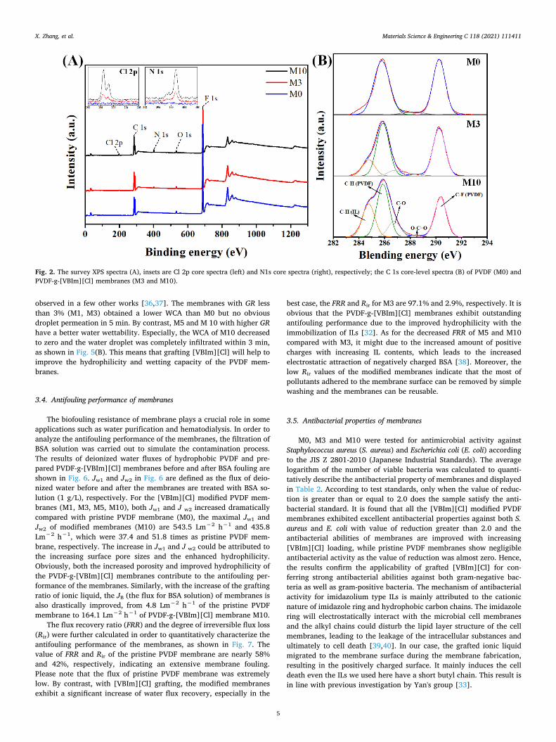
XPS is usually employed to ascertain the chemical compositions ofmembranes surface. In Fig. 2A, the pristine PVDF membrane showsprimary emission peaks corresponding to C 1s (286.3 eV) and F 1s(687.3 eV) and O 1s (532.1 eV). Here, O 1s was mainly due to thereaction between O2 and PVDF main chains during electron-beam ir-radiation in air. Meanwhile, comparing with the pristine PVDF mem-brane (M0), membranes M3 and M10 display additional N 1s peak withbinding energy of 401.1 eV and Cl 2p peak with binding energy of197.1 eV, which confirmed the successful immobilization of ionic li-quid. Moreover, the intensity changes of both N 1s and Cl 2p peaks inmodified membranes were carefully investigated and the results areshown in the inset of Fig. 2A. It shows that both the peak intensities at401.1 eV and 197.1 increased as the increasing ILs GR of the modifiedmembranes, which also suggested the success of introducing ILs in thePVDF membranes. In order to clearly study the chemical composition ofthe membrane surface, the C1s peaks of XPS spectra of PVDF membrane(M0) and PVDF-g-[VBIm][Cl] membranes (M3 and M10) were fittedwith five peaks, respectively, the results are shown in Fig. 2B. As seen,five types of carbon-containing functional groups were located at285.8 eV (CeH, PVDF), 286.7 eV (CeO, PVDF), 288.3 eV (O-C=O,PVDF), 290.3 eV (CF2, PVDF), and CeH (IL), 284.7 eV. To compare the
average concentration of immobilized IL in the bulk material with theconcentration on the surface of membranes, the values of molar ratiosof [C]IL/[C]PVDF was calculated. Here, [C]IL/[C]PVDF of bulk materialswere calculated from the mass ratios of IL and PVDF in theory, while[C]IL/[C]PVDF of surface of the membrane was determined from thefitted XPS C1s core-level spectral area ratios of the respective samples,where [C]IL is the area of the peak at 284.7 eV (CeH, IL), [C]PVDF is thesum of the areas of the other four peaks mentioned above [26]. In thecase of M3, the [C]IL/[C]PVDF of the surface of the membrane was0.158, which was nearly 3.5 times the value of [C]IL/[C]PVDF in the bulk(0.0454), indicating a substantial surface segregation of IL component.The same result was also obtained from M10 where the [C]IL/[C]PVDF ofthe surface of the membrane and bulk were 0.353 and 0.141, respec-tively. The surface enrichment of IL might be attributed to the fact thegrafted ILs component is likely to migrate to the surface of membranesduring the coagulation step of non-solvent induced phase separationmethod due to the low interfacial energy between the hydrophilicgrafted ILs component and water [35]. It is noteworthy that the en-riched ionic liquid moiety on the membrane surface could effectivelybenefit for the functionality of the membranes.
3.2. Morphology of membranes
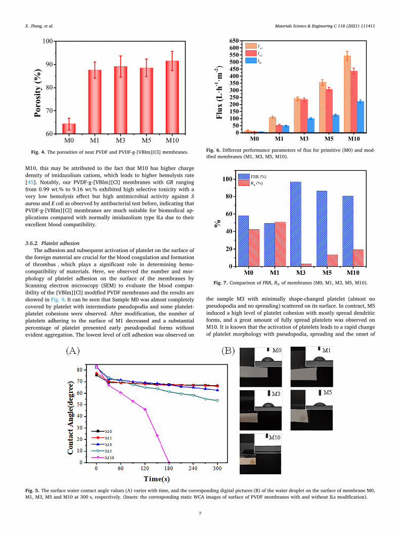
Fig. 3 shows the top surface (as designated as S in the caption) andthe cross section (as designated as C in the caption) SEM images ofobtained membranes. It is clear that the surface of pristine PVDFmembrane was less porous than the PVDF-g-[VBIm][Cl] membrane(Fig. 3 from M0-S to M10-S). Regarding the surface porosity, we usedimage analysis software (Image J) to analyze the area fraction of poreson the membrane surfaces. And the results were 0.49%, 1.57%, 3.20%,6.65% and 8.44%, corresponding to the M0, M1, M3, M5, M10, re-spectively. It indicates that the membrane surface porosity is improvedwith the increasing ILs GR. As shown in the cross-section images (Fig. 3from M0-C1 to M10-C1), all the membranes exhibited a typicallyasymmetric porous structure, consisting of a dense top layer, supportedby finger-like structure and/or sponge-like layer. However, comparedwith Membrane M0, all the modified membranes had more finger-likepores but less sponge-like layer. We speculate that the increasing hy-drophilicity by the ILs grafting might accelerate the solvent (DMF)/non-solvent (deionized water) exchange rate, which decreases solventkinetic hindrance in the phase inversion process. Thus, rapid separationis enhanced, which results in the formation of a fully developed finger-like structure. Moreover, SEM images in high resolution (Fig. 3 fromM0-C2 to M10-C2) reveal that all the modified PVDF membranes hadinter-connective submicro- or nano-sized pores on the walls of finger-like pores, indicating a formation of unique hierarchical pore structure.Specifically, M10 possessed a mostly porous finger-like pore structurewith numerous nano-sized (about 40 nm) pores spreading on the wallsof micro-sized pores. Furthermore, the porosities of membranes weredetermined quantitatively and the results are shown in Fig. 4. As seen,the porosities of membranes had a significant growth from 64.3% (v/v)for M0 to 87.6% (v/v) for M1, while the variation between the modifiedmembranes with different ILs GR was mild. This might be due to thefact that all the modified membranes had a similar finger-like porestructure.
3.3. Surface hydrophilia of membranes
The hydrophilicity of the membrane surface was evaluated by watercontact angle (WCA) measurement, and the results are shown in Fig. 5.All the membranes had a WCA decreasing gradually with the wettingtime. However, with increasing [VBIm][Cl] grafting degrees, thedownward tendency of the [VBIm][Cl] modified membrane becomesmore significant. For the pristine PVDF membrane (M0), it presented ahighest WCA of 77.2° and decreased to 66.1° after 5 min, correspondingto its lowest surface hydrophilicity. This phenomenon was also
Fig. 1. FT-IR spectra of the ionic liquid, the neat PVDF and the PVDF-g-[VBIm][Cl].
Table 1The grafting efficiency and grafting ratio of PVDF-g-[VBIm][Cl].
Membrane PVDF/[VBIm][Cl] (w/w) GE (%) GR(%)
M0 100/0 / 0M1 100/1 99.9 0.99M3 100/3 98.1 2.94M5 100/5 95.7 4.79M10 100/10 91.6 9.16
X. Zhang, et al. Materials Science & Engineering C 118 (2021) 111411
4
observed in a few other works [36,37]. The membranes with GR lessthan 3% (M1, M3) obtained a lower WCA than M0 but no obviousdroplet permeation in 5 min. By contrast, M5 and M 10 with higher GRhave a better water wettability. Especially, the WCA of M10 decreasedto zero and the water droplet was completely infiltrated within 3 min,as shown in Fig. 5(B). This means that grafting [VBIm][Cl] will help toimprove the hydrophilicity and wetting capacity of the PVDF mem-branes.
3.4. Antifouling performance of membranes
The biofouling resistance of membrane plays a crucial role in someapplications such as water purification and hematodialysis. In order toanalyze the antifouling performance of the membranes, the filtration ofBSA solution was carried out to simulate the contamination process.The results of deionized water fluxes of hydrophobic PVDF and pre-pared PVDF-g-[VBIm][Cl] membranes before and after BSA fouling areshown in Fig. 6. Jw1 and Jw2 in Fig. 6 are defined as the flux of deio-nized water before and after the membranes are treated with BSA so-lution (1 g/L), respectively. For the [VBIm][Cl] modified PVDF mem-branes (M1, M3, M5, M10), both Jw1 and J w2 increased dramaticallycompared with pristine PVDF membrane (M0), the maximal Jw1 andJw2 of modified membranes (M10) are 543.5 Lm−2 h−1 and 435.8Lm−2 h−1, which were 37.4 and 51.8 times as pristine PVDF mem-brane, respectively. The increase in Jw1 and J w2 could be attributed tothe increasing surface pore sizes and the enhanced hydrophilicity.Obviously, both the increased porosity and improved hydrophilicity ofthe PVDF-g-[VBIm][Cl] membranes contribute to the antifouling per-formance of the membranes. Similarly, with the increase of the graftingratio of ionic liquid, the JB (the flux for BSA solution) of membranes isalso drastically improved, from 4.8 Lm−2 h−1 of the pristine PVDFmembrane to 164.1 Lm−2 h−1 of PVDF-g-[VBIm][Cl] membrane M10.
The flux recovery ratio (FRR) and the degree of irreversible flux loss(Rir) were further calculated in order to quantitatively characterize theantifouling performance of the membranes, as shown in Fig. 7. Thevalue of FRR and Rir of the pristine PVDF membrane are nearly 58%and 42%, respectively, indicating an extensive membrane fouling.Please note that the flux of pristine PVDF membrane was extremelylow. By contrast, with [VBIm][Cl] grafting, the modified membranesexhibit a significant increase of water flux recovery, especially in the
best case, the FRR and Rir for M3 are 97.1% and 2.9%, respectively. It isobvious that the PVDF-g-[VBIm][Cl] membranes exhibit outstandingantifouling performance due to the improved hydrophilicity with theimmobilization of ILs [32]. As for the decreased FRR of M5 and M10compared with M3, it might due to the increased amount of positivecharges with increasing IL contents, which leads to the increasedelectrostatic attraction of negatively charged BSA [38]. Moreover, thelow Rir values of the modified membranes indicate that the most ofpollutants adhered to the membrane surface can be removed by simplewashing and the membranes can be reusable.
3.5. Antibacterial properties of membranes
M0, M3 and M10 were tested for antimicrobial activity againstStaphylococcus aureus (S. aureus) and Escherichia coli (E. coli) accordingto the JIS Z 2801-2010 (Japanese Industrial Standards). The averagelogarithm of the number of viable bacteria was calculated to quanti-tatively describe the antibacterial property of membranes and displayedin Table 2. According to test standards, only when the value of reduc-tion is greater than or equal to 2.0 does the sample satisfy the anti-bacterial standard. It is found that all the [VBIm][Cl] modified PVDFmembranes exhibited excellent antibacterial properties against both S.aureus and E. coli with value of reduction greater than 2.0 and theantibacterial abilities of membranes are improved with increasing[VBIm][Cl] loading, while pristine PVDF membranes show negligibleantibacterial activity as the value of reduction was almost zero. Hence,the results confirm the applicability of grafted [VBIm][Cl] for con-ferring strong antibacterial abilities against both gram-negative bac-teria as well as gram-positive bacteria. The mechanism of antibacterialactivity for imidazolium type ILs is mainly attributed to the cationicnature of imidazole ring and hydrophobic carbon chains. The imidazolering will electrostatically interact with the microbial cell membranesand the alkyl chains could disturb the lipid layer structure of the cellmembranes, leading to the leakage of the intracellular substances andultimately to cell death [39,40]. In our case, the grafted ionic liquidmigrated to the membrane surface during the membrane fabrication,resulting in the positively charged surface. It mainly induces the celldeath even the ILs we used here have a short butyl chain. This result isin line with previous investigation by Yan's group [33].
Fig. 2. The survey XPS spectra (A), insets are Cl 2p core spectra (left) and N1s core spectra (right), respectively; the C 1s core-level spectra (B) of PVDF (M0) andPVDF-g-[VBIm][Cl] membranes (M3 and M10).
X. Zhang, et al. Materials Science & Engineering C 118 (2021) 111411
5

3.6. Biocompatibility of membranes
3.6.1. HemolysisThe hemolysis rate is usually used to evaluate the hemocompat-
ibility of materials as red blood cells tend to hemolyze when they come
in contact with foreign materials [41–43]. As shown in Fig. 8, all themembranes show non-hemolysis as the hemolysis rate is lower than thepermissible level of 5% [44]. Meanwhile, comparing with pristinePVDF membrane, the hemolysis rate was reduced significantly when[VBIm][Cl] was grafted to PVDF, although there was a slight increase in
Fig. 3. SEM images of the top surface (S), cross section in low resolution (C1) and the walls of finger-like pores in high resolution (C2) of M0, M1, M3, M5, M10.
X. Zhang, et al. Materials Science & Engineering C 118 (2021) 111411
6
M10, this may be attributed to the fact that M10 has higher chargedensity of imidazolium cations, which leads to higher hemolysis rate[45]. Notably, our PVDF-g-[VBIm][Cl] membranes with GR rangingfrom 0.99 wt.% to 9.16 wt.% exhibited high selective toxicity with avery low hemolysis effect but high antimicrobial activity against Saureus and E coli as observed by antibacterial test before, indicating thatPVDF-g-[VBIm][Cl] membranes are much suitable for biomedical ap-plications compared with normally imidazolium type ILs due to theirexcellent blood compatibility.
3.6.2. Platelet adhesionThe adhesion and subsequent activation of platelet on the surface of
the foreign material are crucial for the blood coagulation and formationof thrombus,which plays a significant role in determining hemo-compatibility of materials. Here, we observed the number and mor-phology of platelet adhesion on the surface of the membranes byScanning electron microscopy (SEM) to evaluate the blood compat-ibility of the [VBIm][Cl] modified PVDF membranes and the results areshowed in Fig. 9. It can be seen that Sample M0 was almost completelycovered by platelet with intermediate pseudopodia and some platelet-platelet cohesions were observed. After modification, the number ofplatelets adhering to the surface of M1 decreased and a substantialpercentage of platelet presented early pseudopodial forms withoutevident aggregation. The lowest level of cell adhesion was observed on
the sample M3 with minimally shape-changed platelet (almost nopseudopodia and no spreading) scattered on its surface. In contract, M5induced a high level of platelet cohesion with mostly spread dendriticforms, and a great amount of fully spread platelets was observed onM10. It is known that the activation of platelets leads to a rapid changeof platelet morphology with pseudopodia, spreading and the onset of
M0 M1 M3 M5 M10
60
70
80
90
100Po
rosit
y (%
)
Fig. 4. The porosities of neat PVDF and PVDF-g-[VBIm][Cl] membranes.
Fig. 5. The surface water contact angle values (A) varies with time, and the corresponding digital pictures (B) of the water droplet on the surface of membrane M0,M1, M3, M5 and M10 at 300 s, respectively. (Insets: the corresponding static WCA images of surface of PVDF membranes with and without ILs modification).
M0 M1 M3 M5 M100
50100150200250300350400450500550600650
Flux
(L·h
-1·m
-2)
Jw1
Jw2
JB
Fig. 6. Different performance parameters of flux for primitive (M0) and mod-ified membranes (M1, M3, M5, M10).
Fig. 7. Comparison of FRR, Rir of membranes (M0, M1, M3, M5, M10).
X. Zhang, et al. Materials Science & Engineering C 118 (2021) 111411
7

the release reaction. Briefly, Platelet shapes have been categorized intofive morphological forms to describe the extent of platelet spreadingwhich is corresponding to its increasing activity in the following order:(i) Round or discoid< (ii) Dendritic or early pseudopodial< (iii)
spread dendritic or intermediate pseudopodial< (iv) spreading< (v)fully spread [46]. Therefore, we speculate M3 is less likely to causeblood coagulation due to its minimal level of cell adhesion and no re-sponds to platelet activation. It's worth noting that M10 has a maximalhydrophilicity according to previous WCA investigation, and it shouldhave attached less level of platelets in the view that cell adhesionshould be inhibited by increasing hydrophilicity. In our case, it is ob-vious that surface wettability of prepared membranes doesn't play anabsolutely essential role for PVDF-g-[VBIm][Cl] membranes against theplatelet absorption. In fact, the process of bioadhesion is quite com-plicated and affected by various factors such as compositions of mem-branes, surface charges and so on. To better understanding the way ofplatelet adhere to PVDF-g-[VBIm][Cl] membranes, we carefully ex-amined the surface charge of membranes by measuring zeta-potentialand the results are shown in Fig. 10. We found that [VBIm][Cl] graftingto PVDF gave all the modified membranes positive zeta potential andthe amount of charge increased in line with increasing [VBIm][Cl]content. As we know, platelets are negatively charged within thenormal physiological pH range and likely to attach to positivelycharged surface. We speculated that there is a delicate balance betweensurface hydrophilicity and surface charges for adhesion and activation
Table 2The antibacterial results of PVDF and PVDF-g-[VBIm][Cl]membranes (M3,M10).
Name of testbacteria
Concentrationof bacteria(CFU/mL)
The average of the commonlogarithm of the number of viablebacteria
Reduction
/ At “0 h”contacttime
At “24 h”contacttime
StaphylococcusaureusATCC6538p
1.3 × 10⁶ ControlSample
4.3 4.9 /
M0 / 4.9 0M3 / <−0.2 > 5.1M10 / <−0.2 > 5.1
Escherichia coliATCC 8739
8.3 × 10⁵ ControlSample
4.0 5.6 /
M0 / 5.5 0.1M3 / 1.9 3.7M10 / 0.3 5.3
M0 M1 M3 M5 M100.0
0.5
1.0
1.5
2.0
2.5
3.0
Hem
olys
is (%
)
Fig. 8. The effect of [VBIm][Cl] grafting on the hemolysis (n = 5) for themembranes.
Fig. 9. Scanning electron microscopic observation and the value of platelet adhesion on M0 (A), M1(B), M3(C), M5(D), M10 (E) after 1 h contact of platelet-richplasma with the materials at 37 °C;
Fig. 10. The zeta potentials of PVDF and PVDF-g-[VBIm][Cl]membranes atpH 7.4.
X. Zhang, et al. Materials Science & Engineering C 118 (2021) 111411
8
of platelets on the membranes surface. As a result, M10 had a highplatelet adhesion, which was probably due to that the hydrophilicity ofM10 was not sufficient to offset the effect of the strong attractiveelectrostatic interaction force between the platelets and the imidazo-lium cations. By contrast, for sample M3, the attractive electrostaticinteraction force might be shielded by the hydrate layer on the mem-brane surface significantly, where minimal platelets adhesion and ac-tivation resulted.
3.6.3. Thromboelastography (TEG) assayBesides, a thromboelastography (TEG) assay was performed to
provide more specific information about the effect of prepared mem-branes on the whole blood clotting process, while conventional stan-dard coagulation test such as prothrombin time (PT), activated partialthromboplastin time (APTT), which are plasma tests measuring plasmahemostasis rather than whole blood, and the important role of plateletsin the role of coagulation is ignored [47].
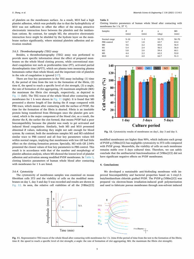
There are four key parameters in the TEG assay including: (1) timeR, the period of time from the test to the formation of the fibrin, (2)time K, the speed to reach a specific level of clot strength, (3) α angle,the rate of formation of clot aggregating, (4) maximum amplitude (MA)the maximum the fibrin clot strength, respectively, as depicted inFig. 11 (left). The TEG traces of the whole blood after contacting withmembranes for 1 h were shown in Fig. 11 (right). It is found that M0presented a shorter length of line during the R range compared withPBS trace, which means after contacting with the surface of PVDF, thetime for the formation of the fibrin is shorted. Fibrin is an insolubleprotein being transferred from fibrinogen once the platelet gets acti-vated, which is the major component of the blood clot, as a result, theshorter the R, the earlier the clot formed, that means PVDF had a poorbiocompatibility because the platelet was ready to get activated andinduced blood coagulation. Similarly, both M5 and M10 presentedabnormal R values, indicating they might not safe enough for bloodsystem. By contrast, both the membrane samples M1 and M3 exhibitedsimilar trace to PBS control and all four key parameters values fellwithin normal ranges, implying that membranes with low GR had littleeffect on the clotting formation process. Specially, M3 with GR 2.94%presented the closest values of four key parameters to PBS control. Thisresult is in accordance with that of the number and morphology ofplatelet adhesion analysis, where M3 showed the lowest level of plateletadhesion and activation among modified PVDF membranes. In Table 3,clotting kinetics parameters of human whole blood after contactingwith membranes for 1 h are listed.
3.6.4. CytotoxicityThe cytotoxicity of membranes samples was examined on mouse
fibroblast cells 3T3 and the viability of cells on the modified mem-branes on day 1, day 3 and day 5 was recorded and results are shown inFig. 12. As seen, the relative cell viabilities of all the [VBIm][Cl]
modified membranes are higher than 90%, which indicates each groupof PVDF-g-[VBIm][Cl] has negligible cytotoxicity to 3T3 cells comparedwith PVDF group. Meanwhile, the viability of cells on each membraneremains stable over 5 days cultured time. Therefore, we can safelyconclude that the antibacterial functionalization of [VBIm][Cl] did nothave significant negative effects on PVDF membranes.
4. Conclusions
We developed a sustainable anti-biofouling membrane with im-proved biocompatibility and bacterial properties based on 1-vinyl-3-butylimidazolium chloride grafted PVDF. The PVDF-g-[VBIm][Cl] wasprepared via electron-beam irradiation-induced graft polymerizationand used to fabricate porous membranes through non-solvent induced
Fig. 11. Representative TEG traces of the whole blood after contacting with membranes for 1 h. (time R:the period of time from the test to the formation of the fibrin;time K: the speed to reach a specific level of clot strength; α angle: the rate of formation of clot aggregating; MA: the maximum the fibrin clot strength).
Table 3Clotting kinetics parameters of human whole blood after contacting withmembranes for 1 h, 37 °C.
Samples R(min)
K(min)
α(deg)
MA(mm)
Normal range 5–10 1–3 53–72 50–70PBS control 5.7 1.8 63.7 63.2M0 4.6 2.2 60.6 56.4M1 5.4 2.4 56.5 56.0M3 5.8 2.0 59.4 60.1M5 4.4 2.2 62.2 53.0M10 4.2 2.0 62.7 51.3
Fig. 12. Cytotoxicity results of membranes on day1, day 3 and day 5.
X. Zhang, et al. Materials Science & Engineering C 118 (2021) 111411
9

phase separation method. Although the membrane morphology ofPVDF-g-[VBIm][Cl] was similar to the pristine PVDF, there were quitedifferent performances between these two types of membranes. TheXPS results revealed that the ionic liquid was enriched on the mem-brane surface, resulting in the improved hydrophilicity and antifoulingproperties. In particular, the PVDF-g-[VBIm][Cl] membranes distinctlyinhibited the both growth of S. aureus and E. coli on the membranesurface and displayed enhanced anti-biofouling and antibacterialproperties. The results of SEM and TEG assay showed that membranewith 2.94 wt.% GR had little effect on the whole blood clotting process.In addition, the PVDF-g-[VBIm][Cl] membranes also exhibited bothlower hemolysis and cytotoxicity compared with the PVDF membranes.These features make the PVDF-g-[VBIm][Cl] membrane pretty attrac-tive as a clinical medical material.
Supplementary data to this article can be found online at https://doi.org/10.1016/j.msec.2020.111411.
Declaration of competing interest
The authors declare that they have no known competing financialinterests or personal relationships that could have appeared to influ-ence the work reported in this paper.
Acknowledgment
This work was financially supported by Zhejiang Provincial NaturalScience Foundation of China (LY15E030014), National Natural ScienceFoundation of China (21674033).
CRediT author statement
Xiaowei Zhang: Data curation, Formal analysis, Investigation,Methodology, Writing - original draft.
Yuanyuan Liang: Conceptualization, Formal analysis,Methodology, Validation, Investigation, Funding acquisition, Writing -review & editing.
Chunjun Ni: Data curation, Formal analysis, Methodology,Software.
Yongjin Li: Conceptualization, Data curation, Funding acquisition,Project administration, Resources, Supervision, Validation, Writing -review & editing.
References
[1] K.H. Oshima, T.T. Evans-Strickfaden, A.K. Highsmith, E.W. Ades, The use of a mi-croporous polyvinylidene fluoride (PVDF) membrane filter to separate con-taminating viral particles from biologically important proteins, Biologicals 24(1996) 137–145.
[2] X.Y. Tan, S.P. Tan, W.K. Teo, K. Li, Polyvinylidene fluoride (PVDF) hollow fibremembranes for ammonia removal from water, J. Membr. Sci. 271 (2006) 59–68.
[3] A. Bottino, G. Capannelli, A. Comite, Novel porous poly(vinylidene fluoride)membranes for membrane distillation, Desalination 183 (2005) 375–382.
[4] F. Liu, N. A. Hashim, Y. T. Liu, M. R. M. Abed, K. Li, Progress in the production andmodification of PVDF membranes, J. Membr. Sci. 375 (2011) 1–27.
[5] A. Naz, R. Sattar, M. Siddiq, Polymer membranes for biofouling mitigation: a re-view, Polym.-Plast. Technol. Mater. 58 (2019) 1829–1854.
[6] X. Shen, Y. Zhao, L. Chen, The construction of a zwitterionic PVDF membranesurface to improve biofouling resistance, Biofouling 29 (2013) 991–1003.
[7] M. Spasova, N. Manolova, N. Markova, I. Rashkov, Superhydrophobic PVDF andPVDF-HFP nanofibrous mats with antibacterial and anti-biofouling properties,Appl. Surf. Sci. 363 (2016) 363–371.
[8] S.Y. Park, J.W. Chung, Y.K. Chae, S.Y. Kwak, Amphiphilic thiol functional linkermediated sustainable anti-biofouling ultrafiltration nanocomposite comprising asilver nanoparticles and poly(vinylidene fluoride) membrane, ACS Appl. Mater.Interfaces 5 (2013) 10705–10714.
[9] O.T. Beek, D. Pavlenko, M. Suck, S. Helfrich, L. Bolhuis-Versteeg, D. Snisarenko,C. Causserand, P. Bacchin, P. Aimar, R.V. Orele, R. Wetzels, P. Verhezen,Y. Henskens, D. Stamatialis, New membranes based on polyethersulfone –SlipSkinTM polymer blends with low fouling and high blood compatibility, Sep.Purif. Technol. 225 (2019) 60–73.
[10] L.J. Zhu, F. Liu, X.M. Yu, L.X. Xue, Poly(lactic acid) hemodialysis membranes withpoly(lactic acid)-block-poly(2-hydroxyethyl methacrylate) copolymer as additive:
preparation, characterization, and performance, ACS Appl. Mater. Interfaces 7(2015) 17748–17755.
[11] N. Hilal, V. Kochkodan, L. Al-Khatib, T. Levadna, Surface modified polymericmembranes to reduce (bio)fouling: a microbiological study using E. coli,Desalination 167 (2004) 293–300.
[12] H.L. Yang, J.C.T. Lin, C. Huang, Application of nanosilver surface modification toRO membrane and spacer for mitigating biofouling in seawater desalination, WaterRes. 43 (2009) 3777–3786.
[13] A. Venault, Y. Chang, D.M. Wang, J.Y. Lai, Surface anti-biofouling control ofPEGylated poly(vinylidene fluoride) membranes via vapor-induced phase separa-tion processing, J. Membr. Sci. 423–424 (2012) 53–64.
[14] G.M. Nisola, J.S. Park, A.B. Beltran, W.J. Chung, Silver nanoparticles in a polyether-block-polyamide copolymer towards antimicrobial and antifouling membranes,RSC Adv. 2 (2012) 2439–2448.
[15] Y. Kim, D. Rana, T. Matsuura, W.J. Chung, Towards antibiofouling ultrafiltrationmembranes by blending silver containing surface modifying macromolecules,Chem. Commun. 48 (2012) 693–695.
[16] C.X. Liu, D.R. Zhang, Y. He, X.S. Zhao, R. Bai, Modification of membrane surface foranti-biofouling performance: effect of anti-adhesion and anti-bacteria approaches,J. Membr. Sci. 346 (2010) 121–130.
[17] W.L. Yang, M. Son, R. Rossi, S.V. Johannes, B.E. Logan, Adapting aluminum-dopedzinc oxide for electrically conductive membranes fabricated by atomic layer de-position, ACS Appl. Mater. Interfaces 12 (2020) 963–969.
[18] J.J. Harrison, R.J. Turner, D.A. Joo, M.A. Stan, C.S. Chan, N.D. Allan, H.A. Vrionis,M.E. Olson, H. Ceri, Copper and Quaternary ammonium cations exert synergisticbactericidal and antibiofilm activity against Pseudomonas aeruginosa, Antimicrob.Agents Chemother. 52 (2008) 2870–2881.
[19] J. Yin, Y. Yang, Z. Hu, B. Deng, Attachment of silver nanoparticles (AgNPs) ontothin-film composite (TFC) membranes through covalent bonding to reduce mem-brane biofouling, J. Membr. Sci. 441 (2013) 73–82.
[20] W.J. Horne, M.A. Andrews, L.L. Terrill, S.S. Hayward, J. Marshall, K.A. Belmore,M.S. Shannon, J.E. Bara, Poly(ionic liquid) superabsorbent for polar organic sol-vents, ACS Appl. Mater. Interfaces 7 (2015) 8979–8983.
[21] P. Virtanen, E. Salminen, J.P. Mikkola, Modeling of supported ionic liquid catalystssystems–from idea to applications, Ind. Eng. Chem. Res. 56 (2017) 12852–12862.
[22] D.K. Chen, W. Ying, Y.L. Ying, X.S. Peng, Enhanced gas separation through nano-confined ionic liquid in laminated MoS2 membrane, ACS Appl. Mater. Interfaces 9(2017) 44251–44257.
[23] M. Wang, H.X. Zhang, G. Thirunavukkarasu, I. Salam, J.R. Varcoe, P. Mardle,X.Y. Li, S.C. Mu, S.F. Du, Ionic liquid-modified microporous ZnCoNC-based elec-trocatalysts for polymer electrolyte fuel cells, ACS Energy Lett. 4 (2019)2104–2110.
[24] H.F.D. Almeida, M.C. Neves, T. Trindade, I.M. Marrucho, M.G. Freire, Supportedionic liquids as efficient materials to remove non-steroidal anti-inflammatory drugsfrom aqueous media, Chem. Eng. J. 381 (2020) 122616.
[25] M. Colonna, C. Berti, E. Binassi, M. Fiorini, S. Sullalti, F. Acquasanta, M. Vannini,D.D. Gioia, I. Aloisio, S. Karanam, D.J. Brunelle, Synthesis and characterization ofimidazolium telechelic poly(butylene terephthalate) for antimicrobial applications,React. Funct. Polym. 72 (2012) 133–141.
[26] J.P. Guan, Y.Y. Wang, S.L. Wu, Y.J. Li, J.Y. Li, Durable anti-superbug polymers:covalent bonding of ionic liquid onto the polymer chains, Biomacromolecules 18(2017) 4364–4372.
[27] J. Zhou, Q.Q. Tao, P.Y. Wang, W.B. Shao, Z.B. Wu, Z. Li, S. Yang, Antimicrobialevaluation and action mechanism of pyridinium-decorated l,4-pentadien-3-onederivatives, Bioorg. Med. Chem. Lett. 28 (2018) 1742–1746.
[28] E. Kim, S.H. Lee, S.J. Lee, O.P. Kwon, H. Yoon, New antibacterial-core structuresbased on styryl quinolinium, Food Sci. Biotechnol. 26 (2017) 521–529.
[29] Y. Xue, H. Xiao, Y. Zhang, Antimicrobial polymeric materials with quaternaryammonium and phosphonium salts, Int. J. Mol. Sci. 16 (2015) 3626–3655.
[30] K.S. Egorova, V.P. Ananikov, Toxicity of ionic liquids: eco(cyto)activity as com-plicated, but unavoidable parameter for task-specific optimization, ChemSusChem7 (2014) 336–360.
[31] Q. Xu, Z. Zheng, B. Wang, H. Mao, F. Yan, Zinc ion coordinated poly(ionic liquid)antimicrobial membranes for wound healing, ACS Appl. Mater. Interfaces 9 (2017)14656–14664.
[32] Z.Q. Zheng, Q.M. Xu, J.N. Guo, J. Qin, H.L. Mao, B. Wang, F. Yan,Structure−antibacterial activity relationships of Imidazolium-type ionic liquidmonomers, poly(ionic liquids) and poly(ionic liquid) membranes: effect of alkylchain length and cations, ACS Appl. Mater. Interfaces 8 (2016) 12684–12692.
[33] J. Qin, J.N. Guo, Q.M. Xu, Z.Q. Zheng, H.L. Mao, F. Yan, Synthesis of pyrrolidinium-type poly(ionic liquid) membranes for antibacterial applications, ACS Appl. Mater.Interfaces 9 (2017) 10504–10511.
[34] C.Y. Xing, Y.Y. Wang, C. Zhang, L.Y. Li, J.Y. Li, Immobilization of ionic liquids ontothe poly(vinylidene fluoride) by electron beam irradiation, Ind. Eng. Chem. Res. 54(2015) 9351–9359.
[35] J.F. Hester, P. Banerjee, A.M. Mayes, Preparation of protein-resistant surface onpoly(vinylidene fluoride) membranes via surface segregation, Macromol. 32 (1999)1643–1650.
[36] B. Deng, M. Yu, X.X. Yang, B.W. Zhang, L.F. Li, Antifouling microfiltration mem-branes prepared from acrylic acid or methacrylic acid grafted poly(vinylidenefluoride) powder synthesized via pre-irradiation induced grafted polymerization, J.Membr. Sci. 350 (2010) 252–258.
[37] Y. Zhang, L. Ye, B.P. Zhang, Y.S. Chen, W.G. Zhao, G. Yang, J. Wang, H.W. Zhang,Characteristics and performance of PVDF membrane prepared by using NaCl coa-gulation bath: relationship between membrane polymorphous structure and organicfouling, J. Membr. Sci. 579 (2019) 22–32.
X. Zhang, et al. Materials Science & Engineering C 118 (2021) 111411
10
[38] H.J. Mo, K.G. Tay, H.Y. Ng, Fouling of reverse osmosis membrane by protein (BSA):effects of pH, calcium, magnesium, ionic strength and temperature, J. Membr. Sci.315 (2008) 28–35.
[39] L.A.T.W. Asri, M. Crismaru, S. Roest, Y. Chen, O. Ivashenko, P. Rudolf, J.C. Tiller,H.C. van der Mei, T.J.A. Loontjens, H.J. Busscher, A shape-adaptive, antibacterial-coating of immobilized quaternary-ammonium compounds tethered on hyper-branched polyurea and its mechanism of action, Adv. Funct. Mater. 24 (2014)346–355.
[40] C. Zhou, F. Wang, H. Chen, M. Li, F. Qiao, Z. Liu, Y. Hou, C. Wu, Y. Fan, L. Liu,S. Wang, Y. Wang, Selective antimicrobial activities and action mechanism of mi-celles self-assembled by cationic Oligomeric surfactants, ACS Appl. Mater.Interfaces 8 (2016) 4242–4249.
[41] N.A. Peppas, R. Langer, New challenges in biomaterials, Science 263 (1994)1715–1720.
[42] P.K. Qi, M.F. Maitz, N. Huang, Surface modification of cardiovascular materials and
implants, Surf. Coat. Technol. 233 (2013) 80–90.[43] G.C. Li, P. Yang, W. Qin, M.F. Maitz, S. Zhou, N. Huang, The effect of coimmobi-
lizing heparin and fibronectin on titanium on hemocompatibility and en-dothelialization, Biomaterials 32 (2011) 4691–4703.
[44] L.H. Li, M. Tu, S.S. Mou, C.G. Zhou, Preparation and blood compatibility of poly-siloxane/liquid-crystal composite membranes, Biomaterials 22 (2001) 2595–2599.
[45] X.C. Zhao, D.W. Lu, Q.S. Liu, Y.L. Li, R. Feng, F. Hao, G.B. Qu, Q.F. Zhou, G.B. Jiang,Hematological effects of gold nanorods on erythrocytes: hemolysis and hemoglobinconformational and functional changes, Adv. Sci. 4 (2017) 1700296.
[46] S.L. Goodman, M. Lelah, L.K. Lambrecht, S.L. Cooper, In vitro vs. ex vivo plateletdeposition on polymer surfaces, Scanning Electron Microsc. 1 (1984) 279–290.
[47] R.K. Ainthan, M. Gnanamani, M. Ganguli, T. Ghosh, D.E. Brooks, S. Maiti,J.N. Kizhakkedathu, Blood compatibility of novel water soluble hyperbranchedpolyglycerol-based multivalent cationic polymers and their interaction with DNA,Biomaterials 27 (2006) 5377–5390.
X. Zhang, et al. Materials Science & Engineering C 118 (2021) 111411
11