Embed Size (px)
Citation preview
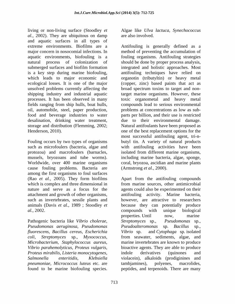
Int.J.Curr.Microbiol.App.Sci (2014) 3(5): 712-725
712
Original Research Article
Anti-biofouling activity of Prodigiosin, a pigment extracted from Serratia marcescens
T.Sathish kumar* and H.Aparna
Department of Microbiology, Madras Christian College (Autonomous), Tambaram, Chennai 600 059, Tamilnadu, India
*Corresponding author
A B S T R A C T
Introduction
Biofilms are complex and dynamic, heterogenous microbial communities containing bacteria, fungi, algae, phytoplanktons
and zooplanktons. The microorganisms in biofilm synthesize and secrete a protective matrix that attaches the biofilm firmly to a
ISSN: 2319-7706 Volume 3 Number 5 (2014) pp. 712-725 http://www.ijcmas.com
K e y w o r d s
Serratia marcescens; Microbial pigments; Anti-Biofouling activity; Staphylococcus aureus, Escherichia coli and Pseudomonas aeruginosa.
The present study aimed at analyzing the antifouling activity of the red pigment of Serratia marcescens obtained from MTCC. The pigment was formed under stationary growth conditions at an optimum temperature of 28oC. It was extracted using standard procedure and a large quantity of pigment (1047.82 units/cell) was obtained. The absorbance maxima of the pigment was found to be at 534 nm, with acidified methanol as blank. This characteristic peak confirmed the presence of red pigment, prodigiosin. The extracted pigment was applied over steel and wood pieces which were immersed in marine water, inoculated with the fouling organisms viz., Staphylococcus aureus, Escherichia coli and Pseudomonas aeruginosa. After a period of 7 days, the biofilm was scraped off from the surfaces and colonies were enumerated. The number of colonies on pigment coated wood and steel were compared with those of methanol coated wood and steel, and analysed statistically. The statistical analysis revealed that the biofilm organisms have more affinity towards wood surface as it is rougher than that of steel. The average number of bacteria settled on wood is ten times more than that of steel. There was significant difference in the reduction of bacterial count when the substrate was coated with prodigiosin, when compared to methanol and control pieces of wood and substrate. The inhibitory activity of the pigment was more efficient against Gram positive bacteria than for Gram negative bacteria. The pigment was also tested for its antifouling activity against the above marine fouling organisms by agar well diffusion method. The pigment exhibited good inhibitory action against Staphylococcus aureus than Escherichia coli and Pseudomonas aeruginosa. The red pigment inhibited Staphylococcus aureus with a minimum zone diameter of 17 mm at a concentration of 50 l, and maximum zone diameter of 20 mm at a concentration of 150 l. Escherichia coli showed a minimum zone of 11 mm at 50 l pigment concentration, while 15 mm zone diameter was recorded with 150 l pigment. Pseudomonas aeruginosa showed a constant diameter of 13 mm at all concentrations of the pigment. The present study showed that the prodigiosin pigment was found to act as a good antifoulant
Int.J.Curr.Microbiol.App.Sci (2014) 3(5): 712-725
713
living or non-living surface (Stoodley et al., 2002). They are ubiquitous on damp and aquatic surfaces in all types of extreme environments. Biofilms are a major concern in nosocomial infections. In aquatic environments, biofouling is a natural process of colonization of submerged surfaces and biofilm formation is a key step during marine biofouling, which leads to major economic and ecological losses. It is one of the major unsolved problems currently affecting the shipping industry and industrial aquatic processes. It has been observed in many fields ranging from ship hulls, boat hulls, oil, automobile, steel, paper production, food and beverage industries to water desalination, drinking water treatment, storage and distribution (Flemming, 2002; Henderson, 2010).
Fouling occurs by two types of organisms such as microfoulers (bacteria, algae and protozoa) and macrofoulers (barnacles, mussels, bryozoans and tube worms). Worldwide, over 400 marine organisms cause fouling problems. Bacteria are among the first organisms to foul surfaces (Rao et al., 2005). They form biofilms which is complex and three dimensional in nature and serve as a focus for the attachment and growth of other organisms, such as invertebrates, sessile plants and animals (Davis et al., 1989 ; Stoodley et al., 2002.
Pathogenic bacteria like Vibrio cholerae, Pseudomonas aeruginosa, Pseudomonas fluorescens, Bacillus cereus, Escherichia coli, Streptomyces sp., Myxococcus, Microbacterium, Staphylococcus aureus, Vibrio parahemolyticus, Proteus vulgaris, Proteus mirabilis, Listeria monocytogenes, Salmonella enteritidis, Klebsiella pneumoniae, Micrococcus luteus etc. are found to be marine biofouling species.
Algae like Ulva lactuca, Synechococcus are also involved.
Antifouling is generally defined as a method of preventing the accumulation of fouling organisms. Antifouling strategies should be done by proper process analysis, integrated and holistic approaches. Most antifouling techniques have relied on organotin (tributyltin) or heavy metal (copper, zinc) based paints that act as broad spectrum toxins to target and non-target marine organisms. However, these toxic organometal and heavy metal compounds lead to serious environmental problems at concentrations as low as sub-parts per billion, and their use is restricted due to their environmental damage. Natural antifoulants have been proposed as one of the best replacement options for the most successful antifouling agent, tri-n-butyl tin. A variety of natural products with antifouling activities have been isolated from different marine organisms, including marine bacteria, algae, sponge, coral, bryozoa, ascidian and marine plants (Armstrong et al., 2000).
Apart from the antifouling compounds from marine sources, other antimicrobial agents could also be experimented on their antifouling activity. Marine bacteria, however, are attractive to researchers because they can potentially produce compounds with unique biological properties. Until now, marine Streptomyces sp., Pseudomonas sp., Pseudoalteromonas sp. Bacillus sp., Vibrio sp. and Cytophaga sp. isolated from seawater, sediments, algae, and marine invertebrates are known to produce bioactive agents. They are able to produce indole derivatives (quinones and violacein), alkaloids (prodiginines and tambjamines), polyenes, macrolides, peptides, and terpenoids. There are many
Int.J.Curr.Microbiol.App.Sci (2014) 3(5): 712-725
714
pigmented microorganisms and their pigments have antimicrobial activity. These pigments could also be incorporated into antifouling coatings in ships and marine equipments.
Among the various pigment producing microbes, Serratia marcescens has been the main focus of this study, as it produces a well-characterized pigment, Prodigiosin. Serratia marcescens is characterized by its ability to produce the red pigment prodigiosin (Khanafari et al., 2006). Serratia sp. have occupied different habitats such as water, surface of plants, animals and insects, soil and hospitalized patients. Serratia sp. are both chromogenic and non - chromogenic. Chromogenic species are usually isolated from the environment from water, soil, plants or insects. The non chromogenic ones are a real hospital threat and are human pathogens. An environmental isolate of S.marcescens was found to be antagonistic against many fungal species like, Alternaria alternate, Curvularia sp., Aspergillus niger, Fusarium oxysporum and Helminthosporium sp. (Parani and Saha, 2009).
The red pigment, Prodigiosin (5[(3-methoxy - 5 - pyrrol - 2- ylidene- pyrrol-2-ylidene) -methyl] -2- methyl-3- pentyl-1Hpyrrole) is a secondary metabolite alkaloid with a unique tripyrrole chemical structure. It has three rings forming a pyrrolylpyrromethane skeleton with a C-4 methoxy group, a molecular formula C20H25N30 and a molecular weight of 323.44 Da. (Harris et al., 2004; Williamson et al., 2006). S. marcescens being facultative, the pigment is produced under both aerobic and anaerobic conditions. Prodigiosin is a promising drug owing to its reported characteristics of having antibacterial, anti-fungal, anti-neoplastic, anti-proliferative, anti-oxidant
and anti-malarial activity (Anita et al., 2006).
The colored pigment of Serratia marcescens is sensitive to UV but it could be modified by addition to marine paints and coatings. The pigment being active against many pathogenic bacteria, it could reduce the fouling effects of marine bacteria. In the present study an attempt has been done to evaluate the antifouling activity of prodigiosin pigment and to establish its efficiency as a good antifoulant.
Materials and Methods
Serratia marcescens was obtained from Microbial Type Culture Collection (MTCC), Chandigarh with ID no. MTCC 8708. The organism was procured as a lyophilized form in a glass vial and it was revived The vial was stored in deep freezer at - 20oC until use. The organism was revived using nutrient broth and nutrient agar. After incubation, the plates and tubes were observed for the growth of pigmented bacteria. To confirm the morphology of Serratia marcescens, Gram s staining method was performed. The smear was observed for the presence of Gram negative bacilli.
Extraction of pigment
Serratia marcescens was cultured in 2% peanut seed medium (Giri et al., 2004). Peanut seeds were finely powdered and 1g was added to 50 ml distilled water in a 250 ml Erylenmeyer flask. In this way, one litre medium was prepared. The pH was adjusted to 7.0. The medium was sterilized at 121oC and 5% broth culture was inoculated. The medium was incubated for 3 days under static condition at 28oC for obtaining high amounts of pigment.

Int.J.Curr.Microbiol.App.Sci (2014) 3(5): 712-725
715
The extraction procedure was followed as per the method of Williams et al. 1955.The pigment was extracted by adding 4 volumes of acetone to the cell suspension. The acetone mixture was shaken for 3 hrs at room temperature, and then centrifuged. The sedimented cell debris was washed twice by resuspending in 50 ml of acetone, shaking for 30 min followed by centrifugation. The washings were combined with the supernatant from the original centrifugation, and the solution was filtered. Pigment was extracted from small portions of the filtrate by mixing thoroughly 1 volume of the acetone solution with 2 volumes of petroleum ether in a separatory funnel. The separating funnel was shaken vigorously for 10
15 min. The pigment was extracted in the petroleum ether phase.
This petroleum ether layer was poured in a petri dish and kept at 30 - 40°C in order to evaporate the solvent completely. 2 ml of acidified methanol was added and the pigment was scraped off from the petridish and stored in a screw capped tube.
Presumptive test for prodigiosin
The culture broth was centrifuged at 4500 rpm for 15 mins. 10 ml of 95% methanol was added to the cell pellet and centrifuged under the same condition. Debris was removed and the 2ml of the supernatant was taken in two test tubes. The content of one of the test tube was acidified with a drop of concentrated HCl and the other alkalinized with a drop of concentrated ammonia solution. The tubes were observed for color change to red or pink colour in the acidified solution and a yellow or tan color in the alkaline solution. This gives a positive presumptive test for prodigiosin (Gerber and Lechevalier, 1976).
Spectral analysis and Estimation of prodigiosin
Spectral analysis was made on dried pigment extracted by the above method by dissolving acidified ethanol (96ml ethanol and 4ml HCl) (Williams et al., 1955). Spectral analysis was made on a UV-Visible spectrophotometer and the extract was scanned in the range of 400
700 nm to find out the maximum absorption spectra. Acidified ethanol was used as a blank.
The bacterial cell absorbance of the culture broth was measured at 620 nm. The relative concentration of prodigiosin produced by liquid grown cultures was quantified as follows: 1 ml sample was harvested by centrifugation at 13,000 rpm for 5 min. The supernatant was discarded and the pellet resuspended in acidified ethanol (4% 1 M HCl in ethanol) to extract prodigiosin from the cells. Cell debris was removed by a second centrifugation step and the supernatant transferred to a cuvette for measurement of absorbance at 534 nm (Slater et al., 2003).
Prodigiosin unit/cell = ([OD534 (1.381 x OD620)]) x 1000/OD620
where, OD - Optical density; OD534 Pigment absorbance ; OD620 Bacterial cell absorbance 1.381 Constant
Biofilm development
The formation of biofilm was studied under laboratory conditions. The biofouling organisms used were Staphylococcus aureus MTCC 3160, Escherichia coli MTCC 9537, and clinical isolate of Pseudomonas aeruginosa obtained from pus culture. Sea water was

Int.J.Curr.Microbiol.App.Sci (2014) 3(5): 712-725
716
sterilized and 500 ml was poured into 3 separate beakers. The number of cells of the indicator organisms were adjusted to 2.8 × 10⁸ cells/ml which was confirmed by obtaining OD value using a colorimeter at 600 nm. 1 ml of each indicator organism was inoculated in the corresponding beaker.
Preparation of biofilm substrate and bacterial enumeration
The substrate used for testing biofilm formation was taken to be stainless steel sheets and wooden blocks. These substrates were sterilized at 121oC. The dimension of stainless sheet sheet and wooden blocks was 2 × 2 . The methanol extract of the pigment was added with 1ml linseed oil which acts as a pigment binder. The pigment was applied onto the steel and wood pieces and left for drying. Controls for wood and steel were prepared without applying pigment. Methanol added with linseed oil was applied onto wood and steel to compare the effect of methanol and pigment on antifouling activity. 3 wooden pieces (control, pigment applied wood and methanol applied wood) and 3 stainless steel pieces (control, pigment applied steel and methanol applied steel) were immersed into each of the beakers containing sea water. The setup was left at room temperature for a period of 7 days for the formation of biofilm. At the end of the 7th
day, the samples were taken from the sea water and viable bacterial count was performed.
The biofilm from wood and steel pieces were scraped off using a sterile spatula into 100 ml of sterile saline to make master dilution. For each sample, for each substrate and for each organism, serial dilution was done in sterile saline tubes.
10-fold dilution was performed by adding 1ml from master dilution to 9 ml of sterile saline. The dilution was done upto 10-5.
Bacterial enumeration was done by spread plate method. Nutrient agar plates were prepared and 0.1 ml of sample from dilutions 10-4 and 10-5 were spread using a flame sterilized glass rod. This procedure was followed for each organism, each substrate and for each sample. All the plates were incubated at 37oC for 24 hrs. Enumeration of viable organisms was carried and the average bacterial counts were recorded. The experiments were conducted in duplicates.
The total number of colony forming units was found out by the formula:
No.of cfu/ml of master dilution = No. of colonies counted × dilution factor/Volume of sample
Antifouling activity of Prodigiosin
Antibacterial assays against 3 fouling organisms viz., Staphyloccus aureus, Escherichia coli and Pseudomonas aeruginosa was performed by the standard agar well diffusion method. The crude methanol extract of pigment was filter sterilized. A 3 hr culture of the indicator organisms were swabbed as lawn culture in the Mueller Hinton agar plate. Wells of 6mm diameter were cut and wells were added with different concentrations (50 l, 100 l and 200 l) of prodigiosin pigment. Also, control plates with different concentrations of methanol were prepared.
The plates were incubated at 37oC for 24 hrs. The plates were observed for zone of inhibition and the zones were measured.

Int.J.Curr.Microbiol.App.Sci (2014) 3(5): 712-725
717
Results and Discussion
The nutrient broth showed uniform red pigmentation throughout. The pigmentation increased day by day and reached a maximum at the 3rd day of incubation. The nutrient agar plate showed round, medium sized, smooth, opaque, convex abundant red pigment producing colonies. The culture was identified to be Gram negative small bacilli. There was no presence of contaminating organisms. By microscopic observation, it was confirmed that the lyophilized culture was a pure culture of Serratia marcescens. The inoculated peanut seed broth was observed for pigment production and it showed increase in the production of pigment day by day. At the 3rd day, deep pink color was observed. The pigment extraction with petroleum ether using a separatory funnel is shown in Figure 1. After drying of the extracted pigment, the plate showed good amounts of the red pigment deposits which was scraped off using acidified methanol and 10 ml was stored in screw capped tube. The dried pigment is shown in Figure 2.
Presumptive test for prodigiosin
On addition of 1drop of concentrated HCl to the methanol extract of pigment and to the cell free supernatant of the nutrient broth culture, the color changed to deep red and pink respectively. When concentrated ammonia solution was added to methanol extract and cell free supernatant, there was a color change to yellow and tan respectively. This showed a positive presumptive test for prodigiosin.
Spectral analysis and estimation of pigment
The pigment dissolved in acidified ethanol showed a characteristic peak at 534 nm
under UV-Vis spectrophotometer analysis. The absorption maxima of the pigment was found to be at 534 nm with a reading of 0.558. The bacterial cell absorbance was found to be 0.23 at OD620. The absorbance maxima of prodigiosin is shown in Figure 3.
The prodigiosin was estimated with the formula:
Prodigiosin unit/cell = ([OD534 (1.381 x OD620)]) x 1000/OD620
= {0.558- (1.381 x 0.23)}x 1000/0.23 = 1047.82
Hence, the prodigiosin was estimated to be 1047.82 unit/cell.
Estimation of viable count of the fouling organisms
At the end of the 7th day, the substrates were removed from the marine water and enumeration of bacteria was done. The number of colonies in pigmented coated substrates were counted in the plates and compared with those in control and methanol coated substrates. There was a difference in the number of colonies formed in controls, methanol coated and pigment coated wood and steel. The experimental setup for biofilm formation has been depicted in Figure 4.
Paired student s T-test was used to analyse and compare the data for significant difference in the mean values of cell numbers. There was significant difference in the reduction of colony forming units in pigment coated substrates when compared to controls. The control and methanol coated substrates could be considered one and the same as the mean values showed no significant differences.

Int.J.Curr.Microbiol.App.Sci (2014) 3(5): 712-725
718
Fig. 1 Extraction of prodigiosin through separatory funnel
Fig.2 Prodigiosin pigment dried on petri plates after pigment extraction

Int.J.Curr.Microbiol.App.Sci (2014) 3(5): 712-725
719
Fig.3 UV Vis spectrophotometric analysis of prodigiosin showing peak at 534 nm
Fig.4 Experimental setup for biofilm formation
S. aureus E. coli
P. aeruginosa
Int.J.Curr.Microbiol.App.Sci (2014) 3(5): 712-725
720
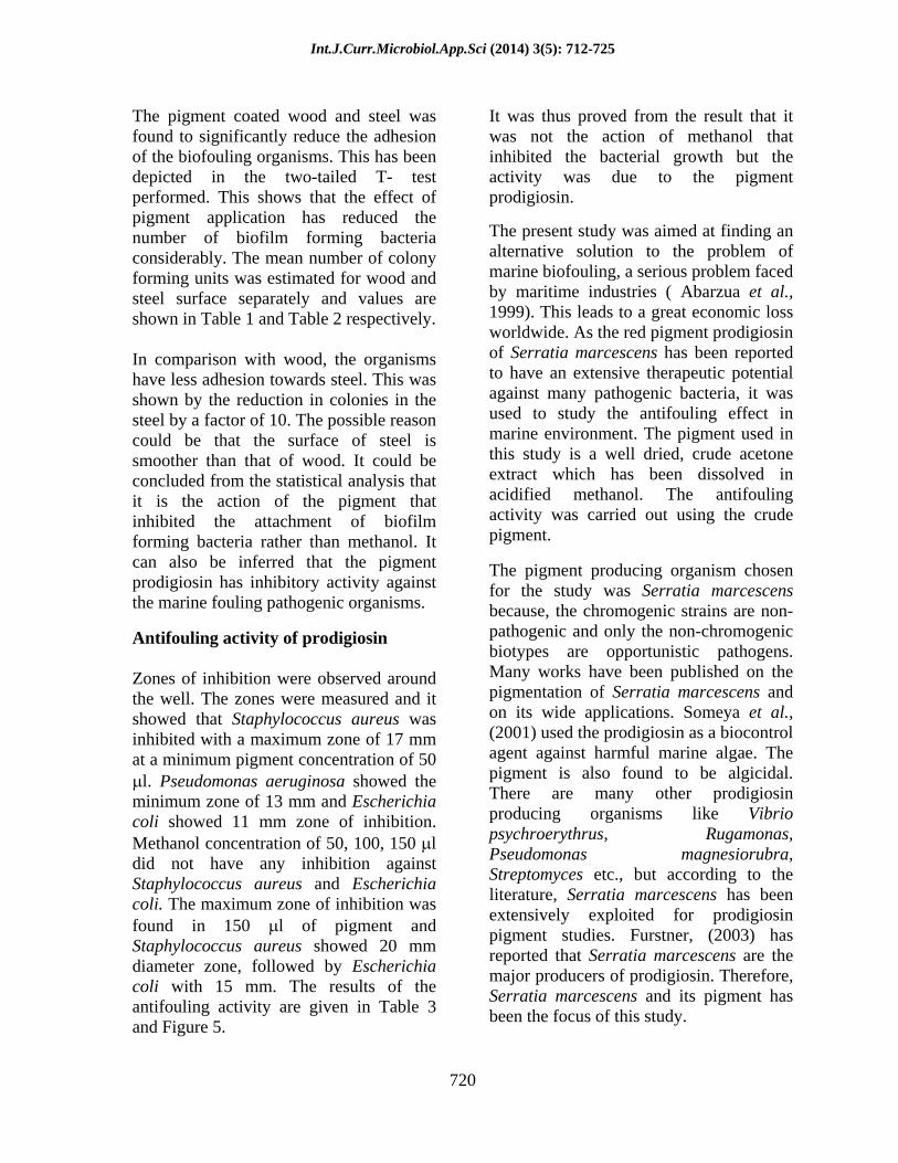
The pigment coated wood and steel was found to significantly reduce the adhesion of the biofouling organisms. This has been depicted in the two-tailed T- test performed. This shows that the effect of pigment application has reduced the number of biofilm forming bacteria considerably. The mean number of colony forming units was estimated for wood and steel surface separately and values are shown in Table 1 and Table 2 respectively.
In comparison with wood, the organisms have less adhesion towards steel. This was shown by the reduction in colonies in the steel by a factor of 10. The possible reason could be that the surface of steel is smoother than that of wood. It could be concluded from the statistical analysis that it is the action of the pigment that inhibited the attachment of biofilm forming bacteria rather than methanol. It can also be inferred that the pigment prodigiosin has inhibitory activity against the marine fouling pathogenic organisms.
Antifouling activity of prodigiosin
Zones of inhibition were observed around the well. The zones were measured and it showed that Staphylococcus aureus was inhibited with a maximum zone of 17 mm at a minimum pigment concentration of 50
l. Pseudomonas aeruginosa showed the minimum zone of 13 mm and Escherichia coli showed 11 mm zone of inhibition. Methanol concentration of 50, 100, 150 l did not have any inhibition against Staphylococcus aureus and Escherichia coli. The maximum zone of inhibition was found in 150 l of pigment and Staphylococcus aureus showed 20 mm diameter zone, followed by Escherichia coli with 15 mm. The results of the antifouling activity are given in Table 3 and Figure 5.
It was thus proved from the result that it was not the action of methanol that inhibited the bacterial growth but the activity was due to the pigment prodigiosin.
The present study was aimed at finding an alternative solution to the problem of marine biofouling, a serious problem faced by maritime industries ( Abarzua et al., 1999). This leads to a great economic loss worldwide. As the red pigment prodigiosin of Serratia marcescens has been reported to have an extensive therapeutic potential against many pathogenic bacteria, it was used to study the antifouling effect in marine environment. The pigment used in this study is a well dried, crude acetone extract which has been dissolved in acidified methanol. The antifouling activity was carried out using the crude pigment.
The pigment producing organism chosen for the study was Serratia marcescens because, the chromogenic strains are non-pathogenic and only the non-chromogenic biotypes are opportunistic pathogens. Many works have been published on the pigmentation of Serratia marcescens and on its wide applications. Someya et al., (2001) used the prodigiosin as a biocontrol agent against harmful marine algae. The pigment is also found to be algicidal. There are many other prodigiosin producing organisms like Vibrio psychroerythrus, Rugamonas, Pseudomonas magnesiorubra, Streptomyces etc., but according to the literature, Serratia marcescens has been extensively exploited for prodigiosin pigment studies. Furstner, (2003) has reported that Serratia marcescens are the major producers of prodigiosin. Therefore, Serratia marcescens and its pigment has been the focus of this study.

Int.J.Curr.Microbiol.App.Sci (2014) 3(5): 712-725
721
Table.1 Comparison of average (mean ± S.D) of cell numbers in control, methanol coated
and pigment coated wood for S. aureus, E. coli and P. aeruginosa
Samples Control Methanol Pigment
Organism
Mean ±SD* Mean ±SD* Mean ±SD*
S.aureus 54.00±2.00 54.00±2.00 18±2.00 E.coli 116.33±6.50 120.00±2.00 21.33±0.57 P.aeruginosa 161.33±4.50 137.33±1.52 23.00±1.00
* - S.D denotes Standard Deviation Inferences of Paired Student s T-test for wood: (a) There is no significant difference in the mean values (mean ± S.D) of control and
methanol coated wood in the case of S. aureus and E. coli at the level P 0.05 (b) There was statistically significant reduction in the colony forming units of pigment coated
wood when compared with control wood for all the three bacteria at P 0.05 (c) P. aeruginosa showed significant difference in the mean values of pigment coated wood
when compared to control and methanol at P
0.05
Table.2 Comparison of average (mean ± S.D) of cell numbers in control, methanol coated and pigment coated steel for S. aureus, E. coli and P. aeruginosa
Samples Control Methanol Pigment Organism
Mean ±SD* Mean ±SD* Mean ±SD*
S.aureus 31.00±1.00 33.33±0.57 1.66±1.15 E.coli 34.33±1.52 31.00±4.00 4.66±2.51 P.aeruginosa 36.66±2.51 14.00±1.00 7.66±1.52
* - S.D denotes Standard Deviation Inferences of Paired Student s T-test for steel: (a) In accordance with the paired Student s T-test, the mean values of control and methanol
coated steel showed no significant difference in case of Escherichia coli at P
0.05 (b) The pigment coated wood showed a significant reduction in the colony forming units
when compared to control at P
0.05
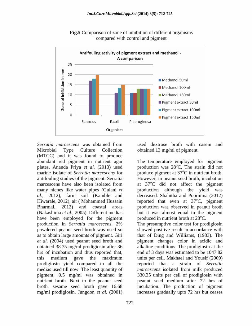
Table.3 Antifouling activity of pigment extract and methanol - A comparison for different organisms
Zone of inhibition in mm Methanol Pigment extract
Organism 50 l 100 l 150 l 50 l 100 l 150 l
S.aureus - - - 17.00 18.00 20.00 E.coli - - - 11.00 13.50 15.00 P.aeruginosa 11.00 11.00 13.00 13.00 13.00 13.00
Int.J.Curr.Microbiol.App.Sci (2014) 3(5): 712-725
722
Fig.5 Comparison of zone of inhibition of different organisms
compared with control and pigment
Serratia marcescens was obtained from Microbial Type Culture Collection (MTCC) and it was found to produce abundant red pigment in nutrient agar plates. Ananda Priya et al. (2013) used marine isolate of Serratia marcescens for antifouling studies of the pigment. Serratia marcescens have also been isolated from many niches like water pipes (Gulani et al., 2012), farm soil (Kamble and Hiwarale, 2012), air ( Mohammed Hussain Bharmal, 2012) and coastal areas (Nakashima et al., 2005). Different medias have been employed for the pigment production in Serratia marcescens. 2% powdered peanut seed broth was used so as to obtain large amounts of pigment. Giri et al. (2004) used peanut seed broth and obtained 38.75 mg/ml prodigiosin after 36 hrs of incubation and thus reported that, this medium gave the maximum prodigiosin yield compared to all the medias used till now. The least quantity of pigment, 0.5 mg/ml was obtained in nutrient broth. Next to the peanut seed broth, sesame seed broth gave 16.68 mg/ml prodigiosin. Jungdon et al. (2001)
used dextrose broth with casein and obtained 13 mg/ml of pigment.
The temperature employed for pigment production was 28oC. The strain did not produce pigment at 37oC in nutrient broth. However, in peanut seed broth, incubation at 37oC did not affect the pigment production although the yield was decreased. Shahitha and Poornima (2012) reported that even at 37oC, pigment production was observed in peanut broth but it was almost equal to the pigment produced in nutrient broth at 28oC. The presumptive color test for prodigiosin showed positive result in accordance with that of Ding and Williams, (1983). The pigment changes color in acidic and alkaline conditions. The prodigiosin at the end of 3 days was estimated to be 1047.82 units per cell. Makhael and Yousif (2009) reported that a strain of Serratia marcescens isolated from milk produced 330.35 units per cell of prodigiosin with peanut seed medium after 72 hrs of incubation. The production of pigment increases gradually upto 72 hrs but ceases
Int.J.Curr.Microbiol.App.Sci (2014) 3(5): 712-725
723
after 92 hrs of incubation. Prodigiosin can exist in two distinct forms, depending upon the hydrogen ion concentration of the solution. In an acid medium the pigment is red and exhibits a sharp spectral peak at 535 nm. In an alkaline medium the pigment is colored orange-yellow and possesses a broader spectral curve centered at 470 nm as reported by Williams et al. (1955).
The prodigiosin pigment showed a characteristic peak at 534 nm in acidified ethanol. This was in accordance with the result of Sundaramoorthy et al. (2009). Mohammed Hussain Bharmal, (2012) also reported a peak of 534 nm in acidified ethanol. So it was confirmed that the pigment was extracted and it corresponds to prodigiosin in this study. The absorption maxima differed upon different blanks and extraction methods. There are reports on absorption maxima at 499 nm by Kamble and Hiwarale, (2012). Priya et al. (2013) reported a peak of 531nm in 95% ethanol and a single peak absorbance in methanol at 535nm (Giri et al., 2004 ; Pradeep et al., 2012).
Shikuma and Hadfield, (2010) reported that E. coli occurred in relatively high abundances in marine biofilms . The mean biofilm abundances of E. coli ranged from 6.43 (SD, 8.99) to 1.25 × 10⁵ (SD, 1.28 × 10⁵) genome copies cm-2 while abundances in the water-column ranged from 2.3 × 10⁻¹ (SD, 2.0 × 10⁻¹) to 1.4 × 10⁴ (SD, 1.8 × 10⁴) genome copies ml-1. Mayavu et al. (2009) have reported that Pseudomonas aeruginosa, Bacillus, E. coli, Staphylococcus aureus, Proteus vulgaris are the pathogens that are found in marine biofilm. Therefore, in this study the pigment activity was tested against these pathogens. Staphylococcus aureus showed maximum inhibition of 17 mm at the least
concentration of the pigment (50 l) and Escherichia coli showed inhibition of 11 mm at the same concentration. There was a difference in the zone of inhibition of methanol and pigment. This shows that it is the activity of the pigment that inhibited the organisms but not the action of methanol. Gulani et al. (2012) showed the activity of prodigiosin against Staphylococcus aureus with 17.5 mm diameter zone.
The statistical data of paired T-test showed the comparison among the controls, methanol coated and pigment coated substrates. There was a significant difference in the pattern of inhibition of Staphylococcus aureus, Escherichia coli and Pseudomonas aeruginosa by the pigment. The biofilm count decreased significantly in pigment coated substrates when compared with controls. The methanol coated substrates showed more or less similar colony counts like that of control and also the difference in colony count was statistically insignificant.
The results obtained using prodigiosin against biofouling showed the broad antibacterial potentials of the red pigment and are in agreement with the previous literature, which revealed the inhibitory effect of prodigiosin against both Gram-positive and Gram-negative bacteria (Mekhael and Yousif, 2009 ; Samrot et al., 2011). Mekhael and Yousif, (2009) have shown higher inhibitory effect of prodigiosins against Gram-positive bacteria than Gram-negative bacteria. In the present study Staphylococcus aureus showed higher zone of inhibition than Escherichia coli and Pseudomonas. Samrot et al. (2011) reported that ethanol: HCl extract of Serratia has antibacterial activity and its zone of inhibition was higher against both Gram-negative (E. coli
Int.J.Curr.Microbiol.App.Sci (2014) 3(5): 712-725
724
and Pseudomonas sp.) and Gram-positive (S. aureus) bacteria.
It is known that the antibacterial activity of prodigiosin is the result of their potential to pass through the outer membrane and to their capacity for inhibiting target DNA modulating enzymes, such as DNA gyrase and topoisomerase IV, which inhibit the cell growth (Berlanaga and Vinas, 2000). Since, the antibacterial activity of a compound may depend on the destruction of the structure or the inhibition of metabolic reaction in a microorganism, it seems that the presence and the level of the antibacterial activity of the red pigment varied significantly with the type of fouling bacteria used. Further, the red pigment caused growth inhibition, and it suggests that the red pigment, prodigiosin of Serratia marcescens is an effective antifouling agent. Further studies could be done to purify the pigment and to incorporate in antifouling coatings and paints to inhibit or delay the formation of biofilm. The prodigiosin being actively inhibitory to Gram positive and Gram negative organisms could be studied for its anti-algal activity to prevent biofilms by algae in future studies.
References
Abarzua S., S.Jakubowski, S.kert and Fuchs, P.1999. Biotechnological investigation for the prevention of marine biofouling: Blue-green algae as potential producers of biogenic agents for the growth inhibition of microfouling organisms. Botanica Marina. 42(5): 459-465.
Anita Khanafari Mahnaz Assadi and Fatemeh Ahmadi Fakhr.2006. Review of prodigiosin, Pigmentation in Serratia marcescens. J. Biol. Sci. 6 (1): 1 13.
Armstrong, E., K.G.Boyd, A.Pisacane, C.J.Peppiatt and Burgess JG. 2000. Marine microbial natural products in
antifouling coatings. Biofouling. 16(2): 215-224.
Berlanaga, M., and M.Vinas. 2000. Role of outer membrane in the accumulation of quinolones by Serratia marcescens. Can. J. Microbiol. 46 :716-721.
Davis AR, Targett NM, McConnel OJ and Young CM. 1989. Epibiosis of marine algae and benthic invertebrates: natural products chemistry and other mechanisms inhibiting settlement and overgreowth. Bioorg Mar Chem 3: 85-114.
Ding MJ and Williams RP. 1983. Biosynthesis of prodigiosin by white strains of Serratia marcescens isolated from patients. Journal of clinical microbiology. 17(3) : 476-480.
Flemming HC. 2002. Biofouling in water systems: cases, causes, countermeasures. Appl. Envir. Biotechnol. 59: 629 640.
Furstner A. 2003. Chemistry and biology of roseophilin and the Prodigiosin alkaloids: A survey of the last 2500 years. Chem Int Ed Engl. 42: 3582-3603.
Gerber NN and Lechevaller MP. 1976. Prodiginine (prodigiosin-like) pigments from Streptomyces and other aerobic Actinomycetes. Can. J. Microbiol. 22:658-667.
Giri AV, Anandkumar N, Muthukumaran G and Pennathur G. (2004) A novel medium for the enhanced cell growth and production of prodigiosinfrom Serratia marcescens isolated from soil. BMC Microbiol, 4: 1-10.
Gulani C, Bhattacharya S and Das A. 2012. Assessment of process parameters influencing the enhanced production of prodigiosin from Serratia marcescens and evaluation of its antimicrobial, antioxidant and dyeing potential. Malaysian Journal of Microbiology. 8(2):116-122.
Harris AK, Williamson NR, Slater H, Cox A, Abbasi S, Foulds I, Simonsen HT, Leeper FJ and Salmond GP. 2004. The Serratia gene cluster encoding biosynthesis of the
Int.J.Curr.Microbiol.App.Sci (2014) 3(5): 712-725
725
red antibiotic, prodigiosin, shows species- and strain-dependent genome context variation. Microbiology 150, 3547 3560.
Henderson P. 2010 Fouling and antifouling in other industries: power stations, desalination plants, drinking water supplies and sensors. In: Biofouling. Wiley-Blackwell, Chichester. 288 305.
Jungdon B, Hyunsoo M, Kyeong-Keun O, Chang-Ho K, Dae SL, Seung WK and Suk-In H. 2001. A novel bioreactor with an internal adsorbent for integrated fermentation and recovery of prodigiosin like pigment produced from Serratia sp. Biotechnol. Letts. 23: 1315 1319.
Kamble KD, Hiwarale VD. Prodigiosin production from Serratia marcescens strains obtained from farm soil. Int. J. Env. Sci. 3(1): 631-638.
Khanafari A, Assadi MM, Fakhr FA. 2006. Review of prodigiosin, pigmentation in Serratia marcescens. Online J Biol Sci. 6: 1-13.
Mayavu P, Sugesh S and Ravindran VJ. 2009. Antibacterial activity of seagrass species against biofilm forming bacteria. Research Journal of Microbiology, 4: 314-319.
Parani K and Saha BK. 2009. Studies on interaction of Serratia marcescens strain (SR1) with fungal pathogens. J.Agri.Environ.Sci. 5(2): 215- 218.
Priya KA, Satheesh S, Ashokkumar B, Varalakshmi P, Selvakumar G and Sivakumar N. 2013. Antifouling Activity of prodigiosin from estuarine isolate of Serratia marcescens CMST 07. Microbiological Research In Agroecosystem Management. 11-21.
Rao D, Webb JS and Kjelleberg S. 2005 Competitive interactions in mixed-species biofilms containing the marine bacterium Pseudoalteromonas tunicata. Appl Environ Microbiol 71(4): 1729-1736.
Samrot VC, Senthil kumar K and Narendra kumar G. 2011. Optimization of prodigiosin production by Serratia
marcescens SU-10 and evalution of its bioactivity. Int. Res. J Biotechnol. 2(5) :128-133.
Shahitha S and Poornima K. 2012. Enhanced production of prodigiosin production in Serratia marcescens. J. Appl. Pharm. Sci. 2(8): 138-140.
Shikuma NJ and Hadfield MG. 2010. Marine biofilms on submerged surfaces are a reservoir for Escherichia coli and Vibrio cholerae. Biofouling. 26(1) : 39-46.
Slater H, Crow M, Everson L and Salmond GP. 2003. Phosphate availability regulates biosynthesis of two antibiotics, prodigiosin and carbapenem, in Serratia via both quorum sensing dependent and independent pathways. Molecular microbiology. 47(2) :303-320.
Someya N, Nakajima M, Hirayae K, Hibi T, Akutsu K. 2001. Synergistic antifungal activity of chitinolytic enzymes and prodigiosin produced by biocontrol bacterium, Serratia marcescens strain B2 against gray mold pathogen, Botrytis cinerea. J Gen Plant Pathol. 67: 312-317.
Stoodley P, Sauer K, Davies DG and Costerton JW. 2002. Biofilms as complex differentiated communities. Ann Rev Microbiol. 56 : 187-209.
Sundaramoorthy N, Yogesh P and Dhandapani R. 2009. Production of prodigiosin from Serratia marcescens isolated from soil. Ind J Sci Technol. 2: 32-34.
Williams RP, Green JA and Rappoport DA. 1955. Studies on pigmentation of Serratia marcescens : Spectral and paper chromatographic properties of prodigiosin. J Bacteriol. 71 :115-120.
Williamson NR, Fineran PC, Gristwood T, Leeper FJ and Salmond GP. 2006. The biosynthesis and regulation of bacterial prodiginines. Nature Rev Microbiol. 4: 887 899.