Embed Size (px)
Citation preview

T H E E Y E S O N H A I R T R A N S P L A N T S U R G E R Y
VOLUME 1 - ISSUE 1 2018
July-September
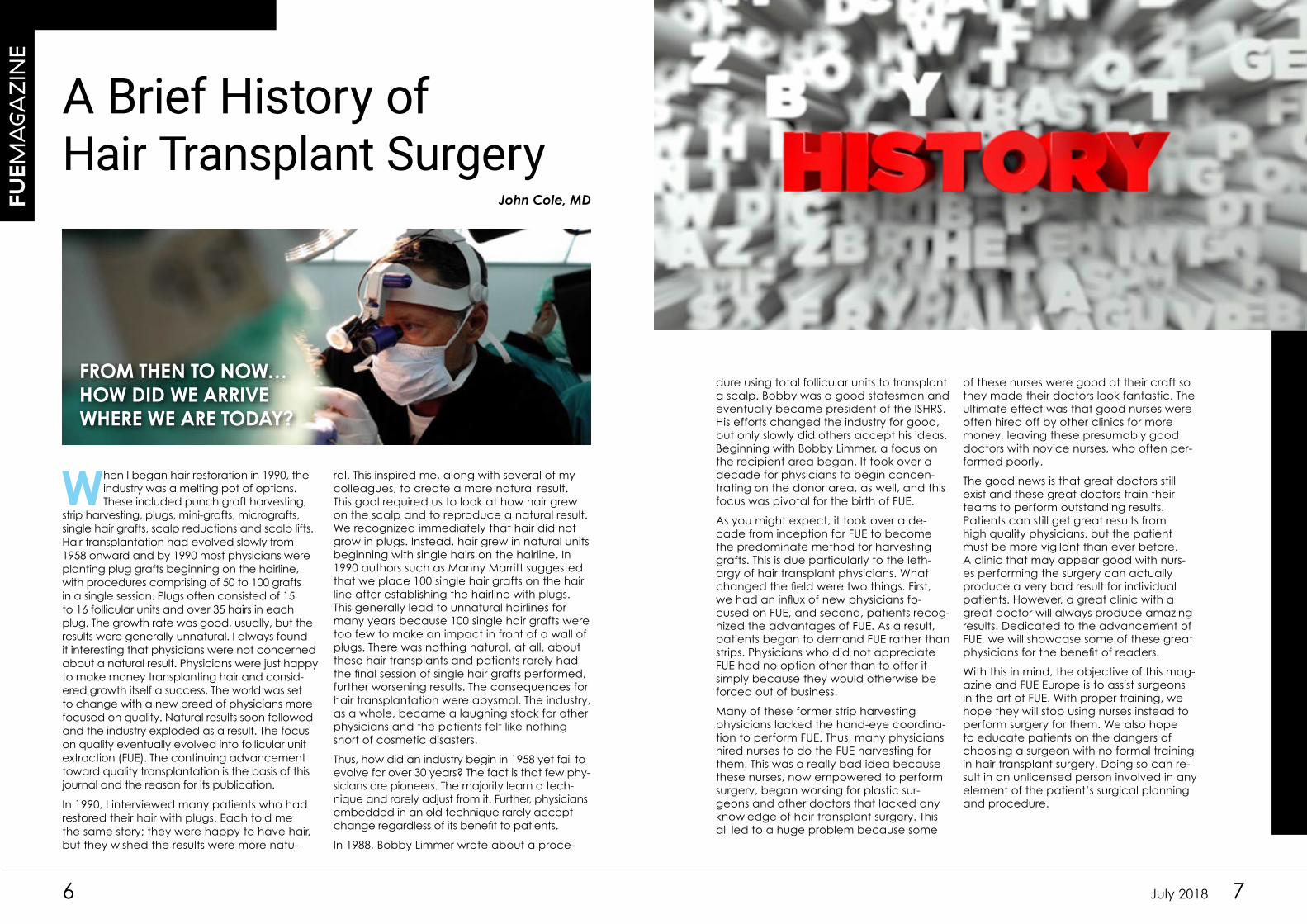
A BRIEF HISTORY OF HAIR TRANSPLANT
SURGERY
ANTI-AGING
MEDICINE FOR HAIR How to Maintain Youthful Hair
OFFICIAL
PUBLICATION
OF FUE EUROPE
A VISIT TO DR. ÖZGÜR Öztan’s office in Ankara, Turkey

3July 20182
FUE Magazine,a New Publication
John Cole, MD
A PERIODICAL DISCUSSING FUE SPECIFICALLY
One might ask “why create a new publication for hair transplantation?” The answer is clear. This journal focuses
on follicular unit extraction (FUE). Another publication, The Hair Transplant Forum International, devotes itself to hair loss and hair restoration. Most of its articles have no bearing on FUE and those that do are often for beginners. Further, the Hair Transplant Forum International also heavily focuses on strip harvesting as it must cater to ISHRS members, many of whom are strip-based physicians. This means its focus must be general rather than focus on one technique.
FUE Europe (FUEE) focuses entirely on FUE. Europe was not the birth place for FUE but its introduction was opportune; many young physicians entered the field of hair restoration surgery in Europe beginning in 2003 and the vast majority elected to pursue FUE. The advancements could be compared to another renaissance. FUE then gained prevalence in Asia, beginning in 2008. USA physicians, meanwhile, did not grasp the benefits of FUE for another five to seven years and still lags far behind. I think this was the first time the USA fell behind the rest of the world in hair restoration surgery.
The ISHRS has many good people in their leadership but its membership is composed of many old, tired, and stagnant minds who prefer strip procedures; they do not grasp the change in the wind. Thus, FUEE has a responsibility to usher the greater practice of FUE for the benefit of patients. FUE Magazine, as a publication, seeks to spur further interest in FUE and inform its practitioners of its latest innovations.
We consider any paper related to FUE from any country of origin. FUE Magazine’s main purpose is to foster compelling articles and new ideas relevant to the technique. We also review articles related to cell-based therapy, antiaging techniques, medical therapy, and ground-breaking ideas. We do not review articles related to strip harvesting or FUT, as it is commonly called. FUT leaves unpredictable scars, distorts hair growth angles, leads to traction alopecia, reduces donor hair mass and has the potential to destroy lives. FUE, meanwhile, is a younger technique that offers far better results. We believe focusing on it is essential for the wellbeing of patients and the advancement of hair transplantation.
CONTENT PAGE6
John Cole, MDA BRIEF HISTORY OF HAIR TRANSPLANT SURGERY
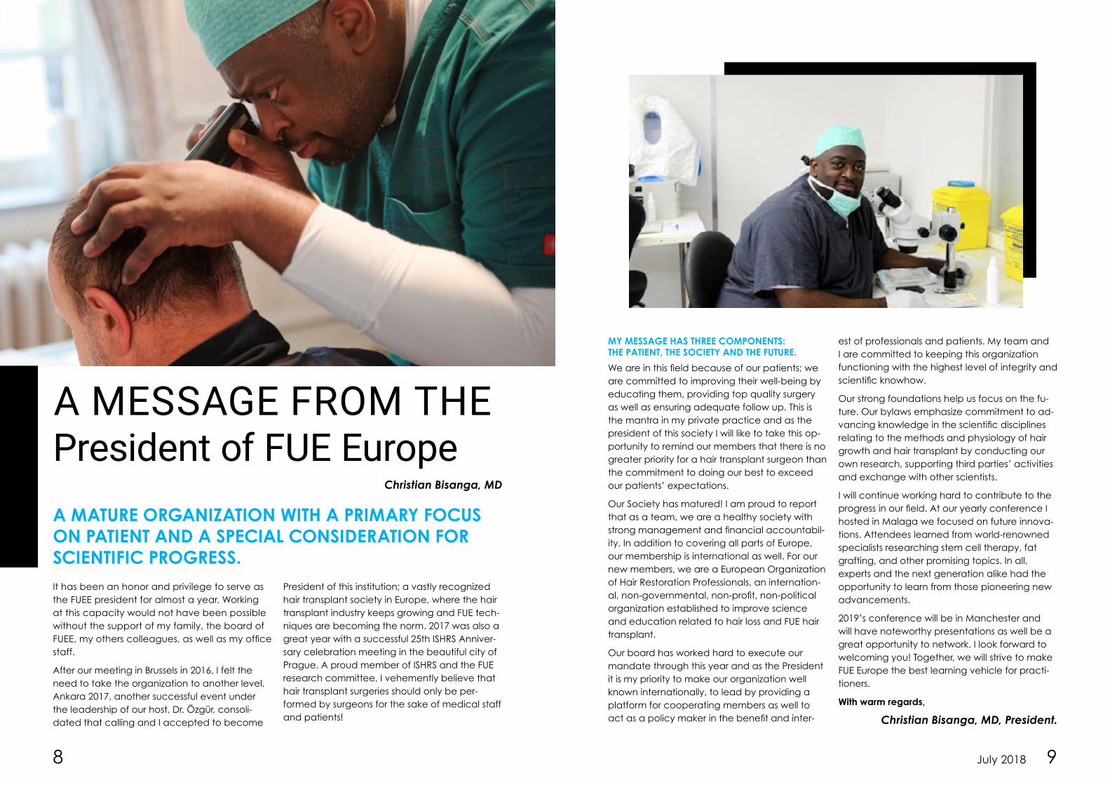
8Christian Bisanga, MD
A MESSAGE FROM THE PRESIDENT OF FUE EUROPE
10John Cole, MD
EDITOR’S NOTES
12Megan A. Cole, PHD,
John P. Cole, MDANTI-AGING MEDICINE FOR HAIR
18John Cole, MD
HAIR MAPPING
34John P. Cole, MD
THE LIMITATIONS OF DONOR SUPPLY IN TREATING
40John Cole, MD
A VISIT TO DR. ÖZTAN ÖZGÜR’SOFFICE IN ANKARA, TURKEY
44John Cole, MD
FUE FIGHTS BACK
48John Cole, MD
THE TRUMPETERS TOOT THEIR HORNS
54John Cole, MD
PAIN MANAGEMENT
64John Cole, MD
A RESPONSE TO DR. JOESEPHITIS
66John Cole, MD
WHAT IS UP WITH FUE EUROPE?

4 5July 2018
FU
EM
AG
AZ
INE
TO CONTRIBUTE AN ARTICLE
TO ADVERTISE
BOARD OF DIRECTORS
President: Dr. Christian Bisanga*Vice President: Reza Azar and Dr. Lars Heitmann
Secretary: Dr. Ludger Mentrup*Director Clinical Research: Dr. John Cole
Director Central Europe: Dr. Lars HeitmannDirector EUROASIA: Dr. Özgür Öztan
Director UK / Northern Europe: Dr. ShamalakDirector Communication/Industry: Marc Costin*
Auditor: Alya GadDirector of Consumer/Patient Affairs, IAHRS: Spencer Kobren
Program Chairman Manchester Asim ShahmalakScientific Director: Dr. John Cole
Program Chairman Ankara Live Surgery Özgür Öztan
6 7July 2018
FU
EM
AG
AZ
INE
A Brief History of Hair Transplant Surgery
John Cole, MD
FROM THEN TO NOW… HOW DID WE ARRIVE WHERE WE ARE TODAY?
When I began hair restoration in 1990, the industry was a melting pot of options. These included punch graft harvesting,
strip harvesting, plugs, mini-grafts, micrografts, single hair grafts, scalp reductions and scalp lifts. Hair transplantation had evolved slowly from 1958 onward and by 1990 most physicians were planting plug grafts beginning on the hairline, with procedures comprising of 50 to 100 grafts in a single session. Plugs often consisted of 15 to 16 follicular units and over 35 hairs in each plug. The growth rate was good, usually, but the results were generally unnatural. I always found it interesting that physicians were not concerned about a natural result. Physicians were just happy to make money transplanting hair and consid-ered growth itself a success. The world was set to change with a new breed of physicians more focused on quality. Natural results soon followed and the industry exploded as a result. The focus on quality eventually evolved into follicular unit extraction (FUE). The continuing advancement toward quality transplantation is the basis of this journal and the reason for its publication.
In 1990, I interviewed many patients who had restored their hair with plugs. Each told me the same story; they were happy to have hair, but they wished the results were more natu-
ral. This inspired me, along with several of my colleagues, to create a more natural result. This goal required us to look at how hair grew on the scalp and to reproduce a natural result. We recognized immediately that hair did not grow in plugs. Instead, hair grew in natural units beginning with single hairs on the hairline. In 1990 authors such as Manny Marritt suggested that we place 100 single hair grafts on the hair line after establishing the hairline with plugs. This generally lead to unnatural hairlines for many years because 100 single hair grafts were too few to make an impact in front of a wall of plugs. There was nothing natural, at all, about these hair transplants and patients rarely had the final session of single hair grafts performed, further worsening results. The consequences for hair transplantation were abysmal. The industry, as a whole, became a laughing stock for other physicians and the patients felt like nothing short of cosmetic disasters.
Thus, how did an industry begin in 1958 yet fail to evolve for over 30 years? The fact is that few phy-sicians are pioneers. The majority learn a tech-nique and rarely adjust from it. Further, physicians embedded in an old technique rarely accept change regardless of its benefit to patients.
In 1988, Bobby Limmer wrote about a proce-
dure using total follicular units to transplant a scalp. Bobby was a good statesman and eventually became president of the ISHRS. His efforts changed the industry for good, but only slowly did others accept his ideas. Beginning with Bobby Limmer, a focus on the recipient area began. It took over a decade for physicians to begin concen-trating on the donor area, as well, and this focus was pivotal for the birth of FUE.
As you might expect, it took over a de-cade from inception for FUE to become the predominate method for harvesting grafts. This is due particularly to the leth-argy of hair transplant physicians. What changed the field were two things. First, we had an influx of new physicians fo-cused on FUE, and second, patients recog-nized the advantages of FUE. As a result, patients began to demand FUE rather than strips. Physicians who did not appreciate FUE had no option other than to offer it simply because they would otherwise be forced out of business.
Many of these former strip harvesting physicians lacked the hand-eye coordina-tion to perform FUE. Thus, many physicians hired nurses to do the FUE harvesting for them. This was a really bad idea because these nurses, now empowered to perform surgery, began working for plastic sur-geons and other doctors that lacked any knowledge of hair transplant surgery. This all led to a huge problem because some
of these nurses were good at their craft so they made their doctors look fantastic. The ultimate effect was that good nurses were often hired off by other clinics for more money, leaving these presumably good doctors with novice nurses, who often per-formed poorly.
The good news is that great doctors still exist and these great doctors train their teams to perform outstanding results. Patients can still get great results from high quality physicians, but the patient must be more vigilant than ever before. A clinic that may appear good with nurs-es performing the surgery can actually produce a very bad result for individual patients. However, a great clinic with a great doctor will always produce amazing results. Dedicated to the advancement of FUE, we will showcase some of these great physicians for the benefit of readers.
With this in mind, the objective of this mag-azine and FUE Europe is to assist surgeons in the art of FUE. With proper training, we hope they will stop using nurses instead to perform surgery for them. We also hope to educate patients on the dangers of choosing a surgeon with no formal training in hair transplant surgery. Doing so can re-sult in an unlicensed person involved in any element of the patient’s surgical planning and procedure.
8 9July 2018
FU
EM
AG
AZ
INE
A MESSAGE FROM THE President of FUE Europe
Christian Bisanga, MD
A MATURE ORGANIZATION WITH A PRIMARY FOCUSON PATIENT AND A SPECIAL CONSIDERATION FORSCIENTIFIC PROGRESS.
It has been an honor and privilege to serve as
the FUEE president for almost a year. Working
at this capacity would not have been possible
without the support of my family, the board of
FUEE, my others colleagues, as well as my office staff.
After our meeting in Brussels in 2016, I felt the
need to take the organization to another level.
Ankara 2017, another successful event under
the leadership of our host, Dr. Özgür, consoli-
dated that calling and I accepted to become
President of this institution; a vastly recognized
hair transplant society in Europe, where the hair
transplant industry keeps growing and FUE tech-
niques are becoming the norm. 2017 was also a
great year with a successful 25th ISHRS Anniver-
sary celebration meeting in the beautiful city of
Prague. A proud member of ISHRS and the FUE
research committee, I vehemently believe that
hair transplant surgeries should only be per-
formed by surgeons for the sake of medical staff
and patients!
MY MESSAGE HAS THREE COMPONENTS: THE PATIENT, THE SOCIETY AND THE FUTURE.
We are in this field because of our patients; we are committed to improving their well-being by
educating them, providing top quality surgery
as well as ensuring adequate follow up. This is
the mantra in my private practice and as the
president of this society I will like to take this op-
portunity to remind our members that there is no
greater priority for a hair transplant surgeon than
the commitment to doing our best to exceed
our patients’ expectations.
Our Society has matured! I am proud to report
that as a team, we are a healthy society with
strong management and financial accountabil-ity. In addition to covering all parts of Europe,
our membership is international as well. For our
new members, we are a European Organization
of Hair Restoration Professionals, an internation-
al, non-governmental, non-profit, non-political organization established to improve science
and education related to hair loss and FUE hair
transplant.
Our board has worked hard to execute our
mandate through this year and as the President
it is my priority to make our organization well
known internationally, to lead by providing a
platform for cooperating members as well to
act as a policy maker in the benefit and inter-
est of professionals and patients. My team and
I are committed to keeping this organization
functioning with the highest level of integrity and
scientific knowhow.
Our strong foundations help us focus on the fu-
ture. Our bylaws emphasize commitment to ad-
vancing knowledge in the scientific disciplines relating to the methods and physiology of hair
growth and hair transplant by conducting our
own research, supporting third parties’ activities
and exchange with other scientists.
I will continue working hard to contribute to the
progress in our field. At our yearly conference I hosted in Malaga we focused on future innova-
tions. Attendees learned from world-renowned
specialists researching stem cell therapy, fat
grafting, and other promising topics. In all,
experts and the next generation alike had the
opportunity to learn from those pioneering new
advancements.
2019’s conference will be in Manchester and
will have noteworthy presentations as well be a
great opportunity to network. I look forward to
welcoming you! Together, we will strive to make
FUE Europe the best learning vehicle for practi-
tioners.
With warm regards,
Christian Bisanga, MD, President.
10 11July 2018
FU
EM
AG
AZ
INE
EDITOR’S NOTESJohn Cole, MD
THE PAST AND THE FUTURE OF FUE EUROPE
FUE Europe has grown significant-ly from the early days when only a handful of founding fathers attend-ed the meetings, but the organiza-tion has gotten off the ground with three back to back to back success-ful annual meetings. FUE Europe is a pure FUE organization. We will not discuss the merits of strip harvest-ing. The International Society of Hair Restoration Surgery (ISHRS) is waiting with open arms if strip surgery is your focus or interest. Having said this, we wish to partner with the ISHRS’s Global Council to promote hair restoration surgery. The FUE Europe organization had another wonderful meeting that was well attended pre-dominantly by Middle Eastern sur-geons interested in FUE. The meeting in Ankara was co-chaired by Shadi Zhari and Özgür Öztan. This meet-ing followed a wonderful meeting chaired by Christian Bisanga.
This year Christian formulated an-
other fantastic meeting in Malaga, Spain. Christian was the co-director of the meeting along with me, but I have to give Christian credit for doing almost all the work this year. I saw his program come alive over the past few months and there were some great faculty members in attendance. I have to say that I’m very pleased to see an increasing amount of European doctors come on board and speak at our meet-ings. We welcome all Europeans fo-cused on FUE to our ranks and invite them to our meetings. This does not suggest that we do not want physi-cians from other parts of the world to attend. Last year’s long-distance attendee was Michael Lee from South Korea.
Teresa Myer was the chairwoman of this year’s meeting in Malaga, Spain. The co-chairwoman was Chiara Insalaco. Both worked to ensure a great meeting in sunny Malaga.
I am also very excited we had Flavia Barasali, Mariana Alves, Ramiro Yane Mana, Georgios Zontos, Emorane Lupanzula, Laura Caicedo, Anasta-sios Vekris, Philippe Ginouves, Felix Popescu, and Ezequiel Panno join us from Europe. From the USA, we had Jeffery Epstein, Sanusi Umar, and Go-rana Kuka-Epstein join us, along with a return of Ryan Welter. From Brazil, Otavio Boaventura and Carlos Calixto joined us. Unfortunately, Otavio had a commitment in Korea last year. Both are becoming famous hair transplant doctors in Brazil. Slowly, we are grow-ing. This is great news and we encour-age all FUE surgeons to join.
This year we had some amazing in-vited speakers that include Professor Cristiana Serrano-Falcon, Pietro Gen-tile, and Angelo Trivisonno. These are people who are actually doing things clinically rather than theorists with no clinical results, who the ISHRS invites to their meetings.
This year’s vice president, Lars Heit-mann, has performed admirably. He helped to implement our financial success as a growing society, as well manage the ship at the helm admira-bly. Some behind the scenes people in our society receive little notice. They include Ludger Mentrup, who serves as our secretary. These individ-uals get little credit nor pay for keep-ing us all on our toes.
NEXT YEAR, MANCHESTER, ENGLAND
Next year the Program Chairman will be Asim Shahmalak, who will host us all in Manchester, England at his new facility. This program also should be wonderful. Please note that we are partnering with Spencer Kobren, who will hold the first IAHRS meeting in conjunction with FUE Europe in Manchester. This could easily be the largest attended FUE meeting in the world. I know that we always leave important names out of the recog-nition, but I encourage readers to check out our website to see the full list of lecturers and attendees. https:// www.fue-europe.com
We greatly encourage FUE surgeons to join our ranks. Slowly, we are gain-ing momentum. I know that with the help of so many, we will continue building our ranks and improving the quality of our meetings. I hope to see all of you in Manchester!
13July 201812
ANTI-AGING MEDICINE FOR HAIR How to Maintain Youthful Hair
Megan A. Cole, PHD,
John P. Cole, MD
Aging is a natural and unavoidable conse-quence of life; however, the appearance of being “aged” need not follow suit.
Indeed, the biology associated with cosmetic aging, the chemical pathways that trigger col-lagen degradation and isolated areas of hyper-pigmentation in the dermis, as well as hair loss and/or greying in the scalp, is a growing area of research, and as scientists unveil key regula-tors of the aging cascade, clinicians become better equipped to rejuvenate wrinkled skin and increase scalp hair density. In the context of anti-aging medicine, the hair follicle is uniquely qualified to provide a wealth of knowledge; it is easily accessible, contains multiple distinct adult stem cell populations, and regenerates itself cyclically. Moreover, the hair follicle constitutes a miniorgan with environmental niches estab-lished for both quasi-permanent, slow-cycling stem cells and transit-amplifying, highly-prolif-erative progenitor cells.1 Areas including the bulge, isthmus, and infundibulum are present in all stages of the hair growth cycle. Unsurprisingly, multiple adult stem cell populations have been identified in these regions, particularly in the outer root sheath region lying directly below the sebaceous gland, or the bulge region. On the other hand, the hair follicle matrix and pre-cor-tex are only observed during the active growth phase of the hair cycle (anagen) where they serve as reservoirs of rapidly dividing keratino-cyte progenitor cells and as differentiation and
melanin synthesis centers, respectively.
THE HAIR CYLE
The hair cycle begins with a short stage of apop-totic-driven hair follicle regression (catagen)
that lasts approximately 2 weeks. During cata-gen, the deeper, highly proliferative structures of the hair follicle, namely the matrix and the precortex, are lost, while the hair shaft, along with the inner and outer root sheaths, regress up towards the scalp surface. Following this peri-od of regression, the hair follicle enters a state of relative quiescence (telogen); the dermal papilla condenses, and the hair shaft is actively held in place by a specialized junction com-plex located at the base of the bulge region. The depigmented, fully keratinized telogen hair shaft is referred to as the “club” hair in homage the characteristic morphology of its club-like base. Notably, the secondary hair germ, which contains bulge stem cell-derived progenitor cells that will eventually give rise to the anagen hair bulb, pigmented hair shaft, and inner root sheath, manifests early in telogen and express-es an impressive quantity of circadian clock target genes. Deletion of two such genes, Bmal1 and Clock, delays the onset of anagen in mice without altering the morphological appearance of the hair follicle once growth finally ensues.2 In-terestingly, knockdown of the circadian protein BMAL1 (or Period 1) significantly prolongs the anagen phase in human hair follicles already in anagen, rendering these two chronobiolog-ical proteins potential drug targets for future hair follicle anti-aging medication(s).3 Late in telogen, the secondary hair germ is activated, and, following a period of rapid proliferation, elongates distally into the subcutaneous tissue, enveloping the dermal papilla and establishing itself as a matrix of proliferative transit-amplifying cells at the base of the follicle. Cell differenti-ation programs are reactivated, giving rise to the inner root sheath and hair shaft, and differ-
entiation of melanocyte precursors leading to melanogenesis occurs. Meanwhile, the club hair is shed (exogen) in an independently-regulated process. Although the duration of catagen and anagen remain fairly consistent from one cycle to the next, each telogen becomes progressive-ly longer than the one before; consequently, a progressive asynchrony in hair follicle cycling is observed with age. Additionally, many hair loss disorders (androgenic alopecia, alopecia areata, telogen effluvium) are characterized by concomitant increases in telogen and reduc-tions in anagen, making the telogen to anagen regulatory pathway(s) of particular importance in the rational design of anti-aging therapies. To date, two separate signaling pathways have been linked to hair follicle regeneration and underpin the readiness of the telogen follicle to enter anagen. Competing gradients of their inhibitory signals [bone morphogenetic protein (BMP) and fibroblast growth factor 18 (Fgf18)] and stimulatory signals [wingless (Wnt) and Fgf7/10] cycle slightly out of phase with one an-other, thereby establishing an early “refractory” telogen follicle and later “competent” telogen follicle.4
THE WNT/B-CATENIN PATHWAY
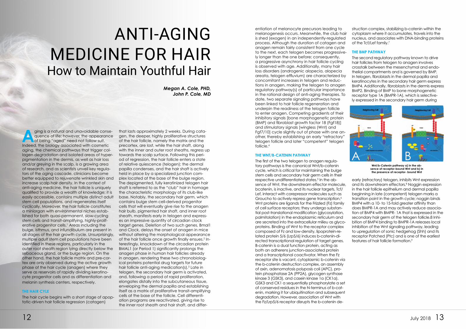
The first of the two telogen to anagen regula-tory pathways is the canonical Wnt/b-catenin cycle, which is critical for maintaining the bulge stem cells and secondary hair germ cells in their respective undifferentiated states.5 In the ab-sence of Wnt, the downstream effector molecule, bcatenin, is inactive, and its nuclear targets, Tcf/Lef, interact with corepressor molecules such as Groucho to actively repress gene transcription.6 Wnt proteins are ligands for the Frizzled (Fz) family of cell surface receptors; they undergo substan-tial post-translational modification (glycosylation, palmitolation) in the endoplasmic reticulum and are secreted into the extracellular milieu as glyco-proteins. Binding of Wnt to the receptor complex composed of Fz and low-density, lipoprotein-re-lated protein 5/6 (Lrp5/6) induces b-catenin-di-rected transcriptional regulation of target genes. B-catenin is a dual function protein, acting as both an adherens junction-associated protein and a transcriptional coactivator. When the Fz receptor site is vacant, cytoplasmic b-catenin via the b-catenin destruction complex, an assembly of axin, adenomatosis polyposis coli (APC), pro-tein phosphatase 2A (PP2A), glycogen synthase kinase 3 (GSK3), and casein kinase 1a (CK1a). GSK3 and CK1 a sequentially phosphorylate a set of conserved residues in the N-terminus of b-cat-enin, marking it for ubiquitination and subsequent degradation. However, association of Wnt with the Fz/Lrp5/6 receptor disrupts the b-catenin de-
struction complex, stabilizing b-catenin within the cytoplasm where it accumulates, travels into the nucleus, and associates with DNA-binding proteins of the Tcf/Lef family.7
THE BMP PATHWAY
The second regulatory pathway known to drive hair follicles from telogen to anagen involves crosstalk between the mesenchymal and endo-thelial compartments and is governed by BMP. In telogen, fibroblasts in the dermal papilla and keratinocytes in the secondary hair germ express BMP4. Additionally, fibroblasts in the dermis express BMP2. Binding of BMP to bone morphogenetic receptor type 1A (BMPR-1A), which is selective-ly expressed in the secondary hair germ during
early (refractory) telogen, inhibits Wnt expression and its downstream effectors.8 Noggin expression in the hair follicle epithelium and dermal papilla beginning in late (competent) telogen marks a transition point in the growth cycle; noggin binds BMP4 with a 10- to 15-fold greater affinity than does BMPR-1A and may actively reduce associa-tion of BMP4 with BMPR- 1A that is expressed in the secondary hair germ of the telogen follicle.8 Inhi-bition of BMP4 binding to BMPR-1A releases local inhibition of the Wnt signaling pathway, leading to upregulation of sonic hedgehog (Shh) and its receptor Patched (Ptc) and is one of the earliest features of hair follicle formation.9
A BWnt/b-Catenin pathway a) in the ab-
sence of receptor-bound Wnt and b) in the presence of receptor- bound Wnt
14 15July 2018
FU
EM
AG
AZ
INE
ACTIVATION OF THE TELOGEN TO ANAGEN TRANISTION
Coordination of the Wnt/b-catenin and BMP path-ways to effect hair follicle regeneration is function not of just what is secreted but also of when and where. Two members of the Tcf/Lef family, Tcf3 and Lef1, are expressed in the hair follicle.10 Tcf3 expres-sion has been identified in the bulge and in the secondary hair germ, where it appears to function independently of the b-catenin interacting domain to suppress features of epidermal terminal differen-tiation, thereby maintaining their stem cell features. Lef1, which requires Wnt signaling and b-catenin stabilization to exert regulatory control over hair dif-ferentiation, is expressed in the dermal papilla and secondary hair germ in late telogen through early anagen without concomitant expression of Shh.11 Dermal papilla cells also begin producing Fgf7/10 ligands and BMP inhibitors in late (competent) tel-ogen, both of which contribute to the progression of the hair follicle into anagen. Specifically, Fgf7/10 stimulate the secondary hair germ and (to a much lesser extent) the bulge to proliferate through FGFR2-111b binding events. This is in stark contrast to the dermal papilla secretory profile in early (re-fractory) telogen, at which point FGF18 is predom-inantly expressed and actively inhibits bulge cells through association with FGFR3.
At the onset of anagen, expression of BMP4 and BMPR-1A are downregulated in the germinative compartment of the hair follicle (i.e., the matrix), leading to a high rate of keratinocyte prolifera-tion. Moreover, in late anagen noggin expres-sion extends from the dermal papilla and cyclic epithelium (i.e., the matrix and precortex) to the surrounding connective tissue. Shh is expressed in unilateral clusters of hair matrix keratinocytes, and
Lef1 is observed in the matrix and precortical zone, prompting entry of progenitor cells within these regions into post-mitotic hair lineages. Bulge stem cells continue to cycle slowly throughout anagen, but the precise mechanism(s) shielding this cell population from the increasing gradient of stimula-tory cues has yet to be determined. However, it is speculated that bulge stem cells sparingly supply cells to extend the outer root sheath and refresh the pool of matrix progenitor cells that terminally differentiate after several proliferative cycles. As a result, the bulge has been deemed the “engine maintaining the (hair growth) process” by Greco et al. in their 2008 Cell Stem Cell journal article (DOI 10.1016/j.stem.2008.12.009).11
VITALITY OF THE HAIR FOLLICLE BULGE REGION
The importance of the bulge in the hair follicle growth cycle cannot be overemphasized. As mentioned previously, cells in the lower portion of the bulge undergo gene expression changes that transform them into the secondary germ cells at the end of catagen. The progeny of multipotent bulge cells generates the new lower anagen folli-cle in response to the cellular and environmental cues discussed above. However, immunohisto-logical studies of alopecic scalps have revealed persistent infiltrates of activated T cells in the bulge region of the transitional scalp, that is, the region of scalp lying at the intersection of balding scalp and hair-retaining scalp.12 Secondary to this prolonged inflammation, infundibula widen and become blocked by laminated keratin, trichogenic ele-ments are replaced with fibrous tracts, and (criti-cally) anagen follicles are rare. Immune response genes are known to be upregulated in early catagen and throughout telogen in normal hair
follicles; the infundibulum is an established early tar-get of acute and transient T- cell mediated-protein expression. Indeed, the formation of desmosomes, the proteins responsible for anchoring telogen “club hairs” in place, is perpetuated via transloca-tion of nuclear factor of activated T cells (NFAT) into the nucleus of bulge cells during telogen.13 Clearly, activation of the immune response is necessary for maintaining hair cycle homeostasis, yet the mech-anistic link between androgens and T cell dysregu-lation remains unclear.
Nevertheless, clinical manifestations of andro-genic alopecia (AA) support an immune-medi-ated attack on the bulge. The time course of AA progresses as follows: hair shafts become void of pigmentation; the diameter of individual hair shafts decreases while total hair count remains stable; finally, hair count begins to decline while follicular unit density remains stable. In the hair follicle re-generation cascade, arrest of melanogenesis (i.e., pigment production) precedes that of keratinocyte proliferation, as evidenced by the unpigmented base of the “club” hair in normal telogen. Thus, one may conclude that in the wake of replicative exhaust, depigmentation would be the first clinical manifestation regardless of the source of melano-cyte stem cells and matrix transit-amplifying pro-genitor cells. However, unlike telogen, AA-associ-ated depigmentation extends the entire hair shaft, indicating that transit-amplifying cell populations are continually being replenished by the bulge while melanocyte stem cells, which are located at the base of the bulge, are not. With the passage of time, the bulge region responsible for fueling matrix keratinocyte populations also become compro-mised, and hair shaft diameter begins to decrease. Once trichogenic elements in the bulge are fully replaced with fibrous tracts, the asynchrony of hair follicle growth becomes apparent. Individual hair follicles within a given follicular unit and receiving progenitor cells from the same bulge region may exist at different states of proliferative potential; therefore, they may reach replicative senescence at significantly later times. That is, if one hair shaft in a follicular unit has been in anagen for 6 years, its matrix cell population may be more “exhausted” than a neighboring hair shaft that has only been in anagen for 6 months. As a result, when the older hair reaches senescence and the fibrous bulge is unable to regenerate the secondary germ during telogen, anagen does not accompany exogen. However, the younger hair may remain intact for several more years since anagen can persist for decades in humans, but follicular unit density will be down, and the growing hair will likely present with a small diameter.
MEDICAL TREATMENTS TO COMBAT HAIR LOSS
How then can clinicians roll back the clock on a cellular destructive process? The answer may be summed up by the age-old adage “an ounce of prevention is worth a pound of cure”. Begin with early intervention. Anti-inflammatory medications such as minoxidil and cyclosporine A would reduce immune-mediated “attack” on bulge stem cells by preventing mast cell degranulation. Furthermore, minoxidil is known to exert anti-proliferative effects on dermal fibroblasts, decreasing collagen synthe-sis and thereby reducing fibrosis in the susceptible hair loss regions. Experimental stage therapies, in-cluding methyl vanillate, aminotic membrane, and WNT-Act, may stimulate anagen-inducing and/or transitional elements. Topical application of methyl vanillate, for example, has been found to increase hair count and hair mass index in women by 6% and 12% following 6 months of use, respectively. The active ingredient is a suspected Wnt-activator given the concomitant 32% increase in Wnt10B ex-pression in the temporal scalp.14 Treatment of mice with amniotic membrane have similarly shown upregulation of anagen stimulatory signals, specifi-cally, increased FGF7 and proliferating cell nuclear antigen. Mice treated with topical amniotic mem-brane expressed similar levels of hair regeneration as those treated with 5% minoxidil.15 Additional drug therapies may target extrafollicular domains, such as adipocyte precursors whose generation begins in late catagen. Release of platelet derived growth factor (PDGF) from these cells is linked to the suppression of BMP and subsequent onset of anagen. Autologous platelet rich plasma (PRP) is rich in PDGF and has been shown to increase hair density by 50% at 6 months. Prescription medica-tions like finasteride have also proven beneficial. Regulation of Wnt signaling in dermal papilla cells has demonstrated an androgen dependence in AA. Dermal papilla harvested from the scalps of AA patients express increased levels of androgen receptor (AR), which is a member of the nucle-ar receptor superfamily that translocates to the nucleus upon binding ligandwhere it functions as a ligand-dependent transcription factor. 16 Thus, in AA patients, increased AR expression is associated with increased translocation of testosterone- (T) or dihydrotestosterone- (DHT) bound AR. This nuclear complex interacts with b-catenin to inhibit Wnt-me-diated transcriptional activity, and the result is keratinocyte growth suppression in the matrix. Additionally, AR enhances nuclear translocation of b-catenin in preadipocytes, ultimately preventing their differentiation. The sum effect is suppressed hair growth in anagen follicles. Since finasteride is a selective 5a-reductase inhibitor that blocks the conversion of T into DHT (which binds AR with a slightly higher affinity than T17), the medication may be considered a Wnt upregulator.17
16 17July 2018
FU
EM
AG
AZ
INE
Oral finasteride formulations have been associat-ed with increased scalp hair density in men, gain-ing widespread attention under the tradename Propecia in the late 1990s. However, the medica-tion does not stop the hair loss process and must be taken perpetually to prevent relapse. Since the oral formulation is associated with a num-ber of undesirable side effects (including sexual dysfunction), patient compliance is an issue that topical finasteride formulations could conceivably bypass. Although topical formulations are in their infancy of use, remarkable improvements in hair density have been observed, and, in that regard, may represent the best available anti-aging medi-cation for hair at present.
In summary, the factors governing the hair follicle cycle are vast and rather complex. Two inter-re-lated pathways (Wnt/b-catenin and BMP) have been examined in great detail, but the exact trigger mechanism pushing refractory telogen follicles to become competent follicles remains unknown. Extrafollicular players, particularly adipocyte precursors, appear to be involved. Similarly, androgen sensitivity is a suspected culprit in immune dysregulation leading to bulge region fibrosis and eventual demise of the entire hair follicle. Therefore, anti-aging efforts should begin early with anti-inflammatory agents, adipocyte supporters (like PDGF), and Wnt cycle promoters (such as topical finasteride, methyl vanillate, and amniotic membrane).
REFERENCES
1. Schneider MR, Schmidt-Ullrich R, Paus R. The Hair Follicle as a Dynamic Miniorgan. Cur-
rent Biology 2009;19:R132-R42.
2. Lin KK, Kumar V, Geyfman M, et al. Circadian Clock Genes Contribute to the Regula-
tion of Hair Follicle Cycling. Plos Genetics 2009;5.
3. Al-Nuaimi Y, Hardman JA, Biro T, et al. A Meeting of Two Chronobiological Systems:
Circadian Proteins Period1 and BMAL1 Modulate the Human Hair Cycle Clock. Journal of
Investigative Dermatology 2014;134:610-9.
4. Geyfman M, Plikus MV, Treffeisen E, Andersen B, Paus R. Resting no more: re-de-
fining telogen, the maintenance stage of the hair growth cycle. Biological Reviews 2015;90:1179-96.
5. Lowry WE, Blanpain C, Nowak JA, Guasch G, Lewis L, Fuchs E. Defining the impact of beta- catenin/Tcf transactivation on epithelial stem cells. Genes & Development
2005;19:1596-611.
6. Reya T, Clevers H. Wnt signalling in stem cells and cancer. Nature 2005;434:843-50.
7. Komiya Y, Habas R. Wnt signal transduction pathways. Organogenesis 2008;4:68-75.
8. Botchkarev VA, Botchkareva NV, Nakamura M, et al. Noggin is required for induction
of the hair follicle growth phase in postnatal skin. Faseb Journal 2001;15:2205-14.
9. Lo Celso C, Prowse DM, Watt FM. Transient activation of beta-catenin signalling in
adult mouse epidermis is sufficient to induce new hair follicles but continuous activation is required to maintain hair follicle tumours. Development 2004;131:1787-99.
10. Merrill BJ, Gat U, DasGupta R, Fuchs E. Tcf3 and Lef1 regulate lineage differentiation
of multipotent stem cells in skin. Genes & Development 2001;15:1688-705.
11. Greco V, Chen T, Rendl M, et al. A two-step mechanism for stem cell activation
during hair regeneration. Journal of Investigative Dermatology 2010;130:S102-S.
12. Jaworsky C, Kligman AM, Murphy GF. Characterization of inflammatory infiltrates in male pattern alopecia - Implications for pathogenesis. British Journal of Dermatology
1992;127:239-46.
13. Sato-Miyaoka M, Hisatsune C, Ebisui E, Ogawa N, Takahashi-Iwanaga H, Mikoshiba K.
Regulation of Hair Shedding by the Type 3 IP3 Receptor. Journal of Investigative Derma-
tology 2012;132:2137-47.
14. Tosti A, Zaiac MN, Canazza A, et al. Topical application of the Wnt/-catenin activa-
tor methyl vanillate increases hair count and hair mass index in women with androgenet-
ic alopecia. Journal of Cosmetic Dermatology 2016;15:469-74.
15. Seo H-S, Lee D-J, Chung J-h, et al. Hominis Placenta facilitates hair re-growth by up-
regulating cellular proliferation and expression of fibroblast growth factor-7. BMC Com-
plementary and Alternative Medicine 2016;16:187.
16. Kitagawa T, Matsuda K, Inui S, et al. Keratinocyte Growth Inhibition through the Mod-
ification of Wnt Signaling by Androgen in Balding Dermal Papilla Cells. Journal of Clinical Endocrinology & Metabolism 2009;94:1288-94.
17. Wilson EM, French FS. Binding properties of androgen receptors - Evidence for iden-
tical receptros in rat testis, epididymis, and prostate. Journal of Biological Chemistry
1976;251:5620-9.
18 19July 2018
FU
EM
AG
AZ
INE
HAIR MAPPINGJohn Cole, MD
John P. Cole, MD, Kyu Ho Lee, MD, Eun Seong Park, MD,
Seong Eun Kang, MD, Chang-Hun Huh,PhD.
A COMPARISON OF CAUCASIAN AND KOREAN HAIRDENSITY, FOLLICULAR DENSITY, AND CALCULATEDDENSITY; A COMPELLING ARGUMENT FOR INDIVIDUALFOLLICULAR GROUP HARVESTINGINTRODUCTION
Numerous investigators have estimated donor area hair density and follicular density over the years. No published study to date has attempt-ed to objectively quantify the total follicular density in the donor area. Numerous individu-als have estimated follicular hair density in the Korean population, but no one has compared Korean densities with Caucasian densities using similar objective methods to measure these densities. The purpose of this study is to better estimate hair density and follicular density in a finite surface area of the donor region and to compare these results in both Korean and Caucasian populations using the same method of measurement.
In addition to estimating the total number of follicular groups in the donor area, this study attempted to define the mean number of hairs per follicular group (calculated density) pre-operatively.1 We also evaluated the mean number of hairs per follicular group extract-ed by a method of individual follicular group harvesting (IFGH), commonly called follicular unit extraction (FUE), known as the Cole Isola-tion Technique (CIT) and compared them to the donor area calculated densities that were determined pre-operatively.2,3 The purpose of this later comparison was to determine if there were specific advantages to this method of IFGH harvesting over strip donor harvesting.
The results of this objective study showed that Korean follicular density and hair density are higher than previously reported in Asian pa-tients. Caucasian hair density and follicular density are higher than in Korean people, but the difference is not as great as previously re-ported. Previous studies have shown that IFGH produces more hair per graft than does strip harvesting. This study reconfirms that IFGH does offer an advantage over traditional strip har-vesting in that it produces more hair per follicu-lar group and a higher percentage of follicular groups containing multiple hairs the distribution of grafts produced.
OBJECTIVE
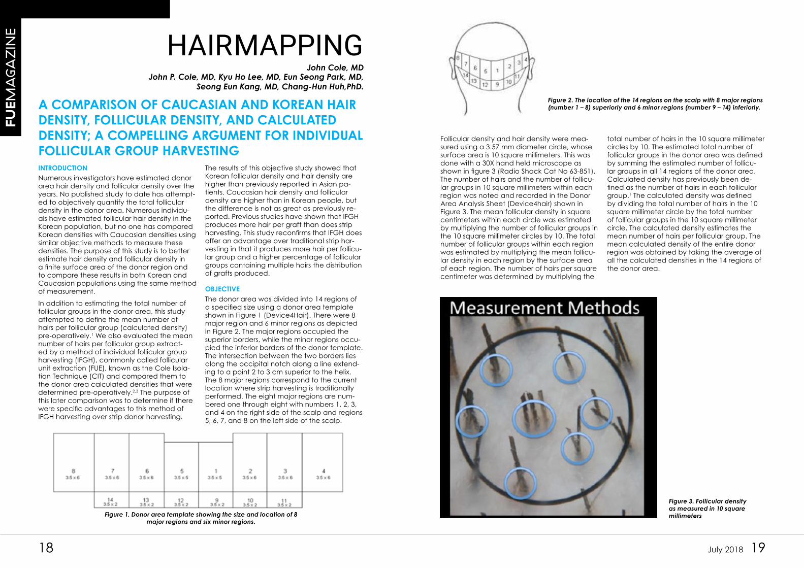
The donor area was divided into 14 regions of a specified size using a donor area template shown in Figure 1 (Device4Hair). There were 8 major region and 6 minor regions as depicted in Figure 2. The major regions occupied the superior borders, while the minor regions occu-pied the inferior borders of the donor template. The intersection between the two borders lies along the occipital notch along a line extend-ing to a point 2 to 3 cm superior to the helix. The 8 major regions correspond to the current location where strip harvesting is traditionally performed. The eight major regions are num-bered one through eight with numbers 1, 2, 3, and 4 on the right side of the scalp and regions 5, 6, 7, and 8 on the left side of the scalp.
Figure 1. Donor area template showing the size and location of 8
major regions and six minor regions.
Figure 2. The location of the 14 regions on the scalp with 8 major regions
(number 1 – 8) superiorly and 6 minor regions (number 9 – 14) inferiorly.
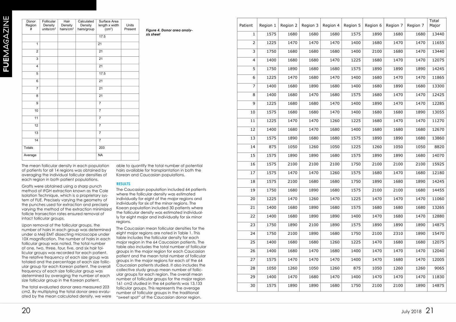
Figure 3. Follicular density
as measured in 10 square
millimeters
Follicular density and hair density were mea-sured using a 3.57 mm diameter circle, whose surface area is 10 square millimeters. This was done with a 30X hand held microscope as shown in figure 3 (Radio Shack Cat No 63-851). The number of hairs and the number of follicu-lar groups in 10 square millimeters within each region was noted and recorded in the Donor Area Analysis Sheet (Device4hair) shown in Figure 3. The mean follicular density in square centimeters within each circle was estimated by multiplying the number of follicular groups in the 10 square millimeter circles by 10. The total number of follicular groups within each region was estimated by multiplying the mean follicu-lar density in each region by the surface area of each region. The number of hairs per square centimeter was determined by multiplying the
total number of hairs in the 10 square millimeter circles by 10. The estimated total number of follicular groups in the donor area was defined by summing the estimated number of follicu-lar groups in all 14 regions of the donor area. Calculated density has previously been de-fined as the number of hairs in each follicular group.1 The calculated density was defined by dividing the total number of hairs in the 10 square millimeter circle by the total number of follicular groups in the 10 square millimeter circle. The calculated density estimates the mean number of hairs per follicular group. The mean calculated density of the entire donor region was obtained by taking the average of all the calculated densities in the 14 regions of the donor area.
20 21July 2018
FU
EM
AG
AZ
INE
Donor Region
#
Follicular Density
units/cm2
Hair Density
hairs/cm2
Calculated Density
hairs/group
Surface Arealength x width
(cm2)Units
Present
17.5
1 21
2 21
3 21
4 21
5 17.5
6 21
7 21
8 21
9 7
10 7
11 7
12 7
13 7
14 7
Totals 203
Average NA
Figure 4. Donor area analy-
sis sheet
The mean follicular density in each population of patients for all 14 regions was obtained by averaging the individual follicular densities of each region in both patient populations.
Grafts were obtained using a sharp punch method of IFGH extraction known as the Cole Isolation Technique, which is a proprietary sys-tem of FUE. Precisely varying the geometry of the punches used for extraction and precisely varying the method of the extraction minimized follicle transection rates ensured removal of intact follicular groups.
Upon removal of the follicular groups, the number of hairs in each group was determined under a Meji EMT dissecting microscope under 10X magnification. The number of hairs in each follicular group was noted. The total number of one, two, three, four, five, and six hair fol-licular groups was recorded for each patient. The relative frequency of each size group was totaled and the percentage of each size follic-ular group for each Korean patient. The overall frequency of each size follicular group was determined by averaging the number of each size follicular group in the Korean patient.
The total evaluated donor area measured 203 cm2. By multiplying the total donor area evalu-ated by the mean calculated density, we were
able to quantify the total number of potential hairs available for transplantation in both the Korean and Caucasian populations.
RESULTS
The Caucasian population included 64 patients where the follicular density was estimated individually for eight of the major regions and individually for six of the minor regions. The Koean population included 30 patients where the follicular density was estimated individual-ly for eight major and individually for six minor regions.
The Caucasian mean follicular densities for the eight major regions are noted in Table 1. This table includes the follicular density for each major region in the 64 Caucasian patients. The table also includes the total number of follicular groups in the major region for each Caucasian patient and the mean total number of follicular groups in the major regions for each of the 64 Caucasian patients studied. It also includes the collective study group mean number of follic-ular groups for each region. The overall mean number of follicular groups for the major region 161 cm2 studied in the 64 patients was 13,133 follicular groups. This represents the average number of follicular groups in the traditional “sweet spot” of the Caucasian donor region.
Patient Region 1 Region 2 Region 3 Region 4 Region 5 Region 6 Region 7 Region 7
Total
Major
1 1575 1680 1680 1680 1575 1890 1680 1680 13440
2 1225 1470 1470 1470 1400 1680 1470 1470 11655
3 1750 1680 1680 1680 1400 2100 1680 1470 13440
4 1400 1680 1680 1470 1225 1680 1470 1470 12075
5 1750 1890 1680 1680 1575 1890 1890 1890 14245
6 1225 1470 1680 1470 1400 1680 1470 1470 11865
7 1400 1680 1890 1680 1400 1680 1890 1680 13300
8 1400 1680 1470 1680 1575 1680 1470 1470 12425
9 1225 1680 1680 1470 1400 1890 1470 1470 12285
10 1575 1680 1680 1470 1400 1680 1680 1890 13055
11 1225 1470 1470 1260 1225 1680 1470 1470 11270
12 1400 1680 1470 1680 1400 1680 1680 1680 12670
13 1575 1890 1680 1680 1575 1890 1890 1680 13860
14 875 1050 1260 1050 1225 1260 1050 1050 8820
15 1575 1890 1890 1680 1575 1890 1890 1680 14070
16 1575 2100 2100 2100 1750 2100 2100 2100 15925
17 1575 1470 1470 1260 1575 1680 1470 1680 12180
18 1575 2100 1680 1680 1750 1890 1680 1890 14245
19 1750 1680 1890 1680 1575 2100 2100 1680 14455
20 1225 1470 1260 1470 1225 1470 1470 1470 11060
21 1400 1680 1890 1680 1575 1680 1680 1680 13265
22 1400 1680 1890 1890 1400 1470 1680 1470 12880
23 1750 1890 2100 1890 1575 1890 1890 1890 14875
24 1750 2100 1890 1680 1750 2100 2310 1890 15470
25 1400 1680 1680 1260 1225 1470 1680 1680 12075
26 1400 1680 1470 1680 1400 1470 1470 1470 12040
27 1575 1470 1470 1470 1400 1470 1680 1470 12005
28 1050 1260 1050 1260 875 1050 1260 1260 9065
29 1400 1470 1680 1470 1400 1470 1470 1470 11830
30 1575 1890 1890 1680 1750 2100 2100 1890 14875
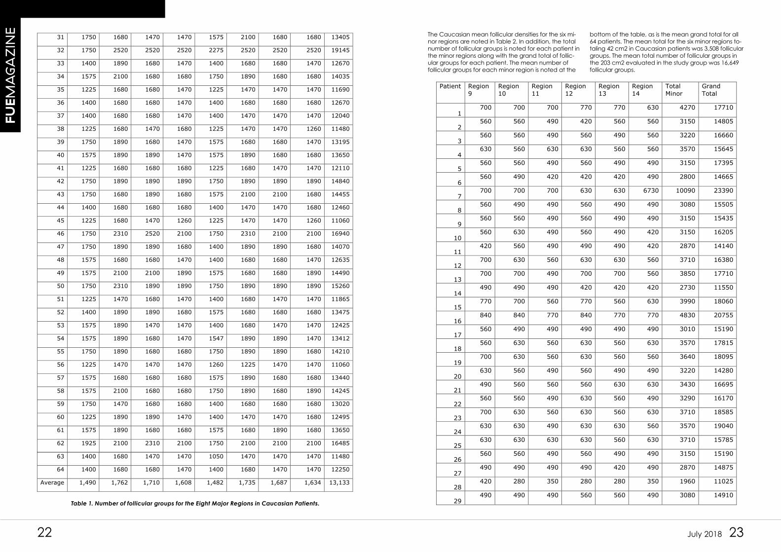
22 23July 2018
FU
EM
AG
AZ
INE
31 1750 1680 1470 1470 1575 2100 1680 1680 13405
32 1750 2520 2520 2520 2275 2520 2520 2520 19145
33 1400 1890 1680 1470 1400 1680 1680 1470 12670
34 1575 2100 1680 1680 1750 1890 1680 1680 14035
35 1225 1680 1680 1470 1225 1470 1470 1470 11690
36 1400 1680 1680 1470 1400 1680 1680 1680 12670
37 1400 1680 1680 1470 1400 1470 1470 1470 12040
38 1225 1680 1470 1680 1225 1470 1470 1260 11480
39 1750 1890 1680 1470 1575 1680 1680 1470 13195
40 1575 1890 1890 1470 1575 1890 1680 1680 13650
41 1225 1680 1680 1680 1225 1680 1470 1470 12110
42 1750 1890 1890 1890 1750 1890 1890 1890 14840
43 1750 1680 1890 1680 1575 2100 2100 1680 14455
44 1400 1680 1680 1680 1400 1470 1470 1680 12460
45 1225 1680 1470 1260 1225 1470 1470 1260 11060
46 1750 2310 2520 2100 1750 2310 2100 2100 16940
47 1750 1890 1890 1680 1400 1890 1890 1680 14070
48 1575 1680 1680 1470 1400 1680 1680 1470 12635
49 1575 2100 2100 1890 1575 1680 1680 1890 14490
50 1750 2310 1890 1890 1750 1890 1890 1890 15260
51 1225 1470 1680 1470 1400 1680 1470 1470 11865
52 1400 1890 1890 1680 1575 1680 1680 1680 13475
53 1575 1890 1470 1470 1400 1680 1470 1470 12425
54 1575 1890 1680 1470 1547 1890 1890 1470 13412
55 1750 1890 1680 1680 1750 1890 1890 1680 14210
56 1225 1470 1470 1470 1260 1225 1470 1470 11060
57 1575 1680 1680 1680 1575 1890 1680 1680 13440
58 1575 2100 1680 1680 1750 1890 1680 1890 14245
59 1750 1470 1680 1680 1400 1680 1680 1680 13020
60 1225 1890 1890 1470 1400 1470 1470 1680 12495
61 1575 1890 1680 1680 1575 1680 1890 1680 13650
62 1925 2100 2310 2100 1750 2100 2100 2100 16485
63 1400 1680 1470 1470 1050 1470 1470 1470 11480
64 1400 1680 1680 1470 1400 1680 1470 1470 12250
Average 1,490 1,762 1,710 1,608 1,482 1,735 1,687 1,634 13,133
Table 1. Number of follicular groups for the Eight Major Regions in Caucasian Patients.
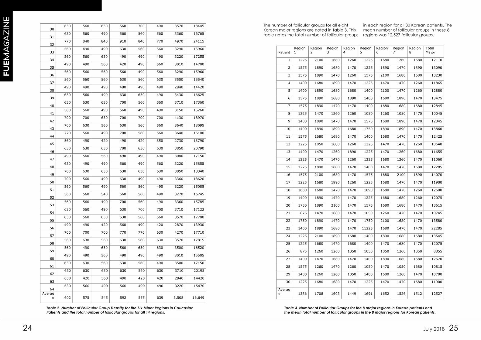
The Caucasian mean follicular densities for the six mi-nor regions are noted in Table 2. In addition, the total number of follicular groups is noted for each patient in the minor regions along with the grand total of follic-ular groups for each patient. The mean number of follicular groups for each minor region is noted at the
bottom of the table, as is the mean grand total for all 64 patients. The mean total for the six minor regions to-taling 42 cm2 in Caucasian patients was 3,508 follicular groups. The mean total number of follicular groups in the 203 cm2 evaluated in the study group was 16,649 follicular groups.
Patient Region
9
Region
10
Region
11
Region
12
Region
13
Region
14
Total
Minor
Grand
Total
1 700 700 700 770 770 630 4270 17710
2 560 560 490 420 560 560 3150 14805
3 560 560 490 560 490 560 3220 16660
4 630 560 630 630 560 560 3570 15645
5 560 560 490 560 490 490 3150 17395
6 560 490 420 420 420 490 2800 14665
7 700 700 700 630 630 6730 10090 23390
8 560 490 490 560 490 490 3080 15505
9 560 560 490 560 490 490 3150 15435
10 560 630 490 560 490 420 3150 16205
11 420 560 490 490 490 420 2870 14140
12 700 630 560 630 630 560 3710 16380
13 700 700 490 700 700 560 3850 17710
14 490 490 490 420 420 420 2730 11550
15 770 700 560 770 560 630 3990 18060
16 840 840 770 840 770 770 4830 20755
17 560 490 490 490 490 490 3010 15190
18 560 630 560 630 560 630 3570 17815
19 700 630 560 630 560 560 3640 18095
20 630 560 490 560 490 490 3220 14280
21 490 560 560 560 630 630 3430 16695
22 560 560 490 630 560 490 3290 16170
23 700 630 560 630 560 630 3710 18585
24 630 630 490 630 630 560 3570 19040
25 630 630 630 630 560 630 3710 15785
26 560 560 490 560 490 490 3150 15190
27 490 490 490 490 420 490 2870 14875
28 420 280 350 280 280 350 1960 11025
29 490 490 490 560 560 490 3080 14910
24 25July 2018
FU
EM
AG
AZ
INE
30 630 560 630 560 700 490 3570 18445
31 630 560 490 560 560 560 3360 16765
32 770 840 840 910 840 770 4970 24115
33 560 490 490 630 560 560 3290 15960
34 560 560 630 490 490 490 3220 17255
35 490 490 560 420 490 560 3010 14700
36 560 560 560 560 490 560 3290 15960
37 560 560 560 630 560 630 3500 15540
38 490 490 490 490 490 490 2940 14420
39 630 560 490 630 630 490 3430 16625
40 630 630 630 700 560 560 3710 17360
41 560 560 490 560 490 490 3150 15260
42 700 700 630 700 700 700 4130 18970
43 700 630 560 630 560 560 3640 18095
44 770 560 490 700 560 560 3640 16100
45 560 490 420 490 420 350 2730 13790
46 630 630 630 700 630 630 3850 20790
47 490 560 560 490 490 490 3080 17150
48 630 490 490 560 490 560 3220 15855
49 700 630 630 630 630 630 3850 18340
50 700 560 490 630 490 490 3360 18620
51 560 560 490 560 560 490 3220 15085
52 560 560 540 560 560 490 3270 16745
53 560 560 490 700 560 490 3360 15785
54 630 560 490 630 700 700 3710 17122
55 630 560 630 630 560 560 3570 17780
56 490 490 420 560 490 420 2870 13930
57 700 700 700 770 770 630 4270 17710
58 560 630 560 630 560 630 3570 17815
59 560 490 630 560 630 630 3500 16520
60 490 490 560 490 490 490 3010 15505
61 630 630 560 630 560 490 3500 17150
62 630 630 630 630 560 630 3710 20195
63 630 420 560 490 420 420 2940 14420
64 630 560 490 560 490 490 3220 15470
Averag
e 602 575 545 592 555 639 3,508 16,649
Table 2. Number of Follicular Group Density for the Six Minor Regions in Caucasian
Patients and the total number of follicular groups for all 14 regions.
Table 3. Number of Follicular Groups for the 8 major regions in Korean patients and
the mean total number of follicular groups in the 8 major regions for Korean patients.
The number of follicular groups for all eight Korean major regions are noted in Table 3. This table notes the total number of follicular groups
in each region for all 30 Korean patients. The mean number of follicular groups in these 8 regions was 12,527 follicular groups.
Patient
Region
1
Region
2
Region
3
Region
4
Region
5
Region
6
Region
7
Region
8
Total
Major
1 1225 2100 1680 1260 1225 1680 1260 1680 12110
2 1575 1890 1680 1470 1225 1890 1470 1890 13090
3 1575 1890 1470 1260 1575 2100 1680 1680 13230
4 1400 1680 1890 1470 1225 1470 1470 1260 11865
5 1400 1890 1680 1680 1400 2100 1470 1260 12880
6 1575 1890 1680 1890 1400 1680 1890 1470 13475
7 1575 1890 1470 1470 1400 1680 1680 1680 12845
8 1225 1470 1260 1260 1050 1260 1050 1470 10045
9 1400 1890 1470 1470 1575 1680 1890 1470 12845
10 1400 1890 1890 1680 1750 1890 1890 1470 13860
11 1575 1680 1680 1470 1400 1680 1470 1470 12425
12 1225 1050 1680 1260 1225 1470 1470 1260 10640
13 1400 1470 1260 1890 1225 1470 1260 1680 11655
14 1225 1470 1470 1260 1225 1680 1260 1470 11060
15 1225 1890 1680 1470 1400 1470 1470 1680 12285
16 1575 2100 1680 1470 1575 1680 2100 1890 14070
17 1225 1680 1890 1260 1225 1680 1470 1470 11900
18 1680 1680 1470 1470 1890 1680 1470 1260 12600
19 1400 1890 1470 1470 1225 1680 1680 1260 12075
20 1750 1890 2100 1470 1575 1680 1680 1470 13615
21 875 1470 1680 1470 1050 1260 1470 1470 10745
22 1750 1890 1470 1470 1750 2100 1680 1470 13580
23 1400 1890 1680 1470 11225 1680 1470 1470 22285
24 1225 2100 1890 1680 1400 1890 1680 1680 13545
25 1225 1680 1470 1680 1400 1470 1680 1470 12075
26 875 1260 1260 1050 1050 1050 1260 1050 8855
27 1400 1470 1680 1470 1400 1890 1680 1680 12670
28 1575 1260 1470 1260 1050 1470 1050 1680 10815
29 1400 1260 1260 1050 1400 1680 1260 1470 10780
30 1225 1680 1680 1470 1225 1470 1470 1680 11900
Averag
e 1386 1708 1603 1449 1691 1652 1526 1512 12527
26 27July 2018
FU
EM
AG
AZ
INE
The number of follicular groups in all six minor regions for the 30 Korean patients studied is noted in Table 4. This table includes the mean number of follicular groups for each of the six regions. The mean number of follicular groups
in the six minor regions for the Korean study group was 3,191 follicular groups. The mean total number of follicular groups in all 14 regions for the Korean study groups was 15,718 follicu-lar groups.
Patient Region
9
Region
10
Region
11
Region
12
Region
13
Region
13
Total
Minor
Grand
Total
1 630 490 560 700 560 630 3570 15680
2 630 630 630 490 560 630 3570 16660
3 560 490 560 560 490 490 3150 16380
4 560 420 490 490 490 350 2800 14665
5 630 490 420 490 630 420 3080 15960
6 560 560 630 630 560 560 3500 16975
7 700 560 420 630 560 490 3360 16205
8 980 980 840 980 700 420 4900 14945
9 630 630 420 630 560 420 3290 16135
10 560 560 560 560 420 490 3150 17010
11 490 490 560 490 420 490 2940 15365
12 490 560 420 490 630 420 3010 13650
13 420 420 420 630 350 420 2660 14315
14 490 560 490 490 630 490 3150 14210
15 980 560 420 840 420 490 3710 15995
16 560 560 210 560 560 245 2695 16765
17 700 490 560 700 560 630 3640 15540
18 630 700 630 560 560 490 3570 16170
19 490 420 420 560 490 490 2870 14945
20 560 560 560 700 490 560 3430 17045
21 490 490 420 490 490 490 2870 13615
22 560 560 420 560 490 490 3080 16660
23 560 560 490 560 560 490 3220 25505
24 560 490 560 630 560 420 3220 16765
25 490 560 490 490 490 560 3080 15155
26 420 420 350 420 350 280 2240 11095
27 560 560 560 490 700 560 3430 16100
28 560 560 490 490 490 560 3150 13965
29 560 420 280 490 420 350 2520 13300
30 560 420 420 560 490 420 2870 14770
Averag
e 586 539 490 579 523 475 3191 15718
Table 4. Number of Follicular Groups for the 6 minor regions in Korean Patients and
the total number of follicular groups for all 14 regions.
Table 5 Comparison of the number of follicular groups in the eight major regions between Caucasian
and Korean patients.
Table 6. Comparison of the six minor regions mean number of follicular groups in Caucasian and
Korean patients along with the total number of follicular groups in the minor region and in the entire
203 cm2 donor area.
Table 7. Comparison of the mean follicular density, the mean hair density, and the mean calculated
density in the 14 regions of the donor area pre-operatively in Caucasian and Korean patients.
Table 8. Mean percentage of follicular groups with respect to the number of hairs in each group in the
30 Korean patients studied
Table 5 is a comparison of the mean follicular density of the eight major regions of Both Cauca-sians and Koreans. It also compares the mean number of follicular groups in the each of the eight major regions of Caucasians and Koreans. The total number of follicular groups in the eight major regions for Koreans and Caucasians is also compared in this table.
Population Region 1
Region 2
Region 3
Region 4
Region 5
Region 6
Region 7
Region 8
Total Major
Caucasian 1,490 1,762 1,710 1,608 1,482 1,735 1,687 1,634 13,133
Korean 1386 1708 1603 1449 1691 1652 1526 1512 12527
Table 6 is a comparison of the mean number of follicular groups in all six minor regions of Cauca-sians and Koreans. It also includes a comparison of the total follicular groups from all 14 regions and the total mean number of follicular groups in both Caucasians and Koreans.
Population Region 9
Region 10
Region 11
Region 12
Region 13
Region 13
Total Minor
Grand Total
Caucasian 602 575 545 592 555 639 3,508 16,649
Korean 586 539 490 579 523 475 3191 15718
Table 7 compares the mean follicular density in both Koreans and Caucasians. The mean follic-ular density was taken pre-operatively with a 10X microscope, which was previously described. The mean follicular density is an average from all 14 regions. Taking the mean from all 14 regions in a similar fashion derived the mean hair density. The mean calculated density was obtained by dividing the mean hair density by the mean follicular density.
Population Mean Follicular Density in cm2
Hair Density in cm2
Mean Calculated Density in mm2
Caucasian 81.37 193.07 2.37
Korean 74.81 165.29 2.21
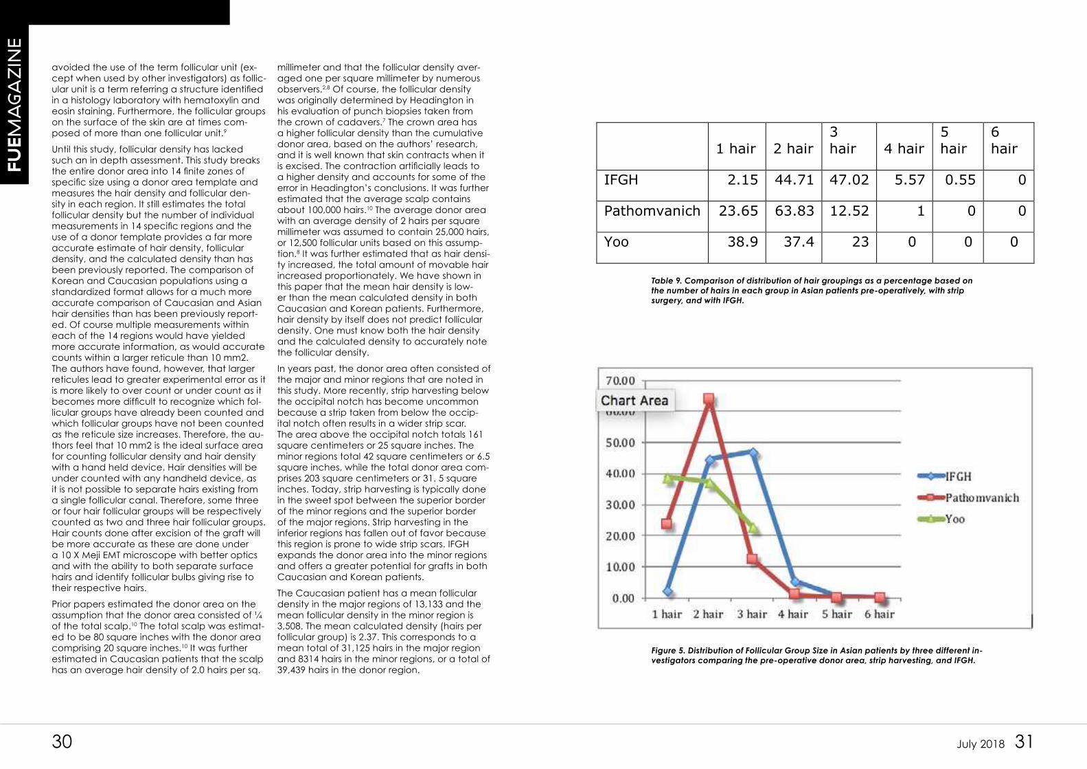
Table 8 notes the distribution of follicular groups by size that were extracted from Korean patients by IFGH in the 30 Korean patients studied. The number of each size was calculated for each of the 30 Korean patients studied and the mean of each size is noted in Table 8.
1 hair 2 hair 3 hair 4 hair 5 hair 6 hair
Percent 2.15 44.71 47.02 5.57 0.55 0
28 29July 2018
FU
EM
AG
AZ
INE
DISCUSSION
Most hair transplant procedures today involve removal of donor tissue from the donor area with a scalpel. The typical donor area runs along the occipital scalp to a point superior to each ear. This line generally ranges from 28 to 30 centimeters. In our experience the mean length was 28 centimeters, but occasionally was up to 30 centimeters. Some physicians ex-tend the line an additional centimeter anterior to the external auditory meatus bilaterally and some make the incision slightly more vertical laterally in an effort to increase the length of this line up to 34 centimeters.4 The typical strip harvest is between one to two centimeters in width.
In this study, the length of the donor area was 28 centimeters, which corresponds to the aver-age length of a strip harvest. Generally speak-ing, the strip incision occurs within the 8 major zones depicted in this study. The eight major zones comprise 161 cm sq. It is felt that one can usually take ½ of this surface area and still have enough hair to cover the resulting strip scar. In other words, it is possible to remove 80.5 square centimeters and still have adequate coverage for the strip scar. Removal of a single strip 28 centimeters long and 1 cm wide removes 28 cm sq, while a 28 cm X 2cm wide strip removes 56 cm sq. The maximal width that can be removed and still leave 80.5 square centime-ters above the incision is 2.875 cm. It may be possible to take as much as 119 square centi-meters, which would leave 42 cm sq above the strip scar and 42 cm sq below the incision scar in the occipital and mastoid regions. This would correspond to a width of 4.2 cm. This would be possible provided there is adequate scalp laxity along with other ideal donor area condi-tions, and the strip scar remains covered. Unfor-tunately, the authors are not familiar with any such endeavor to date. Additional variables to consider with regard to strip scar exposure are the width of the scar, color of the scar, degree of curl, and the caliber of the hair. The width of strip scars is unpredictable. With more and more width excised, strip scars have a tenden-cy to widen up to 0.5 cm in width though some physicians create scars in excess of 1 cm in width. Wider strip scars require a greater num-ber of hairs to conceal them and tend to limit the total amount of donor tissue that can be excised. Coarse hair will generally cover much better than fine hair such that it is easer to conceal a strip scar provided the hair is coarse. Higher hair densities provide more hair mass and make it easier to conceal strip scars. Better hair characteristics and ideal circumstances may some day allow the physician to take up
to 119 square centimeters of donor area, while less favorable characteristics will make it less probable that such widths are possible. The total width of the excision affects the resulting width of the scar. Greater overall widths tend to produce wider scars.
Based on the mean follicular density and the mean calculated density in this study, the aver-age 28 cm X 1 cm strip will produce the follow-ing number of follicular groups and hairs:
Caucasian 2278 FG 5,400 Hairs
Asian 2,095 FG 4,630 Hairs
The following discussion pertains to Caucasians although a similar argument could be gener-ated for Asian patients. The maximal number of grafts than can be obtained in the average patient from 80.5 cm sq is 6,550 and the max-imal number of hairs is 15,524. This probably represents the average maximal number of follicular groups and hairs possible from strip surgery. Graft counts on the other hand are generally higher than the total number of fol-licular groups. This occurs because physicians are rarely involved in the production of their grafts. Surgical technicians with variable levels of experience and training are generally the ones who dissect the strip into individual grafts. Based on numerous studies, experienced tech-nicians produce a mean of 2.0 hairs per graft.3 With the removal of a strip, the total number of available hairs removed does not vary, but the number of grafts produced and the number of hairs transferred varies based on how the grafts are dissected by technicians, who have variable degrees of experience and training. If the average graft contains 2.0 hairs, but the average follicular group contains 2.37 hairs, then 0.37 hairs are removed from each graft. For example, if you have three follicular groups containing a 3, 2, and 3 hair respectively, then the average is 2.67 hairs per group. However if in the dissection process, you turn these three follicular groups into grafts containing 3, 2, 1, and 2 hair respectively, the average becomes 2.0 hairs per graft. This in fact is what happens in many strip surgeries as the percentage of one hair and two hair grafts far exceeds the percentage of 1 and two hair groups in the donor area. The authors term this practice fractionation of the follicular group. Thus, if you assume that you can excise 80.5 cm, un-der acceptable donor circumstances (laxity, density, hair caliber, hair length, wave), then the average strip surgery clinic should produce 7762 grafts averaging 2.0 hairs per graft that total 15,524 hairs. Of course, waste from graft production in strip harvesting is generally not accounted for because physicians do not cut
the grafts, nor do they generally moderate the efficiency of graft production. Therefore, many hairs may be lost due to waste.
The maximal number of grafts from strip surgery ranges between 4500 grafts to 8000 grafts in most strip harvesting clinics. In general, only those with ideal donor laxity and hair charac-teristics can achieve a total of 8000 grafts and it is worth pointing out that it is the scalps with greater laxity that form the widest strip scars. Using the mean follicular density of 81.37 and a length of 28 centimeters, the width of donor area to produce 8000 intact follicular groups is 3.5 cm or 98 cm2 of hair bearing scalp. Suf-fice it to say that not many donor areas can tolerate such a voluminous amount of tissue excised, especially if only a single scar is pro-duced. Most donor areas probably tolerate a hair bearing width between 2.875 cm and 3.0 cm of strip excision in the authors opinion.
Under exceedingly rare circumstances in a patient with average donor characteristics, the total maximum number of follicular groups possible from 119 cm sq is 9569 and the max-imal number of hair is 22,679. This number far exceeds what most physicians have ever achieved in hair restoration surgery. This would leave only 42 cm2 of donor area above the strip scar. The authors are unaware of any strip surgery that has achieved such a width of exci-sion and this number of intact follicular groups.
Knowledge of individual patient characteristics such as their mean follicular density and their mean calculated density would allow the sur-geon to gauge the efficiency of his procedure. For instance, if you produce fewer than 5400 total hairs from a strip of 28 cm2 in a patient with average follicular and calculated densi-ties, then you have waste.
It is often stated among strip surgeons that you cannot produce more grafts from IFGH. It is generally accepted that you can remove up to 50% of a donor area and still have ad-equate coverage.5 If you consider that the average Caucasian donor area contains a mean follicular density of 16,641, one would on average be able to remove 8,321 follicular groups under the 50% rule of thumb. With IFGH you do not need to worry about scalp laxity and you do not need to worry about hiding a strip scar. Thus, you would almost always be able to achieve this number of grafts assuming that it is true that you can remove 50% of the donor density without producing thinness. The range of follicular densities in our 64 Caucasian patient study was 11,025 to 24,115 follicular groups. Accordingly, the range of potential follicular groups possible with IFGH is 5513 to
12,058, assuming you remove only 50% of the follicles. At the time of this publication, the authors are unaware of any patients who have had this many follicular groups extracted from their donor area, however. It is worth emphasiz-ing that in IFGH the mean number of hairs per follicular group is 2.37, while it is 2.0 hairs per graft with strip surgery. Thus, with multiple strip procedures producing between 4500 to 8000 grafts, the average number of hairs possible is 9,000 to 16,000, while with IFGH the potential range of hairs on average is 13,066 to 28,577. The number of potential hairs with IFGH ardent-ly exceeds the capacity of strip surgery and completely avoids the strip scar, the resulting donor area complications, and is independent of scalp laxity. Of course, IFGH averages 2.9 hairs per graft with the first 3000 grafts because it is possible to cherry pick the grafts that con-tain the most hair. In other words, the first 3000 grafts harvested by IFGH will provide up to 8700 hairs on average, whereas, strip harvesting will provide an average of only 6000 hairs from 3000 grafts.
Donor density measurements between observ-ers in both Caucasian and Asian patient pop-ulations have varied over the years based on the investigator and the method of collecting the data. Part of the reason for such variability is a lack of consistency in experimental study design. Many physicians have resorted to an estimation based on phototrichograms.6,15 Such studies are potentially flawed as it is difficult to verify the size of the surface area and it is im-possible to accurately count the hairs in each follicular group. Accurate hair counts depend on a verifiable study area and on hand count-ing each hair in a follicular group. Photographs will always undercount the number of hairs in the follicular groups, as it is impossible to sep-arate hairs in a photograph. Photographs also tend to record a surface area that is larger than the planned area because the photo-graph is taken at an angle to the surface of the skin. Furthermore, the scalp is a curved surface along a three dimensional plane. Photographs are a two dimensional representation of a three dimensional surface.
In 1984, Headington defined the follicular unit as the pilosebaceous unit as disclosed at the mid-dermal level.7 In 1995, Bernstein, et al. de-fined the follicular unit as the cluster of hair as disclosed on the surface of the skin.8 In medi-cine, it is not possible to give two completely different anatomical structures the same name. Therefore, we have more correctly identified the cluster of hair seen on the surface of the skin as the follicular group and completely
30 31July 2018
FU
EM
AG
AZ
INE
avoided the use of the term follicular unit (ex-cept when used by other investigators) as follic-ular unit is a term referring a structure identified in a histology laboratory with hematoxylin and eosin staining. Furthermore, the follicular groups on the surface of the skin are at times com-posed of more than one follicular unit.9
Until this study, follicular density has lacked such an in depth assessment. This study breaks the entire donor area into 14 finite zones of specific size using a donor area template and measures the hair density and follicular den-sity in each region. It still estimates the total follicular density but the number of individual measurements in 14 specific regions and the use of a donor template provides a far more accurate estimate of hair density, follicular density, and the calculated density than has been previously reported. The comparison of Korean and Caucasian populations using a standardized format allows for a much more accurate comparison of Caucasian and Asian hair densities than has been previously report-ed. Of course multiple measurements within each of the 14 regions would have yielded more accurate information, as would accurate counts within a larger reticule than 10 mm2. The authors have found, however, that larger reticules lead to greater experimental error as it is more likely to over count or under count as it becomes more difficult to recognize which fol-licular groups have already been counted and which follicular groups have not been counted as the reticule size increases. Therefore, the au-thors feel that 10 mm2 is the ideal surface area for counting follicular density and hair density with a hand held device. Hair densities will be under counted with any handheld device, as it is not possible to separate hairs existing from a single follicular canal. Therefore, some three or four hair follicular groups will be respectively counted as two and three hair follicular groups. Hair counts done after excision of the graft will be more accurate as these are done under a 10 X Meji EMT microscope with better optics and with the ability to both separate surface hairs and identify follicular bulbs giving rise to their respective hairs.
Prior papers estimated the donor area on the assumption that the donor area consisted of ¼ of the total scalp.10 The total scalp was estimat-ed to be 80 square inches with the donor area comprising 20 square inches.10 It was further estimated in Caucasian patients that the scalp has an average hair density of 2.0 hairs per sq.
millimeter and that the follicular density aver-aged one per square millimeter by numerous observers.2,8 Of course, the follicular density was originally determined by Headington in his evaluation of punch biopsies taken from the crown of cadavers.7 The crown area has a higher follicular density than the cumulative donor area, based on the authors’ research, and it is well known that skin contracts when it is excised. The contraction artificially leads to a higher density and accounts for some of the error in Headington’s conclusions. It was further estimated that the average scalp contains about 100,000 hairs.10 The average donor area with an average density of 2 hairs per square millimeter was assumed to contain 25,000 hairs, or 12,500 follicular units based on this assump-tion.8 It was further estimated that as hair densi-ty increased, the total amount of movable hair increased proportionately. We have shown in this paper that the mean hair density is low-er than the mean calculated density in both Caucasian and Korean patients. Furthermore, hair density by itself does not predict follicular density. One must know both the hair density and the calculated density to accurately note the follicular density.
In years past, the donor area often consisted of the major and minor regions that are noted in this study. More recently, strip harvesting below the occipital notch has become uncommon because a strip taken from below the occip-ital notch often results in a wider strip scar. The area above the occipital notch totals 161 square centimeters or 25 square inches. The minor regions total 42 square centimeters or 6.5 square inches, while the total donor area com-prises 203 square centimeters or 31. 5 square inches. Today, strip harvesting is typically done in the sweet spot between the superior border of the minor regions and the superior border of the major regions. Strip harvesting in the inferior regions has fallen out of favor because this region is prone to wide strip scars. IFGH expands the donor area into the minor regions and offers a greater potential for grafts in both Caucasian and Korean patients.
The Caucasian patient has a mean follicular density in the major regions of 13,133 and the mean follicular density in the minor region is 3,508. The mean calculated density (hairs per follicular group) is 2.37. This corresponds to a mean total of 31,125 hairs in the major region and 8314 hairs in the minor regions, or a total of 39,439 hairs in the donor region.
1 hair 2 hair 3 hair 4 hair
5 hair
6 hair
IFGH 2.15 44.71 47.02 5.57 0.55 0
Pathomvanich 23.65 63.83 12.52 1 0 0
Yoo 38.9 37.4 23 0 0 0
Table 9. Comparison of distribution of hair groupings as a percentage based on
the number of hairs in each group in Asian patients pre-operatively, with strip
surgery, and with IFGH.
Figure 5. Distribution of Follicular Group Size in Asian patients by three different in-
vestigators comparing the pre-operative donor area, strip harvesting, and IFGH.
32 33July 2018
FU
EM
AG
AZ
INE
In the Korean patient, the mean follicular density in the major regions is 12,527 and the mean follicular density in the minor region is 3191. The mean calculated density (hairs per follicular group) is 2.21. This computes to a mean total of 27,685 hairs in the major region and 7,052 hairs in the minor regions or a total of 34, 737 hairs in the donor region. Prior stud-ies in Korean patients indicated a potential between 10,375 and 15, 250 hairs based on patient age.
The mean number of hairs per follicular group in Korean patients undergoing IFGH was shown to be much higher in this study than in previous studies. Pathomvanich found the distribution of follicular units in 30 Asian patients undergoing strip harvesting to be 23.65% 1 hair FU, 63.82% 2 hair FU, 12.52% 3 hair FU, and 0-1% 4 hair FU.11 The 30 Korean patients studied in this paper revealed a much lower percentage of 1 hair FU and a much higher percentage of 3 hair FU and 4 hair FU. In addition rare 5 hair FU were noted. Dr. Yoo found that the occipital region of young male Koreans with androgenic alo-pecia had a follicular distribution of 38.9% 1 hair, 37.4% 2 hair, and 23% 3 hair groups.6 A comparison of IFGH, Dr. Pathomvanich, and Dr. Yoo is depicted in Table 9 and Figure 4. In a previous study of Korean patients Lee found the following distribution with IFGH: 1.5% 1hair, 37.2% 2 hair, 51.7% 3hair, 9.3% 4 hair, 0.4% 4 hair, and 0% 6 hair.3 Clearly, more data is needed to more accurately predict the overall mean with IFGH in Korean patients, but the data continues to confirm that IFGH offers more hair per graft than strip surgery in Asian patients.
Prior studies looking at the mean follicular density in Caucasians by Limmer noted 0.9/ mm2, Bernstein and Rassman 1.0/mm2, and Cole 0.8/mm2.9,8,12. Prior literature in Korean patients suggested a range of 0.55 to 0.64 per mm2 depending on the age of the pa-tient.13 In this study the follicular density based on the total surface area of 203 square cm was 0.81 per mm2 in Caucasian and 0.75 per mm2 in Asian patients. This represents a much more similar follicular density than has been previously reported between Caucasian and Korean patients. Clearly, data in this study far exceeds the estimates previously performed
and shows that Koreans have far more hair in their donor regions than has been previously reported. Koreans are far better candidates for hair transplanted surgery than has been previously reported based on this study.
Prior studies showed that Korean hair densi-ties ranged from 0.83 to 1.22 hair per mm2.13 In this study we found the mean hair density in Asian patients was 1.65 per mm2. Bern-stein and Rassman suggested the mean hair density was 2.0 hair per mm2. In this study, the Caucasian hair density was 1.93 hairs per mm2.
The average number of hairs per graft with strip surgery in the Korean patient is 1.9 hairs.14 In IFGH the average number of hairs per graft is 2.53 in Korean patients. IFGH is particularly suited for the Korean patient as IFGH provides 0.62 more hairs per graft than does strip surgery. This results in a better value to the Korean patient undergoing FUE and greater potential coverage with an equal number of grafts.
Previous studies have shown that the survival rates for IFGH are similar to those in strip har-vesting and that transection rates with IFGH are generally less than in strip harvesting.3 Cumulatively, the data overwhelmingly sup-ports IFGH as the method of choice in donor harvesting.
CONCLUSION
The authors have shown in numerous previ-ous studies that the mean Caucasian fol-licular density is approximately 80 follicular groups per square centimeter. This study con-firms the mean follicular density is similar to the authors’ previous findings. Using a similar protocol, this study showed that the Korean hair density, follicular density, and calculat-ed density are much higher than previously reported. In addition, this study shows that IFGH can produce a much higher percent-age of grafts containing more than one hair in Korean patients. The results document that IFGH provides a much better solution for the Korean patient undergoing hair restoration surgery as the average IFGH graft contains 0.62 more hairs per graft than strip surgery offers to Korean patients.
BIBLIOGRAPHY
1. Unger W, Cole J, Donor Harvesting, Hair Transplantation. W.P. Unger and R.M. Sha-piro, eds. Marcel Dekker: New York, 2004; 307-308.
2. Rassman, WR, et al. Follicuar unit extraction: minimally invasive surgery for hair transplantation. Dermatol Surg. 202: 28(8):720-27
3. Cole, JP, Status of individual follicular group harvesting, Hair Transplant Forum Inter-national, Jan/Feb 2009; 20-24.
4. Tykocinski, A, personal communication February 2009
5. Marritt E. The death of the density debate. Dermatol Surg 1999;25:654-660.
6. Yoo JH, Analysis of hair characteristics in Koreans using phototrichograms, Hair Transplantation. W.P. Unger and R.M. Shapiro, eds. Marcel Dekker: New York, 2004; 892-897.
7. Headington JT. Transverse microscopic anatomy of the human scalp. Arch Derma-tol 1984:120:449-456.
8. Bernstein, RM, Rassman, WR, Szaniawski, W, Halperin, A, Follicular Transplantation, International Journal of Aesthetic and Restorative Surgery, Vol 3, 2, 1995, pp 119-132.
9. Cole JP. Mathematics of follicular transplantation, ISHRS, Washington, DC 1998.
10. Rassman WR, Carson S, Micrografting in Extensive Quantities, Dermatologic Sur-gery, Vol 21, 4, April 1995, pp 306-311.
11. Pathomvanich, D, Hair Replacement in Asians, Stough, D, Haber, B. (eds) pp 149-156.
12. Limmer BL. Personal communication 1998.
13. Korea Dermatology Society, 43.
14. Kuelachi, M. Review of the asian hair surgery workshop. Hair Transplant Forum int’l. 17(3):94-95.
15. Neidel, F, Bretschneider, P, Measuring hair density and mass, Hair Transplantation. W.P.Unger and R.M. Shapiro, eds. Marcel Dekker: New York 2004; 876-886
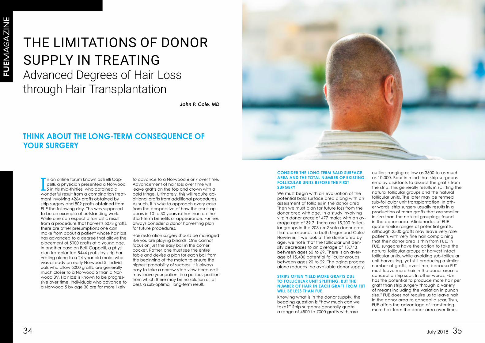
34 35July 2018
FU
EM
AG
AZ
INE
THE LIMITATIONS OF DONOR SUPPLY IN TREATING Advanced Degrees of Hair Loss through Hair Transplantation
John P. Cole, MD
THINK ABOUT THE LONG-TERM CONSEQUENCE OF YOUR SURGERY
In an online forum known as Belli Cap-pelli, a physician presented a Norwood 5 in his mid-thirties, who obtained a
wonderful result from a combination treat-ment involving 4264 grafts obtained by strip surgery and 809 grafts obtained from FUE the following day. This was supposed to be an example of outstanding work. While one can expect a fantastic result from a procedure that harvests 5073 grafts, there are other presumptions one can make from about a patient whose hair loss has advanced to a degree that allows the placement of 5000 grafts at a young age. In another case on Belli Cappelli, a physi-cian transplanted 5444 grafts by strip har-vesting alone to a 24-year-old male, who was already an early Norwood 5. Individ-uals who allow 5000 grafts, are generally much closer to a Norwood 5 than a Nor-wood 3V. Hair loss is known to be progres-sive over time. Individuals who advance to a Norwood 5 by age 30 are far more likely
to advance to a Norwood 6 or 7 over time. Advancement of hair loss over time will leave grafts on the top and crown with a bald fringe. Ultimately, this will require ad-ditional grafts from additional procedures. As such, it is wise to approach every case from the perspective of how the result ap-pears in 10 to 30 years rather than on the short-term benefits or appearance. Further, always consider a donor harvesting plan for future procedures.
Hair restoration surgery should be managed like you are playing billiards. One cannot focus on just the easy ball in the corner pocket. Rather, one must see the entire table and devise a plan for each ball from the beginning of the match to ensure the highest probability of success. It is always easy to take a narrow-sited view because it may leave your patient in a perilous position from which there may be no solution or, at best, a sub-optimal, long-term result.
CONSIDER THE LONG TERM BALD SURFACE AREA AND THE TOTAL NUMBER OF EXISTING FOLLICULAR UNITS BEFORE THE FIRST SURGERY
We must begin with an evaluation of the potential bald surface area along with an assessment of follicles in the donor area. Then we must plan for future loss from the donor area with age. In a study involving virgin donor areas of 477 males with an av-erage age of 39.7, there are 15,200 follicu-lar groups in the 203 cm2 safe donor area that corresponds to both Unger and Cole.1 However, if we look at the donor area by age, we note that the follicular unit den-sity decreases to an average of 13,743 between ages 60 to 69. There is an aver-age of 15,400 potential follicular groups between ages 20 to 29. The aging process alone reduces the available donor supply.
STRIPS OFTEN YIELD MORE GRAFTS DUE TO FOLLICULAR UNIT SPLITTING, BUT THE NUMBER OF HAIR IN EACH GRAFT FROM FUT WILL BE LESS THAN FUE
Knowing what is in the donor supply, the begging question is “how much can we take?” Strip surgeons generally quote a range of 4500 to 7000 grafts with rare
outliers ranging as low as 3500 to as much as 10,000. Bear in mind that strip surgeons employ assistants to dissect the grafts from the strip. This generally results in splitting the natural follicular groups and the natural follicular units. The later may be termed sub-follicular unit transplantation. In oth-er words, strip surgery usually results in a production of more grafts that are smaller in size than the natural groupings found in the donor area. Aficionados of FUE quote similar ranges of potential grafts, although 2500 grafts may leave very rare patients with very fine hair complaining that their donor area is thin from FUE. In FUE, surgeons have the option to take the natural follicular groups or harvest intact follicular units, while avoiding sub-follicular unit harvesting, yet still producing a similar number of grafts, over time, because FUT must leave more hair in the donor area to conceal a strip scar. In other words, FUE has the potential to produce more hair per graft than strip surgery through a variety of means including the variation in punch size.2 FUE does not require us to leave hair in the donor area to conceal a scar. Thus, FUE offers the advantage of transferring more hair from the donor area over time.
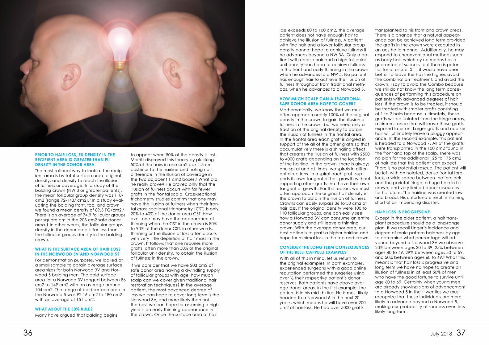
36 37July 2018
FU
EM
AG
AZ
INE
PRIOR TO HAIR LOSS, FU DENSITY IN THE RECIPIENT AREA IS GREATER THAN FU DENSITY IN THE DONOR AREA
The most rational way to look at the recip-ient area is by total surface area, original density, and density to reach the illusion of fullness or coverage. In a study of the balding crown (NW 3 or greater patients), the mean follicular group density was 110/ cm2 (range 72-143/ cm2).3 In a study eval-uating the balding front, top, and crown we found a mean density of 89.3 FG/cm2.4 There is an average of 74.9 follicular groups per square cm in the 203 cm2 safe donor area.1 In other words, the follicular groups density in the donor area is far less than the follicular groups density in the balding crown.
WHAT IS THE SURFACE AREA OF HAIR LOSS IN THE NORWOOD 3V AND NORWOOD 5?
For demonstration purposes, we looked at a small sample to obtain average surface area sizes for both Norwood 3V and Nor-wood 5 balding men. The bald surface area for a Norwood 3V ranged between 86 cm2 to 149 cm2 with an average around 104 cm2. The range of bald surface area in the Norwood 5 was 92.16 cm2 to 180 cm2 with an average of 151 cm2.
WHAT ABOUT THE 50% RULE?
Many have argued that balding begins
to appear when 50% of the density is lost. Marritt disproved this theory by plucking 50% of the hairs in one cm2 box 1.5 cm posterior to the hairline and noting no difference in the illusion of coverage in the two adjacent 1 cm2 boxes.5 What did he really prove? He proved only that the illusion of fullness occurs with far fewer grafts in the frontal scalp. Cross-sectional trichometry studies confirm that one may have the illusion of fullness when their fron-tal cross-sectional trichometry (CST) is only 20% to 40% of the donor area CST. How-ever, one may have the appearance of thinning when the CST in the crown is 80% to 90% of the donor CST. In other words, thinning or the illusion of loss often occurs with very little depletion of hair mass in the crown. It follows that one requires more grafts, often more than 50% of the original follicular unit density, to obtain the illusion of fullness in the crown.
If we consider that we have 203 cm2 of safe donor area having a dwindling supply of follicular groups with age, how much scalp can we cover given traditional hair restoration techniques? In the average patient, the most advanced degree of loss we can hope to cover long term is the Norwood 3V, and more likely than not, the best we can hope for assuming a high yield is an early thinning appearance in the crown. Once the surface area of hair
loss exceeds 80 to 100 cm2, the average patient does not have enough hair to achieve the illusion of fullness. A patient with fine hair and a lower follicular group density cannot hope to achieve fullness if he advances beyond a NW 3A. Only a pa-tient with coarse hair and a high follicular unit density can hope to achieve fullness in the front and early thinning in the crown when he advances to a NW 5. No patient has enough hair to achieve the illusion of fullness throughout from traditional meth-ods, when he advances to a Norwood 5.
HOW MUCH SCALP CAN A TRADITIONAL SAFE DONOR AREA HOPE TO COVER?
Mathematically, we know that we must often approach nearly 100% of the original density in the crown to gain the illusion of fullness in the crown, but we need only a fraction of the original density to obtain the illusion of fullness in the frontal area. In the frontal area each graft is angled in support of the all of the other grafts so that accumulatively there is a shingling affect that creates the illusion of fullness with 2500 to 4000 grafts depending on the location of the hairline. In the crown, there is always one spiral and at times two spirals in differ-ent directions. In a spiral each graft sup-ports its own tangent of hair growth without supporting other grafts that have their own tangent of growth. For this reason, we must often approach the original hair density in the crown to obtain the illusion of fullness. Crowns can easily surpass 36 to 50 cm2 of hair loss. If the original density were 100 to 110 follicular groups, one can easily see how a Norwood 3V can consume an entire donor supply and still leave a thinning crown. With the average donor area, our best option is to graft a higher hairline and hope for minimal loss in the top and crown.
CONSIDER THE LONG TERM CONSEQUENCES OF THE BELLI CAPPELLI EXAMPLES
With all of this in mind, let us return to the original examples. In both examples, experienced surgeons with a good online reputation performed the surgeries using over ½ their respective patient’s donor reserves. Both patients have above aver-age donor areas. In the first example, the patient is in his mid-thirties. He is most likely headed to a Norwood 6 in the next 20 years, which means he will have over 200 cm2 of hair loss. He had over 5000 grafts
transplanted to his front and crown areas. There is a chance that a natural appear-ance can be achieved long term provided the grafts in the crown were executed in an aesthetic manner. Additionally, he may respond to unconventional methods such as body hair, which by no means has a guarantee of success, but there is poten-tial for a rescue. Still, it would have been better to leave the hairline higher, avoid the combination treatment, and avoid the crown. I say to avoid the Combo because we still do not know the long term conse-quences of performing this procedure on patients with advanced degrees of hair loss. If the crown is to be treated, it should be treated with smaller grafts consisting of 1 to 2 hairs because, ultimately, these grafts will be isolated from the fringe areas, a circumstance that will leave these grafts exposed later on. Larger grafts and coarser hair will ultimately leave a pluggy appear-ance. In the second example, this patient is headed to a Norwood 7. All of the grafts were transplanted in the 100 cm2 found in the front and top of the scalp. There was no plan for the additional 125 to 175 cm2 of hair loss that this patient can expect. There is no potential rescue. The patient will be left with an isolated, dense frontal fore-lock, a wide space between the forelock and the parietal fringe, a huge hole in his crown, and very limited donor resources for his future. The hairline was created low and broad. His unfortunate result is nothing short of an impending disaster.
HAIR LOSS IS PROGRESSIVE
Except in the older patient, a hair trans-plant procedure should be a long-range plan. If we recall Unger’s incidence and degree of male pattern baldness by age to determine what percentage will ad-vance beyond a Norwood 3V we observe 20% between ages 30 to 39, 25% between ages 40 to 49, 29% between ages 50 to 59, and 50% between ages 60 to 69.6 What this means is that hair loss is progressive and long term we have no hope to create an illusion of fullness in at least 50% of men who have the good fortune to survive until age 60 to 69. Certainly when young men are already showing signs of advancement to a Norwood 5 in their twenties we must recognize that these individuals are more likely to advance beyond a Norwood 5, making our probability of success even less likely long term.
38 39July 2018
FU
EM
AG
AZ
INE
THE BEST SOLUTION FOR ADVANCED DEGREES OF HAIR LOSS
What should we do and what should we avoid to improve our chances of long-term success? It is known that FUE results in a mean contraction of the donor surface area of 13% and a combination of FUE and strip surgery (Combo therapy) results in a 50% wider scar than strip surgery alone.4 The wider scar most likely results from additional strip wound tension resulting from the donor contraction secondary to the FUE. Addition-al tension and a wider scar in an initial strip surgery suggest that the subsequent surgery will result in an even wider scar. We just do not know at this point so Combo therapy is not an optimal solution in the young patient headed to an advanced degree of hair loss. Regardless, we know that multiple strip surgeries have a propensity to create wider scars. Wider scars require more hair to con-ceal the donor scars. We know from studies that the average CST in donor area is 70, but after strip surgeries the mean donor CST is 52, while the mean CST following FUE is 69.7 My theory is that wound tension from the strip results in traction alopecia in the donor area. Traction alopecia along with a decrease in skin surface area from strips causes a far greater decrease in the CST
from strip surgery than from FUE. Combina-tion of strips and FUE simultaneously further rob the donor area of follicles that may be required to conceal a strip scar later in life. For this reason, strips and combo therapy are not a good idea in younger patients, who very well may advance to a great-er degree of hair loss, or who are already showing signs of advanced degrees of hair loss. In this instance, you are simply setting yourself and your patient up for failure long term. What about FUE? The numbers do not change, but the future hairstyle options are much better. Hair transplant surgery is not a great idea in the young patient destined to severe degrees of hair loss, but we do know that we can perform micropigmenta-tion on hypopigemented extraction sites to produce a more aesthetic donor result that allows us to shave the scalp. In other words, the potential to rescue an advanced de-gree of hair loss is greater with FUE. Still, we must acknowledge that FUE, in the hands of inexperienced physicians, can result in high follicle transection rates that potentially reduce FUE yield.
TRANSPLANTS TEND TO THIN OVER TIME
Anyone who has done hair transplants long enough has seen that results tend to thin
over time, especially in advanced degrees of hair loss. Otar Norwood recognized that the plugs on his Norwood 6 pattern of loss progressing to a Norwood 7 pattern appeared more natural over time due to thinning of his plugs. One theory is that separation of the primary follicle from the secondary follicle can result in premature thinning, but with plugs, we generally did not separate the natural follicular units. Thinning can result in a rescue of sorts as in Dr. Norwood’s case, but it can also create even more dramatic problems long term when we try to treat advanced degrees of hair loss in the young patient.
One may throw a monkey in the mix by suggesting medical therapy such as Pro-pecia. However, we cannot treat patients surgically under the premise that they will always remain on Propecia, that they may always respond to Propecia, that they will not eventually develop unacceptable side effects, that they will not stop Prope-cia in preparation to start a family, or that they will not switch to generic Propecia, which we know often results in treatment failures. According to Wolf, the purity of generics is often only 80%, which means a 1 mg generic finasteride may have only 0.8 mg of active drug.8
THE BEST LONG TERM PLAN
In summary, the donor capacity for the vast majority of men is suitable for 100 cm2 of hair loss. Hair loss to this extent or great-er will require maximal donor area harvest-
ing. Maximal donor area harvesting more commonly results in wider strip scars, which require more donor area hair to conceal them. The total number of follicular groups decreases with age naturally, but donor density decreases even more as a result of strip harvesting, most likely secondary to traction alopecia. A combination of strip and FUE simultaneously results in a 50% increase in scar width. A combination of FUE and strip surgery at any time fur-ther depletes the donor area of follicular groups that may be necessary to conceal wider strip scars. While FUE may allow for more potential grafts and a better aes-thetic result, there still is not enough hair to treat advanced degrees of hair loss without resorting to nontraditional means that have a variable degree of success. All of these factors must play a role in the long-term planning for surgical restoration. Obviously, it is wise for patients to consider supplementing surgical therapy with med-ical therapy, but we cannot presume that patients will remain on effective medical therapy for a lifetime. Therefore, a 20 year or more plan should focus on what we can achieve through surgical restoration alone. This plan should include a focus on methods that minimize scarring while maximizing donor area yields to produce an aesthetically pleasing result that will en-dure for decades.
1. Cole, J., Donor Hair Mapping, in Italian Society of Hair Restoration Surgery. 2014: Siracusia.
2. Cole, J.P., Status of Individual Follicular Group Harvesting. Hair Transplant Forum Interna-tional, 2009. January/February: p. 20-24.
3. Cole, J.P., Regional Variation in Donor Density and Hair Diameter, in ISHRS Meeting. 2000.
4. Cole J, I.C., Toscani, M. , Donor area response to FUE, in ISHRS annual meeting. 2015: Chicago, Illinois.
5. Marritt, E., The death of the density debate. Dermatol Surg, 1999. 25(8): p. 654-60.
6. W.P, U., The incidence and degree of MPB and FPHL at various ages in men and women. 5 ed. 2011.
7. Cole, J., Cross Sectional Trichometry, in ISHRS Annual Conference. 2013: San Francisco.
8. Wolf, B., A top ten list on generics: interesting inside information. personal communica-tion 8-4-2015.
REFERENCES
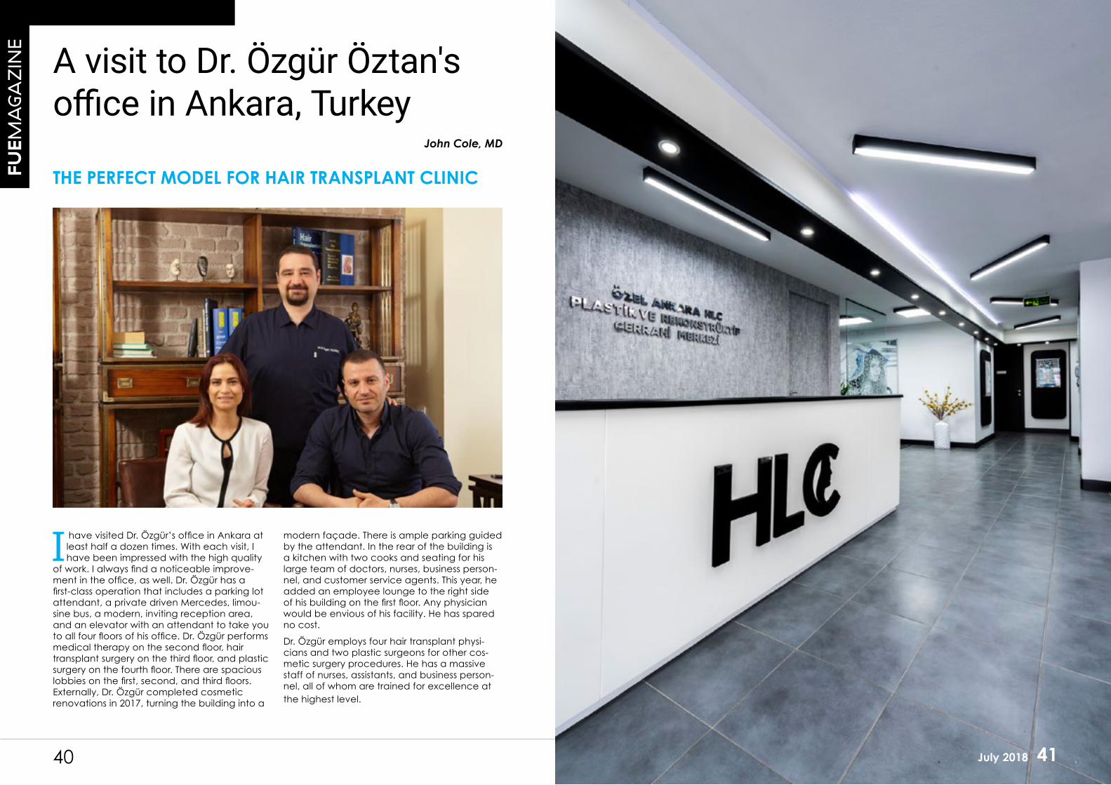
40
FU
EM
AG
AZ
INE
41July 2018
A visit to Dr. Özgür Öztan's office in Ankara, Turkey
John Cole, MD
THE PERFECT MODEL FOR HAIR TRANSPLANT CLINIC
I have visited Dr. Özgür’s office in Ankara at least half a dozen times. With each visit, I have been impressed with the high quality
of work. I always find a noticeable improve-ment in the office, as well. Dr. Özgür has a first-class operation that includes a parking lot attendant, a private driven Mercedes, limou-sine bus, a modern, inviting reception area, and an elevator with an attendant to take you to all four floors of his office. Dr. Özgür performs medical therapy on the second floor, hair transplant surgery on the third floor, and plastic surgery on the fourth floor. There are spacious lobbies on the first, second, and third floors. Externally, Dr. Özgür completed cosmetic renovations in 2017, turning the building into a
modern façade. There is ample parking guided by the attendant. In the rear of the building is a kitchen with two cooks and seating for his large team of doctors, nurses, business person-nel, and customer service agents. This year, he added an employee lounge to the right side of his building on the first floor. Any physician would be envious of his facility. He has spared no cost.
Dr. Özgür employs four hair transplant physi-cians and two plastic surgeons for other cos-metic surgery procedures. He has a massive staff of nurses, assistants, and business person-nel, all of whom are trained for excellence at
the highest level.
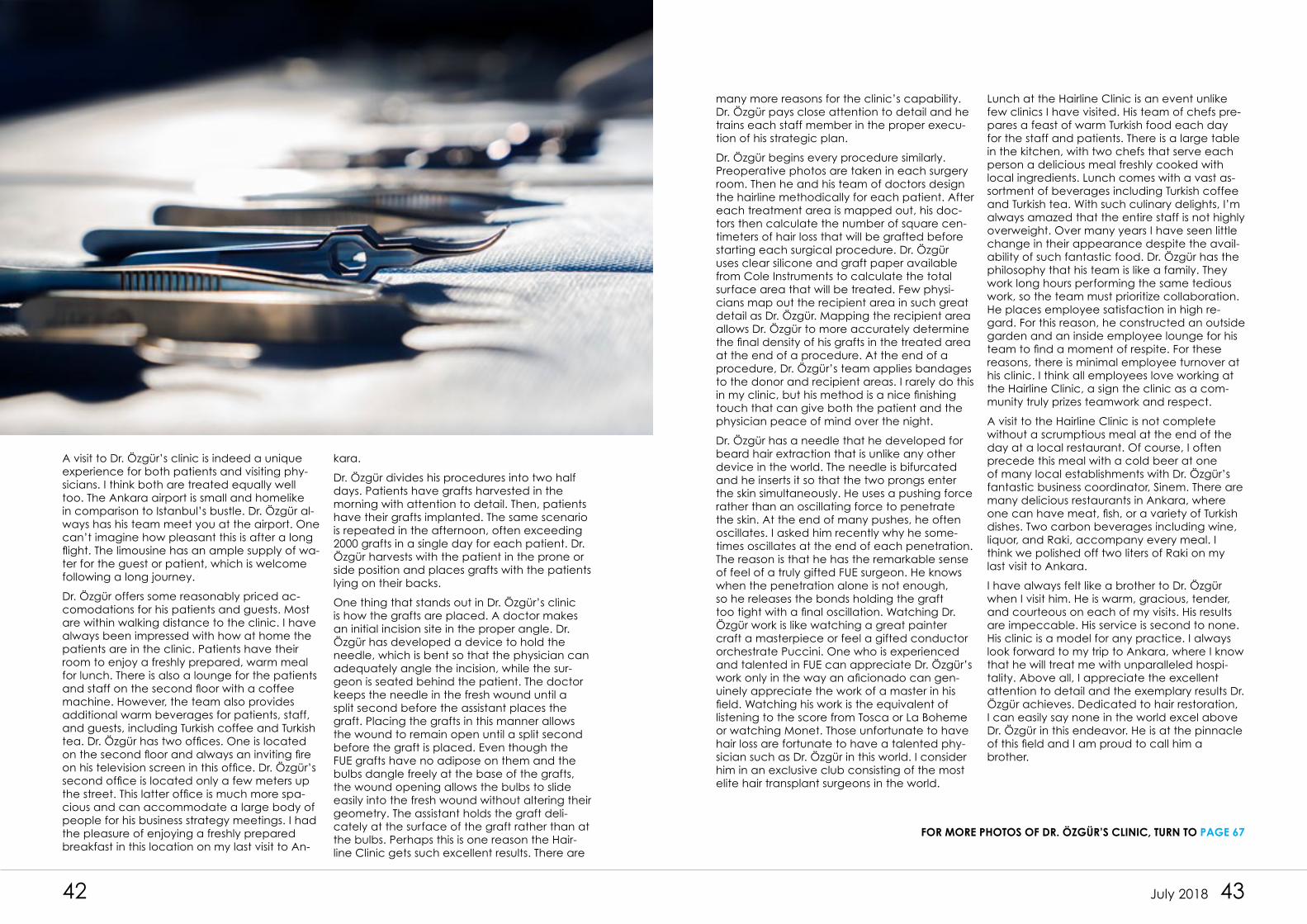
42 43July 2018
FU
EM
AG
AZ
INE
A visit to Dr. Özgür’s clinic is indeed a unique experience for both patients and visiting phy-sicians. I think both are treated equally well too. The Ankara airport is small and homelike in comparison to Istanbul’s bustle. Dr. Özgür al-ways has his team meet you at the airport. One can’t imagine how pleasant this is after a long flight. The limousine has an ample supply of wa-ter for the guest or patient, which is welcome following a long journey.
Dr. Özgür offers some reasonably priced ac-comodations for his patients and guests. Most are within walking distance to the clinic. I have always been impressed with how at home the patients are in the clinic. Patients have their room to enjoy a freshly prepared, warm meal for lunch. There is also a lounge for the patients and staff on the second floor with a coffee machine. However, the team also provides additional warm beverages for patients, staff, and guests, including Turkish coffee and Turkish tea. Dr. Özgür has two offices. One is located on the second floor and always an inviting fire on his television screen in this office. Dr. Özgür’s second office is located only a few meters up the street. This latter office is much more spa-cious and can accommodate a large body of people for his business strategy meetings. I had the pleasure of enjoying a freshly prepared breakfast in this location on my last visit to An-
kara.
Dr. Özgür divides his procedures into two half days. Patients have grafts harvested in the morning with attention to detail. Then, patients have their grafts implanted. The same scenario is repeated in the afternoon, often exceeding 2000 grafts in a single day for each patient. Dr. Özgür harvests with the patient in the prone or side position and places grafts with the patients lying on their backs.
One thing that stands out in Dr. Özgür’s clinic is how the grafts are placed. A doctor makes an initial incision site in the proper angle. Dr. Özgür has developed a device to hold the needle, which is bent so that the physician can adequately angle the incision, while the sur-geon is seated behind the patient. The doctor keeps the needle in the fresh wound until a split second before the assistant places the graft. Placing the grafts in this manner allows the wound to remain open until a split second before the graft is placed. Even though the FUE grafts have no adipose on them and the bulbs dangle freely at the base of the grafts, the wound opening allows the bulbs to slide easily into the fresh wound without altering their geometry. The assistant holds the graft deli-cately at the surface of the graft rather than at the bulbs. Perhaps this is one reason the Hair-line Clinic gets such excellent results. There are
many more reasons for the clinic’s capability. Dr. Özgür pays close attention to detail and he trains each staff member in the proper execu-tion of his strategic plan.
Dr. Özgür begins every procedure similarly. Preoperative photos are taken in each surgery room. Then he and his team of doctors design the hairline methodically for each patient. After each treatment area is mapped out, his doc-tors then calculate the number of square cen-timeters of hair loss that will be grafted before starting each surgical procedure. Dr. Özgür uses clear silicone and graft paper available from Cole Instruments to calculate the total surface area that will be treated. Few physi-cians map out the recipient area in such great detail as Dr. Özgür. Mapping the recipient area allows Dr. Özgür to more accurately determine the final density of his grafts in the treated area at the end of a procedure. At the end of a procedure, Dr. Özgür’s team applies bandages to the donor and recipient areas. I rarely do this in my clinic, but his method is a nice finishing touch that can give both the patient and the physician peace of mind over the night.
Dr. Özgür has a needle that he developed for beard hair extraction that is unlike any other device in the world. The needle is bifurcated and he inserts it so that the two prongs enter the skin simultaneously. He uses a pushing force rather than an oscillating force to penetrate the skin. At the end of many pushes, he often oscillates. I asked him recently why he some-times oscillates at the end of each penetration. The reason is that he has the remarkable sense of feel of a truly gifted FUE surgeon. He knows when the penetration alone is not enough, so he releases the bonds holding the graft too tight with a final oscillation. Watching Dr. Özgür work is like watching a great painter craft a masterpiece or feel a gifted conductor orchestrate Puccini. One who is experienced and talented in FUE can appreciate Dr. Özgür’s work only in the way an aficionado can gen-uinely appreciate the work of a master in his field. Watching his work is the equivalent of listening to the score from Tosca or La Boheme or watching Monet. Those unfortunate to have hair loss are fortunate to have a talented phy-sician such as Dr. Özgür in this world. I consider him in an exclusive club consisting of the most elite hair transplant surgeons in the world.
Lunch at the Hairline Clinic is an event unlike few clinics I have visited. His team of chefs pre-pares a feast of warm Turkish food each day for the staff and patients. There is a large table in the kitchen, with two chefs that serve each person a delicious meal freshly cooked with local ingredients. Lunch comes with a vast as-sortment of beverages including Turkish coffee and Turkish tea. With such culinary delights, I’m always amazed that the entire staff is not highly overweight. Over many years I have seen little change in their appearance despite the avail-ability of such fantastic food. Dr. Özgür has the philosophy that his team is like a family. They work long hours performing the same tedious work, so the team must prioritize collaboration. He places employee satisfaction in high re-gard. For this reason, he constructed an outside garden and an inside employee lounge for his team to find a moment of respite. For these reasons, there is minimal employee turnover at his clinic. I think all employees love working at the Hairline Clinic, a sign the clinic as a com-munity truly prizes teamwork and respect.
A visit to the Hairline Clinic is not complete without a scrumptious meal at the end of the day at a local restaurant. Of course, I often precede this meal with a cold beer at one of many local establishments with Dr. Özgür’s fantastic business coordinator, Sinem. There are many delicious restaurants in Ankara, where one can have meat, fish, or a variety of Turkish dishes. Two carbon beverages including wine, liquor, and Raki, accompany every meal. I think we polished off two liters of Raki on my last visit to Ankara.
I have always felt like a brother to Dr. Özgür when I visit him. He is warm, gracious, tender, and courteous on each of my visits. His results are impeccable. His service is second to none. His clinic is a model for any practice. I always look forward to my trip to Ankara, where I know that he will treat me with unparalleled hospi-tality. Above all, I appreciate the excellent attention to detail and the exemplary results Dr. Özgür achieves. Dedicated to hair restoration, I can easily say none in the world excel above Dr. Özgür in this endeavor. He is at the pinnacle of this field and I am proud to call him a brother.
FOR MORE PHOTOS OF DR. ÖZGÜR’S CLINIC, TURN TO PAGE 67
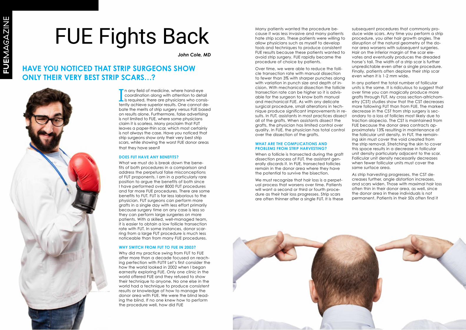
45July 2018
FU
EM
AG
AZ
INE
FUE Fights Back John Cole, MD
HAVE YOU NOTICED THAT STRIP SURGEONS SHOW ONLY THEIR VERY BEST STRIP SCARS…?
In any field of medicine, where hand-eye coordination along with attention to detail is required, there are physicians who consis-
tently achieve superior results. One cannot de-bate the merits of strip surgery versus FUE based on results alone. Furthermore, false advertising is not limited to FUE, where some physicians claim it is scarless. Many proclaim that FUT leaves a paper-thin scar, which most certainly is not always the case. Have you noticed that strip surgeons show only their very best strip scars, while showing the worst FUE donor areas
that they have seen?
DOES FUT HAVE ANY BENEFITS?
What we must do is break down the bene-fits of both procedures in a comparison and address the perpetual false misconceptions of FUT proponents. I am in a particularly rare position to argue the benefits of both since I have performed over 8000 FUT procedures and far more FUE procedures. There are some benefits to FUT. FUT is far less laborious to the physician. FUT surgeons can perform more grafts in a single day with less effort primarily because surgery time on any case is less so they can perform large surgeries on more patients. With a skilled, well-managed team, it is easier to obtain a low follicle transection rate with FUT. In some instances, donor scar-ring from a large FUT procedure is much less noticeable than from many FUE procedures.
WHY SWITCH FROM FUT TO FUE IN 2003?
Why did my practice swing from FUT to FUE after more than a decade focused on reach-ing perfection with FUT? Let’s first consider the how the world looked in 2002 when I began earnestly exploring FUE. Only one clinic in the world offered FUE and they refused to show their technique to anyone. No one else in the world had a technique to produce consistent results or knowledge of how to manage the donor area with FUE. We were the blind lead-ing the blind. If no one knew how to perform the procedure well, how did FUE
Many patients wanted the procedure be-cause it was less invasive and many patients hate strip scars. These patients were willing to allow physicians such as myself to develop tools and techniques to produce consistent FUE results because these patients wanted to avoid strip surgery. FUE rapidly became the procedure of choice by patients.
Over time, we were able to reduce the folli-cle transection rate with manual dissection to fewer than 3% with sharper punches along with variation in punch size and depth of in-cision. With mechanical dissection the follicle transection rate can be higher so it is advis-able for the surgeon to know both manual and mechanical FUE. As with any delicate surgical procedure, small alterations in tech-nique produce significant improvements in re-sults. In FUT, assistants in most practices dissect all of the grafts. When assistants dissect the grafts, the physician has limited control over quality. In FUE, the physician has total control over the dissection of the grafts.
WHAT ARE THE COMPLICATIONS AND PROBLEMS FROM STRIP HARVESTING?
When a follicle is transected during the graft dissection process of FUT, the assistant gen-erally discards it. In FUE, transected follicles remain in the donor area where they have the potential to survive the bisection.
We must recognize that hair loss is a perpet-ual process that worsens over time. Patients will want a second or third or fourth proce-dure as their hair loss progresses. Strip scars are often thinner after a single FUT. It is these
subsequent procedures that commonly pro-duce wide scars. Any time you perform a strip procedure, you alter hair growth angles. The disruption of the natural geometry of the do-nor area worsens with subsequent surgeries. Hair on the inferior margin of the scar ele-vates and eventually produces the dreaded horse’s tail. The width of a strip scar is further unpredictable even after a single procedure. Finally, patients often deplore their strip scar even when it is 1-2 mm wide.
In any patient the total number of follicular units is the same. It is ridiculous to suggest that over time you can magically produce more grafts through FUT. My cross section-altrichom-etry (CST) studies show that the CST decreases more following FUT than from FUE. The marked decrease in the CST from strip surgery is sec-ondary to a loss of follicles most likely due to traction alopecia. The CST is maintained from FUE because the donor area contracts ap-proximately 13% resulting in maintenance of the follicular unit density. In FUT, the remain-ing skin must cover the void created from the strip removal. Stretching the skin to cover this space results in a decrease in follicular unit density particularly adjacent to the scar. Follicular unit density necessarily decreases when fewer follicular units must cover the same surface area.
As strip harvesting progresses, the CST de-creases further, angle distortion increases, and scars widen. Those with maximal hair loss often thin in their donor area, as well, since the donor area in these individuals is not permanent. Patients in their 50s often find it
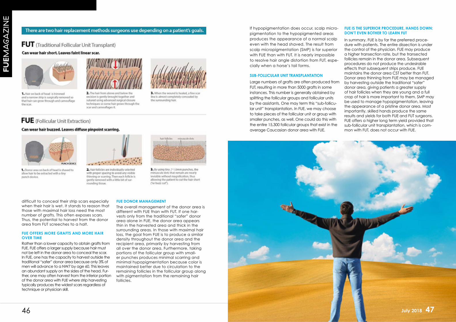
46
FU
EM
AG
AZ
INE
47July 2018
difficult to conceal their strip scars especially when their hair is wet. It stands to reason that those with maximal hair loss need the most number of grafts. This often exposes scars. Thus, the potential to harvest from the donor area from FUT screeches to a halt.
FUE OFFERS MORE GRAFTS AND MORE HAIR OVER TIME
Rather than a lower capacity to obtain grafts from FUE, FUE offers a larger supply because hair must not be left in the donor area to conceal the scar. In FUE, one has the capacity to harvest outside the traditional “safer” donor area because only 3% of men will advance to a NW7 by age 60. This leaves an abundant supply on the sides of the head. Fur-ther, one may often harvest from the inferior portion of the donor area with FUE where strip harvesting typically produces the widest scars regardless of technique or physician skill.
FUE DONOR MANAGEMENT
The overall management of the donor area is different with FUE than with FUT. If one har-vests only from the traditional “safer” donor area alone in FUE, the donor area appears thin in the harvested area and thick in the surrounding areas. In those with maximal hair loss, the goal from FUE is to produce a similar density throughout the donor area and the recipient area, primarily by harvesting from all over the donor area. Furthermore, taking portions of the follicular group with small-er punches produces minimal scarring and minimal hypopigmentation because color is maintained better due to circulation to the remaining follicles in the follicular group along with pigmentation from the remaining hair follicles.
If hypopigmentation does occur, scalp micro-pigmentation to the hypopigmented areas produces the appearance of a normal scalp even with the head shaved. The result from scalp micropigmentation (SMP) is far superior with FUE than with FUT. It is nearly impossible to resolve hair angle distortion from FUT, espe-cially when a horse’s tail forms.
SUB-FOLLICULAR UNIT TRANSPLANTATION
Large numbers of grafts are often produced from FUT, resulting in more than 5000 grafts in some instances. This number is generally obtained by splitting the follicular groups and follicular units by the assistants. One may term this “sub-follicu-lar unit” transplantation. In FUE, we may choose to take pieces of the follicular unit or group with smaller punches, as well. One could do this with the entire 15,300 follicular groups that exist in the average Caucasian donor area with FUE.
FUE IS THE SUPERIOR PROCEDURE, HANDS DOWN: DON’T EVEN BOTHER TO LEARN FUT
In summary, FUE is by far the preferred proce-dure with patients. The entire dissection is under the control of the physician. FUE may produce a higher transection rate, but the transected follicles remain in the donor area. Subsequent procedures do not produce the undesirable effects that subsequent strips produce. FUE maintains the donor area CST better than FUT. Donor area thinning from FUE may be managed by harvesting outside the traditional “safer” donor area, giving patients a greater supply of hair follicles when they are young and a full crop of hair is more important to them. SMP may be used to manage hypopigmentation, leaving the appearance of a pristine donor area. Most importantly, skilled hands produce the same results and yields for both FUE and FUT surgeons. FUE offers a higher long term yield provided that sub-follicular unit transplantation, which is com-mon with FUT, does not occur with FUE.
48 49July 2018
FU
EM
AG
AZ
INE
The Trumpeters Toot their Horns
John Cole, MD
AN OPINION ABOUT THOSE LACKING EXPERIENCE AND SUCCESS IN FUE
In 2016, Drs. Jean Devroye and Arthur Tykos-inski, two novices in FUE, hijacked the ISHRS meeting in Las Vegas with reports they had
solved all our FUE problems. However, they made their proclamations without a single FUE result and in ignorance of the facts. We already had a very successful method to perform FUE. The existing methods and instruments had hun-dreds of thousands of results. The existing FUE doctors were very skilled and talented. What Drs. Devroye and Tykosinski could not compre-hend is they lacked the experience and, per-haps, the skill to perform conventional methods of FUE well. Thus, what these doctors achieved
was an additional method to extract FUE grafts for those lacking the understanding and ca-pacity to perform existing methods of FUE. These two doctors also claimed that transection rates were much lower than all other methods of FUE. When Dr. Devroye finally published a transection rate, nearly a year later, the rate was over 8%. That rate is far higher than many other published transection rates. Lower transection rates come from experience, an understanding of FUE, and proper equipment. In short, almost every claim made by these two in 2015 was false. They also later published that they achieved more hair in each graft, without stating how many hairs in
each graft and without referencing this to any other studies showing up to 2.93 hair per graft. My personal opinion is that the two physicians attempted to ridicule the rest of the doctors, who have thousands of great results from the performance of FUE, by stating that all other methods of FUE were inferior. With this in mind, I will further discuss the advantages, disadvan-tages, and benefits we can anticipate from this method.
A GREAT DEAL OF FUE PROGRESS OCCURRED WELL BEFORE THE HIJACKING
When I began performing FUE in 2002, none of us knew how to best conduct the procedure. What we knew about FUE was very limited. Ray Woods, the father of modern FUE, claimed to have great results in Australia and he refused to teach his procedure to anyone. However, in February 2003 he invited me to come to Syd-ney, Australia to watch him work. I was thrilled at the opportunity, so I flew to Sydney to meet with him. I had lunch with his sister, Angela Campbell at Bondi beach. It was her job to check me out first. I must have passed the first test, so she escorted me to the hotel where Ray Woods was staying with his girlfriend. My introduction to Ray was with him shooting a champagne cork at me, while I was seated in the living room of his suite. I thought we hit it off well and spent the afternoon together, as well had dinner. Multiple times I asked him to tell me about his technique during our conversation. I drew diagrams on a napkin, which Ray always turned into a game of Xs and O’s. I was a bit frustrated but endured the competitions. Unfor-tunately, he refused to show me his procedure after giving me the invitation. I asked him just to let me see his grafts. With strip surgery, we were accustomed to having adipose on the grafts. I was curious if naked grafts without adipose, which is what I was producing from FUE, would grow. He agreed to let me look at his grafts because it would give me a window into his procedure. The next morning, he refused to let me see his grafts, and, thus, I flew home the following day still unsure of the best method to harvest FUE grafts, nor whether “naked” grafts would produce hair. I was most curious whether naked grafts would grow as well as grafts with adipose.
In October 2002, Rassman and Bernstein pub-lished a paper that suggested FUE worked well in only 25 % of patients with transection rates as high as 20% in this group. I think all of us agree that a 20% transection rate is too high and is unacceptable. What Rassman and Bernstein did not describe was how to use the 1 mm punch noted in the study; should we push
the punch, rotate the punch, or oscillate the punch. Furthermore, which punch should we use?
In 2002, there were only a few FUE surgeons in the world. They included Rob Jones, Alan Feller, Ray Woods, and myself. Paul Rose was toying with the procedure, as well. In Decem-ber of 2002, I performed the first FUE case that exceeded 2000 grafts. The patient was in his mid-twenties. I discouraged him from hair transplantation, but the idea of doing a surgery where he would not have a strip scar opened the door for me to perform a procedure on him. I used a 1 mm titanium nitride coated punch on a handle that I devised. I used os-cillation with my finger tips and performed the procedure. The transection rate was higher than I would have liked, but the donor healing was excellent, leaving the patient the opportu-nity to shave his head, which he elected to do after the procedure.
Between this time and September 2003, I per-formed many FUE cases, and my procedures switched from 100% strips to 90% FUE and 10% strips during that year. I developed a depth control punch handle where I could vary the depth of incision by less than one millimeter or by more than one millimeter. By the first of June 2003, I learned that depth control was very important and essential to controlling transec-tion rates. I was the first to describe the impor-tance of depth control, which most doctors have since copied. I also learned that variation in punch size from 0.75 mm to more than 1 mm was also important to control transection rates. I learned how to extract body hair, and I performed the first 7000 graft case in a single session in September 2003. This procedure took me five days. We later developed the skill to perform over 5000 grafts in a single day with speed and excellent results.
In the early years, none of us knew how to perform the procedure, and there was little equipment available to perform the surgery. We didn’t know whether to push the punch or oscillate the punch. Should we use needles? Should we use modified punches? We didn’t know. The only thing I knew for certain was that we were going to find a way. I did. I found many ways. I also developed many instruments to extract grafts, developed the world’s sharp-est punches, and taught many others how to perform FUE. During this same period a few other doctors caught on to FUE and became proficient in it. Today, these are the masters and pioneers of FUE.
50 51July 2018
FU
EM
AG
AZ
INE
In June of 2003, we held the first FUE workshop in Athens, Greece. I introduced FUE to the DHI clinic and they, in turn, trained many physicians in Europe led by George Zontos. These doctors included Jose Lorenzo, though Jose still claims he learned by himself. He did not. Europe was especially important to the growth of FUE. The USA did not adopt nor accept FUE then. How-ever, Europe, Asia, and the Middle East did accept and adopt FUE. Without these regions, it is unlikely FUE would comprise more than 50% of all hair transplant surgeries today. In 2008, I opened the first clinic devoted 100% to FUE in Seoul, Korea. Asia soon caught on fire for FUE. Though Tommy Hwang claims to have trained the first Mongolian clinic in hair transplantation in 2017, he did not. I trained the first clinic in Mongolia in 2013 using FUE and this Mongolian clinic that I trained attended the FUE Europe workshop in Ankara in May 2017. Sadly, the USA did not come around to FUE until 2012.
We allowed Dr. Devroye into our office in 2005 under a non-disclosure agreement. We showed him what we were doing and how we were doing it. We did so with a focus on advancing what we were doing for the rest of the world. We also introduced Dr. Devroye to oscillation because we developed the world’s first func-tioning oscillating device for FUE. Dr. Boudjema published his ideas on oscillation in 2006, but he never built the machine. Instead, the LeadM company in South Korea purchased the idea from Dr. Boudjema in exchange for giving Dr. Boudjema a functioning device. However, LeadM did not produce a device that worked well as neither Dr. Boudjema nor LeadM had any idea what the optimal settings were for oscillation. It was only after we introduced the PCID in 2009 that LeadM added rotation to their poorly performing machine.
In 2005, I presented the excellent results from body hair cases at the ISHRS meeting in Syd-ney. In 2006, we (Patrick Mwamba and I) pre-sented data showing transection rates under 3% with sharp dissection in 200 patients at the ISHRS meeting in San Diego. After this, I ceased attending ISHRS meetings because I was the only guy in the room talking about FUE other than Jim Harris, whose practice was limited to 10% small FUE cases. As popularity for FUE grew, I began attending meetings again in 2009. This did not mean that I did not work to make FUE noteworthy during this span of time. I continued to be very positive about FUE and very nega-tive toward anyone who promoted strip har-vesting. Through the efforts of so many pioneers in FUE, along with my efforts from 2003 until now, FUE has become more popular than strip
surgery across the globe based on an ISHRS census. There are many more physicians who are not members of the ISHRS than there are members of the ISHRS, so the global popularity of FUE is even greater since most non-members of the ISHRS practice it.
The masses wanted a way to efficiently perform FUE. I knew from training that we were limited in advancing the procedure using manual meth-ods. I was very, very good at manual harvest-ing. But I knew that I had to improve mechani-cal harvesting to jump start FUE. From the very beginning, I knew that mechanical harvesting had more pitfalls than manual harvesting. I went through a variety of prototypes before I accepted a machine that would perform FUE and I had to develop the punch to perform FUE mechanically. I designed and filed a patent on this punch in 2006. I would say that our transec-tion rates mechanically were consistently under 5%. We achieved this by varying depth and punch size predominately, but we also needed to alter the angle of approach and the meth-od at times. Sometimes we needed to change the speed of the drill. Sometimes faster and sometimes slower. Sometimes we used oscil-lation. However, in general, we had excellent results.
FAST FORWARD TO 2016
In 2016, Dr. Devroye presented his device and punch. What was new was a flared tip resem-bling a trumpet. Unfortunately, Dr. Devroye created a concave surface in violation of a patent by Dr. Umar. We had an idea for a flare punch that we first introduced in a 2006 patent filing. Dr. Devroye gave me the idea to rede-sign and work with ideas we had way back in 2005 without violating the patent of Dr. Umar. We saw something positive, and we made it better due to Dr. Devroye’s efforts. However, what Dr. Devroye did poorly was that he made it out that sharp dissection does not work only because it did not work in his hands and in the hands of a limited number of physicians, who have inadequate skill and determination in sharp dissection. I remember trying to train one famous strip surgeon in Bangkok on sharp dissection. My grafts were good. Even though he had the punch and the depth already set for him, his technique produced terrible grafts. That is part of the learning curve with sharp dis-section in general. Some catch on quick. Some take longer. Just because you are famous does not ensure you will be competent in either strip harvesting nor FUE harvesting. For reference, the learning curve for manual dissection is even longer. I know because I have trained many physicians in both methods.
ARRIVING AT THE WRONG CONCLUSION
Dr. Devroye suggested that all the work be-tween 2003 and 2016 was unsuccessful only be-cause he could not do it well. I do applaud him for not pursuing something he could not per-form well. However, it was improper for him to suggest that many others using sharp dissection could not perform FUE well. Sharp dissecting surgeons can, and they have produced hun-dreds of thousands of exceptional results during this span of time. When Dr. Devroye jumped on the scene in 2015, he surmised that the sharp dissection doctors all produced poor results only because he delivered poor results. He did his best to make the entire FUE community look like scoundrels and heathens. That was bad for FUE. His claims were also false. He was person-ally biased, self-focused, and utterly wrong in his claims. There are hundreds, if not thousands, of physicians using sharp dissection with fabu-lous results across the globe.
In 2014, Dr. Devroye and I both worked on the same patient in Malaysia. He used his equip-ment, and I used mine. I cut brilliant grafts using sharp dissection in front a meager group of physicians in Malaysia. Dr. Devroye also cut very precise grafts with his system at the time (granted, he did not have the hybrid punch then). However, I cut 3 to 5 grafts for every single graft he cut. I figured out his system and told him that he needed to let the weight of the hand piece do the work. He has since added this advice to his training with the hybrid punch. It was good advice. Still, his method was disappointingly slow, and his system re-mains slow today. Anytime you need to step on a pedal to perform FUE; you are going to be slower. Anytime you use a relatively dull punch, you will be even slower. Patients don’t want to sit in a chair longer than necessary to get their hair restoration and doctors don’t want a team on the clock any longer than necessary. Staff also does not want to work twice as long as required to earn a paycheck. Just because he cannot perform razor-sharp dissection, he should not label the entire sharp dissection world incompetent. Dr. Devroye is the unquali-fied person when it comes to sharp dissection, not the rest of the FUE industry.
THIS SILVER LINING
That mentioned, Dr. Devroye did come up with a compromise for those wishing to enter the field of FUE. He also developed a method to overcome challenging cases where the tran-section rate was unacceptably high, which in my opinion is over 5%. Dr. Devroye recently
published his transection rate at over 8%. This means, in general, his method does not meet my standards. In fact, I bought his punch. I found his punch performed fine in the first 20 grafts, but after this, his punch becomes dull and the grafts became horrible, so I stopped using his punch in this case. I then evaluated his punch. I know the precise geometry of this punch. I also acknowledge the metal. His punch is an inexpensive, soft metal that dulls quickly because the punch cannot maintain an edge. The metal is too soft. His so-called serrated punch is nothing more than a gear on the side that also dulls quickly. Personally, I con-sider his punch scrap metal. I would not sell his punch only because his punch does not meet my standard of quality.
THE NEW GOLD STANDARD
I did recognize a benefit, and I improved on it with a punch that I call the Zero-T punch. The design is totally different than his form in every respect, but the Zero-T punch performs superiorly to his punch in all aspects. The Ze-ro-T punch has a long life, that maintains its sharp cutting edge, it works well in intermittent oscillation, continuous oscillation, intermittent rotation, constant rotation, and intermittent ro-tation followed by oscillation. A variance of this punch will also work exceptionally in long hair FUE. There is nothing “hybrid” about this punch. This is a sharp punch. Because this punch is sharp, the rate of extraction is very fast.
NOW THE REST OF THE STORY
The fact is that FUE took off without Drs. Devor-ye and Tykosinski many years ago and the field grew exponentially without either of them. Their ideas add a new method for us to obtain high quality grafts in complex cases. They should be proud of this. However, they cannot continue to hold the belief that their method is superior to all other methods for harvesting FUE grafts. Devroye’s approach is solely a different meth-od that is useful in some cases in the hands of talented physicians and very useful in the hands of those lacking exceptional talent.
Now here are the facts; Dr. Devroye made a contradictory opinion of sharp dissection after buying only two of my exquisitely sharp serrat-ed tip punches in a single size. One cannot be-come experienced with two single-use punch-es designed to work on an individual patient. Once you sterilize these punches, you risk losing the sharpness of the punch. You also should not use this punch on more than one patient for obvious sterility reasons.
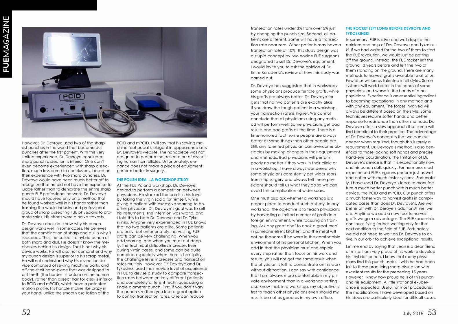
52 53July 2018
FU
EM
AG
AZ
INE
However, Dr. Devroye used two of the sharp-est punches in the world that became dull punches after the first patient. With this very limited experience, Dr. Devroye concluded sharp punch dissection is inferior. One can’t even become experienced with sharp dissec-tion, much less come to conclusions, based on their experience with two sharp punches. Dr. Devroye would have been much better off to recognize that he did not have the expertise to judge rather than to denigrate the entire sharp punch FUE professional network. Dr. Devroye should have focused only on a method that he found worked well in his hands rather than trashing the whole industry and professional group of sharp dissecting FUE physicians to pro-mote sales. His efforts were a naive travesty.
Dr. Devroye does not know why his punch design works well in some cases. He believes that the combination of sharp and dull is why it succeeds. Thus, he called it a hybrid punch of both sharp and dull. He doesn’t know the me-chanics behind his design. That is not why his device works. He will also not comprehend why my punch design is superior to his scrap metal. He will not understand why his dissection de-vice comprised of sewing machine parts, and off-the-shelf hand-piece that was designed to drill teeth (the hardest structure on the human body), rather than dissect hair follicles, is inferior to PCID and mPCID, which have a patented motion profile. His handle shakes like crazy in your hand, unlike the smooth oscillation of the
PCID and mPCID. I will say that his sewing ma-chine foot pedal is elegant in appearance as is Dr. Devroye’s website. The handpiece was not designed to perform the delicate art of dissect-ing human hair follicles. Unfortunately, ele-gance does not make a piece of equipment perform better in surgery.
THE POLISH IDEA ...A WORKSHOP STUDY
At the FUE Poland workshop, Dr. Devroye desired to perform a competition between physicians. He stacked the cards in his favor by taking the virgin scalp for himself, while giving a patient with excessive scarring to an-other physician. Dr. Devroye’s goal was to sell his instruments. The intention was wrong, and I told this to both Dr. Devorye and Dr. Tyko-skinski. Anyone very experienced in FUE knows that no two patients are alike. Some patients are easy, but unfortunately, harvesting FUE grafts can be very challenging. When you add scarring, and when you must cut deep-ly, the technical difficulties increase. Even during virgin cases, and some can be quite complex, especially when there is hair splay, the challenge level increases and transection rates multiply. However, Dr. Devroye and Dr. Tykosinski used their novice level of experience in FUE to devise a study to compare transec-tion rates between entirely different patients and completely different techniques using a single diameter punch. First, if you don’t vary the punch size then you lose a great option to control transection rates. One can reduce
transection rates under 3% from over 5% just
by changing the punch size. Second, all pa-
tients are different. Some will have a transec-
tion rate near zero. Other patients may have a
transection rate of 10%. This study design was
a stupid concept by two novice FUE surgeons
designated to sell Dr. Devorye’s equipment.
I would invite you to ask the opinion of Dr.
Emre Karadeniz’s review of how this study was
carried out.
Dr. Devroye has suggested that in workshops
some physicians produce terrible grafts, while
his grafts are always better. Dr. Devroye for-
gets that no two patients are exactly alike.
If you draw the tough patient in a workshop,
your transection rate is higher. We cannot
conclude that all physicians using any meth-
od will perform well. Some physicians get bad
results and bad grafts all the time. There is a
time-honored fact: some people are always
better at some things than other people are.
Still, any talented physician can overcome ob-
stacles by making changes in their equipment
and methods. Bad physicians will perform
poorly no matter if they work in their clinic or
in a workshop. I have always wondered why
some physicians consistently get wider scars
from strip surgery and always felt these phy-
sicians should tell us what they do so we can
avoid this complication of wider scars.
One must also ask whether a workshop is a
proper place to conduct such a study. In any
workshop, the objective is to teach physicians
by harvesting a limited number of grafts in a
foreign environment, while focusing on train-
ing. Ask any great chef to cook a great meal
in someone else’s kitchen, and the meal will
not be the same if he were in the comfortable
environment of his personal kitchen. When you
add in that the physician must also explain
every step rather than focus on his work and
results, you will not get the same result when
the physician is left to concentrate on his work
without distraction. I can say with confidence that I am always more comfortable in my pri-
vate environment than in a workshop setting. I
also know that, in a workshop, my objective is
first to teach other physicians even should my results be not as good as in my own office.
THE ROCKET LEFT LONG BEFORE DEVROYE AND TYKOSKINSKI
In summary, FUE is alive and well despite the opinions and help of Drs. Devroye and Tykosins-ki. If we had waited for the two of them to start the FUE revolution, we would just be getting off the ground. Instead, the FUE rocket left the ground 13 years before and left the two of them standing on the ground. There are many methods to harvest grafts available to all of us. Few of us will be as talented in all styles. Some systems will work better in the hands of some physicians and worse in the hands of other physicians. Experience is an essential ingredient to becoming exceptional in any method and with any equipment. The forces involved will always be different based on the style. Some techniques require softer hands and better response to resistance than other methods. Dr. Devroye offers a slow approach that some will find beneficial to their practice. The advantage of Dr. Devroye’s concept is that we can cut deeper when required, though this is rarely a requirement. Dr. Devroye’s method is also ben-eficial to those lacking soft hands and precise hand-eye coordination. The limitation of Dr. Devorye’s device is that it is exceptionally slow, and his punch dulls quickly. Furthermore, most experienced FUE surgeons perform just as well and better with much faster systems. Fortunate-ly, I have used Dr. Devorye’s ideas to manufac-ture a much better punch with a much better device, the PCID and mPCID. Our punch offers a much faster way to harvest grafts in compli-cated cases than does Dr. Devroye’s. Are we better off with Dr. Deroye’s additions? Yes, we are. Anytime we add a new tool to harvest grafts we gain advantages. The FUE spaceship continues flying farther, waiting only for the next addition to the field of FUE. Fortunately, we did not need to wait on Dr. Devroye to ar-rive in our orbit to achieve exceptional results.
Let me end by saying that Jean is a dear friend of mine. I am very proud of his work to develop his “hybrid” punch. I know that many physi-cians find this punch useful. I wish he had been fair to those practicing sharp dissection with excellent results for the preceding 15 years. However, I know how proud he is of this punch and his equipment. A little irrational exuber-ance is expected. Useful for most procedures, the modifications I have developed based on his ideas are particularly ideal for difficult cases.
54 55July 2018
FU
EM
AG
AZ
INE
Pain Management John Cole, MD
POST-OPERATIVE PAIN MANAGEMENT IN FUE HAIR TRANSPLANTATION
When I first began performing FUE in 2002, I noted much less pain reported by patients undergoing FUE than FUT. This is
not to suggest that pain is a significant problem with FUT, but pain does occur. Patients who had FUT prior to their first FUE procedure almost all stated that the pain from FUT was significant-ly worse. In fact, more than 99% stated that FUT was more painful than FUE. However, pain can be an issue in patients following the latter. For this reason, I will review some pain manage-
ment options for FUE patients.
WHAT CAUSES PAIN?
Pain may be defined as physical discomfort. There are four types of pain: nociceptive, inflammatory, neuropathic, and centralized. Inflammatory pain is nociceptive pain with localized immune response that generates pro-inflammatory mediators to facilitate tissue repair. Neuropathic pain originates from injury to specific peripheral nervous (PNS) or CNS structures or to all peripheral sensory nerves as with diabetes. Nociceptive pain is a normal acute response to peripheral damage. Cen-tralized pain is peripheral and central sensi-tization without detectable peripheral origin and includes fibromyalgia, irritable bowel syndrome, and tension-type headache and is also called dysfunctional pain. Acute pain from hair transplant surgery may arise from three of the four types of pain in this classifica-tion system (Inflammatory, neuropathic, and nociceptive pain).
The primary area where pain is an issue fol-lowing FUE is the donor area. Less commonly, the recipient area can be a significant source of pain. Nociceptive pain may arise due to tissue injury during the surgery. One question is why some patients experience more pain than others? Another question is how we might reduce the risk for post-operative pain by how we manage the surgery? Some pa-tients seem more sensitive to pain than other patients. It is possible that certain measures
increase the risk of pain. These might include inadvertently hitting a nerve with a needle during anesthesia or tumescence. Another risk is nerve injury (neuropathic pain) due to a deep incision with a punch or needle during graft harvesting or recipient site preparation. In general, most major nerves will be deep-er than the follicle bulb, which is typically 4 to 5.2 mm deep. Obese people will have a cushion of adipose between the bulb and the nerves. In thin people, the adipose is minimal and the nerves reside just deep to the bulbs. Therefore, the greater risk for nerve injury is during tumescence or anesthesia. Minimizing the depth of needle penetration is one pos-sible solution. Knowledge of the anatomy of major nerves is another possibility. Even when we exercise great care, some people seem to have more pain than others.
Cell injury generates enzymes that release arachidonic acid from cell membranes, and arachidonic acid is converted to prostaglan-dins, thromboxanes, leukotrienes, and other eicosanoids. The prostaglandins PGE2 and PGF2a contribute to inflammation and sen-sitize nociceptors innervated by A-delta and C nerve fibers (inflammatory pain). Other prostaglandins are needed for homeostasis. NSAIDs inhibit the enzyme cyclooxygenase (COX) required for synthesis of arachidonic acid into prostaglandins and other eicosa-noids. COX-1 is noncontributory to inflamma-tion but is involved in homeostatic functions and mediates gastrointestinal protection by the gastric mucosa. COX-2 is induced during inflammation and amplifies the inflammatory response. However, COX-2 also contributes to kidney and cardiovascular homeostasis and helps maintain vascular integrity. NSAID anal-gesia also results from central activity, possibly through substance P and NMDA inhibition.
Because pain is a common, yet unpredict-able, side effect of FUE a review of possible treatment options is of value.
NON-STEROIDAL ANTI-INFLAMMATORY DRUGS
NSAIDS are an effective method to control acute pain. There are many NSAIDs on the market globally. Most of them are useful in pain management. NSAIDs are sub-grouped by dominant COX isoenzyme activity. NSAIDs that inhibit COX-1 and 2 are termed nonse-lective NSAIDs. NSAIDs with greater COX-2 inhibition are termed coxibs, COX-2 selective NSAIDs, or COX-2 inhibitors. COX-2 inhibition is not an absolute NSAID characteristic, as COX-2 selective agents also possess varying degrees of COX-1 inhibition. COX-2 inhibi-tion reduces inflammation and pain but can interfere with homeostatic functions such as proper renal blood flow and gastric muco-sa protection. NSAIDs have a ceiling effect, where further dose escalation increases side effects, but not analgesia. Analgesic effica-cy between coxibs and non-selective NSAIDs is comparable and consistent differences in pain reduction for specific NSAIDs have not been discovered. Though we do not know why, up to 70% to 80% of patients favorably respond to a specific NSAID. We cannot predict a response to any specific NSAID, but patients who lack a response to one NSAID will gain full value from another NSAID.
Up to 40% of those taking NSAIDs develop up-per gastrointestinal symptoms, most frequently gastroesophogeal reflux and/or dyspeptic symptoms. Serious and potentially fatal upper gastrointestinal toxicities, including symp-tomatic and/or complicated peptic ulcer, bleeding, perforation, or obstruction, occur in 1% to 2% of NSAID users and are influenced by dose and exposure. COX-2 inhibitors are associated with significantly lower risks of up-per gastrointestinal perforation, obstruction, and bleeding compared with non-selective NSAIDs plus proton pump inhibitors. The high-est risk of serious gastrointestinal morbidity is found with piroxicam, azapropazone, and ketorolac; the lowest risk is with aceclofenac, ibuprofen, and celecoxib. Piroxicam’s half-life of more than 30 hours could explain its pro-pensity for gastrointestinal toxicity and oral ketorolac dosing is limited to five days due to a well-recognized gastrointestinal and renal toxicity profile. Complications from NSAIDs in the lower gastrointestinal tract can be fa-tal. Fortunately, most pain management in the post FUE transplant patient are for short durations, which may minimize gastrointestinal complications.
There is evidence that there is compara-
ble risks of lower gastrointestinal and upper gastrointestinal bleeding and perforation from NSAIDs. Increased gut permeability, gut inflammation, blood loss, anemia, malabsorp-tion, and mucosal ulceration are the most common lower gastrointestinal morbidities.
When prescribing NSAIDs, physicians should assess patient cardiovascular risks. COX- 2 inhibitors were introduced in 1999 in the following order: celecoxib, rofecoxib, and valdecoxib. Rofecoxib and valdecoxib were withdrawn from the market in 2004 and 2005 due to adverse cardiovascular events. While non-selective NSAIDs carry the greatest risk for cardiovascular complications, coxibs carry the greatest risk for cardiovascular complica-tions. However, meta-analysis found compa-rable cardiovascular risk between coxibs and high-dose diclofenac, and possibly ibuprofen. The incidence of major cardiovascular events was increased 33% with diclofenac. Ibuprofen increased the risk of major coronary events, while naproxen did not elevate the risk. The use of any NSAID roughly doubled the risk of heart failure. Another meta-analysis found naproxen the least harmful NAID in cardiovas-cular risk, with coxibs having the highest risk of cardiac infarction. Ibuprofen and diclofenac have the highest risk of stroke. Diclofenac has the highest risk of cardiovascular fatali-ty. Naproxen has the lowest risk of a cardiac event but has a very high risk of gastrointesti-nal complication.
Another potential option is a topical NSAID to improve tolerability. A topical NSAID will penetrate the tissue below the site of appli-cation, which is the location of the source of pain. Diclofenac has the most evidence supporting its use and is the sole NSAID with FDA approval for topical use. Approved for-mulations include diclofenac sodium 1% and 3% gel, diclofenac sodium 1.5% topical solu-tion in 45.5% dimethyl sulfoxide, diclofenac epolamine topical patch,1.3% and diclofenac sodium topical solution 2%. Topical diclofenac has shown efficacy in pain reduction superior to placebo; comparable to topical NSAIDs indomethacin, ketoprofen, piroxicam; and comparable to oral NSAIDs diclofenac, ibu-profen, naproxen. The effectiveness, safety, and tolerability of topical diclofenac support its use for a variety of inflammatory acute and chronic back pain conditions. Topical NSAIDs may work well in the acute post-operative setting, though studies are needed to confirm this.

56 57July 2018
FU
EM
AG
AZ
INE
The chief concerns are the time to onset of pain relief and local irritation in a post-surgi-calsetting. The most common adverse effects with diclofenac are mild site reactions, such as erythema or pruritus. No severe or system gastrointestinal effects have been observed with topical diclofenac or other topical NSAIDs, and skin irritation is significantly less common with patch versus gel. Systemic exposure from diclofenac epolamine topical patch is 1% of that of an oral 75-mg dose. The steady-state plasma concentrations are also significantly lower and unlikely to result in COX-1-mediated effects. From 1993 to 2008, 46 million patients worldwide received a diclofenac epolamine topical patch, and adverse events were reported in 108 patients. Most were skin reactions or lack of efficacy; six were serious gastrointestinal events, but none were believed causally related to the diclofenac epolamine topical patch. The FDA requires that manufactures incorporate stan-dard NSAID warnings of serious liver disease risks into the labeling of topical diclofenac; however, to reach systemic levels from one 150-mg oral dose of diclofenac, 100 patches would need to be worn simultaneously.
There is some evidence that NSAIDS and steroids can reduce the release of growth factors from PRP. In fact, some argue that NSAIDS and steroids should be avoided if PRP is injected because these pharmaceuticals can reduce the activation of platelets. The inflammatory response from PRP is most likely from polymorphonuclear cells rather than from the mononuclear cells and growth fac-tors we seek in PRP; however, the inflamma-tory response may offer some benefit. Thus, we must keep this in mind. Fortunately, with sonicated PRP, platelet activation is not re-quired because sonication bursts the platelets and releases six to eight times the concen-tration of growth factors. NSAIDS are useful in post-operative pain.
ACETAMINOPHEN
Acetaminophen is a nonsalicylate antipyret-ic analgesic that may be used alone or with other analgesics for acute pain. It has no an-tiplatelet effects and gastric mucosa irritation unlike aspirin. There are no anti-inflammatory effects and produces analgesia that is ad-ditive to NSAIDs. The precise mechanism of action of acetaminophen remains unknown. The maximum dose is 3,250 mg due to the risk of liver toxicity based on a more recent rec-ommendation by the Food and Drug Admin-istration (FDA). McNeil pharmaceuticals has
lowered the maximum dose to 3,000 mg in a day in their package insert. The risk of hepa-totoxicity increases with liver impairment and alcoholism. Fatal hepatic necrosis can occur with doses exceeding the maximum, espe-cially in patients with liver disease.
OPIOIDS – THE BASICS
Opioids are approved for the treatment of moderate or severe pain by the FDA. Individu-al patients differ greatly in clinical response to different opioid analgesics and patient pop-ulations show widely variable response to the same opioid and dose. Opioids are a useful tool, so a working knowledge of the various opioids is beneficial to the hair transplant surgeon. However, opioids are not a pana-cea for pain, nor are they safe and effective for every patient. One should be aware that 84.5% of those who died from opioid prescrip-tions were also prescribed benzodiazepines. In 2010, 29% of opioid overdose deaths in-volved alcohol. Studies indicate that fatal respiratory depression events often occur in the first five days of initial opioid therapy; with most in the initial 24 hours. This reflects initia-tion of therapy at too high a starting dose or failure to consider other risk factors, such as co-prescribed CNS sedatives. Thus, caution should be used with the initial dose, with the consumption of alcohol, and with a co-pre-scription of benzodiazepines.
Opioid analgesics produce therapeutic and side effects by mimicking endogenous opioid activity, although some opioids produce an-algesia by activity outside the opioid recep-tor complex. Opioids widely differ in levels of affinity and activation of opioid receptor subtypes. In addition, interindividual variation in analgesic response and side effects is sig-nificant, largely driven by genetic factors. The complex interaction between unique opioid properties and individual patient characteris-tics dictates that a patient-tailored approach is required for opioid selection, dose initiation, and titration to optimize safety, analgesia, and tolerability.
Naturally occurring opioid compounds are produced in plants (opium, morphine) and in the body (the endogenous opioids). Endog-enous opioids are peptides that bind opioid receptors, function as neurotransmitters, and help regulate analgesia, hormone secretion, thermoregulation, and cardiovascular func-tion. The three-primary endogenous opioid peptide families are the endorphins, enkeph-alins, and dynorphins, and the three primary opioid receptor types are mu, kappa, and
delta. A quick overview of this complex pain modulation system is helpful in understanding how opioid analgesics work.
Endogenous opioid peptides are neurotrans-mitter molecules in the opioid receptor complex that produce specific physiologic effects determined by neuronal distributions of the activated opioid receptor type. The endogenous opioid peptides are cleaved from pro-hormone precursors proenkephalin, pro-opiomelanocortin, and prodynorphin. The dneogenous delta opioid receptor pep-tides are metenkephalin and leuenkephalin, cleaved from proenkephalin. Prodynorphin
gives rise to kappa opioid receptor agonists dynorphin A and B. Pro-opiomelancortin en-codes the peptide beta-endorphin, which has agonist activity at all three classical opioid receptors. Some endogenous opioid ligands lack specificity for opioid receptor subtypes, such as b-endorphin and enkephalins.
Endorphins are synthesized in the hypothala-mus and the pituitary gland. Pain, strenuous exercise, excitement, and orgasm stimulate their release, binding, and activation. Endor-phins are popularized as the “natural pain killers” from their ability to induce analgesia and a general feeling of well-being. They are
58 59July 2018
FU
EM
AG
AZ
INE
thought to largely mediate analgesia from acupuncture, massage, hydrotherapy, and transcutaneous electrical nerve stimulation therapy.
Dynorphin peptides are synthesized from the precursor prodynorphin and have primary af-finity and binding at the kappa opioid recep-tor. Dynorphins are distributed throughout the CNS, with the highest concentrations in the brain stem, hypothalamus, and spinal cord. Their physiologic actions are diverse, and their primary function is the modulation of pain response, appetite and weight, circadian rhythm, and body temperature. Dynorphins are linked to stress-induced depression and drug-seeking behavior, and drugs that inhibit dynorphin release are under evaluation for possible use in the treatment of depression related to drug addiction.
Enkephalin peptides, derived from proen-kephalin, are located throughout the brain and spinal cord and are involved in regulat-ing nociception. Enkephalins inhibit neuro-transmission in pain perception pathways, reducing the emotional and physical impact of pain. Enkephalins also reside in the GI tract, where they help regulate pancreatic enzyme secretion and carbohydrate metabolism.
Opioid receptors are expressed throughout the CNS and PNS on key nodes within the pain pathway and are highly concentrated in areas involved with integrating pain in-formation. Opioids vary greatly by receptor affinity, binding, and activity and can bind to produce agonist, partial agonist, or antago-nist receptor activity. As noted, the analgesic activity and the side effects result from mim-icry of endogenous opioids, achieved by the beta-phenylethylamine group moiety shared by endogenous and exogenous opioid re-ceptor ligands that facilitate opioid receptor binding.
Mu receptors are the primary mediators of analgesia produced by opioid analgesics in clinical use. Their greatest CNS concentration is in the thalamus, medulla, periaqueduc-tal gray area, neocortex, amygdala, dorsal horn, inferior and superior colliculi, and brain stem. PNS occupancy includes the peripheral sensory neuron dorsal root ganglion, stomach, duodenum, jejunum, oleum, proximal, and distal colon. Mu receptors in non-neural tissue are found in the vascular cardiac epithelium, keratinocytes, vas deferens, Sertoli cells.
Mu opioid receptors in the amygdala and nucleus accumbens mediate opioid reward response (euphoria). In the brain region,
opioids bind to and activate mu receptors, which inhibit gamma-aminobutyric acid (GABA) to increase dopamine transmission. Mu opioid receptors broadly distributed in the limbic system mediate emotional response to pain and analgesia. In the medial thalamic nuclei, they relay spinothalamic inputs from the spinal cord to the cingulate gyrus and limbic structures.
Kapp opioid receptors bind dynorphin as the primary endogenous ligand. In the CNS, they are highly concentrated in the caudateputa-men, nucleus accumbens, amygdala, brain stem, neural lobe of the pituitary gland, and hypothalamus. In the PNS, these receptors are found in the sensory neuron dorsal root gan-glion, stomach, duodenum, je-junum, oleum, and proximal and distal colon. They are pri-marily found in the limbic system, brain stem, and spinal cord. Their effects include spinal analgesia, sedation, dyspnea, and respiratory depression, dependence, and dysphoria. The kappa opioid receptor subtype k3 is consid-ered the primary analgesic mediator.
Delta receptors are mostly confined to CNS structures of the pontine nuclei, amygdal, olfactory bulbs, and deep cortex, but are also found in the GI tract and the lungs. They me-diate spinal and supraspinal analgesia and the psychomimetic and dysphoric effects of opioid analgesics.
Opioid analgesia results from a complex series of neuronal interactions, largely mediated by the high density of opioid receptors in the dorsal horn of the spinal cord and in subcorti-cal regions of the brain. The analgesic effects of opioids result from two general processes: 1) direct inhibition of ascending transmission of pain signaling from the dorsal horn of the spinal cord, and 2) activation of descending pain control circuits from the midbrain to the dorsal horn of the spinal cord. All three opi-oid receptor types mediate spinal analgesia. Supraspinal analgesia is primarily mediated by mu opioid receptor subtype 1.
The spinal cord dorsal horn is a primary anal-gesic site of opioids and densely populated with mu (70%), delta (20%), and kappa (10%) opioid receptors. Opioid receptors are local-ized on presynaptic afferent fibers, interneu-rons, and postsynaptic projection neurons. Opioids bind to and activate mu receptors, which inhibit the release of pain mediators such as substance P, glutamate, and nitric oxide from nociceptive afferent neurons. Spi-nal level analgesia appears to elevate pain thresholds.
At the supraspinal levels, opioids produce analgesia by attenuation of the subjective evaluation of pain. After morphine is giv-en for severe pain, patients report pain but without the associated anguish and distress. Conscious awareness and pain response are retained but modified by changes in emo-tional response to pain, mediated in part through opioid receptors in the limbic system. The greatest factor that contributes to opioid analgesia is the concentration of the drug on the mu receptor.
OPIOID PHARMACOLOGY
Opioids have been a mainstay of pain treat-ment for thousands of years. The opium poppy, Papaver somniferum, is the oldest and most prevalent source of opium and opioid analgesics. The opium poppy was grown in the Mediterranean region at least as early as 5000 BC and has since been cultivated in a number of regions throughout the world.
The first historical medical reference to opium dates back to the 3rd century BC by Arab physicians experienced in its therapeutic uses. In 1806, Friedrich Serturner reported the isolation of a pure substance in opium that he named morphine, after Morpheus, the Greek god of dreams. Serturner also published the first report of morphine toxicity in 1817. He discussed his experimentation of administer-ing the alkaloid to himself, three young boys, three dogs, and a mouse. One of the dogs died, and the effects of morphine on Ser-turner and his three young volunteers were described as “near-fatal.” In the 1850s, the first recorded morphine overdose fatality was reported by Alexander Wood when perform-ing one of the first morphine injections on his wife, who subsequently died of respiratory depression.
Raw opium contains numerous alkaloids, but only morphine, codeine, thebaine, and pa-paverine have an identified use in medicine. Because the synthesis of morphine is difficult, the opium poppy plant remains the primary source of morphine. Thebaine is a minor con-stituent of opium that chemically resembles morphine and codeine but produces a stimu-lating rather than a calming effect. Hebaine is not used medicinally but is converted into oxymorphone, nalbuphine, naloxone, nal-trexone, and buprenorphine. The numerous synthetic derivatives of morphine and the-baine are produced by relatively simple mod-ifications of the parent molecule. Morphine is transformed into codeine by methyl substitu-tion on the phenolic hydroxyl group and into
diacetylmorphine by acetylation at the 3 and 6 positions to produce heroin.
Opioids may be classified by analgesic po-tency, chemical class, or functional activity. Potency is divided into weak, intermediate, and strong. Functional activity is divided into full agonist, partial agonist, mixed agonist/ antagonist, and antagonist (naloxone). The only weak potency opioid is codeine. Inter-mediate potency opioids include buprenor-phine, pentazocine, butorphanol, nalbuphine, hydrocodone, tramadol, and tapentadol. Strong potency opioids include morphine, oxycodone, hydromorphone, oxymorphone, levorphanol, fentanyl and analogs, metha-done, and meperidine.
Each opioid has a unique analgesic and adverse effect profile. Morphine remains the criterion standard by which the analgesic efficacy of new opioids is measured.
MORPHINE
Morphine (Roxanol, MS Contin, Avinza, Kadi-an, Morphabond, Embeda) was first isolated in 1803 and first introduced as an analgesic to the USA in 1830. Hypodermic syringes were introduced in the mid-19th century. Morphine remains one of the best drugs for alleviating moderate to severe pain despite its discov-ery over two centuries ago. In fact, the World Health Organization has designated morphine as a drug of choice for moderate-to-severe pain. It is a standard from which all other analgesics may be compared. Morphine is a strong mu opioid receptor against and a weak kappa and delta receptor agonist. Morphine may be delivered IV, IM, subcuta-neously, rectally, epidurally, or intrathecal-ly. With IM/SC injection, the onset of effect occurs after 15 to 30 minutes, peak effect in 45 to 90 minutes, and duration of effect is roughly 4 hours. With IV injection, the peak effect occurs in 15 to 30 minutes. When given IV, only a small portion of morphine reaches the CNS due to poor lipid solubility, a high degree of ionization at physiologic pH, pro-tein binding, and rapid metabolism. Morphine produces analgesia, euphoria, and a sensa-tion of warmth. It increases pain threshold and alters the perception of noxious stimuli even at low doses. Continuous, dull pain and pain originating in visceral organs, skeletal muscles, joints, and bone are most responsive to mor-phine. The analgesic and respiratory depres-sant effects of morphine may not correlate with plasma concentrations,because CNS concentration peaks later and decays more slowly than plasma.
60 61July 2018
FU
EM
AG
AZ
INE
When given orally, morphine undergoes ex-tensive first-pass hepatic metabolism, resulting in an elimination half-life of approximately two hours independent of route of administra-tion or formulation. Morphine administered by sublingual and buccal routes has a delayed onset of action compared with oral morphine (due to smaller peak plasma levels, lower bioavailability, and larger interpatient vari-ability). Compared with the oral form, intra-thecal morphine is 100 times more potent and epidural morphine is 10 times more potent. Oral morphine preparations are available in short-acting (SA) and extended release formations (ER), including an ER formulation containing naltrexone to discourage tamper-ing and diversion. Morphine is far more potent of an analgesic than would be needed in typical hair restoration surgery.
HYDROMORPHONE
Hydromorphone (Dilaudid, Exalgo) is a semi-synthetic hydrogenated ketone of mor-phine with primary activity as a mu receptor agonist. It has roughly five to seven times the potency of morphine, with similar effects but possibly less sedation with greater euphoria. Hyrdormorphone can be administered by parenteral, IV, rectal, and oral routes and is considered the best opioid for SC administra-tion. Oral hydromorphone has a bioavailabil-ity of 50% and plasma elimination half-life of 2.5 hours. Its high water solubility permits very concentrated formulations. Hydromorphone produces better analgesia than morphine for acute pain without significant differences in adverse effects. Following oral administration, hydromorphone undergoes hepatic first-pass elimination of approximately 50%. The elimi-nation half-life after IV administration is 2.5 to 3 hours, primarily through the urinary system. The first ER form of hydromorphone was called Palladone, but this was withdrawn from the market due to concerns about potentially fa-tal interactions with alcohol. Exalgo has since been introduced without this concern.
CODEINE
Codeine (Tylenol with Codeine, Capital with Codeine, Vopac) produces analgesia solely through enzymatic conversion into morphine, so it is considered a pro-drug. A pro-drug is a drug ingested in a biologically inactive or less active form that is biotransformed into an active or more active metabolite. The oral bioavailability of codeine is 50% with rough-ly 10% metabolized to morphine. However at least 10% of individuals possess deficient activity of hepatic enzyme necessary to me-
tabolize codeine to morphine due to genetic variation or polymorphism. In these individ-uals, codeine has no analgesic effect and should be avoided.. Codeine may be used orally or IM for mild-to-moderate pain but has a very limited use in severe pain. Codeine is also used as an antitussive and antidiarrheal. Codeine produces minimal euphoria, has a low abuse potential, is less sedating, and is less likely to result in respiratory depression than morphine. Constipation is a common side effect. Because commercially available codeine is combined with acetaminophen or acetylsalicylic acid (ASA) the dosage should be monitored to ensure daily safe lifts are not surpassed. Codeine has an analgesic ceiling, with no additional analgesic effect from dos-es greater than 60 mg. It is commonly used post hair transplant to control pain, but we must remember that up to 10% of all patients will receive no benefit from this mild narcotic.
OXYCODONE
Oxycodone (Oxy IR, Percocet, Tylox, OxyCon-tin, Xtampza ER, Targiniq ER) is a semisynthetic opioid analgesic derived from the natural al-kaloid thebaine and has been in medical use since 1917. Although oxycodone mu receptor affinity is at least 20 times less than morphine, oxycodone possesses high oral bioavailability and delivers analgesia and other subjective effects comparable to oral morphine. Unlike morphine, oxycodone has moderate affinity and agonist activity at the kappa-2b opioid receptor, which contributes to its efficacy in neuropathic pain. Oxycodone is available in SA and ER oral formulations. Oxycodone SA has a half-life of approximately two to four hours and a bioavailability of 50% to 60%. The overall clinical effects of oxycodone reflect primary mu receptor activity, with analgesia, respiratory depression, euphoria, and abuse potential comparable to other mu agonists. Oxycodone differs from morphine by pro-ducing less dysphoria and by rapid transport through the blood-brain barriers, resulting in greater CNS than plasma concentrations, the reverse of morphine. In addition to its low dose combination with acetaminophen, oxy-codone is formulated as the sole analgesic in 10, 20, 40, and 80-mg controlled-release (DR) tablets and 5-mg SA capsules. Sales of oxy-codone CR (OxyContin) 160 mg were discon-tinued over abuse and diversion concerns in 2001.
OXYMORPHONE
Oxymorphone (Numorphan, Opana) was first synthesized in Germany in 1914, patented in
the United States in 1957, and introduced in 1959 for parenteral injection and in supposito-ry form. It then became available as an oral SA opioid, but this was withdrawn from the U.S. market in the early 1970s. Following rein-troduction in 2006 in oral SA and ER formula-tions, its use in the treatment of noncancerous pain has steadily increased. Oxymorphone is a semisynthetic derivative of the parent compound morphine and has a high affinity for the mu opioid receptor and negligible interaction with kappa and delta opioid re-ceptors. The potency is roughly 1.2 times that of morphine, but with less sedative effects. Oxymorphone possesses less protein binding (10% t 12%) than morphine (30% to 35%) and oxycodone (45%), and its highly lipophilic properties provide ease in blood-brain barrier penetration. The oral bioavailability of oxy-morphone is approximately 10%, the lowest of the full agonists. In healthy volunteers, the half-life ranges from 4.2 to 9.4 hours, longer than that of morphine, hydromorphone, and oxycodone. Oxymorphone SA tablets may be given at six hour intervals, whereas the ER formulation is dosed twice daily. Steady-state conditions are achieved after three to four days. Oxymorphone is subject to hepatic first-pass effects and is excreted by the kidneys. As such, this agent has a prolonged half-life and accumulates in patients with renal fail-ure. In patients with hepatic insufficiency, in-creasing the dosing interval is recommended. Oxymorphone is an effective opioid analgesic with a safety profile comparable to other mu agonist opioids. It may have a safety advan-tage in elderly or frail patients from whom adverse drug interactions are concerning. However, in 2017, the FDA requested Opana ER be removed from the market due to abuse concerns.
HYDROCODONE
Hydrocodone (Zohydro ER, Hysingla ER, Lortab, Vicodin) is a semi-synthetic codeine derivative that more closely resembles mor-phine in its pharmacologic profile. Hydroco-done was first used medically as a cough sup-pressant and analgesic in the 1920s. It exhibits a complex pattern of metabolism including demethylation at the 3-carbon position into hydromorphone, which has a stronger mu receptor binding than the parent drug. Thus, similar to codeine, hydrocodone is suggested to be a pro-drug. Its analgesic properties are similar in potency to codeine. Hydrocodone is effective as a cough suppressant and as an analgesic for moderate to moderately severe pain. It is most frequently prescribed in
cominged formulations with ibuprofen (Vico-profen), and antihistamines (Hycomine) and as an antitussive liqud formulation. The hy-drocodone ibuprofen product is intended for short-term (generally less than 10 days) man-agement for acute pain from trauma, muscu-loskeletal or back pain, postoperative pain, abdominal pain, or dental pain.
METHADONE
Methadone (Dolophine, Methadose) was first synthesized as an analgesic in Germany during World War II in response to the difficul-ty obtaining raw opium to synthesize mor-phine. Although chemically unlike morphine or heroin, methadone produces many of the same pharmacologic and clinical effects. It was introduced into the United States in 1947 as the analgesic Dolophine. High-dose methadone can block the effects of heroin and other opioid drugs by diminishing reward and reinforcement effects, and this has been the primary use of methadone in the United States over the last five decades. Methadone is both a mu receptor agonist and a NMDA receptor antagonist and reuptake inhibitor of serotonin and norepinephrine. Methadone produces analgesia very similar to other com-monly used opioids, but its lack of euphoric effects relative to other agents can make it advantageous in some patient populations. The highly variable elimination half-life is 8 to 60 hours, and a single dose analgesia lasts 4 to 8 hours. Methadone requires a thorough understand of its pharmacokinetic properties to safely prescribe. This product should be avoided for hair transplant surgery post-oper-ative pain.
FENTANYL AND ANALOGS
Fentanyl (Duragesic) is a phenylpiperidine- class opioid and is structurally similar to me-peridine. Fentanyl was first synthesized in Belgium in the late 1950s and introduced to the US in the 1960s as an IV anesthetic. Oth-er fentanyl analogues were subsequently introduced, including alfentanil, a ultra-short acting (5 to 10 minutes) analgesic; sufentan-il, an exceptionally potent analgesic (1,000 time more potent than morphine) for use in cardiac surgery; and remifentanil, with similar potency to fentanyl and ultra-short duration of 3 to 10 minutes. Fentanyl has an analgesic potency 80 to 100 times that of morphine. The highly lipophilic nature of the molecule allows rapid blood-brain barrier penetra-tion and quick onset of action (two to three minuteswith IV administration).
62 63July 2018
FU
EM
AG
AZ
INE
Primary clinical effect comes from mu recep-tor agonist activity and to a lesser extent from kappa and delta receptor activity. The phar-macologic profiles of fentanyl and its con-geners are similar to other mu-receptor ago-nists, although fentanyl produces fewer side effects of sedation, nausea, vomiting, urinary retention, or pruritus than morphine or hydro-morphone. This product is more potent than necessary for hair restoration surgery pain.
TRAMADOL
Research efforts into mechanism of pain relief during the 1990s focused on centrally medicated monoamine transmission and its influence on chronic and neuropathic pain. Clinical evidence demonstrated that increas-ing the extracellular concentrations of sero-tonin and norepinephrine in descending pain inhibitory pathways produced an analgesic effect. Norepinephrine is the primary mono-amine contributor to pain signal attenuation and is especially useful in neuropathic pain. Combining an opioid agonist with a mono-amine reuptake inhibitor was hypothesized to produce opioid-sparing effects, increased pain control, and decreased adverse ef-fects. These efforts led to the development of tramadol and tapentadol. Tramadol (Ultram, ConZip) is a synthetic codeine analog from the aminocyclohexanol structural group and a racemic compound. The positive enantio-mer acts as a serotonin reuptake inhibitor, with 30% of the total analgesic effect from weak mu opioid receptor agonism; the nega-tive enantiomer inhibits norepinephrine re-uptake. Tramadol has greater efficacy in neu-ropathic than nociceptive pain. Monoamine reuptake inhibition accounts for tramadol’s efficacy in neuropathic pain. Tramadol is as effective as morphine in mild-moderate pain. Tramadol has a lower abuse potential than other opioids but is associated with significant adverse drug reactions of serotonin syndrome and seizures. Dosage should not exceed 400 mg/day due to the seizure risk, and even dos-es less than 400 mg/day can increase seizure potential in patients with epilepsy or risk-fac-tors for seizure. Seizure risk is elevated by con-current use of selective serotonin reuptake inhibitors (SSRIs), tricyclic antidepressants (TCAs), cyclobenzaprine, and other tricyclic compounds, other opioids, neuroleptics, and certain other drugs. Tramadol should not be used within 14 days of monoamine oxidase inhibitors (MAOIs), as this increases the risk of seizures or serotonin syndrome.
TAPENTADOL
Tapentadol (Nucynta) is a synthetic opioid structurally related to tramadol that was ap-proved in 2009. It was intentionally designed to overcome the barriers to efficacy asso-ciated with tramadol, such as the potential risk for serotonin syndrome. Tapentadol has 18 times less affinity for mu opioid receptors than morphine and is 5 times less potent than oxycodone. Tapentadol has an oral bioavail-ability of 32%, and plasma protein binding is 20%. Time to maximum serum concentration is achieved in 1.25 to 1.5 hours, and half-life is 24 hours. Tapentadol has no active metab-olites and primarily undergoes hepatic me-tabolism via phase II conjugation. Tapentadol selectively inhibits norepinephrine reuptake. In a study of patients with chronic pain receiv-ing tapentadol for up to two years, 88% did not experience opioid withdrawal symptoms on abrupt withdrawal and symptoms were mild-to-moderate among those who did. Analgesic tolerance develops at significantly lower rates with tapentadol than with mor-phine. It has a low risk for drug interactions, does not depend on metabolic activation for efficacy, and shows a lower incidence in adverse GI effects such as nausea, vomiting, and constipation relative to other opioids. A review of prolonged-release (PR) tapen-tadol concludes its broad analgesic efficacy, ease of initiating and titrating in opioid-naïve and opioid-experienced patients, favorable pharmacokinetic profile with few medication interactions, low abuse potential, and low risk of withdrawal after cessation may offer significant advantages over classic opioid an-algesics. Tapentadol is not recommended in patients with severe renal or hepatic impair-ment, because studies are lacking in these patient populations.
MEPERIDINE
Meperidine (Demerol, Meperitab) is a synthet-ic phenylpiperidine derivative with weak mu and kappa receptor agonist activity. It has roughly one-tenth the potency of morphine. In equivalent analgesic doses, meperidine produces comparable sedation and respira-tory depression and possibly greater eupho-ria than morphine, although some patients experience dysphoria. Meperidine may reduce blood pressure, particularly in elderly and hypovolemic patients. Its short analgesic duration (2.5 to 3.5 hours) makes meperidine impractical for persistent pain. Accumulation of the neurotoxic metabolite normeperidine contraindicates its use for longer than 48
hours or at doses of 600 mg or greater over 24 hours in any context. Normeperidine accumu-lation is especially likely in patients with im-paired renal function. Normeperidine toxicity is not reversible with naloxone. Normeperidine should not be administered to patients on MAOIs. Clinical use of normeperidine has de-clined into virtual disuse in recent years.
PROPOXYPHENE
Propoxyphene (Darvon, Darvocet) was first marketed in 1957 to treat mild-to-moderate pain. Propoxyphene primarily binds to mu opioid receptors to produce mild analgesia with potency one-half to one-third that of co-deine. Propoxyphene also became a popular drug of abuse. In 2010, the FDA requested the removal of poropoxyphene from the U.S. mar-ket due to new data showing increased risk for serious abnormal heart rhythms with its use, even at therapeutic doses, and is no longer available in the U.S.A.
LEVO-ALPHA-ACETYLMETHADOL
Levo-alpha-acetylmethadol (LAAM) is a synthetic mu opioid receptor agonist close-ly related to methadone, but with a longer duration of action (48 to 72 hours). LAAM was originally developed by German chemists in 1948 and in 1952 was identified as a product that can prevent opioid withdraw symptoms for more than 72 hours. Concerns over cardio-vascular toxicity led to withdrawal from the U.S.A. market in 2004.
BUPRENORPHINE
Buprenorphine (Belbuca, Suboxone, Subutex, Butrans) is a partial agonist of the mu recep-tor and derived from thebaine. There are injectable, sublingual, and transdermal patch forms of buprenorphine. The transdermal patch is useful for moderate to severe chronic pain requiring continuous opioid analgesia. Buprenorphine was initially an alternative to methadone therapy for heroin addiction.
Mixed Agoinsts and antagonist opioids in-clude Pentazocine (Talwin), Butorphanol (Stadol), and Nalbuphine (Nubain). These are kappa receptor agonists and mu receptor an-tagonists. These analgesics have an analgesia ceiling, but side effects increase with increas-ing dosage. Pentazocine is 25% to 50% the po-tency of morphine, and moderate analgesia is produced with an oral dose of 50mg.
Butorphanol (Stadol) is available in injectable and nasal spray. Butorphanol is more suitable for acute rather than chronic pain. Butor-phanol has a pharmacologic profile similar
to pentazocine. Butorphanol has analgesic potency five to eight times greater than morphine when given parenterally. The nasal spray is useful for migraines.
Nalbuphine (Nubain) has a potency similar to morphine. Nubain has primary activity as a kappa opioid receptor agonist, a mu opioid receptor partial antagonist, and delta recep-tor activity. With IV administration, onset is 5 to 10 minutes, duration is 3 to 6 hours, and elimination half-life is roughly 5 hours. The most common side effect is sedation. It pro-duces less dysphoria than other mixed ago-nist- antagonists and may produce euphoria. Respiratory depression is similar to morphine at equianalgesic doses. Nubain is reversed by naloxone.
OPIOID ANTAGONISTS
Opioid Antagonists bind and inactivate opi-oid receptors. Naltrexone (ReVia, Depade) and naloxone (Narcan) are both used to reverse over-dosage of morphine and hero-in. Both displace mu receptor agonists and abruptly halt mu receptor agonist effects and induce opioid withdrawal. Naloxone has a higher affinity for mu receptors over kappa and delta receptors. While naloxone may be taken orally, the first pass liver metabolism is greater than 95% so this medication should be used IV or IM. Naltrexone is comparable in ac-tivity to Naloxone, but has a longer duration of action and higher oral bioavailability (40%). Other opioid antagonists include methylnal-trexone, alvimopan, and naloxegol, which are are useful in mu receptor GI tract side effects such as constipation.
IN CONCLUSION
There are many options for treating post-op-erative pain from hair transplant surgery. Fortunately, pain is generally mild. In some instances, the pain can be significantly more intense. For these individuals, lower grade narcotics are an option along with a sleeping medication.
64 65July 2018
FU
EM
AG
AZ
INE
A FALSE COMPARISON OF INSTRUMENTS AND MEHODS
The ISHRS recently published an article by Drs. Joesephitis and Shapiro comparing a 0.9 mm serrated sharp punch from Cole
Instruments to a 0.9 mm dull punch from Jim Harris. At no time did the ISHRS seek a reply to this article. This is a major problem for both the ISHRS and the Hair Transplant Forum Interna-tional. The ISHRS haphazardly also made this presentation an opening day event for the 2015 ISHRS general meeting to denigrate sharp dissection with FUE. The chairwoman’s goal was to present anything negative about FUE in an effort to resurrect strip harvesting. Many told me this was the worst ISHRS meeting they had
ever attended. I agree.
The key component of the study design was the false conclusion that the authors were experienced in both sharp and dull techniques. They did not mention whether a manual or me-chanical method of extraction was used. There was no discussion of incision depth. The meth-ods were sloppy, evasive, and inaccurate.
The overwhelming problem with the study was that the authors are not experienced in the use of very sharp punches nor in the serrated punches that Cole Instruments manufactures. These physicians are highly experienced in the use of dull punches from Harris. No physician can compare something that they are sea-
soned at versus something that they have little experience performing.
USELESS RESULTS
Let me show you the shipments to the Shapiro office from us over the duration of time, which they have purchased from Cole Instruments.
2014 – Two shipments
One 10-pack each of the 0.80 serrated (Novem-ber 18) and the 0.90 serrated (September 26).
2015 – One shipment in July
One 10-pack each of the 0.85 regular, 0.85 serrated, and 0.90 regular.
2016 – One shipment (November 7)
One 10-pack of the 0.85 serrated. Prior to publication of their article, the Shaprio group purchased a total of 30 serrated punches. In their study, they treated 20 consecutive pa-tients by harvesting 200 grafts each with a 0.9 mm serrated punch, yet they bought only ten 0.9 mm serrated punches. They used 10 punches to treat 20 patients, and there is no way to know how many more patients these 10 punches were used on over the span of one year. In summary, it is impossible to know how many other patients were treated with these same single-use punches between the date
A Response to Dr. Joesephitis
John Cole, MD
they were bought in September 2014 and the date the results were presented was Septem-ber, 2015. This means they had to use several of these single use devices on multiple patients. Heat sterilization by itself can alter the geome-try of a punch if the punch is absentmindedly cleaned and packaged prior to sterilization. Use alone dulls a sharp punch. In other words, the researchers used duller punches in at least ½ of their cases, and they may have used dull-er punches on all of the cases. This fact alone makes their results useless.
The total purchase of 30 serrated punches over a span of three years hardly makes anyone experienced in sharp dissection. I have used two to four serrated tip punches every single surgery day. Sharp dissection is a completely different technique than dull dissection. The punch design is different for both sharp and dull punches. The method of extraction is different for both styles of punches. Exquisite-ly sharp punches perform poorly when dull. In fact, once the transection rate increases, I throw away the sharp punch and get out a fresh punch to continue working on the same patient. Sharp punches are not designed to be used when they have become dull either from use or misuse.
A REALLY BAD MEETING FOR FUE
I am particularly disappointed because I pointed all of these issues out following Dr. Joesephitis’s presentation in Chicago. This study is far more a comparison of a physician’s specialty experience vs. a physician’s limited experience. Had Dr. Joesephitis presented the data in this light, it would have been plausible. As presented, the comparison of sharp vs dull is useless. Instead, the program chairwom-an, the ISHRS president, and the future ISHRS president allowed a very poor paper to be presented and also squashed any objective critique. The program chairwoman and presi-dent of the ISHRS do not perform FUE and they do not understand FUE. The chairwoman was a poor choice to chair a meeting that included FUE and a poor choice to join the ISHRS BOG since she does not perform every step of hair transplant surgery and because she does not perform FUE. I am a firm believer that anyone chairing a meeting should be well versed in every step of the surgery we perform today. Furthermore, the chair must understand all as-pects of hair restoration surgery. Due to a lack of understanding about FUE, the chairwoman included very little about FUE in her meeting while focusing on strip harvesting. What FUE
information she did include was worthless, igno-rant, and predominantly negative.
The Chicago meeting was a real turning point for the ISHRS. This is when they began to limit confrontation about bad science from those in the audience. It was a progression in their effort to control messaging, especially about FUE. Pri-or to this, the ISHRS splintered the membership by eliminating some of the best FUE minds from their organization. Today, the ISHRS believes that any organization that does not teach strip surgery cannot be recognized by the ISHRS. In short, the ISHRS is at war with FUE. Unfortu-nately, the ISHRS is fighting to preserve the life of the strip surgeon rather than to promote the patients’ best interests. This is a war the ISHRS will not win but they continue to stumble like a drunk sailor in their efforts. In this instance, the effort was to allow bad science to present a negative light on FUE by a relative novice in hair transplant surgery.
FUE MAGAZINE EXPOSES BAD SCIENCE
There were numerous conclusions that Dr. Joesephitis and Dr. Shapiro made in both their presentation and article. Unfortunately, none of their conclusions are valid based on their poor study methods. I do applaud both Drs. Shapiro and Joesephitis for accumulating a vast ex-panse of data. However, when the methods do not validate the conclusions the data is then of no value. I encourage them to repeat their study after they become experienced in sharp dissection. Unfortunately, this will not occur because they do not purchase sharp punches in a volume necessary to obtain quality data. I often use three or more sharp punches in every case. Their first step, to me, is to begin buying new sharp punches in a quantity that verifies the truth of their conclusions. Then, they should spend a year or two performing sharp dissec-tion. Only after this, should they attempt anoth-er study. For now, both their presentation and conclusions are just blabber, rubbish. Unfortu-nately, blabber is something the ISHRS seems to condone at the moment as long as the blab-ber supports their agenda, which seems to be to squash FUE as a whole.
UPDATE
After a hiatus of almost two years, Dr. Josephi-tis is attempting to purchase additional sharp punches. I presume he plans another really bad study to complement his last abysmal ef-fort to become an authority on hair restoration surgery.
66
FU
EM
AG
AZ
INE
67July 2018
So what is up with FUE Europe (FUEE)? More than I can share. We have worked to gain a spot on the back cover of the ISHRS
International Forum, but thus far we can’t find a place. It seems the ISHRS wants us to cease including nurses as members and to promote strips such as FUT. Come on. FUEE is for FUE. However, we can talk about strips. We can even teach strips because, collectively, we have performed over 100,000 of these poten-tially disastrous procedures. We do mention strips. We state that all physicians and all pa-tients should avoid them. However, some of our membership still performs strip procedures due
to demand and expertise.
Ok, the ISHRS will not promote us and our meetings. We understand this. Instead, the ISHRS wants to promote mediocre meetings like their gathering at Dubai. Dubai is a great place to visit but the meeting was not fan-tastic because of the ISHRS members who attended. If you want to see Dubai, go to Dubai. If you wanted to learn FUE, you should not have wasted your time and money on this meeting. Consider the faculty who spoke. Many of them don’t even perform FUE. The ISHRS seems to insist on force-feeding us a steady diet of topics relevant to strip surgery.
The fact, meanwhile, is that many ISHRS mem-bers are scrambling to find a place in FUE after denigrating the procedure for so long. For many of these new comers to FUE, Dubai was their first FUE workshop. Does being on a FUE workshop let you hold yourself out as an expert in FUE? No. Many of these individuals know nothing about FUE. Only a few of this esteemed faculty had a thorough knowledge of it. Of the them, only one was a pioneer in the progress of FUE.
Also, what about the FUE workshop’s price? Participants paid extravagant fees to learn from speakers with rudimentary knowledge.
Unfortunately, this is what the ISHRS has be-come. They have run off all the experts of FUE to focus on maintaining the status quo of strip surgery. This favoritism is a disgrace that has become an undertoe at the ISHRS since their current and recent leadership.
WHAT ABOUT FUE EUROPE?
Now let us change our thought process from a negative to a positive. What did you expe-rience in Malaga? You had the FUE masters from all over the world attending this meeting as faculty. You got to rub elbows with the masters and you got to glance inside their brains. What is more, we have very few strip oriented surgeons. Most of the faculty has completely abandoned strip surgery because strip surgery is inferior.
You had tremendous photo opportunities with this faculty that actually pioneered FUE rath-er than a photo with someone that doesn’t perform FUE. If you want a selfie with a ghost, take it yourself at home or photoshop it. If you wanted a selfie with a FUE master, Malaga was the place to be.
Now, here is what we had for you in Malaga. We had two days of didactic lectures from the basics to the latest technology in cell-based therapy and FUE. We had one day of live surgery. As participants can attest, one day of live surgery is enough.
The best aspect of this meeting was the price. FUEE is not exorbitant. FUEE is charging a rea-sonable price to learn from the most experi-enced FUE doctors in the world.
The board at FUE Europe will decide how to manage the assistant members over time. Remember that the ISHRS once included any assistant who wanted to be a member. The ISHRS continued this policy too long, primarily because the leadership of the ISHRS did not
THE ROAD TO A SUCCESSFUL HAIR TRANSPLANT SOCIETY
What is Up with FUE Europe?
John Cole, MD
foresee that FUE would replace strip surgery. Those of us involved in FUE did see the risk and many technicians began to offer their ser-vices to doctors at our ISHRS meetings. Only after many years did the ISHRS put an end to assistant-based membership without physician sponsorship. However, this did not solve any problems nor prevent the expansion of assis-tant-based surgery. Like all the actions the ISHRS took to limit the practice of medicine by unlicensed assistants, the measure only dam-aged the society as a whole.
We too have a problem that needs to be addressed with assistant membership. We will address it over time; however, we know the best course is to train physicians properly in FUE. Our primary focus is the advancement of FUE. We are not interested in telling physicians how to manage their practice. In addition, we are not concerned about preserving the financial prosperity of strip surgeons. This is un-like the ISHRS, which seems more focused on their strip members than their FUE members.
Our meetings are focused on FUE. We are not inviting strip surgeons like the Lisbon meeting. Nor are we allowing strip talks like the ISHRS meeting in Dubai. The World FUE Institute props up their faculty by inviting famous strip surgeons. Come on! Who wants to learn any-thing from a strip surgeon? The ISHRS invites
mostly strip specialists. Anyone focused on FUE will find most of their insight peripheral at best.
FUE Europe is FUE without the politics and with a focus on FUE. Those who want to learn FUE from those practicing it will find FUEE is the only option in the world that currently suits them.
UPCOMING MEETINGS
FUE Europe has a hands-on FUE workshop in Ankara, Turkey November 23 to 24. This is an excellent opportunity for anyone wanting to learn FUE or improve their FUE skills. Our next meeting will be in Manchester, UK from June 6-9. The Manchester meeting could be the largest meeting ever; we will be coordinat-ing with the first meeting of the International Alliance of Hair Restoration Surgeons. The goal of this collaboration will be to teach the best FUE practices and techniques and to bring awareness of the danger of physicians employing unlicensed assistants who know nothing about hair transplant surgery. FUEE makes the point to educate physicians in high quality FUE and to warn patients of the risks of unlicensed surgery by assistants working for physicians, most of whom likely have no train-ing in the technique. The 2019 conference will broadcast live and Spencer Kobren will host. I hope to see you there!
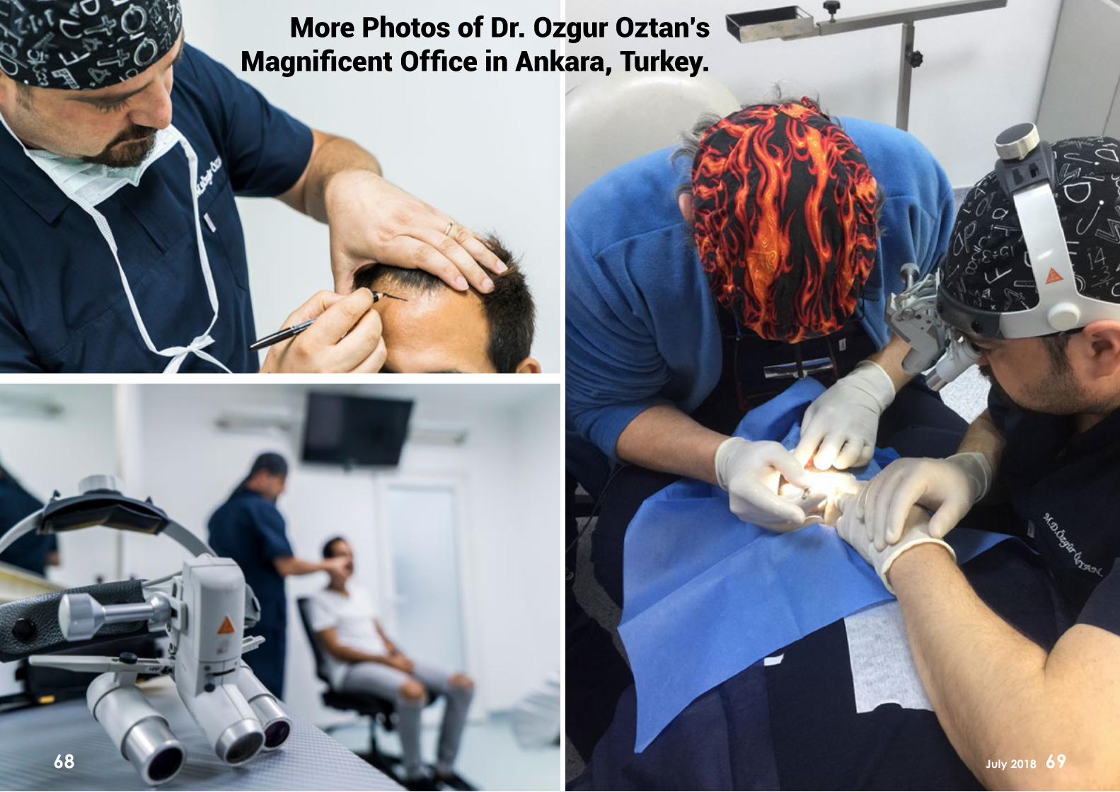
68 69July 2018
More Photos of Dr. Ozgur Oztan’s Magnificent Office in Ankara, Turkey.
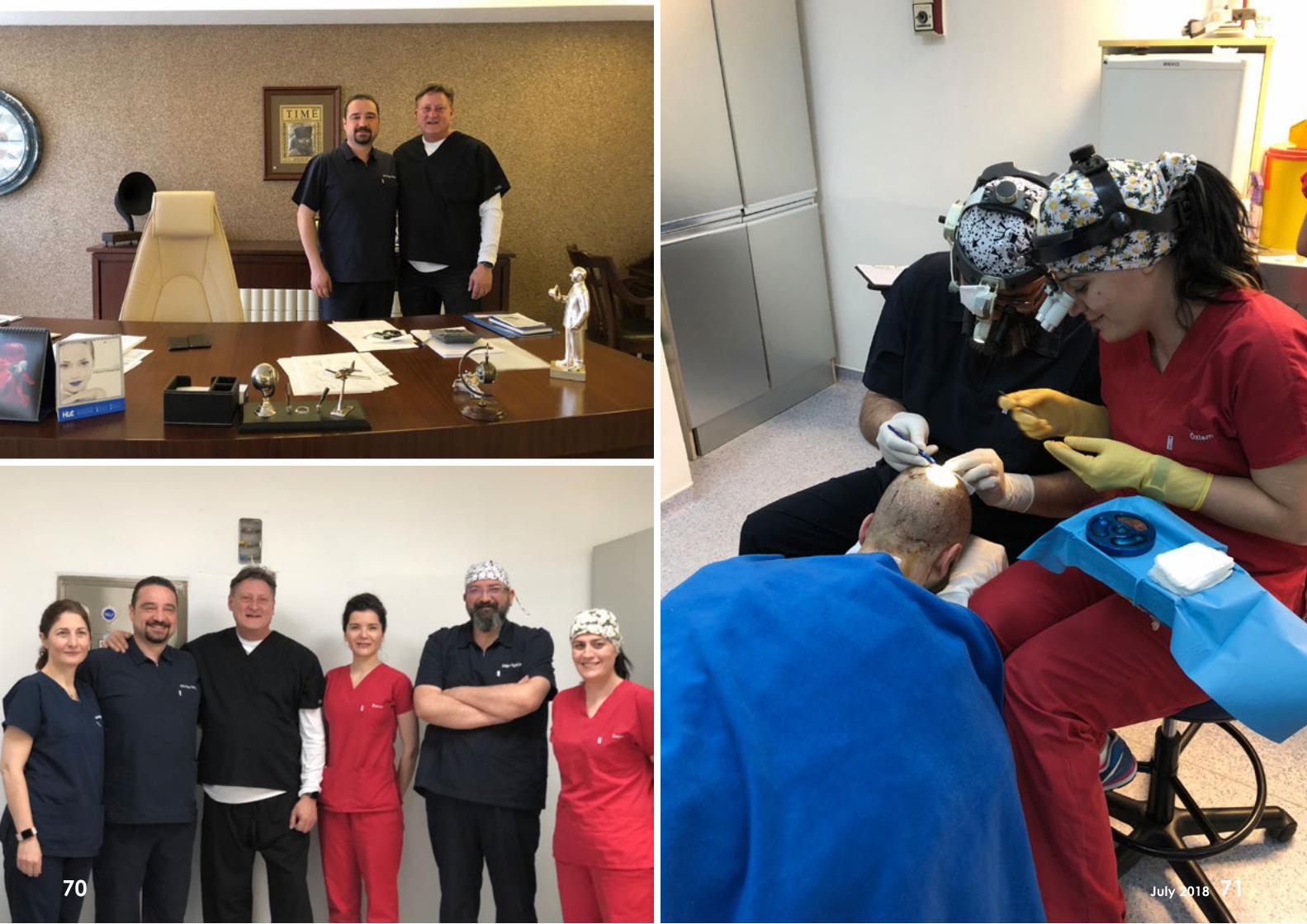
70 71July 2018
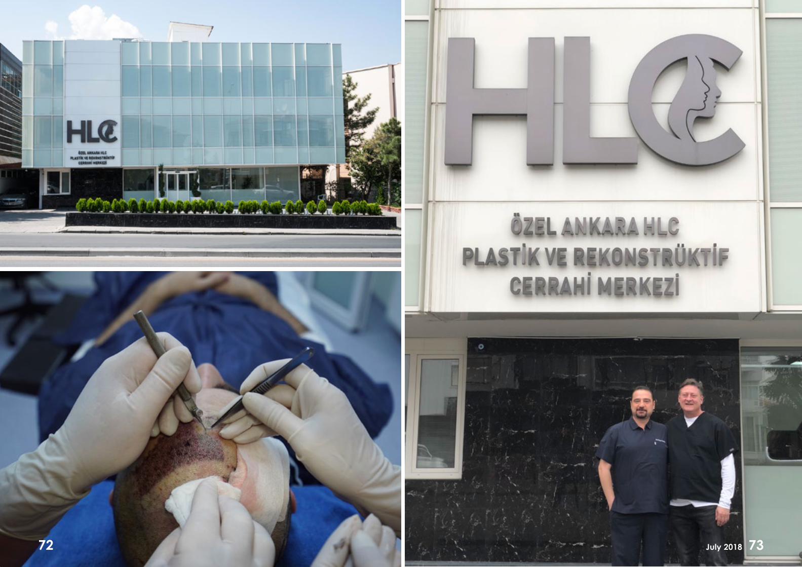
72 73July 2018

74 75July 2018

76 77July 2018
FU
EM
AG
AZ
INE
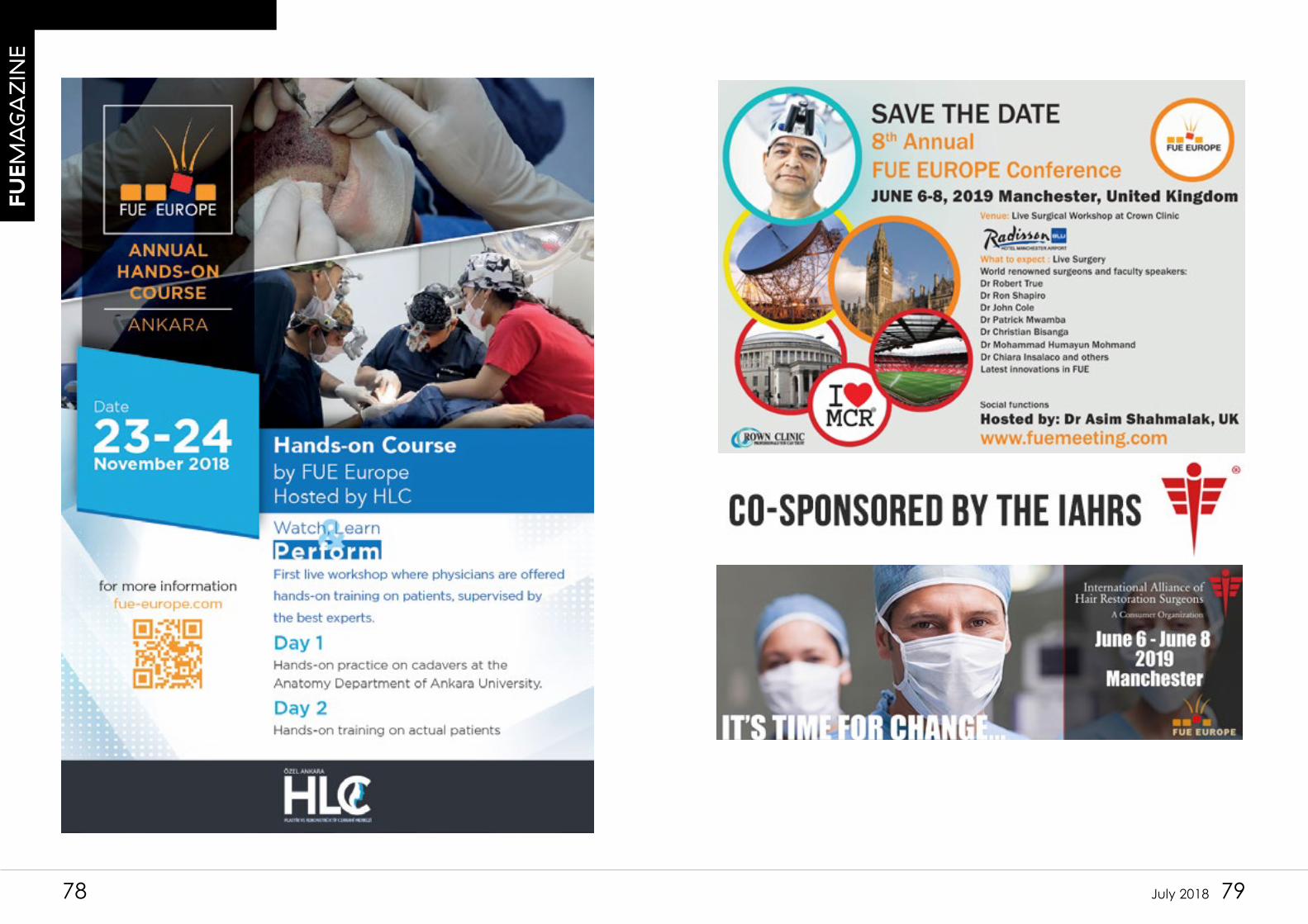
78 79July 2018
FU
EM
AG
AZ
INE