Embed Size (px)
Citation preview
45
Institute of Experimental Morphology, Pathology and Anthropology with Museum Bulgarian Anatomical SocietyActa morphologica et anthropologica, 25 (1-2)Sofia • 2018
Anthropology and Anatomy
Results from Anthropological Analysis of Bone Remains in Grave No 1 from Archaeological Site at Kremikovtsi Monastery „St. Georgi Pobedonosets“, SofiaNadezhda Atanassova*, Vladislav Todorov
Institute of Experimental Morphology, Pathology and Anthropology with Museum, Bulgarian Academy of Sciences, Sofia, Bulgaria
* Corresponding author e-mail: [email protected]
In 2015 were held excavations of the archeological site in Kremikovtsi Monastery. Well preserved grave (no. 1) with skeleton in anatomical order, orientated West-East, was uncovered in rock base. In the area of the skull, a structure of four reused bricks was established. Radiocarbon dating shows that the grave no. 1 is from the second half of 15th - first quarter of 16th c. The aim of the present study is to provide de-tailed anthropological information for the skeleton from grave no. 1: age-at-death, sex, stature, body mass, pathological changes and anatomical variations. In view of the high stature, body mass, massive and long bones with a strong relief, it can be concluded that the male buried individual had a very well developed musculoskeletal system and strenuous physical activity in lifetime. A large number of paleopathological changes have been diagnosed on the skull and trunk bones.
Key words: Kremikovtsi Monastery, Ottoman period, human skeleton, anthropological analysis, paleo-pathology
IntroductionKremikovtsi Monastery „St. Georgi Pobedonosets“ (St. George the Victorious) is a reco gnizable monument of the Bulgarian cultural heritage. It is located 3 km above Sofiaʼs quarter of Kremikovtsi, north-east of the center of Sofia city, in the southern slopes of Stara planina mountain.
Scientific interest about this Monastery was generated at the end of the 19th cen-tury [8, 24, 33] and continued throughout the 20th century [5, 15, 17, 21, 22]. Publica-tions described different aspects related to the history, the architecture of the buildings and stylistic features of the frescoes in the old church „St. Georgi Pobedonosets“. In

46
1987, excavations were carried out in conjunction with restoration work in naos (inner chamber) of the church [19, 20]. Four child skeletons have been discovered by Dr. Slav-cho Cholakov [20]. Investigators suggested that two of these children buried in 1493, belonged to the family of Radivoy - ctitor of the church [20]. In 2014, an archaeological observation was made during the construction of a church drainage where archaeologi-cal materials were not found [28].
In 2015 under the direction of archaeologist Vladislav Todorov excavations are extended, covering the area north of the old church [26]. During the field work a ne-cropolis is registered. It was compromised by the construction of monastery buildings during the Revival [26] and anthropogenic activities in the 20th century. Scattered hu-man bones from individuals of different ages and sexes are registered. There was also a fully preserved inhumated skeleton in an anatomical order (grave no. 1), orientated West-East. The burial ritual is Christian. In the area of the skull, a structure of four re-used bricks is established. Radiocarbon analysis shows that the grave no. 1 dated from the second half of the 15th century – first quarter of the 16th century.
The aim of the present study is to provide detailed anthropological information for the skeleton from grave no. 1, Kremikovtsi Monastery - the condition of the presented bones, age-at-death, sex, stature, body mass, pathological changes and anatomical vari-ations of the buried individual.
Material and MethodsThis paper includes results of detailed anthropological investigation of inhumated male skeleton from grave no.1 at archaeological site in Kremikovtsi Monastery, Sofia. The bones are dated from the Scottish Universities Environmental Research Centre via radiocarbon analysis in the second half of the 15th - first quarter of the 16th century. The skeletal remains are in very good condition. Their cortical layers are well preserved which allows establish-ment of paleopathological changes. The skull is fully represented. The postcranial skeleton is also completed preserved with the exception of some left tarsal bones.
The age-at-death of buried individual is established after symphyseal relief [27] and ossification degree of cranial sutures [1, 6, 13, 30]. Epiphyseal union [25], dental attrition [4, 32] and other general age indicators [3, 6, 7] are also used.
The sex of the buried individual is identified mainly from pelvic girdle bones - the shape of incisura ischiadica major and foramen obturatum, the value of the pubic angle, the breadth and length of the sacrum [3, 12]. The morphology of cranial elements and the measurements of trunk bones are also applied [3, 9, 12].
Assessment of the individual body mass is done after the biomechanical methods [23]. Stature reconstruction is based on the length of the long bones of the limbs by the formulae of Pearson-Lee [18] and Trotter-Gleser [29].
The pathological traces on the bones and normal anatomical variations are as-sessed after different paleopathological methods [2, 11, 14, 16].
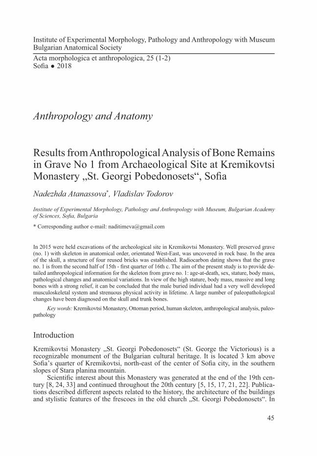
Results and DiscusionPosition of the skeleton in situ (Fig. 1)The buried individual was placed in supine position (lying on the back). A structure of four reused bricks covered the skull which lies on the occipital bone. The upper limbs are flexed in the elbow joints. The forearms are parallel and lie on the lower half of the
47
chest. The hands of the buried individual have been laid with their palms up. Lower limbs are stretched out.
Age-at-death estimationBased on the relief of symphyseal surface of the pubic bone and the degree of ossifica-tion (synostosis) of the coronal, sagittal and lambdoid cranial sutures, the age-at-death of individual from grave no. 1 is identified in age group Maturus - between 45 and 49 years.
Sex determinationThe sex is defined as male, based on a large number of scopical features of postcra-nial skeleton and skull: acute subpubic angle (arcus pubicus); the greater sciatic notch (incisura ischiadica major) is narrow; flat auricular surface of ilium (facies auricularis ossis ilii); high and narrow pelvis major; narrow and long sacral bone (os sacrum); the upper orbital margin (margo supraorbitalis) is rounded; the end of zygomatic arch (arcus zygomaticus) continues behind the external auditory opening (porus acusticus externus); mastoid process (processus mastoideus) is large and has a strong relief; the forehead is low; glabella and arcus supercilialis are moderately pronounced; the chin is square; an acute angle of the mandible (angulus mandibulae). The long bones of the limbs are massive with a great length and a strong expressed relief.
Sex is determined also as male by some measurements of postcranial bones (Table 1).
Fig. 1. Position of the skeleton in situ (grave no. 1)
48
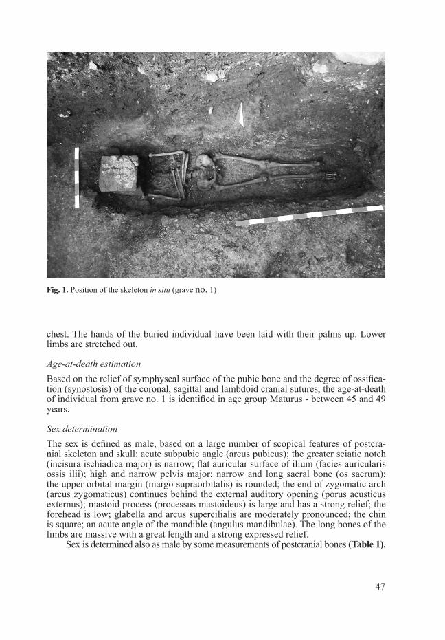
Metrical and scopical characteristic of the skullInvestigated skull is characterized by “large” absolute measurements of the cranial part and “medium” length and height of the face. Orbital height is “very small”, while nasal width and height are “very large” and “large”. The length of the palate is also “large”. The lower jaw is with a “large” angular width, a “small” chin height and a “very small” branch width. The indexing characterization showed that the skull is tall and wide, with a slight protuberance of the face.
The scopical skull decription is as follows: vertical norm is ovoid; occiput is flat with a fluent transition in a profile, the occipital bone being slightly protruding and moderate in relief (Fig. 2); mastoid process (processus mastoideus) is large, with a strong relief and a downward direction; frontal eminences (tubera frontalia) are less pronounced; glabella and arcus supercilialis are moderately pronounced; the shape of the face is ellipsoid; orbits are rectangular with a vertical slope; nasal bones are sym-metrical, narrow, with medium length and slight shedding; nasal aperture has blunt mar-gins and lower end – anthropina; cheekbones (ossa zygomatica) are slightly protruding and with vertical profiling; fossa canina dextra is medium deep and fossa canina sinistra is shallow, both of them with a slight expressed relief; mandibular bone is small, mode rately massive, with a strong relief and a narrow branch (ramus mandibulae). This jaw is deformed by the advanced edentulous ante mortem; the chin is moderately pro-nounced, triangular, wide-based and without sinking. Occlusion could not be determined due to edentulism of the upper jaw.
Bone (mm) Sex Bone (mm) Sex
Epycond.D hum. dx 72.0 ♂ (Bass 2005) D caput radii dx 25.0 ♂ (Kühl 1985)
Epycond.D hum. sin 70.0 ♂ (Bass 2005) D caput radii sin 24.5 ♂ (Kühl 1985)
Vert D caput humeri dx 47.5 ♂ (Bass 2005) Trans. D caput hum. dx 47.0 ♂ (Bass 2005)
Vert D caput humeri sin 48.0 ♂ (Bass 2005) Trans. D caput hum. sin 47.0 ♂ (Bass 2005)
D caput femоris dx 51.0 ♂ (Bass 2005) D caput femoris sin. 51.5 ♂ (Bass 2005)
Epycond.D fem. dx 87.0 ♂ (Bass 2005) Epycond.D fem.sin 87.0 ♂ (Bass 2005)
L clavicula dex 149.0 ♂ (Bass 2005) L clavicula sin 148.0 ♂ (Bass 2005)
Table 1. Measurements of postcranial bones used for sex determination of the individual from grave no. 1, Kremikovtsi Monastery
Dentition: permanent, incomplete:
right left
0 0 0 0 0 0 0 0 0 0 0 4 0 0 0 0
0 0 0 R 4 3 2 0 0 0 3 4 5 0 0 01 - a permanent tooth preserved in the alveolus; 0 - tooth lost before death (ante mortem) or tooth agenesis; R - preserved tooth root
Abrasion of the teeth - moderately (3-4 degree).
49
Stature and body mass estimationThe stature of male from grave no. 1 is reconstructed by the lengths of all long bones of the limbs (Table 2). Stature reconstructed by both formulae [18, 29] falls into the ca-tegory “tall” after Martin-Saller’s rubrications [12] for the European population. Heads of both femoral bones are preserved, making it possible to assess the body mass of the individual - 77.01 kg.
Bone Length (сm) Stature
Humerus dexter 34,0 169,04 cm (Pearson-Lee 1935 )175,17 cm ±4,05 (Trotter-Gleser 1952)
Humerus sinister 34,0 169,04 cm (Pearson-Lee 1935 )175,17 cm ±4,05 (Trotter-Gleser 1952)
Radius dexter 25,5 175,40 cm ±4,32 (Trotter-Gleser 1952)
Radius sinister 25,5 175,40 cm ±4,32 (Trotter-Gleser 1952)
Femur dexter 48,9 173,24 cm (Pearson-Lee 1935 )177,79 cm ±3,27 (Trotter-Gleser 1952)
Femur sinister 48,8 173,05 cm (Pearson-Lee 1935)177,55 cm±3,27 (Trotter-Gleser 1952)
Tibia dextra 41,0 181,94 cm±3,37 (Trotter-Gleser 1952)
Tibia sinistra 41,0 181,94 cm±3,37 (Trotter-Gleser 1952)
Mean value for the stature: 171,09 cm (after Pearson-Lee-Lee 1935 )177,55 cm±3,48 (after Trotter-Gleser 1952)
Fig. 2. Skull in norma lateralis (grave no.1)
Table 2. Lengths of long bone of the limbs used for reconstruction of the stature of the individual from grave no. 1, Kremikovtsi Monastery
50
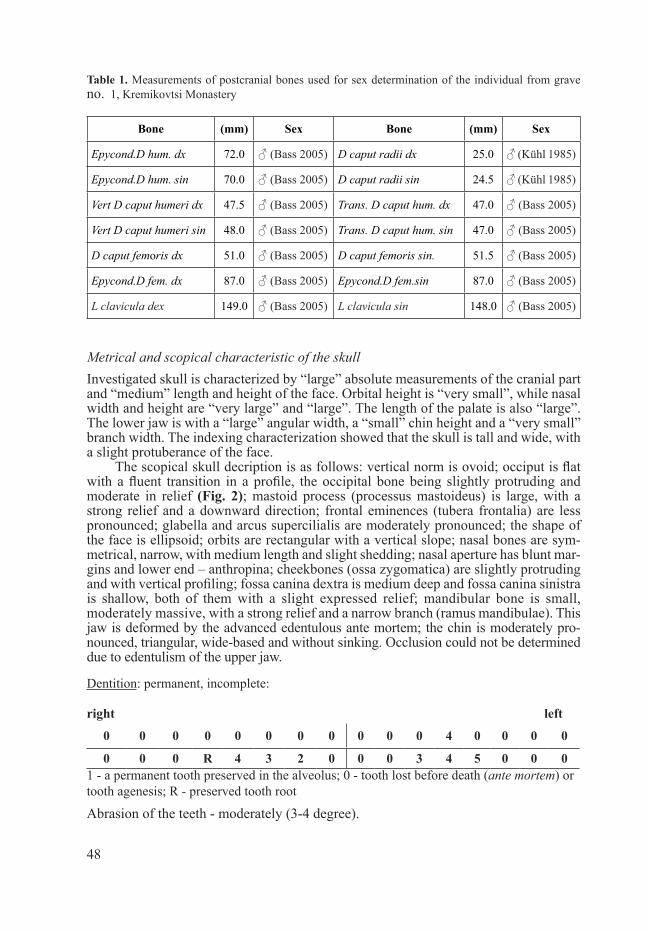
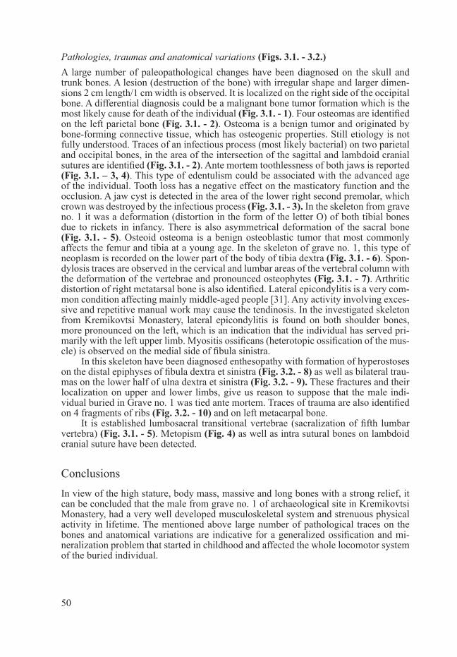
Pathologies, traumas and anatomical variations (Figs. 3.1. - 3.2.)A large number of paleopathological changes have been diagnosed on the skull and trunk bones. A lesion (destruction of the bone) with irregular shape and larger dimen-sions 2 cm length/1 cm width is observed. It is localized on the right side of the occipital bone. A differential diagnosis could be a malignant bone tumor formation which is the most likely cause for death of the individual (Fig. 3.1. - 1). Four osteomas are identified on the left parietal bone (Fig. 3.1. - 2). Osteoma is a benign tumor and originated by bone-forming connective tissue, which has osteogenic properties. Still etiology is not fully understood. Traces of an infectious process (most likely bacterial) on two parietal and occipital bones, in the area of the intersection of the sagittal and lambdoid cranial sutures are identified (Fig. 3.1. - 2). Ante mortem toothlessness of both jaws is reported (Fig. 3.1. – 3, 4). This type of edentulism could be associated with the advanced age of the individual. Tooth loss has a negative effect on the masticatory function and the occlusion. A jaw cyst is detected in the area of the lower right second premolar, which crown was destroyed by the infectious process (Fig. 3.1. - 3). In the skeleton from grave no. 1 it was a deformation (distortion in the form of the letter O) of both tibial bones due to rickets in infancy. There is also asymmetrical deformation of the sacral bone (Fig. 3.1. - 5). Osteoid osteoma is a benign osteoblastic tumor that most commonly affects the femur and tibia at a young age. In the skeleton of grave no. 1, this type of neoplasm is recorded on the lower part of the body of tibia dextra (Fig. 3.1. - 6). Spon-dylosis traces are observed in the cervical and lumbar areas of the vertebral column with the deformation of the vertebrae and pronounced osteophytes (Fig. 3.1. - 7). Arthritic distortion of right metatarsal bone is also identified. Lateral epicondylitis is a very com-mon condition affecting mainly middle-aged people [31]. Any activity involving exces-sive and repetitive manual work may cause the tendinosis. In the investigated skeleton from Kremikovtsi Monastery, lateral epicondylitis is found on both shoulder bones, more pronounced on the left, which is an indication that the individual has served pri-marily with the left upper limb. Myositis ossificans (heterotopic ossification of the mus-cle) is observed on the medial side of fibula sinistra.
In this skeleton have been diagnosed enthesopathy with formation of hyperostoses on the distal epiphyses of fibula dextra et sinistra (Fig. 3.2. - 8) as well as bilateral trau-mas on the lower half of ulna dextra et sinistra (Fig. 3.2. - 9). These fractures and their localization on upper and lower limbs, give us reason to suppose that the male indi-vidual buried in Grave no. 1 was tied ante mortem. Traces of trauma are also identified on 4 fragments of ribs (Fig. 3.2. - 10) and on left metacarpal bone.
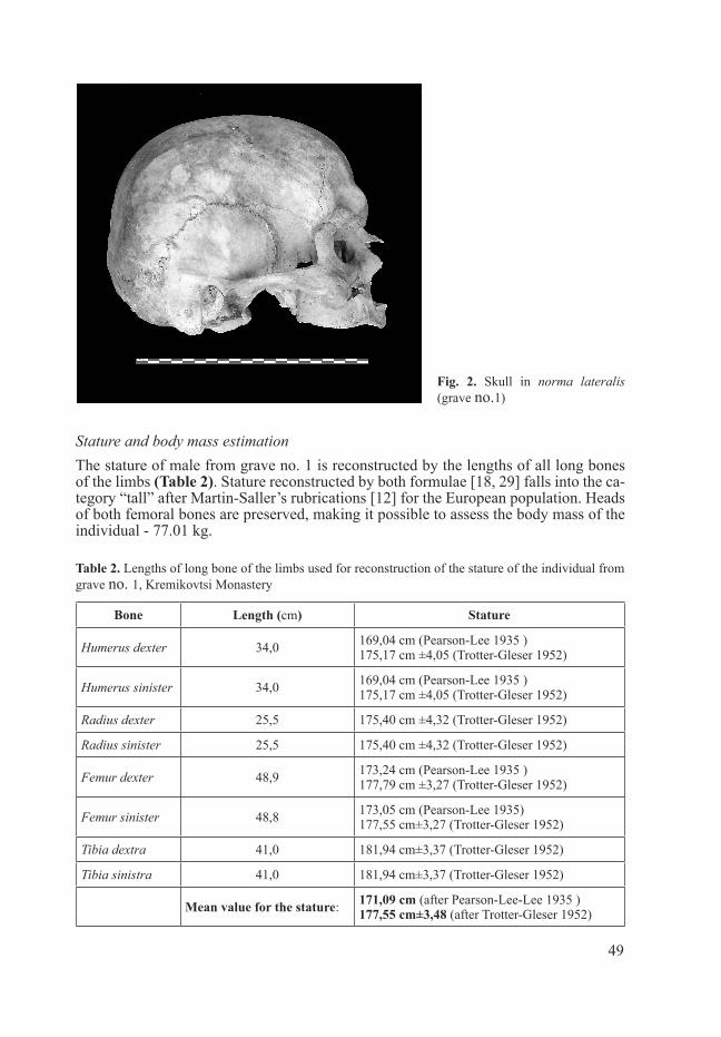
It is established lumbosacral transitional vertebrae (sacralization of fifth lumbar vertebra) (Fig. 3.1. - 5). Metopism (Fig. 4) as well as intra sutural bones on lambdoid cranial suture have been detected.
ConclusionsIn view of the high stature, body mass, massive and long bones with a strong relief, it can be concluded that the male from grave no. 1 of archaeological site in Kremikovtsi Monastery, had a very well developed musculoskeletal system and strenuous physical activity in lifetime. The mentioned above large number of pathological traces on the bones and anatomical variations are indicative for a generalized ossification and mi-neralization problem that started in childhood and affected the whole locomotor system of the buried individual.

51
1
2
3
4
5 7
6
8
9
10
Fig. 3.1. Bone diseases on the skeleton from grave No 11 – Lesion on squama occipitalis; 2 - Osteoma and traces from an infectious process on the skull; 3 - Toothlessness (edentulism) ante mortem and cyst on the mandibular bone; 4 - Toothlessness (edentulism) ante mortem of maxillary bone; 5 - Asymmetrical deformation of sacral bone and lum-bosacral transitional vertebrae; 6 - Osteoid osteoma on the lower part of the body of right tibia; 7 - Spondylosis in the cervical area of columna vertebralis;
Figure 3.2. Bone diseases and trau-mas on the skeleton from grave no 18 - Enthesopathy on the distal epi-physes of fibula dextra et sinistra; 9 – Traces of fracture on ulna dextra et sinistra; 10 - Traces of fracture on ribs
Fig. 4. Metopism (unfused sutura metopica) on frontal bone of investigated skeleton

52
R e f e r e n c e s
1. Alekseev, V., G. Debets. Craniometry, methods of anthropological study. Moscow, Nauka, 1964, 228 p. [in Russian].
2. Aufderheide, C., C. Rodriguez-Martin. The Cambridge Encyclopedia of Human Paleopathology. Cambridge University Press, 1998, 478 p.
3. Bass, W. Human osteology: a laboratory and field manual of the human skeleton. University of Mis-soury, Special publication no. 2 of the Missouri Archaeological Society, 2005, 365 p.
4. Brothwell, D. R. The relationship of tooth wear to aging. - In: Age markers in the human skeleton (Ed. M. İşcan), Illinois, Springfield, 1989, 303-316.
5. Dimitrova, Z., Z. Ganeva, G. Stoyanova. Sofia Monasteries. Origin, development, structure and character. Sofia, Publishing Center at the Ministry of Culture, 1992, 130 p. [in Bulgarian].
6. Ferembach, D., I . Schwidetzky, M. Stloukal. Recommendations for age and sex diagnosis of skel-etons. - J. Hum. Evol., 9, 1980, 517-549.
7. Gerasimov, M. Reconstruction of the face by the skull. Moscow, Nauka, 1955, 586 p. [in Russian].8. Irechek, K. Principality of Bulgaria, II. Traveling in Bulgaria. Plovdiv, Hristo Danov Publishing
House, 1899, 943 p. [in Bulgarian].9. Kühl, R. Skeletal remains from praehistorical cremations and their possibilities for interpretation
with with attention to the special problems in Schleswig-Holstein. - Communications of the An-thropological Society in Wien (MAGW), 115, 1985, 113-137 [in German].
10. Lovejoy, C., R. Meindl, T. Pryzbeck, R. Mensforth. Chronological metamorphosis of the auricular surface of the ilium: A new method for the determination of adult skeletal Age-at-death. - Am. J. Phys. Anthropol., 68, 1985, 15-28.
11. Mann, R., D. Hunt. Photographic regional atlas of bone disease. A guide to pathologicand normal variation in the human skeleton. Illinois, Charles Thomas Publisher Ltd., 2005, 416 p.
12. Martin, R., K. Saller. Lehrbuch der Anthropologie in sistematischer Darstellung, Band I. Stuttgart, G. Fischer Verlag, 1957, 429 –595.
13. Meindl, R., C. Lovejoy. Ectocranial suture closure: a revised method for the termination of skeletal age-at-death based on lateral-anterior sutures. - American Journal of Physical Anthropology, 68, 1985, 57-66.
14. Merbs, C., F. Trauma. Reconstruction of Life (Eds. M.Y. Iscan, K. Kennedy), New York, Alan R. Liss, 1989, 161-189.
15. Mihailov, S. Ctitor portrait in Kremikovtsi church in light of the Bulgarian-Romanian cultural rela-tions in the fifteenth century. – Archaeology, 2, 1960, 23-29 [in Bulgarian].
16. Ortner, D. J., J. Putschar. Identification of pathological conditions in human skeletal remains. Washington, Smithsonian Institution Press, 1981, 499 p.
17. Paskaleva-Kabadieva, K. The church “St. George” in Kremikovtsi Monastery. Sofia, Publishing house Bulgarian artist, 1980, 164 p. [in Bulgarian].
18. Pearson, K. Mathematical Contributions to the Theory of Evolution. V. On the Reconstruction of the Stature of Prehistoric Races. - Philosophical Transactions of the Royal Society of London. Series A, Containing Papers of a Math. or Phys. Character (1896-1934), 192, 1899, 169-244.
19. Pobornikova, R. New archeological studies of three medieval monasteries from Sofia. – Serdica – Sredetc - Sofia, 2, 1994, 117-144 [in Bulgarian].
20. Pobornikova, R. Archaeological excavations in naos of the medieval church „St. George“ in Kremikovtsi Monastery. - Contributions to Bulgarian Archeology, III-IV, 2006, 173-179 [in Bulgarian].
21. Protich, A. A portrait model for Bulgarian masters in 18th-19th century. - Yearbook of the National Museum, 4, 1922, 21-25 [in Bulgarian].
22. Protich, A. Denationalization and revival of our art from 1393 to 1879. - Bulgaria 1000 years, 1930, 434-435 [in Bulgarian].
23. Ruff, C. B., W. W. Scott, A. Y. Liu. Articular and diaphyseal remodeling of the proximal femur with changes in body mass in adults. - Am. J. Phys. Anthropol., 86(3), 1991, 397–413.
24. Shindarov, I. Some notes about Kremikovtsi Monastery near Sofia. - Collection of folk perceptions, XV, 1898, 304-309 [in Bulgarian].
25. Stull, K., D. James. Determination of age-at-death using the acetabulum of the os coxa. - In: Age Estimation of the Human skeleton (Eds. K. Latham, M. Finnegan), Springfield, 2010, 134-146.

53
26. Todorov, V., N. Atanassova. Archaeological excavations at Kremikovtzi Monastery “Saint George The Victorious”, Sofia municipality, “Kremikovtzi” district. - Archaeological Discoveries and Ex-cavations in 2015. 2016, 748-750 [in Bulgarian, with English summary].
27. Tood, T. W. Age changes in the pubic bone I: the male white pubic. - American Journal of Physical Anthropology, 3, 1920, 285-334.
28. Tranteev, B. The church “St. George” in PI with identifier 68134.8229.9 at Manastirsky livadi local-ity, Kremıvotzi district, Sofia city. - Archaeological Discoveries and Excavations in 2014. 2015, 765-766 [in Bulgarian].
29. Trotter, M., G. Gleser. Estimation of Stature from Long Bones of American Whites and Negroes. - American Journal of Physical Anthropology, 10, 1952, 463-514.
30. Valois, H. V. Lʼomoplate humaine: Étude anatomique et anthropologique. - Buletins et Mémoires de la Société dʼAnthropologie de Paris, 3, 1932, 3-153.
31. Vaquero-Picado, A., R. Barco, S. A. Antuña. Lateral epicondylitis of the elbow. - EFORT Open Rev, 1, 2016, 391-397.
32. Zubov, А. Odontology. Methods of anthropological investigations. Moscow, Nauka, 1968, 200 p. [in Russian].
33. Unknown author. Description of Kremikovtsi Monastery “St. George”. - Religious stories, 9-10, 1896, 376-384 [in Bulgarian].