Embed Size (px)
Citation preview

Anthrax
Nina Marano DVM, MPH, Dipl. ACVPMMeningitis and Special Pathogens Branch
National Center for Infectious Diseases

Overview
• History, etiology, pathogenesis• Burden of natural disease• Disease forms• Recognition and diagnosis• Differential diagnosis• Treatment• Prevention

History
• Malignant Pustule, Malignant Edema, Siberian plague, Black Baine, Ragpicker's disease, Woolsorter's disease
• Described since ancient times
• Medieval Period - multiple pandemics
• 1876 - The first microbial etiology (Koch)
• 1881 - The first live bacterial vaccine (Pasteur)
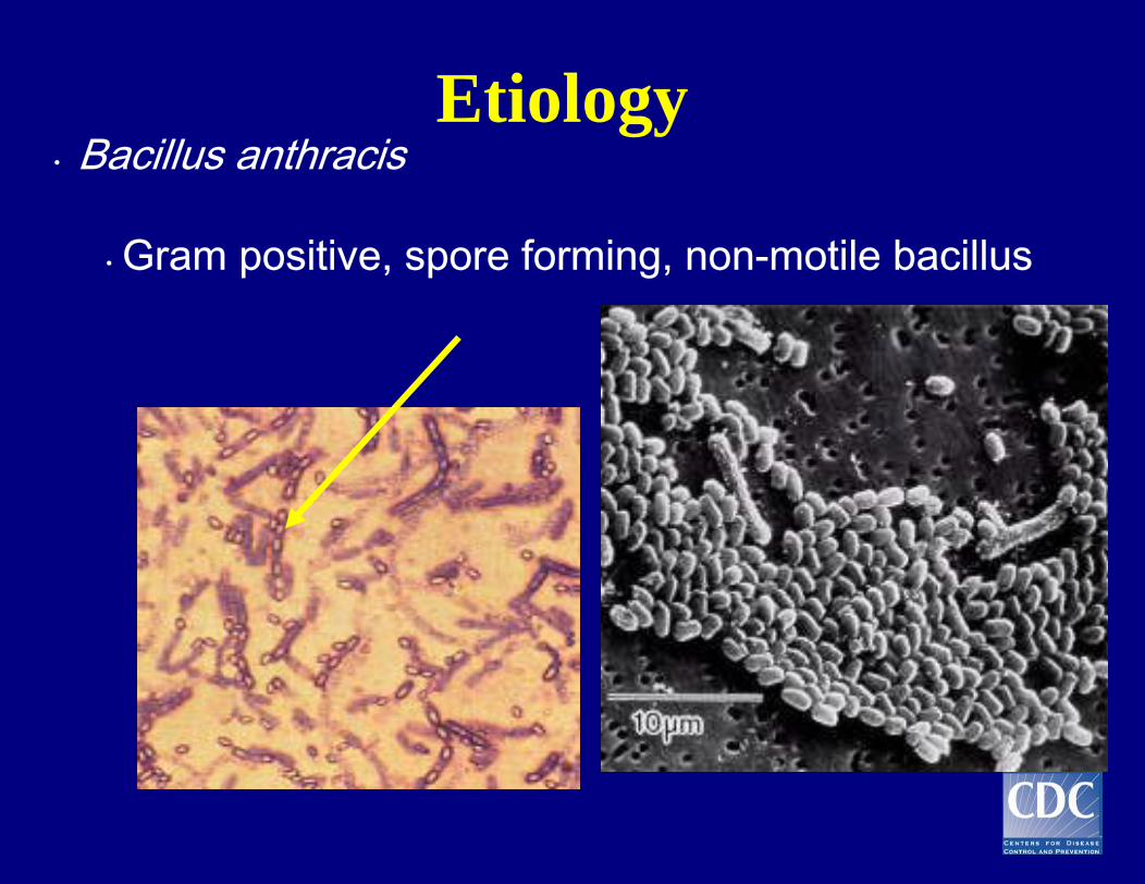
Etiology• Bacillus anthracis
• Gram positive, spore forming, non-motile bacillus

Bacillus anthracis: Microbiology
Tacky (egg white) Non-hemolytic
Tails (medusa head) colony morphology
Penicillin susceptible (97%)
Phage susceptible

Anthrax Binary ToxinsProtective Antigen
(PA, 83kDa)Receptor binding & toxin internalisation
Lethal Factor
(LF, 90kDa)Endopeptidase
Edema Factor
(EF, 89kDa)Adenylyl cyclase
Lethal Toxin Edema Toxin
ATP -->cAMP
Cytokine modulation
Edema
MAPKK cleavage
MØ lysis
Cytokine modulation
Fatal toxic shock

Anthrax Toxin - Mode of Action(from Leppla, 1999)
MEK-1
CaM
EF or LF
Target CellTarget Cell
PA83
Furin PA20
PA63
ATP
cAMP
EndocytosisEndocytosisLFLF
EFEF
TranslocationTranslocation
H+
Pathogenesis of Anthrax
NEJM 1999; 341: 815- 826
Spores

Anthrax in Animals Rapid progression from febrile illness to death with hemorrhage
Dogs and pigs get a pharyngeal form – more resistant

Epidemiology and Transmission in Humans
Spores live in the soil for many yrs: at least 60 yrs
Animals ingest spores
Humans become infected from animal products
- Cutaneous: direct contact
- Gastrointestinal: ingestion of infected meat
- Inhalation: inhalation of aerosolized spores
Generally, not transmissible person-to-person

Why is Anthrax a Threat Agent?
• Persistence of endospore in environment
• Pathogenicity• Delayed onset of recognizable
symptoms renders treatment ineffective
• Can be manufactured using standard laboratory equipment
• No recognizable color, taste, or odor

2001 Anthrax Threat Letters
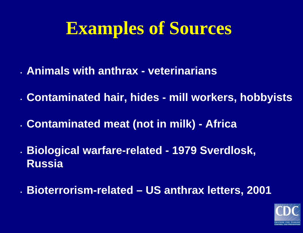
Examples of Sources
• Animals with anthrax - veterinarians
• Contaminated hair, hides - mill workers, hobbyists
• Contaminated meat (not in milk) - Africa
• Biological warfare-related - 1979 Sverdlosk, Russia
• Bioterrorism-related – US anthrax letters, 2001

Burden of Natural Disease• Disease absent/sporadic in Northern Europe• More common in Greece, Italy, Spain, Turkey, Yugoslavia• U.S. - South Dakota, Nebraska, Oklahoma, Texas• Enzootic in Central America, Peru, Bolivia, Venezuela• Hyperendemic in Middle Eastern and adjoining countries of former USSR republics
• Largest recent epidemic: Zimbabwe,1978-80 - 10,000human cases
• Reporting deficiencies due to decreasing veterinary experience in case recognition and civil unrest
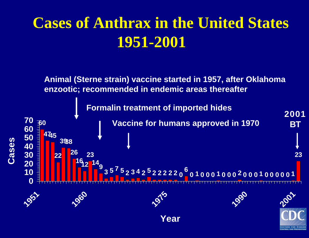
Cases of Anthrax in the United States 1951-2001
604745
22
393826
161223
1493 5 7 5 2 3 4 2 5 2 2 2 2 2 0
60 1 0 0 0 1 0 0 0 2 0 0 0 1 0 0 0 0 0 1
23
010203040506070
1951
1960
1975
1990
2001
Year
Cas
es
Animal (Sterne strain) vaccine started in 1957, after Oklahoma enzootic; recommended in endemic areas thereafter
Formalin treatment of imported hides
Vaccine for humans approved in 19702001
BT

Cutaneous Anthrax• 95% of human cases
• 1 - 2 days post exposure, papule develops (2 - 19 days)
• 2 - 4 days, ulcer surrounded by vesicles
• Black eschar forms - painless with edema
• Most common site is head, forearm, hands
• Untreated: 5-20% case-fatality rate

Anthrax: CutaneousDay 6Vesicle
developmentDay 2 Day 4
Day 10
Eschar formation

Anthrax: Cutaneous

Cutaneous Anthrax Resulting from Bioterrorism, NYC, October, 2001

Diagnosis of Cutaneous Anthrax
Eschar formationCulture of vesicular fluid or exudateBlood cultureBiopsyPolymerase chain reactionImmunofluorescence and immunohistochemistry

Differential Diagnosisof Cutaneous Anthrax
• Spider bite• Rickettsialpox• Varicella zoster• Herpes simplex• Staphylococcal or streptococcal cellulitis• Ecthyma gangrenosum• Ulceroglandular tularemia• Plague

Gastrointestinal Anthrax• Ingestion
– Rare natural incidence• Undercooked, ground beef
– Most simple, high-consequence application method
• Possible route of choice for criminals and non-state sponsored terrorists
– Symptoms• Nausea 2-5 days after ingestion
– Mortality• Up to 50% without treatment
Intestinal lesion of GI anthrax

Inhalation AnthraxInhalation of sporesIncubation, 2-3 days (range up to 60 days)Spores engulfed by macrophages and transported to mediastinal and peribronchial lymph nodesInsidious onset: malaise, low grade fever, nonproductive coughAbrupt development of respiratory distressHemorrhagic mediastinitisHematogenous spreadMeningitis in 50% Case fatality rate before 2001 – 90%

Inhalation Anthrax

Inhalation Anthrax: Diagnosis
Chest radiographs - widened mediastinum, pleural effusionsBlood or cerebrospinal fluid culture and Gram stainPolymerase chain reaction (PCR)Immunofluorescence and immunohistochemistry

Differential Diagnosisof Inhalation Anthrax
• Q fever• Viral pneumonia• Histoplasmosis
(fibrous mediastinitis)• Coccidioidomycosis
• Mycoplasmal pneumonia
• Legionnaires’ disease
• Psittacosis• Tularemia

Distinguishing Anthrax from Influenza-like Illness (ILI)
• ILI: – Nasal congestion and rhinorrhea– ILI not usually associated with radiographic findings of
pneumonia– Person-to-person spread– Rapid influenza testing and viral culture useful to
indicate whether viruses are circulating among specific populations
• Anthrax: – Abnormal chest radiographs – No person-to-person spread

Treatment of Inhalation or Complicated Cutaneous Anthrax
Strategy: • Early treatment • Combination therapy • Avoid penicillins, at
least early• ? Antitoxin (not
available)• ? Steroids• Treat for 60 days total
Assumptions:• Rapid progression /
systemic• Beta-lactamases
were present in isolates from FL, NY, DC (2001)
• Toxin – mediated• High fatality

Recommended Initial* Anthrax Treatment
Therapy DurationCutaneous Ciprofloxacin 14 days
500mg BID POOR
Doxycycline100 mg BID PO
Inhalation Ciprofloxacin 400 mg IV BID 14 days,OR may switch
Doxycycline 100 mg BID IV to PO when clinically
appropriate
Ciprofloxacin or doxycycline also recommended as initial therapyfor children in appropriate doses
*Until antibiotic susceptibility test results available

Recommended Postexposure Antibiotic Prophylaxis for Prevention of Inhalation Anthrax
Initial Therapy DurationAdults Ciprofloxacin 60 days(including pregnant 500 mg PO BID women and OR
immunocompromised) Doxycycline100 mg PO BID
Children Ciprofloxacin 60 days10-15 mg/kg PO Q 12 hrs
ORDoxycycline:
>8 yrs and >45 kg: 100 mg PO BID>8 yrs and <45 kg: 2.2 mg/kg PO BID
<8 yrs: 2.2 mg/kg PO BID

Control / Prevention
- Reducing infection in livestock
- Supervised slaughter and meat inspection
- Reducing exposure through import restriction, biosafety precautions, education
- Vaccination of high-risk human populations
- Treatment and post-exposure prophylaxis
Human disease is controlled by:
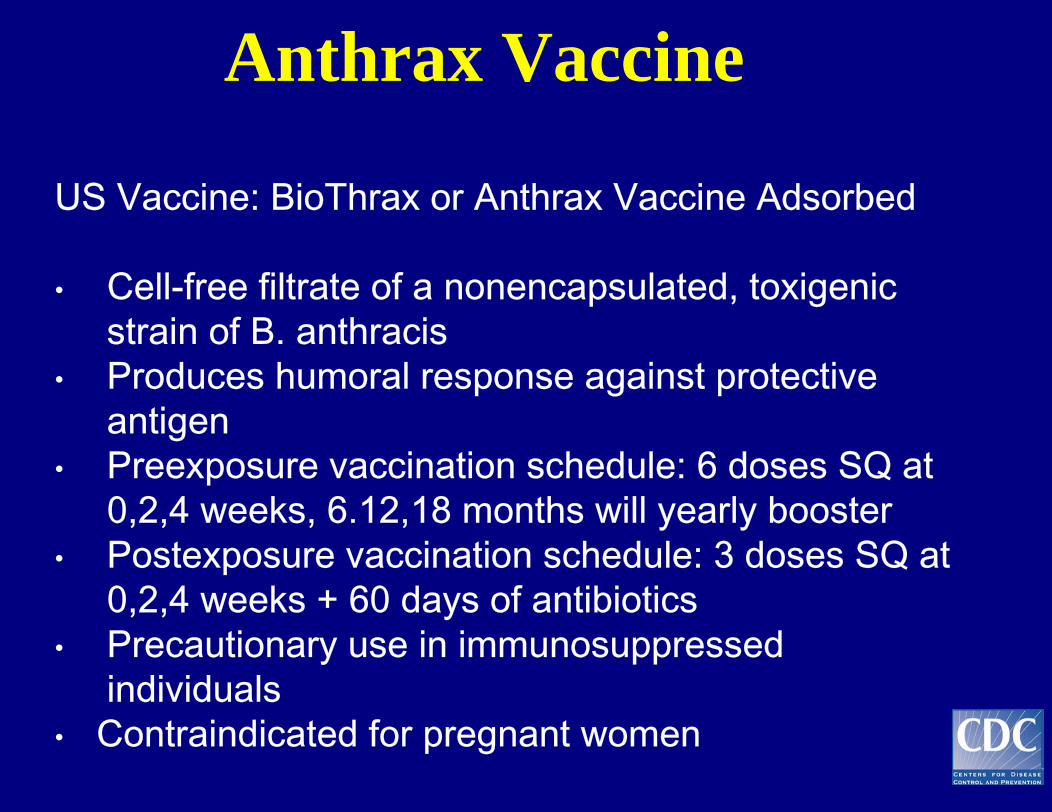
Anthrax Vaccine
US Vaccine: BioThrax or Anthrax Vaccine Adsorbed
• Cell-free filtrate of a nonencapsulated, toxigenicstrain of B. anthracis
• Produces humoral response against protective antigen
• Preexposure vaccination schedule: 6 doses SQ at0,2,4 weeks, 6.12,18 months will yearly booster
• Postexposure vaccination schedule: 3 doses SQ at0,2,4 weeks + 60 days of antibiotics
• Precautionary use in immunosuppressed individuals
• Contraindicated for pregnant women

Human Anthrax Vaccine Trial
Brachman PS et al. Field Evaluation of a Human Anthrax Vaccine. 1962. Am J Pub Health 52: 632-645.
– Randomized adjuvant control trial using alum vaccine
– 4 mills processing goat hair
– 379 vaccinated and 870 controls
- 3 cases (cutaneous) in vaccinated group: 2 had not completed series
- 23 cases in unvaccinated group, 5 inhalation
- 93% efficacy (95%CI = 65% to 95%)

Animal Studies of Post-exposure Prophylaxis
- Henderson, et al (1956): Alum precipitate PA filtrate vaccine
Methods: 5 days of penicillin compared to penicillin with postexposure vaccination
Results: 9 of the 10 receiving only penicillin died, while all of the macaques receiving penicillin and vaccine survived
- Friedlander et al (1993): Aluminum hydroxide PA filtrate vaccine
Methods: 30 days of various antibiotics compared to 30 days of doxycycline with postexposure vaccination
Results: 9 of the 10 animals in the doxycycline-alone arm survived, while all receiving doxycycline and vaccine survived

Anthrax Vaccine Safety
• Mild local reactions (tenderness, swelling, nodule formation) occur in 30% - 60% of recipients
• Large local reactions occur in < 1% of vaccinees• Systemic reactions: 5%-35% experience muscle ache,
joint ache, headache, malaise, fever• Serious side effect profile similar to other vaccines
given to adults (influenza and hepatitis)

Deaths with and without post-exposure prophylaxis following an anthrax release
Start of post-attack treatment (days)
-5
5
15
25
35
N o P ro g ra m 1 4 1 5 1 6 1 7 1 8 1 9 2 0
Source: Kaufmann AF, Meltzer MI, Schmid GP. Emerging Infect Dis 1997; 3:83-94.
With chemoprophylaxisWithout
1 2 3 4 5 6 7
15%
83%
Dea
ths (
1000
,s/10
0,00
0)

Case Study 1• It is 5pm on a Friday afternoon in December and you
are getting ready to go a Christmas party with your
family. Your last appointment of the day is a 53 year
old male office worker with the following
complaints:
– Headache, malaise, muscle ache, feels feverish and having
some difficulty breathing
• Your exam findings: absence of breath sounds on the
right side of the thorax, crackles on the left side,
body temperature 37.5° C

Chest X-Ray Results
What is on your list of differential diagnoses?

• You hospitalize the patient; hiscondition worsens rapidly with dyspnea, cyanosis, and hemoptysis
• What samples should you consider collecting?

Specimen Selection is ImportantAnthrax • Blood or cerebrospinal fluid – gram stain• Pleural fluid – request immunohistochemical staining
Tularemia• Serum for antibody titer• Pharyngeal wash or sputum specimen for culture, direct
flourescent antibody, organism gram stains poorly
Pneumonic Plague• Sputum/throat or bronchial washings- request Wayson
stain to see bipolar organism, or direct flourescentantibody of smear

Case Study 2: A US tourist comes to see you with this lesion on her arm.What questions would you ask this patient?

Seven days after her first visit, she comes to see you again. The lesion now looks like this – what is on your list of differential diagnoses?

DO NOT PANIC !
• Individuals must be exposed to B. Anthracis spores
• To cause disease, B. anthracis spores must enter the skin, be swallowed, or inhaled
• Disease can be prevented after exposure to anthrax spores by early treatment with appropriate antibiotics
• Anthrax is NOT spread from person to person

Anthrax Information
• www.aad.org/BioInfo/Biomessage2• www.who.int/emc-documents/zoonoses/docs• www.bt.cdc.gov

Acknowledgements
• Marc Fischer MD MPH• David Ashford DVM MPH DSci• Conrad Quinn PhD• Tanya Popovic PhD• Nancy Rosenstein MD• Bradley Perkins MD• David Stephens MD• Richard Besser MD