Embed Size (px)
Citation preview
Volume 18 Number 2 / April 2014 Lingao et al 191
Literature Search
The authors searched PubMed (MEDLINE) for English-language articles for theperiod1946-2013using combinationsof the following search terms: orbital compression syndrome, sicklecell disease, epidural hematoma, and cephalohematoma.
References
1. van Beers EJ, van Tuijn CF, Nieuwkerk PT, Friederich PW,Vranken JH, Biemond BJ. Patient-controlled analgesia versus contin-uous infusion of morphine during vaso-occlusive crisis in sickle celldisease, a randomized controlled trial. Am JHematol 2007;82:955-60.
2. Ganesh A, Al-Zuhaibi S, Pathare A, et al. Orbital infarction in sicklecell disease. Am J Ophthalmol 2008;146:595-601.
3. Ganesh A, William RR, Mitra S, et al. Orbital involvement in sicklecell disease: a report of five cases and review literature. Eye (Lond)2001;15:774-80.
4. KaracostasD, ArtemisN, PapadopoulouM,Christakis J. Case report:epidural and bilateral retroorbital hematomas complicating sickle cellanemia. Am J Med Sci 1991;302:107-9.
5. Ozkavukcu E, Fitoz S, Yagmurlu B, Ciftci E, Erden I, Ertem M.Orbital wall infarction mimicking periorbital cellulitis in a patientwith sickle cell disease. Pediatr Radiol 2007;37:388-90.
6. Curran EL, Fleming JC, Rice K, Wang WC. Orbital compressionsyndrome in sickle cell disease. Ophthalmology 1997;104:1610-15.
7. MankadVN,Williams JP,HarpenMD, et al.Magnetic resonance im-aging of bone marrow in sickle cell disease: clinical, hematologic, andpathologic correlations. Blood 1990;75:274-83.
8. Kim SK,Miller JH.Natural history and distribution of bone and bonemarrow infarction in sickle hemoglobinopathies. JNuclMed 2002;43:896-900.
9. Buyukdereli G, Guney IB, OzerdemG, Kesiktas E. Evaluation of vas-cularized graft reconstruction of the mandible with Tc-99m MDPbone scintigraphy. Ann Nucl Med 2006;20:89-93.
10. Fukumoto M, Yoshida S, Yoshida D, Kishimoto S. Dual-isotopeSPECT of skull-base invasion of head and neck tumors. J NuclMed 1995;36:1741-6.
Anterior lentiplane associatedwith posterior lenticonus andmicrocorneaMichelle D. Lingao, MD,a Grace T. Liu, MD,a,b,c
Kammi Gunton, MD,a,d and Alex V. Levin, MD,MHSca,d
Author affiliations: aPediatric Ophthalmology and Ocular Genetics, Wills Eye Institute,Philadelphia, Pennsylvania; bPediatric Ophthalmic Consultants, New York, New York;cDepartment of Ophthalmology, New York University, Langone Medical Center New York,New York; dThomas Jefferson University, Philadelphia, PennsylvaniaFunded in part by the Foerderer Fund (AVL), the Alfiero and Lucia Palestroni
Foundation (GTL), and an Alcon Fellowship Grant (MDL, GTL).Submitted June 7, 2013.Revision accepted November 1, 2013.Correspondence: Alex V. Levin,MD,MHSc, Chief, Pediatric Ophthalmology and Ocular
Genetics, Wills Eye Institute, 840 Walnut St., Philadelphia, Pennsylvania 19107-5109(email: [email protected]).J AAPOS 2014;18:191-192.Copyright � 2014 by the American Association for Pediatric Ophthalmology and
Strabismus.1091-8531/$36.00http://dx.doi.org/10.1016/j.jaapos.2013.11.009
Journal of AAPOS
We report a 12-year-old boy with a rare condition consisting offamilial bilateral anterior lentiplane (a flat anterior lens capsule)posterior lenticonus, and microcornea.
Case Report
A12-year-old boy was referred to the Wills EyeInstitute, Philadelphia, for evaluation for possiblecataract surgery. He had been followed since in-
fancy for bilateral congenital cataracts and high hyperopia.The cataracts were initially visually insignificant but even-tually caused symptomatic decreased visual acuity in theleft eye. The patient was the product of an uncomplicatedfull-term pregnancy and delivery. Physical examinationwas normal. He was otherwise healthy.
A multigenerational pedigree revealed no consanguin-ity. His 13-year-old sister had congenital cataracts,microcephaly, microphthalmia (axial length, 19.91 mmin the right eye and 20.31 mm in the left eye), andlearning disability; she underwent lens extraction at 2weeks of age and successful rehabilitation with contactlenses.
On his ophthalmological examination, the best-correctedvisual acuity (17.0010.75� 100 right eye,19.7512.00�95 left eye) was 20/30 and 20/600 respectively. Pupillaryresponses were normal; there was no afferent pupillarydefect.
Slit-lamp examination showed a right posterior subcap-sular cataract within a posterior lenticonus with spokelikesubcapsular opacities radiating from the center of theposterior capsule (Figure 1) and a more diffuse posteriorsubcaspular cataract within a large posterior lenticonus inthe left eye. There was also a left anterior subcapsularcataract. Corneal diameters were 9.5 mm in each eye.Intraocular pressure was 14 mm Hg in each eye byGoldmann applanation tonometry. Dilated fundus exami-nation was normal, but the view of the left retina wasobscured by the cataract.
Immersion A-scan biometry (Eyecubed V4; Ellex,Minneapolis, MN) revealed axial length of 22.45 mm inthe right eye and 22.52 mm in the left eye. Keratometry(Eyecubed V4 ) showed 37.79 � 38.84 D in the right eyeand 37.09 � 38.93 D in the left eye. Anterior chamberdepth was 2.90 mm on the right and 3.18 mm on the leftleft. Lens thickness was 3.62 mm on the right and 3.13mm on the left. Ultrasound biomicroscopy (SKU24-6300; Accutome, Malvern, PA) of both eyes showed aflat anterior surface of the lens (Figure 2).
The patient underwent left cataract extraction withanterior vitrectomy. Intraoperatively, the flattened lenssurface was not discernible. The anterior capsule seemedfriable, and a tag broke off during manual capsulorhexis.Capsulorhexis was completed despite a rent that remainedconfined to the anterior lens surface. Because of his ante-rior segment dimensions, capsule abnormalities, andpositive family experience with contact lenses, he was left
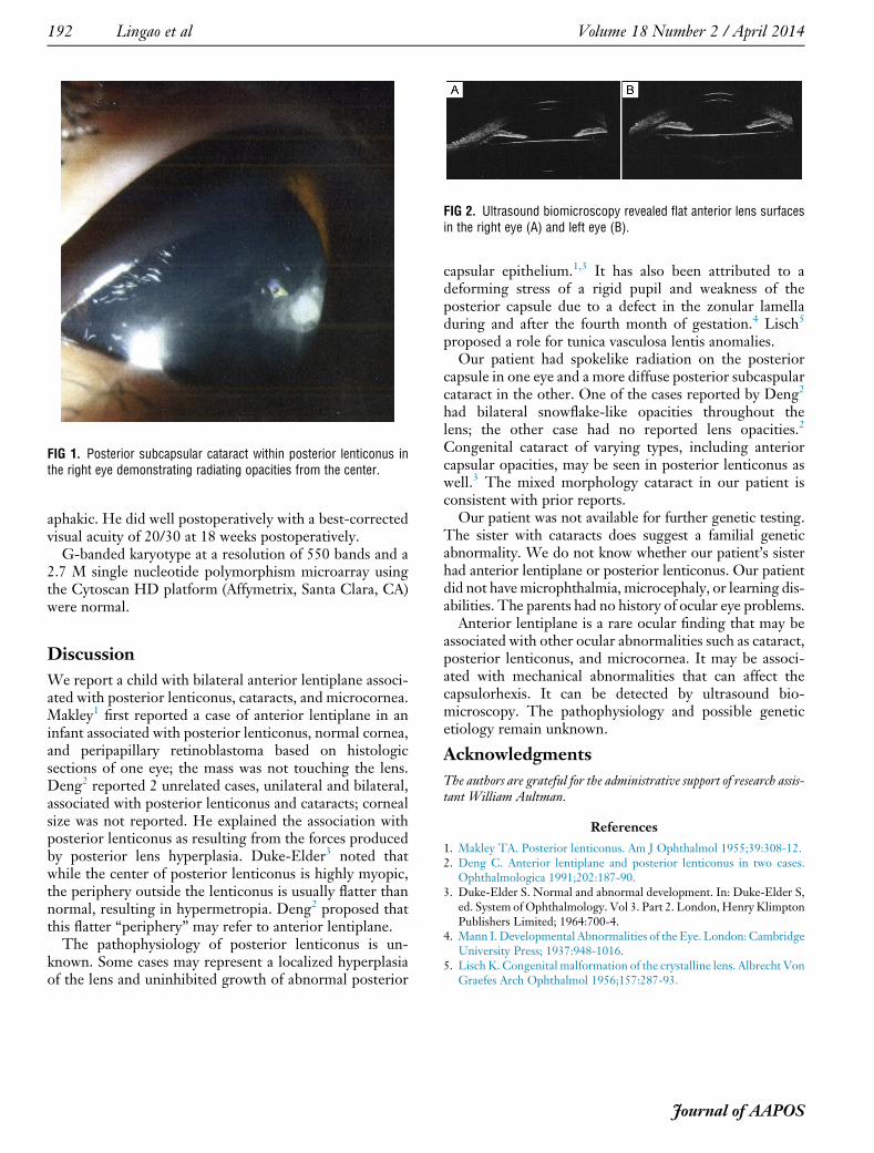
FIG 1. Posterior subcapsular cataract within posterior lenticonus inthe right eye demonstrating radiating opacities from the center.
FIG 2. Ultrasound biomicroscopy revealed flat anterior lens surfacesin the right eye (A) and left eye (B).
192 Lingao et al Volume 18 Number 2 / April 2014
aphakic. He did well postoperatively with a best-correctedvisual acuity of 20/30 at 18 weeks postoperatively.
G-banded karyotype at a resolution of 550 bands and a2.7 M single nucleotide polymorphism microarray usingthe Cytoscan HD platform (Affymetrix, Santa Clara, CA)were normal.
Discussion
We report a child with bilateral anterior lentiplane associ-ated with posterior lenticonus, cataracts, and microcornea.Makley1 first reported a case of anterior lentiplane in aninfant associated with posterior lenticonus, normal cornea,and peripapillary retinoblastoma based on histologicsections of one eye; the mass was not touching the lens.Deng2 reported 2 unrelated cases, unilateral and bilateral,associated with posterior lenticonus and cataracts; cornealsize was not reported. He explained the association withposterior lenticonus as resulting from the forces producedby posterior lens hyperplasia. Duke-Elder3 noted thatwhile the center of posterior lenticonus is highly myopic,the periphery outside the lenticonus is usually flatter thannormal, resulting in hypermetropia. Deng2 proposed thatthis flatter “periphery” may refer to anterior lentiplane.
The pathophysiology of posterior lenticonus is un-known. Some cases may represent a localized hyperplasiaof the lens and uninhibited growth of abnormal posterior
capsular epithelium.1,3 It has also been attributed to adeforming stress of a rigid pupil and weakness of theposterior capsule due to a defect in the zonular lamelladuring and after the fourth month of gestation.4 Lisch5
proposed a role for tunica vasculosa lentis anomalies.Our patient had spokelike radiation on the posterior
capsule in one eye and a more diffuse posterior subcaspularcataract in the other. One of the cases reported by Deng2
had bilateral snowflake-like opacities throughout thelens; the other case had no reported lens opacities.2
Congenital cataract of varying types, including anteriorcapsular opacities, may be seen in posterior lenticonus aswell.3 The mixed morphology cataract in our patient isconsistent with prior reports.
Our patient was not available for further genetic testing.The sister with cataracts does suggest a familial geneticabnormality. We do not know whether our patient’s sisterhad anterior lentiplane or posterior lenticonus. Our patientdid not havemicrophthalmia, microcephaly, or learning dis-abilities. The parents had no history of ocular eye problems.
Anterior lentiplane is a rare ocular finding that may beassociated with other ocular abnormalities such as cataract,posterior lenticonus, and microcornea. It may be associ-ated with mechanical abnormalities that can affect thecapsulorhexis. It can be detected by ultrasound bio-microscopy. The pathophysiology and possible geneticetiology remain unknown.
Acknowledgments
The authors are grateful for the administrative support of research assis-tant William Aultman.
References
1. Makley TA. Posterior lenticonus. Am J Ophthalmol 1955;39:308-12.2. Deng C. Anterior lentiplane and posterior lenticonus in two cases.
Ophthalmologica 1991;202:187-90.3. Duke-Elder S. Normal and abnormal development. In: Duke-Elder S,
ed. System of Ophthalmology. Vol 3. Part 2. London,Henry KlimptonPublishers Limited; 1964:700-4.
4. Mann I.Developmental Abnormalities of the Eye. London:CambridgeUniversity Press; 1937:948-1016.
5. Lisch K. Congenital malformation of the crystalline lens. Albrecht VonGraefes Arch Ophthalmol 1956;157:287-93.
Journal of AAPOS