Embed Size (px)
Citation preview
Technical Note
From KaKansas City
The authand publicavailable fo
ReceivedAddress
Parkwaymatthewdag
� 2017Elsevier. Thcreativecom
2212-628https://do
Anterior Cruciate Ligament Reconstruction WithSuture Tape Augmentation
Matt Daggett, D.O., M.B.A., Andrea Redler, M.D., and Kevin Witte, D.O., M.B.A.
Abstract: The advent of suture tape augmentation has led to increased use in knee, elbow, and ankle ligament repairsand reconstructions. Recent biomechanical analysis of the use of suture tape augmentation have shown superior strengthcharacteristics compared with repair or reconstruction alone. Despite its increased use in extra-articular ligament pro-cedures, its use as an augment to anterior cruciate ligament reconstruction has not been widely described. This articledetails a simple technique to incorporate the use of suture tape augmentation during concurrent anterior cruciate ligamentreconstruction using hamstring autograft.
nterior cruciate ligament (ACL) injury is among1
Athe most common orthopaedic injuries. Despitethe presence of many different techniques and tech-nological advances in ACL reconstruction, a significantnumber of patients still suffer reruptures of the ACLgraft as well as decreased ability to return to sport atvarying speeds.2
The use of synthetic grafts for ACL reconstruction orrepair has historically been met with skepticism andmixed results.3-7 Most notably, synthetic grafts resultedin effusion, pain, and oftentimes explantation of thedevice.3 Recently, authors have reported increasedbiomechanical strength of ligament repair with the useof suture tape augmentation.3-5 The use of a suture tapeaugmentation has been published in the repair/reconstruction of medial knee injuries,6,8 ulnarcollateral ligament,9 and ACL repair or reconstructionwith allograft,10,11 This article presents a technique thatenhances a previously published technique for ACLreconstruction12 with the addition of suture tape
nsas City University of Medicine and Biosciences (M.D., K.W.),, Missouri, U.S.A.; and Sapenzia University (A.R.), Rome, Italy.ors report that they have no conflicts of interest in the authorshipation of this article. Full ICMJE author disclosure forms arer this article online, as supplementary material.September 5, 2017; accepted October 24, 2017.correspondence to Matt Daggett, D.O., M.B.A., 2000 SE BlueSuite 230, Lee’s Summit, MO 64063, U.S.A. E-mail:[email protected] the Arthroscopy Association of North America. Published byis is an open access article under the CC BY-NC-ND license (http://mons.org/licenses/by-nc-nd/4.0/).7/171100i.org/10.1016/j.eats.2017.10.010
Arthroscopy Techniques, Vol -, N
augmentation (Internal Brace; Arthrex, Inc, Naples,FL). The ACL remnant is preserved and reconstructionis performed with a hamstring autograft. The suturetape augmentation is incorporated into the hamstringautograft construct and is independently fixatedalongside the ACL graft.
Surgical Technique
Patient SetupThe patient is placed supine on an operative table in
the standard arthroscopy position with a lateral postjust proximal to the knee for access into the medialcompartment and prevention of hip external rotation.A foot bump is also placed to keep the knee flexion at90� (Video 1). In this way, the knee can be movedfreely through the full range of motion (ROM).
IncisionsStandard anterolateral and anteromedial arthroscopic
incisions are made on the anterior knee. A diagnosticarthroscopy is performed, and any concurrent intra-articular pathology addressed. The femoral origin isidentified and prepared while the ACL remnant ispreserved using the previously published ACL recon-struction technique.12
Graft Harvest/PreparationThe semitendinosus is harvested using an open-ended
tendon stripper (Pigtail Hamstring Tendon Stripper;Arthrex, Naples, FL). The attachment site is maintained.The gracilis is then whip-stitched using a fiber loopsuture (Arthrex). The semitendinosus (ST) is thenmeasured from its insertion and marked using a skinmarker at 4.5 cm and 11 cm for a female and 5.5 cm
o - (Month), 2017: pp e1-e5 e1
Fig 1. Once the hamstring graft is prepared as shown in thisright knee, a no. 2 FiberWire is sutured through the base ofthe semitendinosus (asterisk) and the needle removed to actas a tension suture for graft fixation and passage. A Lab-ralTape (Arthrex) (arrowhead) is then shuttled through eachhole of the button of the Tightrope (Arthrex) fixation device(circle).
Fig 3. The guidewire and subsequent drills are taken from theexternal cortex into the anterior cruciate ligament remnant atlow rpm to reduce iatrogenic damage to the remnant duringanterior cruciate ligament reconstruction of the right knee inthe supine position.
e2 M. DAGGETT ET AL.
and 12 cm for a male. A Tightrope device (Arthrex) isplaced on the ST at the distal mark, and graft foldedonto itself and tagged with No. 2-0 FiberWire (Arthrex)sutures at the proximal mark. The graft is then tripledover itself and tubularized with No. 2-0 FiberWire. ANo. 2 Fiberwire is then sutured through the base of theST and needle removed to act as a tension suture forgraft fixation and passage. A LabralTape (Arthrex) isthen shuttled through each hole of the button of theTightrope (Arthrex) fixation device (Fig 1). Shuttling ofthe LabralTape through the button can be assisted by anitinol suture passer. The graft is then sized, andreturned inside the incision site while tunnel prepara-tion occurs.
Drilling of the Tibial ACL TunnelDrilling of the tibial ACL tunnel is performed in an
ACL remnantesparing manner.12 The tibial guide isplaced at 60� at the ACL insertion (Fig 2) and guidewiretaken from the external cortex into the ACL remnant at
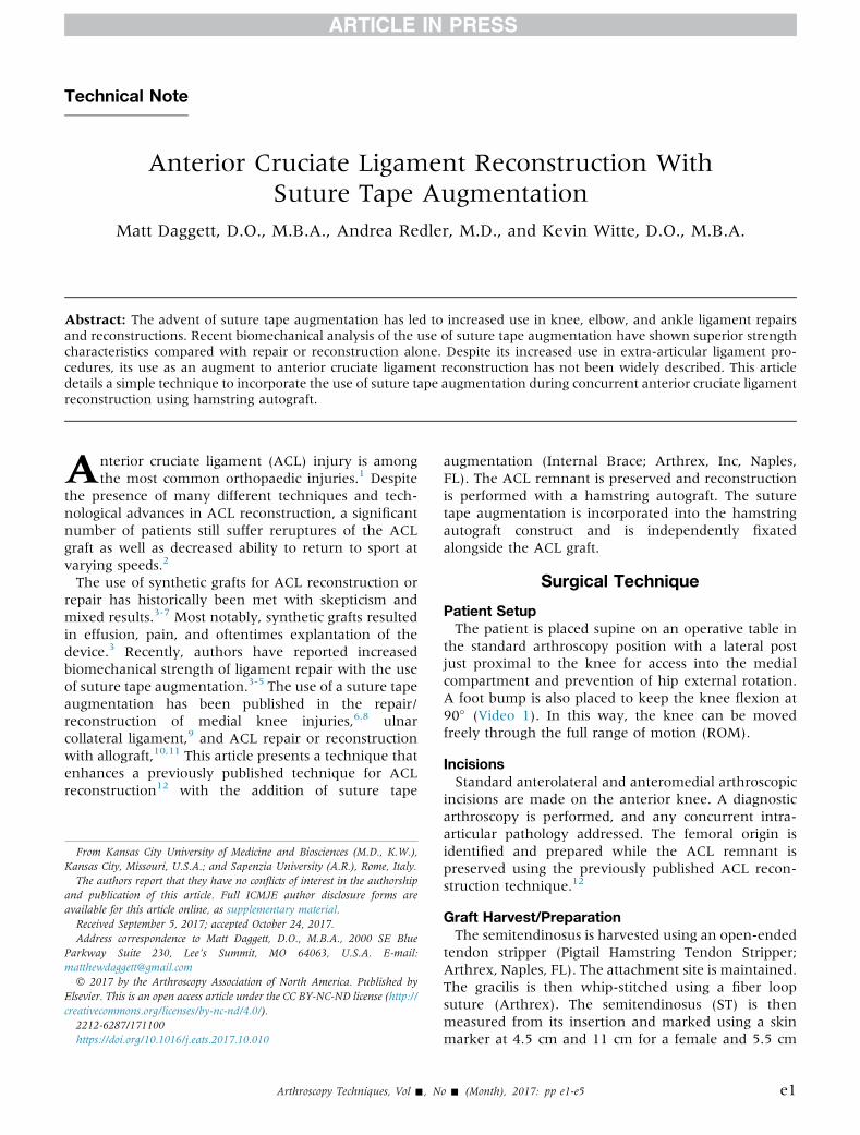
Fig 2. The tibial guide is placed at 60� (arrowhead) at theanterior cruciate ligament insertion (asterisk) during anteriorcruciate ligament reconstruction in a right knee.
low rpm to reduce iatrogenic damage to the remnant(Fig 3). Sequential reaming is performed using a size 6reamer and then increasing to the previously measuredACL graft size.
Drilling of the Femoral ACL/Anterolateral LigamentTunnelUsing the FlipCutter guide (Arthrex), the femoral
tunnel is drilled through an accessory lateral incisionusing an outside-in technique. After anatomic place-ment of the retrograde drill at the ACL insertion withinthe intercondylar notch (Fig 4), the tunnel is preparedto a depth of 20 mm (Fig 5).
Graft PassageA TigerStick (Arthrex) is then placed into the femoral
tunnel and taken transtibially out the tibial tunnel. Thesuture is then used to shuttle the ACL graft transtibiallyinto and then out of the femoral tunnel. The corticalbutton is visualized arthroscopically under direct visu-alization, and as the button is shuttled through thetunnel, it is then deployed on the lateral cortex (Fig 6).Once secured, the graft is then advanced into the
Fig 4. Anatomic placement of the retrograde drill (asterisk) atthe anterior cruciate ligament insertion within the inter-condylar notch with the outside-in drill guide (arrowhead) isplaced during anterior cruciate ligament reconstruction of aright knee.
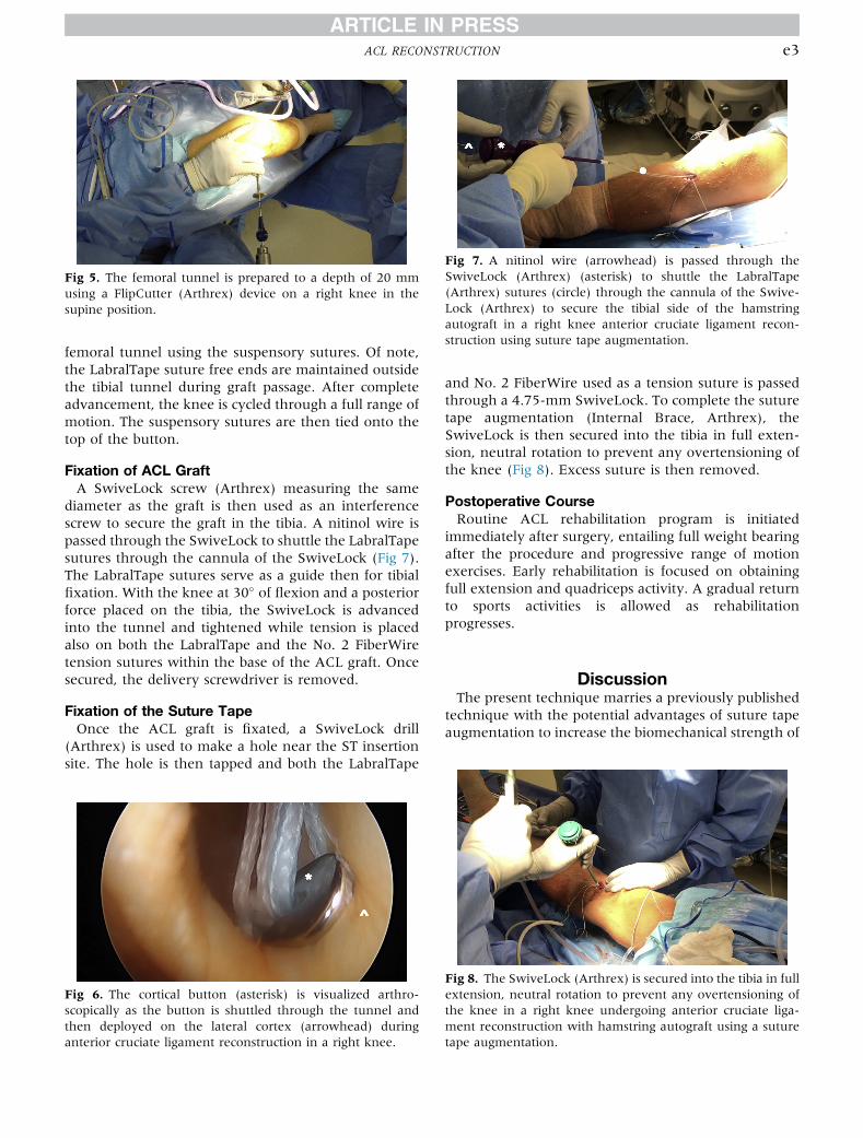
Fig 5. The femoral tunnel is prepared to a depth of 20 mmusing a FlipCutter (Arthrex) device on a right knee in thesupine position.
Fig 7. A nitinol wire (arrowhead) is passed through theSwiveLock (Arthrex) (asterisk) to shuttle the LabralTape(Arthrex) sutures (circle) through the cannula of the Swive-Lock (Arthrex) to secure the tibial side of the hamstringautograft in a right knee anterior cruciate ligament recon-struction using suture tape augmentation.
ACL RECONSTRUCTION e3
femoral tunnel using the suspensory sutures. Of note,the LabralTape suture free ends are maintained outsidethe tibial tunnel during graft passage. After completeadvancement, the knee is cycled through a full range ofmotion. The suspensory sutures are then tied onto thetop of the button.
Fixation of ACL GraftA SwiveLock screw (Arthrex) measuring the same
diameter as the graft is then used as an interferencescrew to secure the graft in the tibia. A nitinol wire ispassed through the SwiveLock to shuttle the LabralTapesutures through the cannula of the SwiveLock (Fig 7).The LabralTape sutures serve as a guide then for tibialfixation. With the knee at 30� of flexion and a posteriorforce placed on the tibia, the SwiveLock is advancedinto the tunnel and tightened while tension is placedalso on both the LabralTape and the No. 2 FiberWiretension sutures within the base of the ACL graft. Oncesecured, the delivery screwdriver is removed.
Fixation of the Suture TapeOnce the ACL graft is fixated, a SwiveLock drill
(Arthrex) is used to make a hole near the ST insertionsite. The hole is then tapped and both the LabralTape
Fig 6. The cortical button (asterisk) is visualized arthro-scopically as the button is shuttled through the tunnel andthen deployed on the lateral cortex (arrowhead) duringanterior cruciate ligament reconstruction in a right knee.
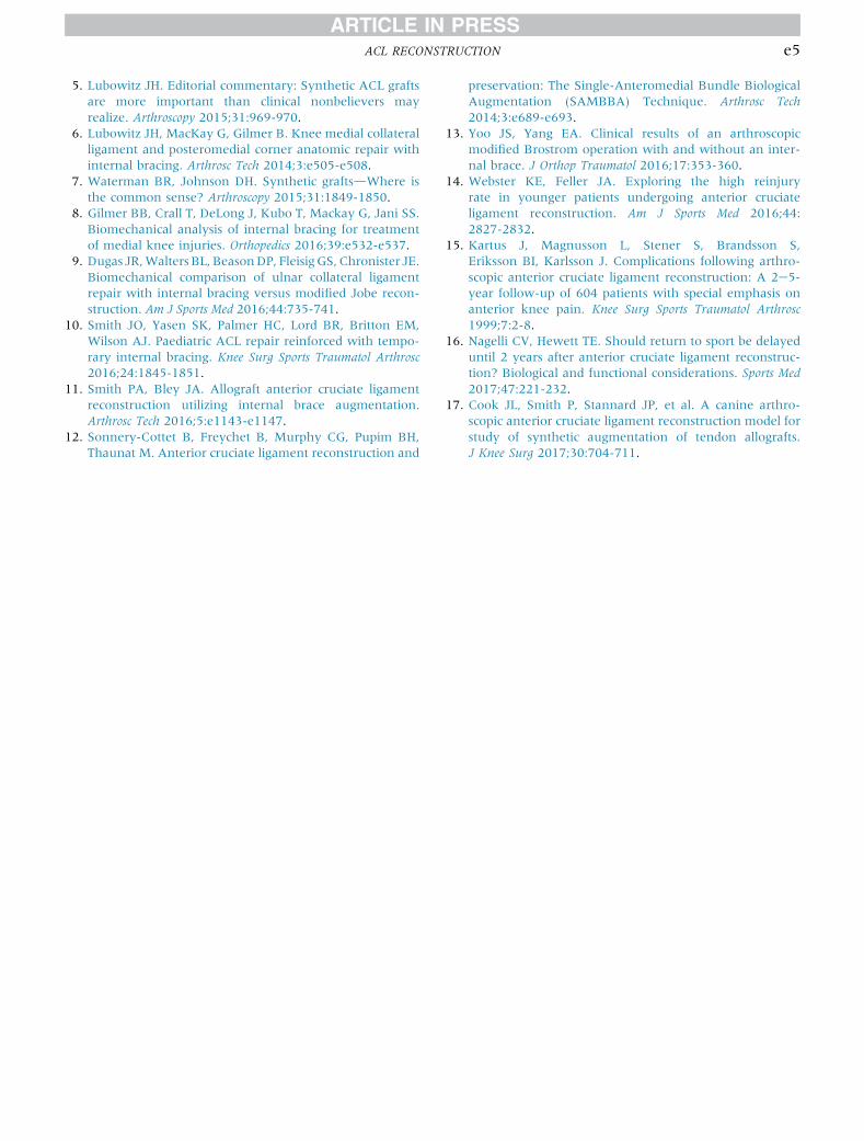
and No. 2 FiberWire used as a tension suture is passedthrough a 4.75-mm SwiveLock. To complete the suturetape augmentation (Internal Brace, Arthrex), theSwiveLock is then secured into the tibia in full exten-sion, neutral rotation to prevent any overtensioning ofthe knee (Fig 8). Excess suture is then removed.
Postoperative CourseRoutine ACL rehabilitation program is initiated
immediately after surgery, entailing full weight bearingafter the procedure and progressive range of motionexercises. Early rehabilitation is focused on obtainingfull extension and quadriceps activity. A gradual returnto sports activities is allowed as rehabilitationprogresses.
DiscussionThe present technique marries a previously published
technique with the potential advantages of suture tapeaugmentation to increase the biomechanical strength of
Fig 8. The SwiveLock (Arthrex) is secured into the tibia in fullextension, neutral rotation to prevent any overtensioning ofthe knee in a right knee undergoing anterior cruciate liga-ment reconstruction with hamstring autograft using a suturetape augmentation.
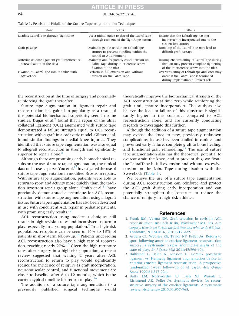
Table 1. Pearls and Pitfalls of the Suture Tape Augmentation Technique
Stage Pearls Pitfalls
Loading LabralTape through TightRope Use a nitinol guide to thread the LabralTapethrough each end of the TightRope button
Ensure that the LabralTape has notinadvertently incorporated one of thesuspension sutures
Graft passage Maintain gentle tension on LabralTapesutures to prevent bundling within thetunnel or ACL remnant
Bundling of the LabralTape may lead todifficult graft passage
Anterior cruciate ligament graft interferencescrew fixation in the tibia
Maintain and frequently check tension onLabralTape during interference screwfixation of the tibia
Incomplete tensioning of LabralTape duringfixation may prevent complete tighteningof the interference screw into the tibia
Fixation of LabralTape into the tibia withSwiveLock
Perform in full extension and withouttension on the LabralTape
Overtensioning of LabralTape and knee mayoccur if the LabralTape is tensionedduring implantation of SwiveLock
e4 M. DAGGETT ET AL.
the reconstruction at the time of surgery and potentiallyreinforcing the graft thereafter.Suture tape augmentation in ligament repair and
reconstruction has gained in popularity as a result ofthe potential biomechanical superiority seen in somestudies. Dugas et al.9 found that a repair of the ulnarcollateral ligament (UCL) augmented with suture tapedemonstrated a failure strength equal to UCL recon-struction with a graft in a cadaveric model. Gilmer et al.found similar findings in medial knee injuries. Theyidentified that suture tape augmentation was also equalto allograft reconstruction in strength and significantlysuperior to repair alone.8
Although there are promising early biomechanical re-sults on the use of suture tape augmentation, the clinicaldata on its use is sparse. Yoo et al.13 investigated theuseofsuture tape augmentation in modified Brostrom repairs.With suture tape augmentation, patients were able toreturn to sport and activity more quickly than the tradi-tion Brostrom repair group alone. Smith et al.11 havepreviously demonstrated a technique for ACL recon-struction with suture tape augmentation using allografttissue. Suture tape augmentation has also been describedin use with concurrent ACL repair in pediatric patients,with promising early results.10
ACL reconstruction using modern techniques stillresults in high revision rates and inconsistent return toplay, especially in a young population.2 In a high-riskpopulation, rerupture can be seen in 16% to 18% ofpatients in short-term follow-up.14 Patients undergoingACL reconstruction also have a high rate of reopera-tion, reaching nearly 27%.15 Given the high rerupturerates after surgery in a high-risk population, a recentreview suggested that waiting 2 years after ACLreconstruction to return to play would significantlyreduce the incidence of reinjury as graft incorporation,neuromuscular control, and functional movement arecloser to baseline after 6 to 12 months, which is thecurrent typical timeline for return to play.16
The addition of a suture tape augmentation to apreviously published surgical technique would
theoretically improve the biomechanical strength of theACL reconstruction at time zero while reinforcing thegraft until mature incorporation. The authors alsobelieve the load to failure of this construct is signifi-cantly higher in this construct compared to ACLreconstruction alone, and are currently conductingresearch to investigate this further.Although the addition of a suture tape augmentation
may expose the knee to new, previously unknowncomplications, its use has been studied in canines andprevented early failure, complete graft to bone healing,and functional graft remodeling.17 The use of suturetape augmentation also has the theoretical potential tooverconstrain the knee, and to prevent this, we fixatethe LabralTape in full extension and without excessivetension on the LabralTape during fixation with theSwiveLock (Table 1).We believe the use of a suture tape augmentation
during ACL reconstruction can reinforce and protectthe ACL graft during early incorporation and canpotentially strengthen the construct to reduce thechance of reinjury in high-risk athletes.
References1. Frank RM, Verma NN. Graft selection in revision ACL
reconstruction. In: Bach Jr BR, Provencher MT, eds. ACLsurgery: How to get it right the first time and what to do if it fails.Thorofare, NJ: SLACK, 2010;217-229.
2. Ardern CL, Webster KE, Taylor NF, Feller JA. Return tosport following anterior cruciate ligament reconstructionsurgery: a systematic review and meta-analysis of thestate of play. Br J Sports Med 2011;45:596-606.
3. Dahlstedt L, Dalen N, Jonsson U. Goretex prostheticligament vs. Kennedy ligament augmentation device inanterior cruciate ligament reconstruction. A prospectiverandomized 3-year follow-up of 41 cases. Acta OrthopScand 1990;61:217-224.
4. Batty LM, Norsworthy CJ, Lash NJ, Wasiak J,Richmond AK, Feller JA. Synthetic devices for recon-structive surgery of the cruciate ligaments: A systematicreview. Arthroscopy 2015;31:957-968.
ACL RECONSTRUCTION e5
5. Lubowitz JH. Editorial commentary: Synthetic ACL graftsare more important than clinical nonbelievers mayrealize. Arthroscopy 2015;31:969-970.
6. Lubowitz JH, MacKay G, Gilmer B. Knee medial collateralligament and posteromedial corner anatomic repair withinternal bracing. Arthrosc Tech 2014;3:e505-e508.
7. Waterman BR, Johnson DH. Synthetic graftsdWhere isthe common sense? Arthroscopy 2015;31:1849-1850.
8. Gilmer BB, Crall T, DeLong J, Kubo T, Mackay G, Jani SS.Biomechanical analysis of internal bracing for treatmentof medial knee injuries. Orthopedics 2016;39:e532-e537.
9. Dugas JR,WaltersBL, BeasonDP, FleisigGS, Chronister JE.Biomechanical comparison of ulnar collateral ligamentrepair with internal bracing versus modified Jobe recon-struction. Am J Sports Med 2016;44:735-741.
10. Smith JO, Yasen SK, Palmer HC, Lord BR, Britton EM,Wilson AJ. Paediatric ACL repair reinforced with tempo-rary internal bracing. Knee Surg Sports Traumatol Arthrosc2016;24:1845-1851.
11. Smith PA, Bley JA. Allograft anterior cruciate ligamentreconstruction utilizing internal brace augmentation.Arthrosc Tech 2016;5:e1143-e1147.
12. Sonnery-Cottet B, Freychet B, Murphy CG, Pupim BH,Thaunat M. Anterior cruciate ligament reconstruction and
preservation: The Single-Anteromedial Bundle BiologicalAugmentation (SAMBBA) Technique. Arthrosc Tech2014;3:e689-e693.
13. Yoo JS, Yang EA. Clinical results of an arthroscopicmodified Brostrom operation with and without an inter-nal brace. J Orthop Traumatol 2016;17:353-360.
14. Webster KE, Feller JA. Exploring the high reinjuryrate in younger patients undergoing anterior cruciateligament reconstruction. Am J Sports Med 2016;44:2827-2832.
15. Kartus J, Magnusson L, Stener S, Brandsson S,Eriksson BI, Karlsson J. Complications following arthro-scopic anterior cruciate ligament reconstruction: A 2e5-year follow-up of 604 patients with special emphasis onanterior knee pain. Knee Surg Sports Traumatol Arthrosc1999;7:2-8.
16. Nagelli CV, Hewett TE. Should return to sport be delayeduntil 2 years after anterior cruciate ligament reconstruc-tion? Biological and functional considerations. Sports Med2017;47:221-232.
17. Cook JL, Smith P, Stannard JP, et al. A canine arthro-scopic anterior cruciate ligament reconstruction model forstudy of synthetic augmentation of tendon allografts.J Knee Surg 2017;30:704-711.