Embed Size (px)
Citation preview
ANTERIOR APPROACH TOTAL HIP REPLACEMENTMINIMIZING THE TIME FROM REPLACEMENT TO RECOVERY
A description of the Matta Method™
JOEL M. MATTA, M.D. HIP DISORDERS: PRESERVATION, REPLACEMENT AND FRACTURES

When I conducted my first instructional course for surgeons in 2003 Anterior Approach was utilized by less than 1% of surgeons in the USA. Today, it is estimated that approximately 1/3 of hip replacement surgeons utilize Anterior Approach.
The important number however is what percentage of patients have their hip replaced from Anterior and this is probably a higher number than for total hip replacement has grown dramatically and this trend continues because of its definite advantages for patients, even those in need of bilateral procedures. Rehabilitation is simplified and accelerated with less pain and a shorter need for walking aids, dislocation risk is reduced, leg length is more accurately controlled, and the incision is small. However, results may vary depending on surgeon experience and specific methodology when performing Anterior Approach.
This trend to Anterior Approach, initially driven by surgeons and patients who saw the benefits is now supported by a growing body of scientific evidence. I believe that Anterior Approach will replace Posterior Approach as the most common technique for Hip Replacement. Why the 20 year process of change? To quote Max Planck, “A new scientific truth does not triumph by convincing its opponents and making them see the light, but rather because its opponents eventually die, and a new generation grows up that is familiar with it.”
My message to surgeons adopting Anterior Approach today: Don’t try to do Anterior Approach like you did Posterior Approach. It is possible but the patients won’t derive the full benefits. Therefore, besides the less invasive incision, Anterior Approach has been a stimulus for creation and utilization of technical innovations that enhance its safety and efficacy. Benefits are derived from use of the Hana® orthopedic table rather than an assistant manipulating the leg, real time assessment of component position and leg length with the image intensifier and associated computer software rather than mechanical guides and feeling the patient’s ankles to judge leg length, improved hip replacement implants, and automated bony instrumentation using the Kincise™ rather than pounding with a hammer. The following describes Matta Method™ for Anterior hip replacement and supporting references.
Joel Matta, MD
A N T E R I O R A P P R O A C H T O T A L H I P R E P L A C E M E N T
W W W . H I P A N D P E L V I S . C O M 1
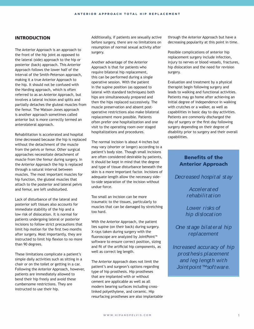
Benefits of the Anterior Approach
Decreased hospital stay
Accelerated rehabilitation
Lower risks of hip dislocation
One stage bilateral hip replacement
Increased accuracy of hip prosthesis placement and leg length with
Jointpoint™ software.
INTRODUCTION
The Anterior Approach is an approach to the front of the hip joint as opposed to the lateral (side) approach to the hip or posterior (back) approach. This Anterior Approach follows the lower half of the interval of the Smith-Peterson approach, making it a true Anterior Approach to the hip. It should not be confused with the Harding approach, which is often referred to as an Anterior Approach, but involves a lateral incision and splits and partially detaches the gluteal muscles from the femur. The Watson-Jones approach is another approach sometimes called anterior but is more correctly termed an anterolateral approach.
Rehabilitation is accelerated and hospital time decreased because the hip is replaced without the detachment of the muscle from the pelvis or femur. Other surgical approaches necessitate detachment of muscle from the femur during surgery. In the Anterior Approach the hip is replaced through a natural interval between muscles. The most important muscles for hip function, the gluteal muscles that attach to the posterior and lateral pelvis and femur, are left undisturbed.
Lack of disturbance of the lateral and posterior soft tissues also accounts for immediate stability of the hip and a low risk of dislocation. It is normal for patients undergoing lateral or posterior incisions to follow strict precautions that limit hip motion for the first two months after surgery. Most importantly, they are instructed to limit hip flexion to no more than 90 degrees.
These limitations complicate a patient’s simple daily activities such as sitting in a chair or on the toilet or getting in a car.Following the Anterior Approach, however, patients are immediately allowed to bend their hip freely and avoid these cumbersome restrictions. They are instructed to use their hip.
Additionally, if patients are sexually active before surgery, there are no limitations on resumption of normal sexual activity after surgery.
Another advantage of the Anterior Approach is that for patients who require bilateral hip replacement, this can be performed during a single operative session. With the patient in the supine position (as opposed to lateral with standard techniques) both hips are simultaneously prepared and then the hips replaced successively. The muscle preservation and absent post-operative restrictions also make bilateral replacement more possible. Patients often prefer one hospitalization and one visit to the operating room over staged hospitalizations and procedures.
The normal incision is about 4-inches but may vary (shorter or longer) according to a patient’s body size. Though small incisions are often considered desirable by patients, it should be kept in mind that the degree and type of tissue disturbance beneath the skin is a more important factor. Incisions of adequate length allow the necessary side-to-side separation of the incision without undue force.
Too small an incision can be more traumatic to the tissues, particularly to muscles that can be damaged by stretching too hard.
With the Anterior Approach, the patient lies supine (on their back) during surgery. X-rays taken during surgery with the fluoroscope are analyzed by JointPoint™ software to ensure correct position, sizing and fit of the artificial hip components, as well as correct leg length.
The Anterior Approach does not limit the patient’s and surgeon’s options regarding type of hip prosthesis. Hip prostheses that are implanted with or without cement are applicable as well as all modern bearing surfaces including cross-linked polyethylene, and ceramic. Hip resurfacing prostheses are also implantable
through the Anterior Approach but have a decreasing popularity at this point in time.
Possible complications of anterior hip replacement surgery include infection, injury to nerves or blood vessels, fractures, hip dislocation and the need for revision surgery.
Evaluation and treatment by a physical therapist begin following surgery and leads to walking and functional activities. Patients may go home after achieving an initial degree of independence in walking with crutches or a walker, as well as capabilities in basic day to day activities. Patients are commonly discharged the day of surgery or the first day following surgery depending on their degree of disability prior to surgery and their overall capabilities.
A N T E R I O R A P P R O A C H T O T A L H I P R E P L A C E M E N T
W W W . H I P A N D P E L V I S . C O M2
Background of Anterior Approach Hip Replacement
Anterior Approach hip replacement was first performed in Paris,
France by Prof. Robert Judet using the Judet orthopedic table in
1947. Since the first surgery over 70 years ago this procedure has
been performed consistently by a small group of surgeons in Paris
including Thierry Judet, son of Robert.
In 1996 Dr. Joel Matta, who had observed anterior hip
replacement in Paris, rethought his approach to hip replacement.
By abandoning the posterior approach and adopting the Anterior
Approach his goals were: lower chance of dislocation, enhance recovery rate, and increase accuracy of hip prosthesis placement and leg length. Clinical research over the following
years has shown that these goals are achieved with Anterior
Approach performed with the proper technique and technology.
Anterior Approach enhancing technologies with design input
from Dr. Matta include: The HANA® Table, JointPoint™ software,
Kincise™ adaptors for Anterior Approach, surgical instruments
for Anterior Approach, and the Actis® femoral hip replacement
prosthesis made by DePuy Synthes. Dr. Matta holds national and
international patents regarding some of the above technologies.
DePuy Synthes, a Johnson & Johnson company, is an educational
partner to Dr. Matta and conducts over 200 Anterior Approach
educational events every year for surgeons which are branded
Matta Method™ and reach over 700 surgeons.
Dr. Matta began his series of Anterior Approach hips in 1996 and
has used it for a consecutive series of over 4000 patients since
then. The Matta Method™ has evolved over the past 22 years with
improvements in technique and technology.
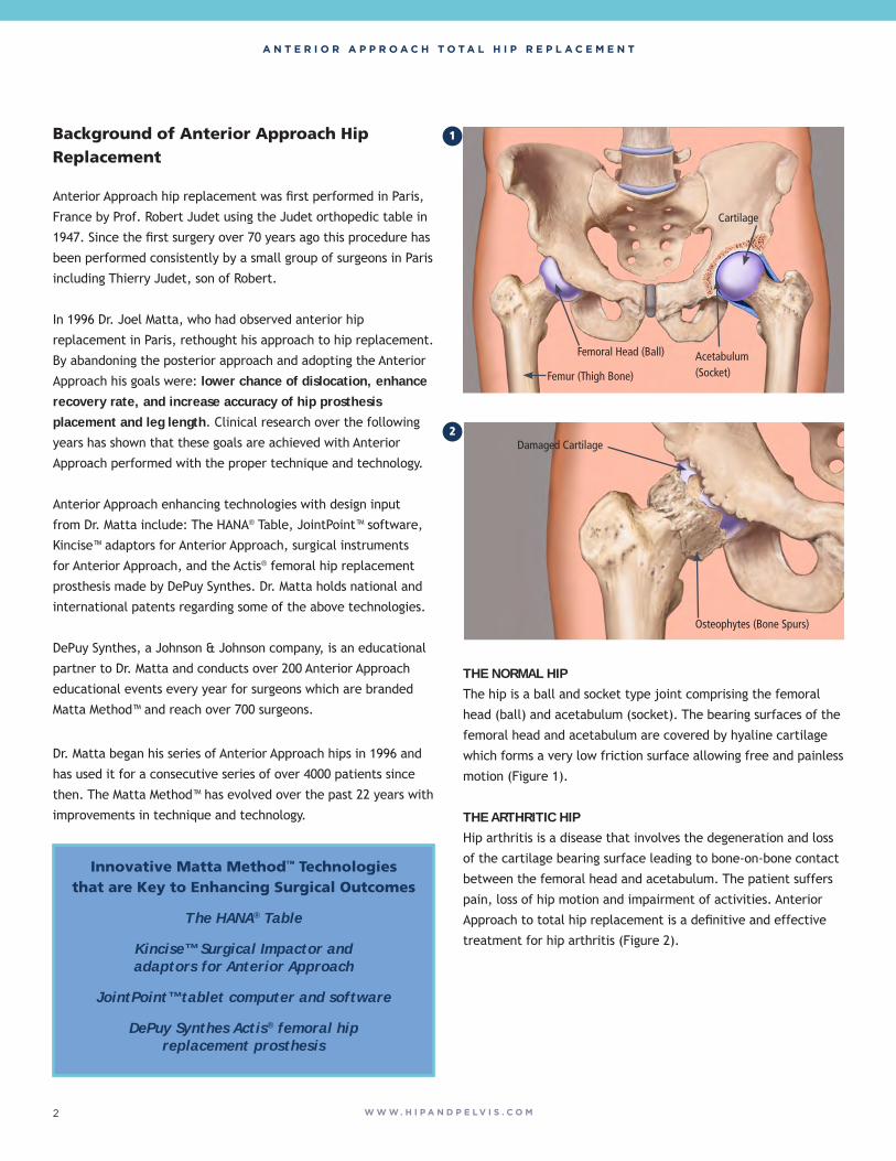
THE NORMAL HIPThe hip is a ball and socket type joint comprising the femoral
head (ball) and acetabulum (socket). The bearing surfaces of the
femoral head and acetabulum are covered by hyaline cartilage
which forms a very low friction surface allowing free and painless
motion (Figure 1).
THE ARTHRITIC HIPHip arthritis is a disease that involves the degeneration and loss
of the cartilage bearing surface leading to bone-on-bone contact
between the femoral head and acetabulum. The patient suffers
pain, loss of hip motion and impairment of activities. Anterior
Approach to total hip replacement is a definitive and effective
treatment for hip arthritis (Figure 2).
Damaged Cartilage
Osteophytes (Bone Spurs)
Femur (Thigh Bone)
Cartilage
Femoral Head (Ball) Acetabulum(Socket)
1
2
Innovative Matta Method™ Technologies that are Key to Enhancing Surgical Outcomes
The HANA® Table
Kincise™ Surgical Impactor and adaptors for Anterior Approach
JointPoint™ tablet computer and software
DePuy Synthes Actis® femoral hip replacement prosthesis

A N T E R I O R A P P R O A C H T O T A L H I P R E P L A C E M E N T
W W W . H I P A N D P E L V I S . C O M 3
Matta Method™ Anterior Approach Surgical Procedure
THE OPERATING TABLEFollowing anesthesia, the patient is placed on the HANA® or
PROfx® table. The unique capabilities of the table facilitate
surgery through this smaller and less invasive approach. The
carbon fiber spars that support the legs move appropriately and
manipulate the operated leg during surgery. Additionally, the
table has a sterile robotic hook attachment that reaches inside
the incision to lift and hold the femur in an accessible position.
(Figure 3).
THE APPROACHThe Anterior Approach is an approach to the front of the hip joint
as opposed to the lateral (side) approach to the hip or posterior
(back) approach (Figure 4). This Anterior Approach follows
the lower half of the interval of the Smith-Peterson approach,
making it a true Anterior Approach to the hip. It should not be
confused with the Harding approach, which is often referred to
as an Anterior Approach, but involves a lateral incision and splits
and partially detaches the gluteal muscles from the femur. The
Watson-Jones approach is another approach sometimes called
anterior but is more correctly termed an antero-lateral approach.
The hip is exposed by following a natural plane between muscles
and without detachment of muscle or tendons from the bone.
The femoral capsule is opened and the femoral neck is exposed
(Figure 5).
The femoral neck is cut and the arthritic femoral head removed
(Figure 6).
Anterior Approach Incision
Femoral Capsule
3
4
5
6
A N T E R I O R A P P R O A C H T O T A L H I P R E P L A C E M E N T
W W W . H I P A N D P E L V I S . C O M4
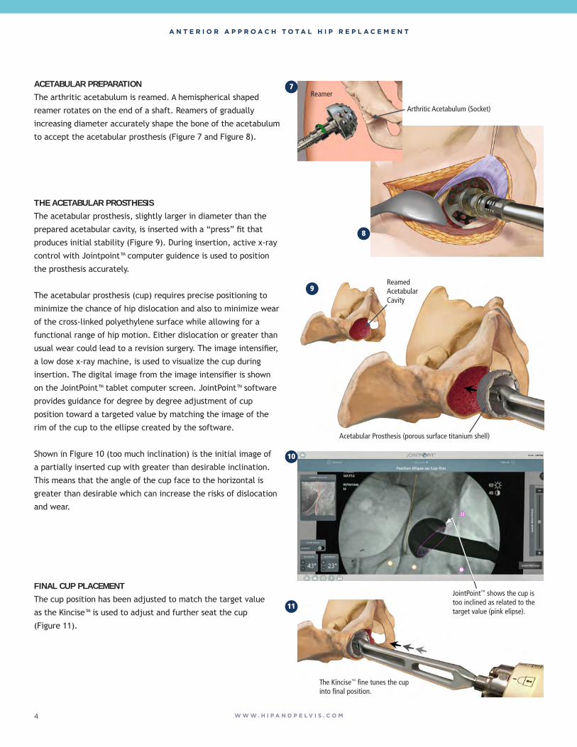
THE ACETABULAR PROSTHESISThe acetabular prosthesis, slightly larger in diameter than the
prepared acetabular cavity, is inserted with a “press” fit that
produces initial stability (Figure 9). During insertion, active x-ray
control with Jointpoint™ computer guidence is used to position
the prosthesis accurately.
The acetabular prosthesis (cup) requires precise positioning to
minimize the chance of hip dislocation and also to minimize wear
of the cross-linked polyethylene surface while allowing for a
functional range of hip motion. Either dislocation or greater than
usual wear could lead to a revision surgery. The image intensifier,
a low dose x-ray machine, is used to visualize the cup during
insertion. The digital image from the image intensifier is shown
on the JointPoint™ tablet computer screen. JointPoint™ software
provides guidance for degree by degree adjustment of cup
position toward a targeted value by matching the image of the
rim of the cup to the ellipse created by the software.
Shown in Figure 10 (too much inclination) is the initial image of
a partially inserted cup with greater than desirable inclination.
This means that the angle of the cup face to the horizontal is
greater than desirable which can increase the risks of dislocation
and wear.
FINAL CUP PLACEMENTThe cup position has been adjusted to match the target value
as the Kincise™ is used to adjust and further seat the cup
(Figure 11).
ACETABULAR PREPARATIONThe arthritic acetabulum is reamed. A hemispherical shaped
reamer rotates on the end of a shaft. Reamers of gradually
increasing diameter accurately shape the bone of the acetabulum
to accept the acetabular prosthesis (Figure 7 and Figure 8).
Reamer
Arthritic Acetabulum (Socket)
Acetabular Prosthesis (porous surface titanium shell)
Reamed Acetabular Cavity
JointPoint™ shows the cup istoo inclined as related to thetarget value (pink elipse).
The Kincise™ fine tunes the cup into final position.
8
7
9
10
11

A N T E R I O R A P P R O A C H T O T A L H I P R E P L A C E M E N T
W W W . H I P A N D P E L V I S . C O M 5
JointPoint™ confirms that cup inclination and anteversion
corresponds to the desired target position (Figure 12).
Long term stability relies on the biologic process of bonding of
the bone to the porous acetabular surface. Following insertion
of this titanium acetabular “shell”, the bearing surface
(polyethylene, or ceramic) is inserted (Figure 13).
FEMORAL PREPARATIONThe table rotates the leg externally (foot pointed outward) and
extends the hip, lowering the foot towards the floor to allow
femoral access through this small approach. The femoral hook
raises the femur to a more accessible position and holds the
femur securely. Using rhythmic impaction, a specialized tool
called the Kincise™ Surgical Impactor, inserts a broach into the
femoral canal. Progressively larger broaches are then inserted.
The broach size is limited by the hard outer cortical bone
(Figure 14).
SIZINGFollowing insertion of the final broach the Kincise™ is detached.
The broach is temporarily left in as a “trial” femoral prosthesis
and a trial neck and head are attached to its upper end.
(Figure 15).
The image of the rim of the cup matches the target.
Acetabular liner (bearing surface)
Kincise™ Surgical Impactor
Broach
Femoral Hook
Trial Head
15
14
13
12
A N T E R I O R A P P R O A C H T O T A L H I P R E P L A C E M E N T
W W W . H I P A N D P E L V I S . C O M6
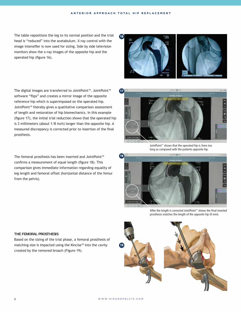
The table repositions the leg to its normal position and the trial
head is “reduced” into the acetabulum. X-ray control with the
image intensifier is now used for sizing. Side by side television
monitors show the x-ray images of the opposite hip and the
operated hip (figure 16).
The digital images are transferred to JointPoint™. JointPoint™
software “flips” and creates a mirror image of the opposite
reference hip which is superimposed on the operated hip.
JointPoint™ thereby gives a quatitative comparison assessment
of length and restoration of hip biomechanics. In this example
(figure 17), the initial trial reduction shows that the operated hip
is 3 millimeters (about 1/8 inch) longer than the opposite hip. A
measured discrepancy is corrected prior to insertion of the final
prosthesis.
The femoral prosthesis has been inserted and JointPoint™
confirms a measurement of equal length (figure 18). This
comparison gives immediate information regarding equality of
leg length and femoral offset (horizontal distance of the femur
from the pelvis).
THE FEMORAL PROSTHESISBased on the sizing of the trial phase, a femoral prosthesis of
matching size is impacted using the Kincise™ into the cavity
created by the removed broach (Figure 19).
JointPoint™ shows that the operated hip is 3mm too long as compared with the patients opposite hip.
After the length is corrected JointPoint™ shows the final inserted prosthesis matches the length of the opposite hip (0 mm).
16
17
18
19
A N T E R I O R A P P R O A C H T O T A L H I P R E P L A C E M E N T
W W W . H I P A N D P E L V I S . C O M 7
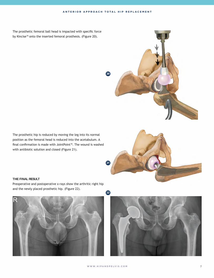
The prosthetic femoral ball head is impacted with specific force
by Kincise™ onto the inserted femoral prosthesis. (Figure 20).
The prosthetic hip is reduced by moving the leg into its normal
position as the femoral head is reduced into the acetabulum. A
final confirmation is made with JointPoint™. The wound is washed
with antibiotic solution and closed (Figure 21).
THE FINAL RESULTPreoperative and postoperative x-rays show the arthritic right hip
and the newly placed prosthetic hip. (Figure 22).
20
21
22
A N T E R I O R A P P R O A C H T O T A L H I P R E P L A C E M E N T
W W W . H I P A N D P E L V I S . C O M8
The following references report on Anterior Approach hip replacement consistent with and supportive of safety and efficacy of the Matta Method™. Over the past 15 years there has been a growing number of peer reviewed references pertaining to Anterior Approach that are positive though neutral and negative reports have also been published. Multiple and variable methodologies for performing Anterior Approach cannot produce consistent results. Medical findings and opinion are also rarely uniform. There has been however, strong and steady growth of Anterior Approach over the past 15 years, and a growing number of surgeon advocates.
References1. Petis SM, et al. “In Hospital Cost Analysis of THA: Does Surgical Approach Matter?” The Journal of Arthroplasty 2016; (31)” 53-58.
The authors found that overall costs (intraoperative costs and hospital stay) were significantly less for the anterior vs lateral and anterior vs posterior approach. Patients in the Anterior Approach group also consumed significantly fewer narcotics than the posterior and lateral cohorts.
2. Barrett WP, et al. “Prospective Randomized Study of Direct Anterior vs Posterolateral Approach for Total Hip Arthroplasty.” The Journal of Arthroplasty 2013; (28): 1634-1638.
This study compared the Anterior Approach and the posterolateral approach and found that Anterior Approach patients were discharged significantly sooner and had shorter length of stay than posterolateral approach patients. They also performed better during the immediate post-operative period. They walked farther, had lower pain scores, and needed less pain medication. Post-operatively the anterior group had significantly more patients who were walking unlimited and using stairs normally at 6 weeks post-operatively compared to the posterolateral group.
3. Barnett, Steven L. et al. Is the Anterior Approach Safe? Early Complication Rate Associated With 5090 Consecutive Primary Total Hip Arthroplasty Procedures Performed Using the Anterior Approach. The Journal of Arthroplasty, Volume 31, Issue 10, 2291 – 2294.
This large consecutive multi-center study found that Anterior Approach is a safe surgical procedure with low rates of complications. The complications included: 0.8% intra-op femur fracture, 0.1% post-op femur fracture, 0.1% deep infection, 0.2% hip dislocation, 0.06% motor nerve palsy, 0.3% deep venous thrombosis.
4. Kamath A, Chitnis A, Holy C, et al. Medical resource utilization and costs for total hip arthroplasty: benchmarking an Anterior Approach technique in the Medicare population. J Med Econ. 2017; 1-7.
The purpose of this study was to benchmark healthcare resource utilization and costs for 1,794 patients with THA via Anterior Approach relative to matched patients. The authors found that patients with Anterior Approach had significantly lower mean hospital length of stay vs patients in the control group (2.06 vs 2.98 days) and the adjusted proportion of patients discharged to home was nearly 20 percentage points higher in the anterior group. Post-acute claim payments for AA patients were nearly 50% lower than those for control patients.
5. Taunton M, Sierra R, Kaufman K, et al. A randomized clinical trial of direct Anterior Approach and mini-posterior approach total hip arthroplasty: which provides better early functional recovery? Orthopaedic Proceedings. 2018 100-B:SUPP_1, 46-46.
The anterior patients in this study had faster recovery than the mini-posterior approach patients; time to discontinue walker (10 vs. 14.5 days), time to discontinue all gait aids (17.3 vs 23.6 days), ascend stairs with gait aid (5.4 vs. 10.3 days), and to walk 6 blocks (20.5 vs. 26.0 days). Activity monitoring at two weeks postoperatively favored Anterior Approach; mean steps per day (3,897 vs 2,235 days), percent of day active (10.5% vs 6.9%). Narcotics were discontinued earlier for the anterior patients (9.1 vs 14 days). At two months, physical functioning as measured by the SF-12 score, favored Anterior Approach with the component change at 2 months (15 vs 10).
6. Miller LE, Gondusky JS, Bhattacharyya S, Kamath AK, Boettner F, Wright J. Does Surgical Approach Affect Outcomes in Total Hip Arthroplasty Through 90 Days of Follow-Up? A Systematic Review With Meta-Analysis. J Arthroplasty. 2017: 33(4); 1296-1302.
This systematic review included prospective studies comparing postoperative outcomes through 90 days of Anterior Approach vs posterior approach in primary THA. The study showed that the Anterior Approach was associated with lower pain severity, lower narcotic usage, and improved hip function compared to posterior group.
7. Bourne MH, et al. “A comparison between direct anterior surgery of the hip (DASH) and the anterolateral (AL) surgical approach to THA: Postoperative outcomes.” 2010 AAOS New Orleans, LA, Poster #014
The study compared the clinical and functional outcomes at six weeks, six months, and one year for patients having had total hip replacement through anterior vs anterolateral approach. The scores measured were Harris Hip score, Harris Hip pain score, percent walking more than six blocks, need for support devices, stair climbing, and the ability to put on socks and shoes. In all six scores measured, Anterior Approach was superior to the anterolateral approach at six weeks and six months. At one year the scores measured were as good or better, and only 5% of anterior patients were sedentary compared with 33% of anterolateral.
8. Matta, J.M.: Anterior Approach for Total Hip Replacement: Background and Operative Technique, Chapter 8 p.121-140; MIS Techniques in Orthopaedics (Scuderi, G.R.; Tria, A.J.; Berger, R.A. editors); copyright Springer Science + Business Media, Inc.
9. Matta, J.M.; Ferguson, T.A.: The Anterior Approach for Hip Replacement. Orthopedics, vol 28, no. 9; p. 927- 928, September 2005.
10. Yerasimides, J.G.; Matta, J.M.: Primary Total Hip Arthroplasty with a Minimally Invasive Anterior Approach. Seminars in Arthroplasty, vol 16, no. 3, p. 186-190; September 2005.
11. Matta J.M.; Shahrdar, C.; Ferguson, T.A.: Single-Incision Anterior Approach for Total Hip Arthroplasty on an Orthopaedic Table. Clin Orthop Rel Res, no. 441, p 115-124, December 2005.
12. Finerman, G.A.M., Dorey, F.J., Grigoris, P., McKellop, H.A.; Total Hip Arthroplasty Outcomes, Churchill Livingstone, Inc.; 1998.

Our Healthcare Professional Team
All our office and hospital staff are familiar with Dr. Matta’s procedure and work together as a team. Those which comprise the team are:
DR. MATTA’s OFFICE
Fellow: an orthopedic surgeon in a training position under Dr. Matta’s supervision who assists with examinations, in-hospital care, surgery and research
Physician Assistant: conducts patient and family contact prior to surgery, in-hospital, and following surgery; organization of operative procedure
Practice Manager: office administration
Research Assistant: documents results and conducts research projects
OTHER PHYSICIANS
Hospitalists: pulmonary and critical care specialists who perform complete medical evaluations prior to surgery; in-hospital management of medications including antibiotics, pain management and venous thromboembolic preventions
Anesthesiologist: performs all anesthesia procedures
HOSPITAL HEALTHCARE PROFESSIONALS
Hospital Staff Nurse: attends to the patient’s needs and dispenses medication while in the hospital or ambulatory surgical center
Physical Therapist: works with the patient to regain joint motion and muscle strength and to ambulate with assistive devices (crutches, walker, or wheelchair)
Authored by Joel Matta, M.D. Illustrations by Ed Zilberts MABC, CMI, FMI

JOEL M. MATTA, M.D. HIP DISORDERS: PRESERVATION, REPLACEMENT AND FRACTURES
THE STEADMAN CLINIC 181 W. MEADOW DRIVE, SUITE 400 • VAIL, COLORADO 81657OFFICE: 970-479-5843 WWW.HIPANDPELVIS.COM
Transforming
Medical
Outcomes
Restoring
Active
Lifestyles